Author: tmpl_admin

  • Chiropractic and Pain Science, Genetics, and Physiology

    Chiropractic and Pain
    Science, Genetics, and Physiology

    Background

    Some may not know that before an individual can enter chiropractic college/university, they must first attend undergraduate college. The vast majority of modern chiropractors have undergraduate degrees, primarily in the sciences (science is a requirement for chiropractic college acceptance). In fact, many chiropractic colleges/universities also have programs to educate and grant accredited undergraduate science degrees.

    After undergraduate college, chiropractic college/university is an additional 4-5 year program. After completing the program and having passed all national and state board examinations, the graduate is awarded the degree of DC, which stands for Doctor of Chiropractic. Yet, while being educated, instructors often note that the DC degree actually stands for “Doctor of Chronic.” This is because chiropractors thrive on chronic pain patients, especially chronic spinal pain patients.

    There is no doubt that chiropractic and spinal manipulation works well in the treating of chronic spinal pain problems, including in patients who have failed to achieve an acceptable clinical response to other more traditional approaches to their problems (1, 2, 3, 4, 5, 6, 7, 8, 9). There are a number of published plausible explanations for these outcomes, including:

    • Improved motion disperses the accumulation of algogenic inflammatory chemicals that otherwise would contribute to the firing of the pain afferents (10).
    • Improved motion initiates a neurological sequence of events that “closes” the pain gate (2, 3).
    • Spinal adjusting (specific manipulation) activates the suprasegmental descending inhibitory control of pain (11, 12, 13).

    A new plausible explanation is evolving, and this article explores it: Genetics.

    ••••••••••

    Questions

    Why do two individuals of similar physique, age, fitness, etc., injured in the same accident, have completely different post-event pain presentations?

    Why do two individuals of similar physique, age, fitness, etc., after receiving similar injury (let’s say a sprained ankle or a whiplash neck injury), have very different pain recovery times? (One may recover in days to weeks, the other takes months or even years).

    Are there scientific, physiological explanations that account for individual variation in pain recovery rates?

    Why do so many chronic pain patients gravitate towards chiropractic? And what are some of the evolving explanations as to why chiropractic helps such individuals?

    ••••••••••

    Biology 101

    Our structure, hormones, neurotransmitters, enzymes, and more, are made up of proteins. All told, the human body uses about 22,000 proteins.

    Proteins are derived from building blocks called amino acids.

    The amino acids that are used to build our proteins come from our diet. (Some of these amino acids are essential, meaning that they must come from our diet; others can be manufactured in our body from other molecules, primarily other amino acids).

    When humans consume protein in the diet, our digestive system breaks the protein down into its individual amino acids.

    These dietary amino acids are reassembled into the approximate 22,000 proteins that the human body requires to run efficiently.

    The reassembly of these amino acids into the functional proteins of life is the job of the human genome, our genetic material. Our genes are essentially a protein assembly factory.

    Stated differently, approximately 22,000 human genes code for (assemble) approximately 22,000 proteins.

    Our genetic material is often referred to as DNA, an abbreviation for deoxyribonucleic acid. A component of this deoxyribonucleic acid is the nucleotide bases.

    Even though there are only four different nucleotide bases that make up the human genome, their total number is approximately 3 billion. Essentially, approximately 3 billion nucleotide bases code for (assembling) approximately 22,000 proteins. (Not all nucleotide bases code for protein, some is quiescent, or non-coding, or “junk [probably not]” or not yet understood).

    All of this genetic material (nucleotide bases) is packed into 23 pairs of chromosomes. Each cell in our body (about 75 trillion of them) has a complete copy of all 23 chromosomes with its approximate 3-billion nucleotide bases. (There are two exceptions. Red blood cells [approximately 25 trillion of them] contain no genetic material. Sex cells have only one copy of the 23 chromosomes, not a pair; the pair is reestablished when the parents conceive a baby).

    When assembling a protein, each amino acid building block is assigned to three adjacent nucleotide bases. The three nucleotide bases that are hooked up to one amino acid is call a codon. The section of a chromosome that codes (assembles) a single protein is called a gene. An example of putting it all together:

    • Insulin is a protein.
    • Insulin is comprised of 51 amino acids.
    • The gene that codes (assembles) for insulin is 153 nucleotide bases long (51 X 3).

    Damage to the nucleotide bases of the DNA alters the assembly of the proteins of life. This is the rationale for minimizing exposure to ionizing radiation, free radicals, toxic chemicals, etc.

    All proteins of life are important. The emphasis of this paper is a special class of proteins called enzymes.

    Enzymes are proteins that act to help the body complete metabolic/biochemical processes. Our life is not possible without these enzymes. The biochemistry of life, without enzymes, would require so much heat or so much pressure that either would kill us.

    All enzymes are proteins, meaning that we have a gene that codes for (assembles) them. When reading science, one can always identify enzymes because they end in the letters ase.

    All episodes of CSI or any other crime television program emphasize there are differences in each of our DNA, or specifically in the nucleotide base sequence of our DNA. These differences account for the differences between people, such as height, eye-skin-hair color, etc. The point of this discussion is that in a similar fashion, each of us has a different expression (assembling) of the genes that code for (assembles) enzymes. One such enzyme has become particularly important in the understanding of the benefits of the chiropractic adjustment in the treatment of chronic pain.

    ••••••••••

    Physiology

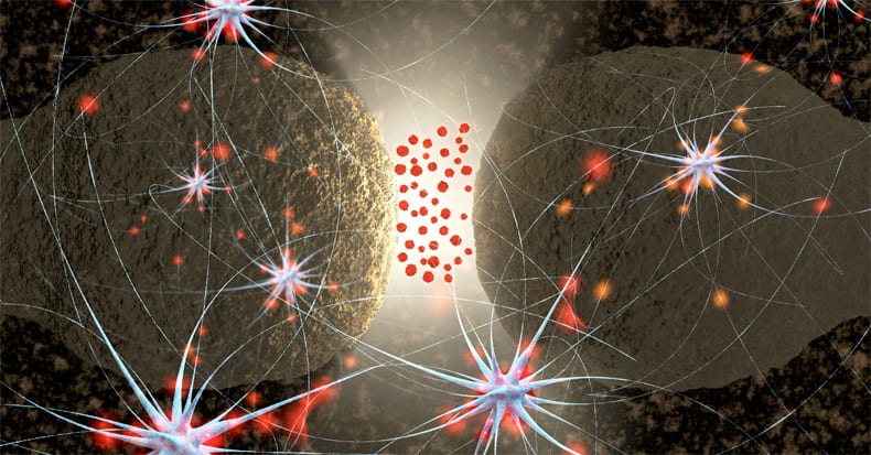
    Pain is an electrical signal in the brain. Oversimplified, five structures are involved for the pain electrical signal to reach the brain:

    The Receptor

    The receptor converts an environmental stress into the electrical signal.

    • For chronic low back pain the receptor is primarily in the intervertebral disc.
    • For chronic neck pain the receptor is primarily in the facet.
    • Receptor thresholds are usually linked to inflammation.

    The Primary Afferent Neuron

    The primary afferent neuron brings the electrical signal from the receptor to the spinal cord.

    • The threshold of the primary afferent neuron is influenced genetically. [Key Concept For This Paper]

    The Synapse

    The synapse is the gap between the primary afferent neuron and the second order afferent neuron.

    The Second Order Afferent Neuron

    The second order afferent neuron brings the electrical signal from the synapse to the brain for the perception of pain.

    The Brain for the perception of pain

    The brain perceives the electrical signal of pain.

    • Several brain relays are involved and influence both pain location and suffering.

    The brain perceives the electrical signal of pain.

    Important for this discussion, the threshold and sensitivity of the primary afferent neuron is influenced by a chemical called norepinephrine (noradrenaline). Norepinephrine is produced by post-ganglionic sympathetic neurons. Norepinephrine makes the primary afferent neuron more sensitive, or making more painful, increasing the pain electrical signal to the brain:

    Norepinephrine makes the primary afferent neuron more sensitive, or making more painful, increasing the pain electrical signal to the brain

    ••••••••••

    Genetics

    Increasingly, individual differences in the intensity, duration and recovery from pain syndromes is being attributed to genetic differences, and specifically to the production of particular enzymes that influence the electrical threshold and transmission of the pain neurons. The genetics of pain is detailed in the 5th edition of the book Wall and Melzack’s Textbook of Pain, 2006 (14). Chapter 9 is titled “The Genetics of Pain.” The summary of this book includes:

    “Pain is associated with considerable variability between individuals. Humans exhibit robust differences in their thresholds and tolerances to controlled noxious stimuli, in their analgesic response to drugs, and in their susceptibility to (and severity of) clinical pain syndromes.”

    Pain genetics is better understood in the 2014 book by Judy Foreman, titled “A Nation in Pain.” Her pertinent chapter, chapter 3 is also titled “The Genetics of Pain.” (15) Ms. Foreman notes that genetics influence both the susceptibility and sensitivity to pain, which she defines as:

    Genetic susceptibility means the likelihood that you’ll get a chronic pain condition.

    Genetic sensitivity means how much it hurts if you do have a chronic pain condition.

    On this topic, Ms. Foreman states:

    “Scientists now think that genes control perhaps 50 percent of susceptibility to chronic pain.”

    “Across a number of different kinds of pain, genes seem to be at least half the driver of how much pain you experience.”

    In her review of the literature, Ms. Foreman notes that a “favorite gene of pain geneticists is COMT.”

    COMT is an enzyme. It’s the abbreviation for:

    Catechol-Oxygen-Methyltransferase = Catechol-O-Methyltransferase

    In review, note that this enzyme ends in the suffix ase, further establishing that it is a protein, and as such there is a gene that codes for (assembles) it. And all genes express differently person to person. Apparently the individual expression of COMT can significantly alter one’s sensitivity to pain. Ms. Foreman states:

    “We all have the COMT gene, but some people have a form of the gene with high activity, and some, the form with low activity.”

    “High activity is better! High COMT means low pain and low COMT means high pain.”

    “The luckiest 40 percent of Caucasians have the high-activity form and are relatively unsusceptible to pain.” These individuals are only half as likely as others to develop chronic pain syndromes.

    COMT works in part by making enzymes that get rid of stress hormones like norepinephrine. “Since norepinephrine acts directly on nervesóthus boosting painógetting rid of norepinephrine can reduce pain.”

    “There’s another key finding emerging from the COMT research. The hormone estrogen decreases COMT activity. Because lower COMT means more pain, this may partly explain why women, who have more estrogen, experience more pain than men.”

    In review, pain sensitivity is controlled genetically by the production of enzymes, and one such enzyme is COMT. Chronic pain is linked to the neurotransmitter norepinephrine, which is produced by the post-ganglionic sympathetic efferent neurons. Norepinephrine makes pain afferents more sensitive. Consequently, more neurotransmitter norepinephrine means more pain; less neurotransmitter norepinephrine means reduced pain. COMT degrades norepinephrine and results in less pain.

    The evidence that increased genetic production of COMT inhibits pain, and reduced genetic production of COMT increases pain, is quite strong. A search of the National Library of Medicine (April 10, 2017) using the key words “COMT and Pain” located 233 studies pertaining to the topic. A sampling of these studies includes:

    COMT val158met genotype affects mu-opioid neurotransmitter responses to a pain stressor. Science, 2003

    Genetic influence on variability in human acute experimental pain sensitivity associated with gender, ethnicity and psychological temperament. Pain, 2004

    Genetic basis for individual variations in pain perception and the development of a chronic pain condition. Hum Mol Genet, 2005

    Catechol-O-methyltransferase gene polymorphisms are associated with multiple pain-evoking stimuli. Pain, 2006

    Genetic variation in the catechol-O-methyltransferase (COMT) gene and morphine requirements in cancer patients with pain. Mol Pain, 2008

    Variation in the COMT gene: implications for pain perception and pain treatment. Pharmacogenomics, 2009

    Catechol-O-methyltransferase and pain. nt Rev Neurobiol, 2010

    Effect of catechol-O-methyltransferase polymorphism on response to propranolol therapy in chronic musculoskeletal pain: a randomized, double-blind, placebo-controlled, crossover pilot study. Pharmacogenet Genomics, 2010

    Catechol O-methyltransferase haplotype predicts immediate musculoskeletal neck pain and psychological symptoms after motor vehicle collision. J Pain, 2011
    COMT genetic variants and pain. Drugs Today (Barc), 2011

    Genetic contribution of catechol-O-methyltransferase variants in treatment outcome of low back pain: a prospective genetic association study. BMC Musculoskelet Disord, 2012

    Complex multilocus effects of catechol-O-methyltransferase haplotypes predict pain and pain interference 6 weeks after motor vehicle collision. Neuromolecular Med, 2014
    A novel catechol-O-methyltransferase variant associated with human disc degeneration. Int J Med Sci, 2014

    Genes associated with persistent lumbar radicular pain; a systematic review. BMC Musculoskelet Disord., 2016

    The inference from these studies is that chronic pain susceptibility may be determinable through genetic testing.

    ••••••••••

    The Chiropractic Connection

    Increased production and release of the neurotransmitter norepinephrine increases one’s sensitivity to pain.

    COMT is an enzyme that degrades the neurotransmitter norepinephrine. Increased COMT decreases norepinephrine. Reduced norepinephrine reduces pain sensitivity.

    The chiropractic connection is, that it is now conclusive that chiropractic spinal adjusting (specific manipulation) reduces the production of the pain sensitizing neurotransmitter norepinephrine. Improvements in spinal mechanical function inhibit the production of norepinephrine, which would make it easier for one’s genetic ability to produce COMT to further reduce the presence of norepinephrine.

    Chiropractic spinal adjusting inhibits the production of norepinephrine.

    COMT degrades the norepinephrine that is produced.

    Both reduce the sensitivity of the pain afferents, reducing the perception of pain in the brain.

    Studies showing that spinal adjusting reduces the production of norepinephrine have been in the literature for decades (16, 17, 18, 19). The most often cited explanation for this is that there exists a reflex between the articular mechanoreceptors and the sympathetic nervous system pre-ganglionic cell bodies in the gray matter of the spinal cord. This connection has been proven to exist in two-legged animals (20).

    The most recent studies showing that chiropractic spinal adjusting inhibits the production of neurotransmitter norepinephrine (2011 and 2017),
    use sophisticated modern technology (radioactive glucose, PET scans, and levels of salivary amylase). The authors are from the Division of Cyclotron Nuclear Medicine, Tohoku University (Graduate School of Medicine), Sendai, Japan. Their first study was published in the journal Alternative Therapies Health Medicine, and titled (21):

    Cerebral Metabolic Changes in Men
    After Chiropractic Spinal Manipulation for Neck Pain

    The study conclusion includes:

    “In summary, the present study demonstrated sympathetic relaxation [inhibition] and corresponding regional brain metabolic changes, as well as reduced muscle tone and decreased pain intensity following a chiropractic spinal manipulation.”

    This group’s second study was published in the journal Evidence-Based Complementary and Alternative Medicine, and titled (22):

    Glucose Metabolic Changes in the Brain and Muscles of Patients with Nonspecific Neck Pain Treated by Spinal Manipulation Therapy: A [18F]FDG PET Study

    The study conclusions include:

    “Brain processing after spinal manipulative therapy may lead to physiological relaxation via a decrease in sympathetic nerve activity.”

    “Our assessment of body responses in this study showed relaxation of muscle tension and decreased salivary amylase levelsóphenomena that are associated with reduced sympathetic nerve activity.”

    Spinal manipulative therapy stimuli to the joints may result in “decreased sympathetic nerve activity.”

    The study has four important findings:

    • Chiropractic adjusting inhibits muscle tone, improving ranges of motion.
    • Chiropractic adjusting inhibits sympathetic tone.
    • Chiropractic adjusting inhibits pain.
    • The brain is affected by chiropractic adjusting.

    Conclusion

    The contention that chiropractic spinal adjusting reduces musculoskeletal pain is now irrefutable. The physiological mechanisms by which chiropractic adjusting helps pain patients are still being investigated and are not fully understood. It is probable that multiple mechanisms are occurring simultaneously. Inhibition of sympathetic nervous system tone with a concomitant reduction of the pain enhancing neurotransmitter norepinephrine, the topic of this paper, has both supportive evidence and biological plausibility.

    REFERENCES

    1. Edwards BC; Low back pain and pain resulting from lumbar spine conditions: a comparison of treatment results; Australian Journal of Physiotherapy; September 1969; Vol. 15; No. 33; pp. 104-110.
    2. Kirkaldy-Willis WH, Managing Low Back Pain, Churchill Livingstone, 1983, p. 19.
    3. Kirkaldy-Willis WH, Cassidy JD; Spinal Manipulation in the Treatment of Low Back Pain; Canadian Family Physician; March 1985; Vol. 31; pp. 535-540.
    4. Meade TW, Dyer S, Browne W, Townsend J, Frank OA; Low back pain of mechanical origin: Randomized comparison of chiropractic and hospital outpatient treatment; British Medical Journal; Volume 300, June 2, 1990, pp. 1431-7.
    5. editors; Chiropractors and Low Back Pain; Lancet; July 28, 1990; Vol. 336(8714);p. 220.
    6. Woodward MN, Cook JCH, Gargan MF, Bannister GC; Chiropractic treatment of chronic ëwhiplash’ injuries; Injury; Vol. 27; No. 9; November 1996; pp. 643-645.
    7. Khan S, Cook J, Gargan M, Bannister G; A symptomatic classification of whiplash injury and the implications for treatment; The Journal of Orthopaedic Medicine; Vol. 21; No. 1; 1999, pp. 22-25.
    8. Giles LGF, Muller R; Chronic Spinal Pain: A Randomized Clinical Trial Comparing Medication, Acupuncture, and Spinal Manipulation; Spine; July 15, 2003; Vol. 28; No. 14; pp. 1490-1502.
    9. Muller R, Lynton G.F. Giles LGF, DC, PhD; Long-Term Follow-up of a Randomized Clinical Trial Assessing the Efficacy of Medication, Acupuncture, and Spinal Manipulation for Chronic Mechanical Spinal Pain Syndromes; Journal of Manipulative and Physiological Therapeutics, January 2005, Vol. 28, No. 1; pp. 3-11.
    10. Cyriax, James; Textbook of Orthopaedic Medicine, Diagnosis of Soft Tissue Lesions; Bailliere Tindall; Vol. I; eighth edition; 1982.
    11. Vicenzino B, Collins D, Wright A; The Initial Effects of a Cervical Spine Manipulative Physiotherapy Treatment on the Pain and Dysfunction of Lateral Epicondylalgia; Pain; November 1996; Vol. 68; No. 1; pp. 69-74.
    12. Savva C, Giakas G, Efstathiou M; The role of the descending inhibitory pain mechanism in musculoskeletal pain following high-velocity, low amplitude thrust manipulation: a review of the literature; Journal of Back Musculoskeletal Rehabilitation; 2014; Vol. 27; No. 4; pp. 377-382.
    13. Garcia DV, Dierckx R, Otte a, Holstege G; PET and SPECT in Neurology, Chapter 46 “Whiplash: Real or Not Real: A Review and New Concept”; Springer-Verlag; 2014, pp. 947-963.
    14. Mogil JS, Mitchell B. Max MB; “The Genetics of Pain,” Chapter 9, in Wall and Melzack’s Textbook of Pain, 5th edition, edited by McMahon SB, Koltzenberg M; Elsevier Churchill Livingstone; 2006.
    15. Judy Foreman; A Nation in Pain; Healing Our Biggest Health Problem; Oxford University Press; 2014.
    16. Korr IM; Proprioceptors and somatic dysfunction; J Am Osteopath Assoc. 1975 Mar;74(7):638-50.
    17. Korr IM; The spinal cord as organizer of disease processes: III. Hyperactivity of sympathetic innervation as a common factor in disease; J Am Osteopath Assoc. 1979 Dec;79(4):232-7.
    18. Budgell B, Hirano F; Innocuous mechanical stimulation of the neck and alterations in heart-rate variability in healthy young adults; Autonomic Neuroscience; Aug 13, 2001; Vol. 9l No. 1-2; pp. 96-99.
    19. Koch LE, Koch H, Graumann-Brunt S, Stoll D, Ramirez JM, Saternus KS; Heart rate changes in response to mild mechanical irritation of the high cervical spinal cord region in infants; Forensic Science International; Vol. 128; No. 3; August 28, 2002; pp. 168-176.
    20. Jiang H, Moreau M, Raso J, Russell G, Bagnall K; Identification of the location, extent, and pathway of sensory neurologic feedback after mechanical stimulation of a lateral spinal ligament in chickens; Spine; January 1, 1997; Vol. 22; No. 1; pp. 17-25.
    21. Ogura T, Tashiro M, Masud M, Watanuki S, Shibuya K, Yamaguchi K, Itoh M, Fukuda H, Yanai K; Cerebral metabolic changes in men after chiropractic spinal manipulation for neck pain; Alternative Therapies Health Medicine; Nov-Dec 2011; Vol. 17; No. 6; pp. 12-17.
    22. Inami A, Ogura T, Watanuki S, Masud M, Shibuya K, Miyake M, Matsuda R, Hiraoka K, Itoh M, Fuhr AW, Yanai K, Tashiro M; Glucose Metabolic Changes in the Brain and Muscles of Patients with Nonspecific Neck Pain Treated by Spinal Manipulation Therapy: A [18F]FDG PET Study; Evidence-Based Complementary and Alternative Medicine; Volume 2017 [epub].
  • Acute Non-Traumatic Neck Pain: Understanding the Meniscoid Entrapment Syndrome and Specific Spinal Manipulation

    Acute Non-Traumatic Neck Pain: Understanding the Meniscoid Entrapment Syndrome and Specific Spinal Manipulation

    Kim was 19 years old, athletic, physically fit, and had never suffered from a neck injury or an episode of prior neck pain; her past history was completely unremarkable.

    While showering earlier that morning, she turned to reach for a bar of soap. She immediately experienced a sharp zap of left–sided neck pain. The pain was sharp and intense, 8/10 on a visual analogue scale, and persistent. To function at any level, she had to tilt her head to the right; any straightening of her neck caused immediate intense worsening of her pain.

    Assuming she had somehow pinched a neck nerve, Kim’s mother made an appointment to see a local chiropractor.

    Other than an inability to left laterally flex her neck because of severe pain, all examination and radiographic findings were unremarkable.

    ••••••••••

    All mechanical joints require lubrication in order for them to glide and function efficiently, and to protect the joint from wear/tear and damage. This includes both inanimate joints (such as joints in automobiles and hinges on doors), and all of the joints in the human body.

    Inanimate joints are lubricated with oil or grease. Human joints are lubricated with synovial fluid. The synovial fluid is held around the joint by dense ligamentous type of fibers called the joint capsule. The joint (articular) capsule is firmly attached to the adjacent bones to insure the synovial fluid does not seep away.

    The oil or lubricant used in inanimate joints needs to be replaced (refreshed) or “topped off” as its supplies are depleted with use. We are all familiar with the occasional oil change that our automobiles require.

    Human joint synovial fluid is replaced and enhanced as a consequence of proper joint motion. The joint synovial fluid is made from the blood that supplies the fibrous joint (articular) capsule. The inner wall of the joint (articular) capsule has a special group of cells called the synovial membrane. With motion, the synovial membrane produces the synovial fluid that lubricates the joint.

    Although this is not the primary intent of this article, it is important to understand that when a joint does not move regularly through a complete range of motion, the quality of the synovial fluid is reduced, contributing to joint pathology, arthritis, and symptomology. This is one of the mechanisms by which chiropractic adjusting of joints (specific manipulation) helps people; the improved joint motion enhances synovial fluid production and as such enhances joint function, resulting in improved function and reduction of symptoms.

    Importantly, parts of the synovial membrane actually project deeper into the joint cavity and lie between the articulation surfaces of the joints. This deeper inner component of the synovial membrane is classically called the meniscus. With certain quick or unexpected motions, the meniscus can become entrapped between the joint surfaces, an experience that is extremely painful.

    Importantly, parts of the synovial membrane actually project deeper into the joint cavity and lie between the articulation surfaces of the joints. This deeper inner component of the synovial membrane is classically called the meniscus. With certain quick or unexpected motions, the meniscus can become entrapped between the joint surfaces, an experience that is extremely painful.

    joint capsule

    ••••••••••

    Perhaps the most authoritative book written pertaining to the spine is Schmorl’s and Junghanns’ The Human Spine in Health and Disease. Georg Schmorl (1861-1932) was a German physician and pathologist. Herbert Junghanns (1902-1986) was the Chief of the Occupational Accident Hospital, Surgical Clinic, and Head of the Institute for Spinal Column Research, in Frankfurt, Germany. The fifth edition of their book was translated into English in 1971 (1). This book contains 500 figures of radiographs, histological sections, photographs, and drawings. The book has more than 500 pages and approximately 5,000 references in the bibliography.

    In their book, Drs. Schmorl and Junghanns note:

    “Like other body articulations, the apophyseal joints are endowed with articular capsules, reinforcing ligaments and menisci-like internal articular discs.” p. 251

    “Like any other joint, the motor segment may become locked. This is usually associated with pain.”

    Chiropractors refer to such events as subluxations. These motor unit disturbances can cause torticollis and lumbago.

    “Various processes may cause such ‘vertebral locking.’ It may happen during normal movement. The incarceration of an articular villus or of a meniscus in an apophyseal joint may produce locking.” pp. 221-222

    If a joint is suddenly incarcerated within the range of its physiologic mobility, it is an “articular locking or a fixed articular block.”

    “Such articular locking is also possible in the spinal articulations (apophyseal joints, intervertebral discs, skull articulations, lumbosacral articulations). They may be mobilized again by specific therapeutic methods (stretching, repositioning, exercises, etc.). Despite many opinions to the contrary, this type of locking is today increasingly recognized by physicians.  Many physicians are employing manipulations which during the past decades were the tools of lay therapists only (chiropractors, osteopaths).  However, these methods have at times been recommended by physicians.  They have also been known in folk medicine and in medical schools of antiquity.” p. 376

    Accompanying this quote and other pertinent discussions are two photographs of anatomical sections through the facet joints showing these “menisci-like internal articular discs,” or meniscus. They also included three radiographs and one drawing showing abnormal gapping of an articulation as a consequence of meniscus entrapment in a facetal articulation. They note that such a meniscoid incarceration can cause acute torticollis, and they show a “follow-up roentgenogram after manual repositioning” resulting in “immediate relief of complaints and complete mobility.” p. 222

    ••••••••••

    In September 1975, the Department of Physical Medicine and Rehabilitation of the California College of Medicine organized an international conference concerning Approaches to the Validation of Manipulation Therapy. The conference was held at the University of California, Irvine. Twenty-one global top experts on spinal manipulation participated in the conference. The proceedings from the conference were published in 1977 (2). Chapter 14 of the conference proceedings was authored by physician James Fisk from New Zealand, and is titled (3):

    An Evaluation of Manipulation in the Treatment of the
    Acute Low Back Pain Syndrome in General Practice

    In this chapter, Dr. Fisk, in the section regarding possible lesions that cause low back pain, lists:

    “Entrapment of a Meniscoid”

    This chapter states:

    “These meniscoids are richly innervated by pain producing nerve endings. There should be a rapid response to manipulation.”

    ••••••••••

    In 1977, the National Institute of Neurological and Communicative Disorders and Stroke of the National Institutes of Health (USA), funded a grant for a Research Workshop on Neurobiologic Mechanisms in Manipulative Therapy. The workshop was held at Michigan State University, and had 38 worldwide participant experts. The Proceedings for the workshop were published the following year, in 1978 (4). The first chapter of the workshop proceedings is authored by Dr. Karel Lewit from Czechoslovakia, and is titled (5):

    The Contribution of Clinical Observation to
    Neurobiological Mechanisms in Manipulative Therapy

    In this chapter, Dr. Lewit notes:

    “The meniscoid has a soft base and a hard edge.”

    “If the meniscoid is caught between the gliding surfaces of the joint facets the hard edge produces a cavity in the cartilage in which it is trapped.”

    “The implications for the theory of manipulation are obvious: if we separate the joint facets the meniscoid can slip out.”

    Dr. Lewit includes a similar discussion in his 1985 text titled Manipulative Therapy in Rehabilitation of the Locomotor System (6).

    ••••••••••

    Also in 1985, 30 distinguished international multidisciplinary experts collaborated on a text titled Aspects of Manipulative Therapy (7). The comments in this text pertaining to the interarticular meniscus include:

    “Histologically, meniscoids are synovial tissue. Their innervation is derived from that of the capsule.”

    The current hypothetical model of the mechanism involved in acute joint locking is based on a phenomenon in which the “meniscoid embeds itself, thereby impeding mobility.”

    “It is highly probable that the meniscoids do play an important role in acute joint locking, and this is confirmed by the observation that all the joints afflicted by this condition are equipped with such structures.” pp. 90-91

    ••••••••••

    In 1986, physical therapist Gregory Grieve authored a text titled Modern Manual Therapy of the Vertebral Column (8). In the chapter titled “Acute Locking of the Cervical Spine” the text notes that a cause of acute cervical joint locking includes:

    “Postulated mechanical derangements of the apophyseal joint include nipped or trapped synovial fringes, villi or meniscoids.”

    ••••••••••

    In 1988, Rene Cailliet, MD, published the fourth edition of his book Low Back Pain Syndrome (9). At the time, Dr. Cailliet was the retired Chairman of the Department of Physical Medicine and Rehabilitation at the medical school at the University of Southern California. Dr. Cailliet states:

    “A meniscus exists within facet joints that becomes entrapped.”

    Dr. Cailliet also includes an explanation of the benefits of spinal manipulation to treat the meniscal entrapment.

    ••••••••••

    In the second edition of his reference text, Common Vertebral Joint Problems, Gregory Grieve includes a section on the meniscoid block. While discussing Sudden Backache or Acute Lumbago, Greive’s text states (10):

    “The small ‘meniscoid’ structures in the facet-joints are apparently susceptible to temporary impaction at times, with the chronic sequelae of the joint tissue damage probably adding to the natural process of senescence.” p. 201

    “The patient with sudden backache due to presumed locking of a facet-joint is usually a young female but may be a young man, and there is often a degree of hypermobility.  They often excel at athletics or ballet dancing, and during some activity which may be reaching up to open a window or adjust curtains, a lumbar synovial joint locks.  No outside force is applied, the condition being consequent upon a body movement involving reaching or stretching.  It is reasonable to suppose that the opposed joint faces of the hypermobile segment come apart more easily than usual, and the normally slight negative pressure within the joint cavity is further lowered by the greater distraction.  A villus of synovial tissue is presumably ‘sucked in and nipped,’ and thus the meniscoid structure is impacted between joint surfaces.  On resumption of normal posture the pain of impaction induces reactive muscle spasm, fixing the articulation rigidly to produce a locked joint.” p. 407

    ••••••••••

    In her 1994 text Physical Therapy of the Cervical and Thoracic Spine, professor of physiotherapy from the University of South Australia, Ruth Grant writes (11):

    “Acute locking can occur at any intervertebral level, but is most frequent at C2-C3. Classically, locking follows an unguarded movement of the neck, with instant pain over the articular pillar and an antalgic posture of lateral flexion to the opposite side and slight flexion, which the patient is unable to correct.  Locking is more frequent in children and young adults. In many, the joint pain settles within 24 hours without requiring treatment (because the joint was merely sprained or because it unlocked spontaneously), but other patients will require a localized manipulation to unlock the joint.” p. 298

    ••••••••••

    In his 2004 text titled The Illustrated Guide to Functional Anatomy of the Musculoskeletal System, Rene Cailliet, MD also comments on the anatomy of the interarticular meniscus, stating (12):

    “The uneven surfaces between the zygapophyseal processes are filled by an infolding of the joint capsule, which is filled with connective tissue and fat called meniscoids. These meniscoids are highly vascular and well innervated.” p. 95

    Presumably it is this innervation that produces the pain associated with meniscoid entrapments.

    ••••••••••

    Nikolai Bogduk, PhD, MD, is a most distinguished and respected physician, anatomist, and researcher. In the fourth edition of his textbook Clinical Anatomy of the Lumbar Spine and Sacrum (13), Dr. Bogduk writes:

    “The largest of the meniscoid structures are the fibro-adipose meniscoids. These project from the inner surface of the superior and inferior capsules. They consist of a leaf-like fold of synovium which encloses fat, collagen and some blood vessels.”

    “Fibro-adipose meniscoids are long and project up to 5 mm into the joint cavity.” p. 35

    “A relatively common clinical syndrome is ‘acute locked back.’  In this condition, the patient, having bent forward, is unable to straighten because of severe focal pain on attempted extension.”

    “Maintaining flexion is comfortable for the patient because that movement disengages the meniscoid. Treatment by manipulation becomes logical.” p. 195

    ••••••••••

    The January 15, 2007 publication of the top ranked orthopaedic journal Spine contains an article titled (14):

    High-Field Magnetic Resonance Imaging of Meniscoids in the Zygapophyseal Joints of the Human Cervical Spine

    The authors note that meniscoids of the cervical facet joints contain nociceptors and may be a source of cervical facet joint pain; neck pain can be caused by pathological conditions of the meniscoids within the zygapophysial joints. They state:

    “Cervical zygapophysial joints are well documented as a possible source of neck pain, and it has been hypothesized that pathologic conditions related to so called meniscoids within the zygapophysial joints may lead to pain.”

    Meniscoids may become entrapped between the articular cartilages of the facet joints. This causes pain, spasm, reduced movement, and “an acute locked neck syndrome.”  “Spinal adjusting can solve the problem by separating the apposed articular cartilages and releasing the trapped apex.”

    Also, importantly, these authors note that meniscoid anatomy and pathology can be visualized with proton density weighted MRI imaging, especially with high-field MRI of 3.0 T strength.

    ••••••••••

    In 2011, the journal Manual Therapy published a study titled (15):

    Synovial folds – A pain in the neck?

    The authors note that synovial meniscoid folds have been identified in all synovial articulations of the cervical spine, stating:

    “Intra-articular synovial folds are formed by folds of synovial membrane (synovium) that project into the joint cavity and are found in synovial articulations throughout the vertebral column.”

    “An abnormal joint movement may cause a synovial fold to move from its normal position at the articular margins to become imprisoned between the articular cartilage surfaces causing pain and articular hypomobility accompanied by reflex muscle spasm.”

     “Synovial fold entrapment has been used to explain the pathophysiology of torticollis and the relief of pain and disability following spinal manipulation.”

    ••••••••••

    In December of 2016, the European Spine Journal early published (epub) an article titled (16):

    Cervical Spine Meniscoids:
    An Update on Their Morphological Characteristics
    Potential Clinical Significance

    These authors note that the cervical spine meniscoids are intra-articular folds of synovial membrane that may have clinical significance in neck pain. They were first anatomically documented in 1855. Cervical spine meniscoids “have been identified within cervical zygapophyseal, lateral atlanto-axial and atlanto-occipital joints, and have been hypothesised to be of clinical significance in neck pain through their mechanical impingement or displacement.”

    Immunohistochemistry studies show that cervical spine meniscoids are innervated with nociceptive nerves, and as such they have a role in cervical pain and pathology. Meniscoid entrapment may be responsible for acute torticollis.

    Advances in MRI technology have allowed for the imaging of cervical spine meniscoids, especially with 3-T MRI. (T = Tesla, the unit of magnet strength)

    In terms of clinical management, these authors advocate manual therapy, stating:

    “It is suggested that rotation or traction-based manual therapy techniques may encourage the entrapped meniscoid to return to its resting place, accounting for the reported clinical effectiveness of such treatment.”

    “Rotation or traction-based manual therapy techniques have been theorized as treatments for impinged or displaced meniscoids.”

    ••••••••••

    Clinical Applications

    Decades of evidence support the perspective that the inner aspect of the facet capsules have a process that extends into and between the facet articular surfaces, and that this tissue is innervated with nociceptors (pain nerves). This evidence includes anatomical sections, histological sections, MR imaging, and clinical evaluations. The meniscoid can become entrapped between the facet articulating surfaces, producing pain, spasm, and antalgia.

    A classic cervical spine clinical presentation would be that of an acute torticollis triggered by a routine motion (like turning and reaching for a bar of soap while in the shower). If the meniscoid is entrapped on the left side of the cervical spine, the patient would present with an antalgia of right lateral flexion; in other words, the patient bends away from the side of entrapment. The patient’s primary pain symptoms will be on the side of entrapment, in this example, the left side. Active range of motion examination will show that the patient is capable of additional lateral flexion to the right, but will not laterally flex to the left because of increased pain; once again this is because the meniscus is entrapped on the left side and left lateral flexion increases meniscus compression, pain, and spasm. This is also why the patient is antalgic to the right; such positioning reduces left sided meniscus compression, pain, and spasm.

    antalgic to the right

    antalgic to the right - cervical spine view

    Additional clinical evaluation will reveal no sings of radiculopathy; no alterations of superficial sensation in a dermatomal pattern, and no signs of motor weakness or altered deep tendon reflexes. An important clinical feature is that the patient will not laterally flex the cervical spine to the left because of increased pain and spasm.

    The best adjustment approach for the synovial fold entrapment syndrome is to adjust on the “low side of the rainbow,” i.e. to make the antalgic lean worse. This approach is most likely to gap the contralateral facet joint, freeing the entrapped synovial fold. The result is immediate improvement of antalgic lean and improvement in the range of motion.

    ••••••••••

    Kim’s chiropractor diagnosed a “meniscoid entrapment.” A single adjustment to the “down” side of the blocked joint resulted in an audible release, and an immediate reduction in pain and improvement in the cervical range of motion.

    A follow-up visit the next day showed lasting improvement, but with an approximate subjective and objective residual pain and stiffness of 20%. A second similar adjustment was given.

    A third follow-up visit was scheduled for two days away and showed a complete resolution of all signs and symptoms.

    REFERENCES

    1. Junghanns H; Schmorl’s and Junghanns’ The Human Spine in Health and Disease; Grune & Stratton; 1971.
    2. Buerger AA and Tobis JS; Approaches to the Validation of Manipulation Therapy; Thomas, 1977.
    3. Fisk JW; “An Evaluation of Manipulation in the Treatment of the Acute Low Back Pain Syndrome in General Practice” in Approaches to the Validation of Manipulation Therapy; Thomas, 1977.
    4. Korr IM; Neurobiologic Mechanisms in Manipulative Therapy; Plenum; 1978.
    5. Lewit K; “The Contribution of Clinical Observation to Neurobiological Mechanisms in Manipulative Therapy” in Korr IM; Neurobiologic Mechanisms in Manipulative Therapy; Plenum; 1978.
    6. Lewit K; Manipulative Therapy in Rehabilitation of the Locomotor System; Butterworths; 1985.
    7. Idczak GD; Aspects of Manipulative Therapy; Churchill Livingstone; 1985.
    8. Grieve G; Modern Manual Therapy of the Vertebral Column; Churchill Livingstone; 1986.
    9. Cailliet R; Low Back Pain Syndrome; FA Davis, 1988.
    10. Grieve G; Common Vertebral Joint Problems, second edition; Churchill Livingstone; 1988.
    11. Grant R; Physical Therapy of the Cervical and Thoracic Spine, second edition; Churchill Livingstone, 1994.
    12. Cailliet R; The Illustrated Guide to Functional Anatomy of the Musculoskeletal System, American Medical Association, 2004.
    13. Bogduk N; Clinical Anatomy of the Lumbar Spine and Sacrum, fourth edition; Elsevier, 2005.
    14. Friedrich KM. MD, Trattnig S, Millington SA, Friedrich M, Groschmidt K, Pretterklieber ML; High-Field Magnetic Resonance Imaging of Meniscoids in the Zygapophyseal Joints of the Human Cervical Spine; Spine; January 15, 2007, Volume 32(2), January 15, 2007, pp. 244-248.
    15. Webb AL, Collins P, Rassoulian H, Mitchell BS; Synovial folds – A pain in the neck?; Manual Therapy; April 2011; Vol. 16; No. 2; pp. 118-124.
    16. Farrell SF, Osmotherly P, Cornwall J, Sterling M, Rivett DA; Cervical Spine Meniscoids: An Update on Their Morphological Characteristics Potential Clinical Significance; European Spine Journal December 19, 2016 [Epub]
  • Safety Comparison: Allopathic vs. Chiropractic Healthcare

    Safety Comparison: Allopathic vs. Chiropractic Healthcare

    All healthcare disciplines are associated with risks of injury or death. Rare but sensational occurrences are often exploited by the media, leading the public to believe that rare events are commonplace. In contrast, common occurrences are often under reported, leading the public to have higher confidence in the safety of certain healthcare disciplines and procedures.

    How Safe Is Medical Care?

    The risks of medical healthcare (allopathic healthcare) were shockingly revealed in 1994 when Harvard’s Lucian Leape, MD, indicated that medical error was responsible for 180,000 deaths per year (1, 2). Dr. Leape’s analogy was that this was “the equivalent of three jumbo-jet crashes every 2 days,” killing all on board (2). Dr. Leape’s revelation was published in the prestigious Journal of the American Medical Association, and titled:

    Error in Medicine

    Four years after Dr. Leape’s headlines pertaining to error in medicine, Jason Lazarou, MD (neurologist) and colleagues from the University of Toronto published a study in the Journal of the American Medical Association titled (3):

    Incidence of Adverse Drug Reactions in Hospitalized Patients
    A Meta-analysis of Prospective Studies

    The objective of this study was to estimate the incidence of serious and fatal adverse drug reactions (ADR) in hospital patients. Serious ADRs were defined as those that required hospitalization, were permanently disabling, or resulted in death. The authors performed a meta-analysis of 39 prospective studies done in the United States over a period of 32 years on the incidences of Adverse Drug Reactions (ADRs). The goal of this study was to “estimate injuries incurred by drugs that were properly prescribed and administered.” If the event was determined to be a “Possible ADRs” it was excluded from this study. The authors noted:

    “We estimated that in 1994 overall 2,216,000 (1,721,000-2,711,000) hospitalized patients had serious ADRs and 106,000 (76,000-137,000) had fatal ADRs, making these reactions between the fourth and sixth leading cause of death.”

    “We have found that serious ADRs are frequent and more so than generally recognized. Fatal ADRs appear to be between the fourth and sixth leading cause of death. Their incidence has remained stable over the last 30 years.”

    “It is important to note that we have taken a conservative approach, and this keeps the ADR estimates low by excluding errors in administration, overdose, drug abuse, therapeutic failures, and possible ADRs. Hence, we are probably not overestimating the incidence of ADRs.”

    This study on ADRs excluded medication errors “to show that there are a large number of serious ADRs even when the drugs are properly prescribed and administered.”

    “The incidence of serious and fatal ADRs in US hospitals was found to be extremely high.”

    The incidence of hospital adverse drug reactions detailed in the Lazarou and colleague’s study is stunning:

    • 106,000 yearly deaths; these deaths rank between the 4th and the 6th leading causes of death yearly.
    • 2,216,000 yearly events that required hospitalization to recover or resulted in a lifelong disability.

    Importantly, these numbers require additional discussion. These statistics pertain only to hospitalized patients; they did not assess similar such events occurring outside of the hospital setting, in locations such as nursing homes, extended care facilities, at home, etc.

    Additionally, and more startling, these deaths and serious adverse events occurred as a consequence of taking the correct drug for the correct diagnosis in the correct dosage. As such, these deaths and serious adverse events are not considered to be as a consequence of error. Rather, they are considered to be “fallout” of a health care delivery discipline that is heavily dependent upon pharmacology.

    Dr. Leape’s premise of “Error in Medicine” was updated in 2016, showing that the problem has not improved in the past two decades. Published in the British Medical Journal, Johns Hopkins University School of Medicine professor Martin A. Makary (MD, MPH) and research fellow Michael Daniel (medical student) produced an article titled (4):

    Medical Error
    The Third Leading Cause of Death in the United States

    This title is misleading, and lay publications were mislead into believing that medical error was the 3rd leading cause of yearly death in the United States. The article clearly notes that the 3rd leading cause of death in the United States is error in hospitals. Once again, the data did not assess medical error deaths occurring outside of the hospital setting, in nursing homes, extended care facilities, at home, etc. Nor did it assess “fallout” deaths either in the hospital or outside the hospital setting.

    In this study, Makary and Daniel analyzed the scientific literature on hospital medical errors to identify its contribution to US deaths. In their appraisal of the magnitude of the problem, they note:

    “We calculated a mean rate of death from [hospital] medical error of 251,454 a year.”

    “We believe this understates the true incidence of death due to medical error because the studies cited rely on errors extractable in documented health records and include only inpatient deaths.”

    [Hospital] “medical error is the third biggest cause of death in the US and therefore requires greater attention.”

    As noted, the 251,000 deaths from medical error underestimates the actual number because it represents only medical error deaths that occur in the hospital setting. Medical error deaths occurring in non-hospital facilities or at home are not included in the estimated number.

    ••••••••••


    How Safe Are Pain Medications?

    The primary reason people seek chiropractic care is for pain. Chiropractic is considered an alternative therapy for pain management, and especially for spinal pain (5, 6). An important study looking at some of the risks associated with the chronic use of nonsteroidal anti-inflammatory drugs (NSAIDs) for pain was published by M. Michael Wolfe, MD, and colleagues, from Stanford’s Medical School and Boston University School of Medicine, and published in the New England Journal of Medicine in 1999. The article was titled (7):

    Gastrointestinal Toxicity of
    Nonsteroidal Anti-inflammatory Drugs

    The authors make the following points:

    “It has been estimated conservatively that 16,500 NSAID-related deaths occur among patients with rheumatoid arthritis or osteoarthritis every year in the United States.”

    “If deaths from gastrointestinal toxic effects of NSAIDs were tabulated separately in the National Vital Statistics reports, these effects would constitute the 15th most common cause of death in the United States.”

    “Yet these toxic effects remain largely a ‘silent epidemic,’ with many physicians and most patients unaware of the magnitude of the problem.”

    “Furthermore, the mortality statistics do not include deaths ascribed to the use of over-the-counter NSAIDs.”

    The authors note that Cox-2 inhibitors (a prescription form of NSAID) have been available in the US since February 1999, in the hope that they will have a reduced capacity to cause injury to the gastroduodenal mucosa. However, Cox-2 inhibitors are also known to cause defects in renal function, alter the regulation of bone resorption, impair female reproductive physiology, and increase the rate of thrombotic events in patients with increased risk of cardiovascular disease.

    In 2003, researchers from the University of Queensland, Australia, published a study in the Journal Spine, titled (8):

    Chronic Spinal Pain:
    A Randomized Clinical Trial Comparing
    Medication, Acupuncture, and Spinal Manipulation

    In this study, the spinal manipulation was performed by licensed chiropractors (two visits per week). The medications used were Celebrex or Vioxx, both prescription NSAIDs. The acupuncture (also two visits per week) was performed by an experienced acupuncturist. The study evaluated 115 chronic neck and back pain patients. The treatment interventions extended over a 9-week period. These authors made the following observations and statements:

    “Adverse reactions to nonsteroidal antiinflammatory (NSAID) medication have been well documented.”

    “Gastrointestinal toxicity induced by NSAIDs is one of the most common serious adverse drug events in the industrialized world.”

    “The newer COX-2-selective NSAIDs are less than perfect, so it is imperative that contraindications be respected.”

    There is “insufficient evidence for the use of NSAIDs to manage chronic low back pain.”

    “The highest proportion of early (asymptomatic status) recovery was found for manipulation (27.3%), followed by acupuncture (9.4%) and medication (5%).”

    “Manipulation yielded the best results over all the main outcome measures.”

    “The consistency of the results provides evidence that in patients with chronic spinal pain, manipulation, if not contraindicated, results in greater short-term improvement than acupuncture or medication.”

    “The results of this efficacy study suggest that spinal manipulation, if not contraindicated, may be superior to needle acupuncture or medication for the successful treatment of patients with chronic spinal pain syndrome.”

    “Medication apparently did not achieve a marked improvement in chronic spinal pain and caused adverse reactions in 6.1% of the patients.”

    “In summary, the significance of the study is that for chronic spinal pain syndromes, it appears that spinal manipulation provided the best overall short-term results, despite the fact that the spinal manipulation group had experienced the longest pretreatment duration of pain.”

    Highlights of this study show that chiropractic spinal manipulation is five times more effective than prescription NSAIDs in the treatment of chronic low back and neck pain, and the results from spinal manipulation were accomplished without any reported adverse events. In contrast, for the patients taking the drugs, more experienced an adverse event (6.1%) than those who became asymptotic (5%) over the nine-week clinical trial.

    Importantly, when this study was published in 2003, Vioxx had been on the market since 1999, four years. The following year, 2004, Vioxx was pulled off the market due to an unacceptable incidence of fatal heart attacks and strokes (9, 10). It has since been established that in the five years that Vioxx was on the market it caused more US deaths (about 60,000) than the Viet Nam war did in 10 years (about 58,000).

    How Safe Is Chiropractic?

    The most sensational adverse event with a supposed link to chiropractic spinal manipulation is vertebral artery dissection. Allegations of vertebral artery dissection caused by chiropractic spinal manipulation have appeared in the published literature for decades. However, recent large critical reviews of the topic have appeared in the scientific literature, and they question the causation between cervical spine manipulation and cervical artery dissection. Some of this literature is reviewed below.

    ••••••••••

    In 2008, Dr. David Cassidy and colleagues published the most comprehensive study at that time pertaining to the risk of vertebral artery dissection as related to chiropractic cervical spine manipulation. The article was published in the journal Spine, and titled (11):

    Risk of Vertebrobasilar Stroke and Chiropractic Care:
    Results of a Population-Based Case-Control and Case-Crossover Study

    This study included all residents of Ontario, CAN, over a period of 9 years, amounting to 109,020,875 person years of observation. Associations between chiropractic visits and vertebral artery dissection versus primary care physician (PCP) visits and vertebral artery dissection were compared.

    The authors noted:

    “We found no evidence of excess risk of vertebral artery stroke associated with chiropractic care.”

    “Neck pain and headache are common symptoms of vertebral artery dissection, which commonly precedes vertebral artery stroke.”

    “The increased risks of vertebral artery stroke associated with chiropractic and primary care physicians visits is likely due to patients with headache and neck pain from vertebral artery dissection seeking care before their stroke.”

    “Because patients with vertebrobasilar artery dissection commonly present with headache and neck pain, it is possible that patients seek chiropractic care for these symptoms and that the subsequent vertebral artery stroke occurs spontaneously, implying that the association between chiropractic care and vertebral artery stroke is not causal.”

    “Our results suggest that the association between chiropractic care and vertebral artery stroke found in previous studies is likely explained by presenting symptoms attributable to vertebral artery dissection.”

    ••••••••••

    In January of 2011, the Journal of Manipulative and Physiological Therapeutics published a population-based case series using administrative health care records of all Ontario, CAN, residents hospitalized with vertebral artery stroke between April 1, 1993, and March 31, 2002, titled (12):

    A Population-based Case-series of Ontario Patients who
    Develop a Vertebrobasilar Artery Stroke After Seeing a Chiropractor

    These authors note:

    “The current evidence suggests that association between chiropractic care and vertebrobasilar artery (VBA) stroke is not causal. Rather, recent epidemiological studies suggest that it is coincidental and reflects the natural history of the disorder.”

    “Because neck pain and headaches are symptoms that commonly precede the onset of a VBA stroke, these patients might seek chiropractic care while their stroke is in evolution.”

    ••••••••••

    Also in January 2011, The Open Neurology Journal published an “open access” editorial by Drs. Dean Smith and Gregory Cramer, titled (13):

    “Spinal Manipulation is Not an Emerging Risk Factor for Stroke
    Nor is it Major Head/Neck Trauma. Don’t Just Read the Abstract!”

    Dean L. Smith is Clinical Faculty, Department of Kinesiology and Health, Miami University, Oxford, Ohio, and Gregory D. Cramer is Professor and Dean of Research, National University of Health Sciences, Lombard, Illinois. Their editorial includes:

    We would like to address two points in this letter:

    1) The current best-evidence indicates no causal relationship between spinal manipulation (‘chiropractic maneuver’ in the paper) and vertebrobasilar artery (VBA) stroke, and,

    2) Spinal manipulation or ‘chiropractic maneuvers’ are not major head/neck trauma as suggested in abstract of this article.

    “First, evidence is mounting that the association between spinal manipulation and stroke is coincidental rather than causal and reflects the natural history of the disorder.”

    “The largest population-based study to date was conducted by Cassidy et al. and included all vertebrobasilar artery (VBA) strokes in Ontario, Canada over a period of 9 years. The authors found no evidence of excess risk (i.e. no risk) of VBA stroke associated with chiropractic care.”

    “The prevailing hypothesis is that patients with vertebral artery dissections often have initial symptoms that cause them to seek care from a chiropractic or medical physician and the stroke is independent of their visit.”

    “The latest scientific evidence questions whether spinal manipulation is a risk factor at all for cervical artery dissection.”

    “Chiropractic spinal manipulations may very well be a demerging risk factor for stroke since there may not be any risk.”

    “The evidence, albeit limited to date, suggests that spinal manipulative treatments produce stretches of the vertebral artery that are much smaller than those that are produced during normal everyday movements, and thus they appear harmless.”

    “Spinal manipulations delivered by licensed chiropractors do not fulfill the criteria for major trauma and should not be considered major trauma.”

    ••••••••••

    The biomechanics of cervical spine manipulation and vertebral artery stress is important. The world leader on this type of biomechanical assessment is Walter Herzog, PhD, from the University of Calgary, CAN. In 2012, Dr. Herzog and colleagues published a study in the Journal of Electromyography and Kinesiology titled (14):

    Vertebral Artery Strains During High-speed,
    Low Amplitude Cervical Spinal Manipulation

    Dr. Herzog notes that spinal manipulative therapy (SMT) is recognized as an effective treatment modality for many back, neck and musculoskeletal problems. Yet, one of the major issues of the use of SMT is its safety, especially with regards to neck manipulation and the risk of stroke. It has been assumed [wrongly as per this study] that the vertebral artery (VA) experiences considerable stretch during extension and rotation of the neck, which may lead to occlusions and damage to the VA, predisposing the patient to stroke. Therefore, this study presents the first ever data on the mechanics between C2/C1 during cervical SMT performed by chiropractic clinicians.

    The authors compared the results of human VA strains during high-speed, low-amplitude SMTs administered by qualified chiropractic clinicians and compared them to the strains encountered during full range of motion (ROM) tests. They used a total of 3,034 segment strains obtained during SMTs and 2,380 segment strains obtained during full ROM testing, making this is an extensive study. Dr. Herzog and colleagues conclude:

    “VA strains obtained during SMT are significantly smaller than those obtained during diagnostic and range of motion testing, and are much smaller than failure strains.”

    “We conclude from this work that cervical SMT performed by trained clinicians does not appear to place undue strain on VA, and thus does not seem to be a factor in vertebro-basilar injuries.”

    “In summary, the maximal strain values for the ROM testing at each segmental level were always greater than the corresponding strain values for the SMTs, suggesting that neck SMTs impose less stretch than turning your head, or extending your neck while looking up at the sky.”

    “Therefore, based on the mechanical tests performed here, one should be able to conclude that stretching of VA during neck SMTs does not cause any damage of the VAs.”

    “The VA is never really strained during spinal manipulative treatments but that the VA is merely taking up slack as the neck and head are moved during SMT, but that there is no stress and thus no possibility for microstructural damage.”

    “The results from this study demonstrate that average and maximal VA strains during high-speed low-amplitude cervical spinal manipulation are substantially less than the strains that can be achieved during ROM testing for all vertebral artery segments.”

    “We conclude that cervical spinal manipulations, as tested here, are safe from a mechanical point of view for normal, healthy VA.”

    ••••••••••

    In 2015, a study was published in the journal Chiropractic & Manual Therapies, and titled (15):

    Chiropractic Care and the Risk of Vertebrobasilar Stroke:
    Results of a Case–control Study in U.S. Commercial
    and Medicare Advantage Populations

    The main purpose of this study was to replicate the case–control epidemiological design study published by Cassidy, et al. in 2008 (11), and to investigate the association between chiropractic care and vertebral artery stroke; and compare it to the association between recent primary care physician (PCP) care and vertebral artery stroke. The authors assessed commercially insured and Medicare Advantage (MA) health plan members in the U.S. The data set included health plan members located in 49 of 50 states (excluded North Dakota) and encompassed national health plan data for 35,726,224 commercial and 3,188,825 MA members. Hence, this study looked at approximately 39 million people, making this the largest case–control study to investigate the association between chiropractic manipulation and vertebral artery stroke. These authors concluded:

    “There was no association between chiropractic visits and VBA stroke found for the overall sample, or for samples stratified by age.”

    “We found no significant association between exposure to chiropractic care and the risk of vertebral artery stroke. We conclude that manipulation is an unlikely cause of vertebral artery stroke.”

    “Our results increase confidence in the findings of a previous study [11], which concluded there was no excess risk of vertebral artery stroke associated with chiropractic care compared to primary care.”

    ••••••••••

    In 2016, a study from the Department of Neurosurgery, Penn State Hershey Medical Center, and the Department of Neurosurgery, Johns Hopkins University School of Medicine, was published in the journal Cureus, and titled (16):

    Systematic Review and Meta-analysis of Chiropractic Care
    and Cervical Artery Dissection:
    No Evidence for Causation

    The authors note that case reports and case control studies have suggested an association between chiropractic neck manipulation and cervical artery dissection (CAD), but a causal relationship has not been established. These authors evaluated the evidence related to this topic by performing a systematic review and meta-analysis of published data on chiropractic manipulation and cervical artery dissection (CAD). These authors state:

    “We found no evidence for a causal link between chiropractic care and CAD. This is a significant finding because belief in a causal link is not uncommon, and such a belief may have significant adverse effects such as numerous episodes of litigation.”

    “In spite of the very weak data supporting an association between chiropractic neck manipulation and CAD, and even more modest data supporting a causal association, such a relationship is assumed by many clinicians. In fact, this idea seems to enjoy the status of medical dogma.”

    “Excellent peer reviewed publications frequently contain statements asserting a causal relationship between cervical manipulation and CAD. We suggest that physicians should exercise caution in ascribing causation to associations in the absence of adequate and reliable data. Medical history offers many examples of relationships that were initially falsely assumed to be causal, and the relationship between CAD and chiropractic neck manipulation may need to be added to this list.”

    “There is no convincing evidence to support a causal link, and unfounded belief in causation may have dire consequences.”

    “The quality of the published literature on the relationship between chiropractic manipulation and CAD is very low. Our analysis shows a small association between chiropractic neck manipulation and cervical artery dissection. This relationship may be explained by the high risk of bias and confounding in the available studies, and in particular by the known association of neck pain with CAD and with chiropractic manipulation.”

    “The association between a chiropractor visit and dissection may be explained by” understanding that “patients with cervical artery dissection more frequently have headache and neck pain” and understanding that “patients with headache and neck pain more frequently visit chiropractors.”

    “Because (on average) patients with headache and neck pain visit chiropractors more frequently, and patients with cervical artery dissection more frequently have headache and neck pain, it appears that those who visit chiropractors have more cervical artery dissections.”

    SUMMARY

    The data presented here indicates that medical care is incredibly dangerous. The authors estimate that hospital errors kill 251,000 Americans yearly (the 3rd leading cause of yearly US deaths), and hospital non-error “fallout” kills an additional 106,000 Americans yearly (the 4th -6th leading cause of yearly US deaths). These numbers total 357,000 yearly hospital medical deaths. It is reasonable to assume that a similar number of deaths occur outside of the hospital setting (nursing homes, extended care facilities, at home, etc.).

    In contrast, chiropractic spinal manipulation, even to the cervical spine, is incredibly safe. In a typical year there are zero reported deaths linked to chiropractic care, and if one such death is alleged it tends to make sensational news. Chiropractic students and chiropractors are extensively trained in spinal anatomy and spinal biomechanics. They are also extensively trained in the science and art of spinal adjusting (specific directional manipulation). They are taught to avoid injury risk, and to recognize serious events that are in progress, making the appropriate referral.

    Even the use of prescription NSAIDs for pain results in the deaths of 16,500 Americans yearly (the 15th leading cause of yearly US deaths). The concern is that in the randomized clinical trial reviewed, chiropractic spinal adjusting was better than five times more effective in alleviating chronic back and neck pain as compared to these drugs, and this was achieved with no side effects. Importantly, the one-year follow-up to this study showed the benefits of chiropractic to be stable (17).

    REFERENCES

    1. Leape LL, Lawthers A, Brennan TA, Johnson WG; Preventing medical injury; Qual Rev Bull 1993;19; pp. 144-149.
    2. Leape LL; Error in Medicine; Journal of the American Medical Association; December 21, 1994; Vol. 272; No. 23; pp. 1851-1857.
    3. Lazarou J, Pomeranz BH, Corey PN; Incidence of Adverse Drug Reactions in Hospitalized Patients: A Meta-analysis of Prospective Studies; Journal of the American Medical Association; Vol. 279; No. 15; April 15, 1998; pp. 1200-1205.
    4. Makary MA, Daniel M; Medical Error: The Third Leading Cause of Death in the United States; British Medical Journal; May 3, 2016; Vol. 353; i2139.
    5. Kirkaldy-Willis WH, Cassidy JD; Spinal Manipulation in the Treatment of Low back Pain; Canadian Family Physician; March 1985; Vol. 31; pp. 535-540.
    6. Ogura T, Tashiro M, Masud M, Watanuki S, Shibuya K, Yamaguchi K, Itoh M, Fukuda H, Yanai K; Cerebral metabolic changes in men after chiropractic spinal manipulation for neck pain; Alternative Therapy Health Medicine; November-December 2011; Vol. 17; No. 6; pp. 12-17.
    7. Wolfe MM, Lichtenstein DL, Singh G; Gastrointestinal Toxicity of Nonsteroidal Anti-inflammatory Drugs; The New England Journal of Medicine; June 17, 1999; Vol. 340; No. 24; pp. 1888-1899.  
    8. Giles GFL, Muller R; Chronic Spinal Pain: A Randomized Clinical Trial Comparing Medication, Acupuncture, and Spinal Manipulation; Spine; July 15, 2003; Vol. 28; No. 14; pp. 1490-1502.
    9. Karha J, Topol EJ; The sad story of Vioxx, and what we should learn from it; Cleveland Clinical Journal of Medicine; December 2004; Vol. 71; No. 12; pp. 938-9.
    10. Topol EJ, Arthritis Medicines and Cardiovascular Events—”House of Coxibs” Journal of the American Medical Association; January 2819 2005; Vol. 293; No. 3; pp. 366-368.
    11. Cassidy JD, Boyle E, Côté P, Yaohua H, Hogg-Johnson S, Silver FL, Bondy SJ; Risk of Vertebrobasilar Stroke and Chiropractic Care: Results of a Population-Based Case-Control and Case-Crossover Study; Spine; Volume 33(4S); February 15; 2008 pp. S176-S183.
    12. Choi S, Boyle E, Cote P, Cassidy JD; A population-based case-series of Ontario patients who develop a vertebrobasilar artery stroke after seeing a chiropractor; Journal of Manipulative and Physiological Therapeutics; 2011; Vol. 34; No. 1; pp. 15-22.
    13. Smith DL, Cramer GC; LETTER TO THE EDITOR: Spinal Manipulation is Not an Emerging Risk Factor for Stroke Nor is it Major Head/Neck Trauma. Don’t Just Read the Abstract!; The Open Neurology Journal; 2011; 5; pp.  46-47.
    14. Herzog W, Leonard TR, Symons B, Tang C, Wuest S; Vertebral artery strains during high-speed, low amplitude cervical spinal manipulation; Journal of Electromyography and Kinesiology; October 2012; Vol. 22; No. 5; pp. 740-746.
    15. Thomas M Kosloff, David Elton, Jiang Tao and Wade M Bannister; Chiropractic Care and the Risk of Vertebrobasilar Stroke: Results of a Case–control Study in U.S. Commercial and Medicare Advantage Populations; Chiropractic & Manual Therapies 2015; 23:19; pp. 1-10.
    16. Church EW, Sieg EP, Zalatimo O, Hussain NS, Glantz M, Harbaugh RE; Systematic Review and Meta-analysis of Chiropractic Care and Cervical Artery Dissection: No Evidence for Causation; Cureus; February 16, 2016; Vol. 8; No. 2; e498.
    17. Muller R, Giles LGF; Long-Term Follow-up of a Randomized Clinical Trial Assessing the Efficacy of Medication, Acupuncture, and Spinal Manipulation for Chronic Mechanical Spinal Pain Syndromes; Journal of Manipulative and Physiological Therapeutics; January 2005; Vol. 28; No. 1; pp. 3-11.

    “Authored by Dan Murphy, D.C.. Published by ChiroTrust® – This publication is not meant to offer treatment advice or protocols. Cited material is not necessarily the opinion of the author or publisher.”

  • Managing Pain with the Chiropractic Adjustment

    Managing Pain with the Chiropractic Adjustment

    Pain is a big problem in America. Of the 238 million adults in America, approximately 116 million suffer from chronic pain (1, 2, 3). The majority of pain and its suffering are musculoskeletal, and is quantified by body region as follows (4):

    Lower-Back Pain 28.1%
    Knee Pain 19.5%
    Severe Headache 16.1%
    Neck Pain 15.1%
    Shoulder Pain 09.0%
    Finger Pain 07.6%
    Hip Pain 07.1%

    As noted, the most common location for chronic pain is the low back. Back pain and neck pain combined account for approximately 43% of chronic pain.

    It is no longer controversial to understand that chiropractic and spinal manipulation are safe and effective treatments for neck and back pain (5, 6, 7, 8, 9, 10, 11, 12, 13). It is also commonplace for spine pain clinical practice guidelines to include chiropractic and/or spinal manipulation (14, 15, 16).

    Treating chronic spinal pain has always been a challenge for health care providers. An advance in the battle was achieved in 1973. Candace Beebe Pert (June 26, 1946 – September 12, 2013) was an American neuroscientist and pharmacologist who discovered the opiate receptor, the cellular binding site for endorphins in the brain (17). In 1974 Candace Pert earned a Ph.D. in pharmacology from Johns Hopkins University School of Medicine, where she worked in the laboratory of Solomon Snyder and discovered the brain’s opiate receptor. Pert’s and Snyder’s discovery of the opiate receptor drastically changed the approach to the management of chronic pain sufferers.

    By 1977, researchers determined that the opiate receptors in the upper brain stem (mesencephalon) were responsible for controlling pain throughout the body (18). The specific location for these opiate receptors was the grey matter surrounding the cerebral aqueduct, known as periaqueductal gray matter:

    Mesencephalon
    Periaqueductal Gray Matter
    Axial View

    posterior
    Mesencephalon Periaqueductal Gray Matter Axial View
    anterior

    The authors from the 1977 article inserted electrodes into the periaqueductal gray matter of the mesencephalon of six cancer patients suffering from intractable pain. They state (18):

    “Relief of intractable pain was produced in six human patients by stimulation of electrodes permanently implanted in the periventricular and periaqueductal gray matter.”

    In his 1979 book, The Brain, The Last Frontier, Richard Restak, MD, reviews this research, stating (19):

    “Within the periaqueductal gray, a deep-seated brainstem area lying along the floor of the third ventricle, neurosurgeons at the University of California in San Francisco placed indwelling stimulating electrodes for pain relief in six patients afflicted with chronic, unremitting pain. Whenever the patients began to experience pain, they were able to shut it off via the activation of a battery-operated stimulator about the size of a pack of cigarettes. After activating the stimulator, all six patients—in accordance with earlier findings in other pain patients—experienced dramatic, long-lasting, and repeatable pain relief.”

    “In order to test the hypothesis that pain relief was genuine and not just an example of a ‘placebo response,’ one patient was outfitted with a stimulator containing a ‘dead’ battery. The patient, a fifty-one-year-old woman with severe back and leg pain caused by cancer of the colon, anxiously reported that her pain had returned and the stimulator ‘wasn’t working.’ Replacement of a new battery led to immediate pain relief.”

    Periaqueductal gray matter stimulation for pain control initiates what is today know as activating the Descending Pain Inhibitory Control System, and it has continued to receive reference text support decades later:

    • Barr’s The Human Nervous System, An Anatomical Viewpoint, John A Kiernan, Lippincott-Raven, 1998, pp351-352.

    Descending pathways modify the activity of all ascending systems, including responses to noxious stimuli.

    The best understood of these is the raphespinal tract, which arises from neurons in the raphe nuclei of the medullary reticular formation, mainly those of the nucleus raphes magnus, and use serotonin as a neurotransmitter.

    The nucleus raphes magnus is itself influenced by descending fibers from the periaqueductal gray matter of the midbrain.

    Electrical stimulation of the nucleus raphes magnus or the periaqueductal gray matter causes profound analgesia.

    An electrode sterotaxically implanted into the periaqueductal gray matter enables a patient to relieve pain instantly when switching on an electrical stimulator.  The analgesia often lasts for several hours after cessation of the stimulation.

    • The Human Brain, An Introduction To Its Functional Anatomy, John Nolte, Mosby, 1999, pp. 270-271, p. 274.

    Electrical stimulation (through implanted electrodes) of the periaqueductal gray of the midbrain causes analgesia so profound that major surgery can then be performed without the aid of an anesthetic.

    Stimulation of the periaqueductal gray of humans can ameliorate intractable pain.

    The periaqueductal gray receives inputs from the hypothalamus and several cortical areas.

    Opium and its derivatives, especially morphine, have long been used for pain control, and one way they work is by activating the periaqueductal gray-raphe nucleus pain-control system at multiple levels.

    Opiate receptors are found in abundance in the periaqueductal gray, nucleus raphe magnus, and superficial laminae of the posterior horn.

    Opiate receptors are also found at a number of other sites in the CNS, accounting for some of the other effects of morphine and related drugs.

    • Principles of Neural Science, Eric Kandel, et.al., 2000, pp. 482-485.

    Stimulation of the periaqueductal gray region produces a profound analgesia.

    Periaqueductal gray neurons make excitatory connections with neurons of the rostroventral medulla, in particular with serotonergic neurons in the midline of the nucleus raphe magnus.

    Opiates such as morphine and codeine are effective analgesic agents.

    The periaqueductal gray region is the most sensitive site for eliciting opiate (such as morphine) induced analgesia.

    Morphine and other opiates produce analgesia by activating descending inhibitory pathways. Morphine is a chemical that binds to opioid peptide receptor sites and causes powerful analgesia.

    Opiate receptors are found in many other sites of the central and peripheral nervous systems, which explains the many other physiological effects of systemically administered morphine.

    • Functional Neuroscience, Oswald Steward, Springer, 2000, pp. 218-219.

    An important aspect of nociception is that transmission along pain pathways is modulated by descending systems.

    Stimulation of the periaqueductal gray matter leads to a loss of sensitivity to painful stimuli.

    This finding led to the development of therapies for intractable pain that involved stimulating the periaqueductal gray with implanted electrodes. Such stimulation alleviates pain.

    Descending modulation of pain sensitivity is mediated by the descending projections from the periaqueductal gray.

    The descending circuits that modulate transmission along nociceptive pathways are thought to operate in part by using opioid peptides as their neurotransmitter.

    Opiate-containing neurons in the periaqueductal gray and medulla activate neurons that give rise to the descending pathways.

    Together, these studies indicate the following:

    • Opiate receptors, when activated, inhibit pain.
    • The periaqueductal gray matter of the mesencephalon is densely populated with opiate receptors.
    • Electrical stimulation of the periaqueductal gray matter opiate receptors causes quick and complete pain relief throughout the body.

    As such, one might ask, why are not all chronic pain patients treated with electrical stimulation to the periaqueductal gray matter of the mesencephalon? Although effective, there are serious logistical problems with such an approach to pain control. Inserting an electrode into the periaqueductal gray matter necessitates breaching the integrity of the blood brain barrier. The blood brain barrier integrity is of upmost importance in protecting the integrity of the brain and spinal cord (20):

    The blood brain barrier confers “precise control of central nervous system homeostasis allows for proper neuronal function and also protects the neural tissue from toxins and pathogens, and alterations of these barrier properties are an important component of pathology and progression of different neurological diseases.”

    As a consequence of this logistical hurdle, the primary contemporary clinical approach to chronic pain treatment by activating the periaqueductal gray matter descending pain inhibitory control system is pharmacology, and over the past decade that has been an over-emphasization in the use of opioid pharmacology. This has created a modern day nightmare because:

    • As noted above, opiate receptors are found throughout the nervous system. Besides suppressing pain, opiate pharmacology is notorious for causing euphoria and serious, life threatening, addictions.
    • Chemical activation of the opiate receptors causes rapid receptor adaptation. This means that after a short time of using these pharmacological products, their benefit diminishes. Provider choices are to increase the dose or add a second opiate drug. Sadly, this approach broadens the receptor adaptation issue, and a dangerous scenario may follow:
    • The June 15, 2015 cover article of the political magazine Time states (21):

    “They’re the most powerful painkillers ever invented. And they’re creating the worst addiction crisis America has ever seen.”

    “…9.4 million Americans take opioids for long-term pain.”

    “Doctors so frequently prescribe the drugs known as opioids for chronic pain from conditions like arthritis, migraines and lower back injuries that there are enough pills prescribed every year to keep every American adult medicated around the clock for a month.”

    “Now 4 of 5 [80%] heroin addicts say they came to the drug from prescription painkillers.”

    “The American Academy of Neurology last year concluded that the risks of long-term opioid treatment for headaches and chronic low-back pain likely outweigh the benefits.”

    • The November/December issue of the RandReview published an article titled Opioid Rising, which notes (22):

    Heroin kills more than 8,000 Americans yearly, but opioid prescription painkillers kill more double that number.

    “The Centers for Disease Control and Prevention now considers opioid drug abuse to be a full-fledged epidemic.”

    • Recent publications indicate that 78 Americans die daily from opioid painkillers overdoses (23).

    Potential Solutions

    The spine is a very complex system. As a profession, chiropractic has many specializations, including some who specialize in biomechanical problems of the neck, and others that specialize in biomechanical problems of the low back. An interesting anecdote is the clinical observation of spinal manipulation in one region of the body alleviating pain in a different, untreated region of the body; this can pertain to other spinal regions, extremities, head, etc. It is for this reason that many chiropractors adjust (specific manipulation) only proven biomechanical lesions, regardless of the patient’s pain presentation.

    This approach to treating the spine (treat the biomechanical problem v. the place of pain) is not new. Reference texts have for decades noted that the spine is a single functioning unit; this means that necks and back are functionally integrated, and vise versa:

    • The editors of The Spine, Richard Rothman, MD, PhD, and Frederick Simeone, MD, published the second edition of their book in 1982 (24). Their book also includes 30 distinguished contributing authors. Chapter 2 of the book is titled (25):

    “Applied Anatomy of the Spine”

    This chapter is written by Wesley Parke, PhD. In 1982, Dr. Parke was Professor and Chairman, Department of Anatomy, University of South Dakota School of Medicine. In this chapter, Dr. Parke writes:

    “Although the 23 or 24 individual motor segments must be considered in relation to the spinal column as a whole, no congenital or acquired disorder of a single major component of a unit can exist without affecting first the functions of the other components of the same unit and then the functions of other levels of the spine.”

    I believe that the point of Dr. Parke’s comments is that although spinal biomechanical function and pathology is often discussed in terms of the segmental motor unit and all of its components, that in fact the entire spinal column is an integrated functioning unit. Specifically, this would indicate that a cervical spine disorder could influence the function and symptomatology of the lower back, and visa versa.

    The concept of the entire spine acting as a single integrated functioning entity is further supported by the reference text written by rheumatologist John Bland, MD, in his 1987 text:

    Disorders of the Cervical Spine

    Dr. Bland is a Professor of Medicine at the University of Vermont College of Medicine.  Dr. Bland writes:

    “We tend to divide the examination of the spine into regions: cervical, thoracic, and lumbar spine clinical studies.

    This is a mistake.

    The three units are closely interrelated structurally and functionally – a whole person with a whole spine.

    The cervical spine may be symptomatic because of a thoracic or lumbar spine abnormality, and vice versa!

    Sometimes treating a lumbar spine will relieve a cervical spine syndrome, or proper management of cervical spine will relieve low backache.”

    In addition to these biomechanical considerations, published studies are showing a neurological explanation for the reason that spinal adjusting is capable of relieving pain in body regions that are not treated. Interestingly, these explanations involve the activation of the periaqueductal gray matter descending pain inhibitory control system.

    In 1996, Bill Vicenzino, David Collins, and Anthony Wright from the University of Queensland published a study in the journal Pain, titled (27):

    The Initial Effects of a Cervical Spine Manipulative Physiotherapy Treatment on the Pain and Dysfunction of Lateral Epicondylalgia

    Importantly, these authors were treating elbow pain in patients who are not suffering from radiculopathy or referred pain without touching or treating the offending elbow. In contrast, treatment was directed towards the dysfunctional joints of the patient’s cervical spine. The most commonly found biomechanical dysfunction was hypomobility of the joints of the lower cervical spine. The authors made the following comments:

    “This study has demonstrated a clear hypoalgesic effect of a manipulative therapy technique [applied to the cervical spine] in the period immediately following its application in a group of patients with lateral epicondylalgia.”

    “A significant treatment effect beyond placebo or control was demonstrated.”

    “The beneficial effects of treatment [cervical manipulation] may continue after its application.”

    The author’s theoretical model to explain their results involved manipulative therapy activation of the hypoalgesic effects of the endogenous supraspinal pain inhibitory systems.

    “The [manipulative] treatment technique used in this study provided a non-noxious sensory input at the cervical spine which resulted in a reduction of elbow pain that outlasted the duration of its application.” “This is thought to activate the descending pain inhibitory system as a major component of their pain-relieving effects.”

    The descending pain inhibitory system is activated by stimulation of the periaqueductal gray (PAG).

    “These findings indicate that manipulative therapy may constitute an adequate physical stimulus for activating descending pain inhibitory system.”

    A common finding in other studies “was the predominance of hypomobility at the lower cervical motion segments.” It is feasible that part or all of the impairments in this study were “projected from the hypomobile cervical spine motion segment(s), and that the improvements gained following application of the [manipulative] technique resulted from treating the source of the pain.”

    “Manipulative therapy [may] recruit the descending pain inhibitory system, through which it exerts a portion or all of its pain relieving effects. That is, manipulative therapy applied to the cervical spine produces a sensory input which could be sufficient to activate descending pain inhibitory system.”

    “In a group of patients with lateral epicondylalgia, a manipulative therapy treatment technique applied to the lower cervical spine produced hypoalgesia at the elbow as manifest by increased pressure pain threshold, increased grip strength, improved neurodynamics and reduced pain over a 24 h period. This finding substantiates clinical observations that manipulative therapy is capable of producing improvements in pain and function immediately following application.”

    Another study indicating that spinal manipulation is capable of activating the descending pain control system was published in the Journal of Back Musculoskeletal Rehabilitation in 2014, and titled (28):

    The role of the Descending Inhibitory Pain Mechanism
    in Musculoskeletal Pain Following High-Velocity,
    Low Amplitude Thrust Manipulation: A Review of the Literature

    The objective of this review was to investigate the role of the Descending Inhibitory Pain Mechanism in musculoskeletal pain following high-velocity, low amplitude thrust manipulation, as well as to identify the pain-relieving importance of this technique within clinical practice. The authors of this article make the following comments and conclusions:

    “Although the antinociceptive effect of high-velocity, low amplitude thrust manipulation has been recognized by numerous systematic reviews, the underlying mechanism for manipulation-related pain relief remains poorly understood. An increasing number of studies have explored its analgesic mechanism suggesting that the excitation of the descending inhibitory pain mechanism might play the most important role for musculoskeletal pain relief.”

    “Findings from current literature support that high-velocity, low amplitude thrust manipulation has a profound influence on nociceptive stimulus via the possible activation of the descending inhibitory pain mechanism. It seems that the application of this technique activates the periaqueductal gray region area of the midbrain, stimulates the noradrenergic descending system and at the level of the spinal cord, the nociceptive afferent barrage is reduced and mechanical hypoalgesia is induced.”

    “The clinical importance of the activation of the descending inhibitory pain mechanism should not be ignored since the resulted analgesic effect of this technique can provide a window of opportunity to restore impaired physical performance and disability.”

    Concluding Remarks

    For decades, the periaqueductal gray matter descending pain inhibitory control system and their links to opiate receptors has been understood. Although it is proven that this descending pain control system can be activated with implanted electrodes, the inherent substantial and serious risks of such a procedure has minimized this approach.

    As an alternative approach to activating this same system, opiate pharmacology has become the treatment of choice. However, this approach has resulted in a modern day epidemic and crisis, including receptor adaptation, addiction, substantial increased risk of heroin and other drug addiction, and death; side consequences include crime, destroyed families and incarceration; a social nightmare.

    The evidence that chiropractic spinal adjusting can activate the descending pain control system, especially upper cervical chiropractic adjusting (29) is an important finding. Perhaps, all chronic pain patients should be evaluated and treated chiropractically for spinal biomechanical dysfunctions. Chiropractic has proven to be helpful in chronic pain patients with almost no risk to the patient.

    REFERENCES

    1. Judy Foreman; A Nation in Pain; Healing Our Biggest Health Problem; Oxford University Press; 2014.
    2. Foreman J; Why Women are Living in the Discomfort Zone; More Then 100 Million American Adults Live with Chronic Pain—Most of them Women. What will it take to bring them relief?; January 31, 2014.
    3. Pho, K; USA TODAY, The Forum; September 19, 2011; pg. 9A.
    4. Wang S; Why Does Chronic Pain Hurt Some People More?; Wall Street Journal; October 7, 2013.
    5. Kirkaldy-Willis WH, Cassidy JD; Spinal Manipulation in the Treatment of Low Back Pain; Canadian Family Physician, March 1985, Vol. 31, pp. 535-540.
    6. Meade TW, Dyer S, Browne W, Townsend J, Frank OA; Low back pain of mechanical origin: Randomized comparison of chiropractic and hospital outpatient treatment; British Medical Journal; Volume 300, June 2, 1990, pp. 1431-7.
    7. Woodward MN, Cook JCH, Gargan MF, Bannister GC; Chiropractic treatment of chronic ‘whiplash’ injuries; Injury; Vol. 27; No. 9; November 1996; pp. 643-645.
    8. Khan S, Cook J, Gargan M, Bannister G; A symptomatic classification of whiplash injury and the implications for treatment; The Journal of Orthopaedic Medicine; Vol. 21; No. 1; 1999, pp. 22-25.
    9. Giles LGF, Muller R; Chronic Spinal Pain: A Randomized Clinical Trial Comparing Medication, Acupuncture, and Spinal Manipulation; Spine, July 15, 2003; 28(14):1490-1502.
    10. Muller R, Lynton G.F. Giles LGF, DC, PhD; Long-Term Follow-up of a Randomized Clinical Trial Assessing the Efficacy of Medication, Acupuncture, and Spinal Manipulation for Chronic Mechanical Spinal Pain Syndromes; Journal of Manipulative and Physiological Therapeutics, January 2005, Volume 28, No. 1.
    11. Kirkaldy-Willis WH, Managing Low Back Pain, Churchill Livingstone, 1983, p. 19.
    12. Cifuentes M, Willetts J, Wasiak R; Health Maintenance Care in Work-Related Low Back Pain and Its Association With Disability Recurrence; Journal of Occupational and Environmental Medicine; April 14, 2011; Vol. 53; No. 4; pp. 396-404.
    13. Senna MK, Machaly SA; Does Maintained Spinal Manipulation Therapy for Chronic Nonspecific Low Back Pain Result in Better Long-Term Outcome?
    14. Randomized Trial; SPINE; August 15, 2011; Volume 36, Number 18; pp. 1427–1437.
    15. Foreman SM, Croft AC; Whiplash injuries: The cervical acceleration/deceleration syndrome; 3rd Ed., Philadelphia: Lippincott Williams & Wilkins, 2002:525-526.
    16. Roger Chou, MD; Amir Qaseem, MD, PhD, MHA; Vincenza Snow, MD; Donald Casey, MD, MPH, MBA; J. Thomas Cross Jr., MD, MPH; Paul Shekelle, MD, PhD; and Douglas K. Owens, MD, MS; Diagnosis and Treatment of Low Back Pain; Annals of Internal Medicine; Volume 147, Number 7, October 2007, pp. 478-491.

    Save

  • Neck Pain, The Eyes, and The Ears (Balance)

    Neck Pain, The Eyes, and The Ears (Balance)

    The Posterior Cervical Sympathetic Syndrome
    The Syndrome of Barre-Lieou

    The blood flows to the brain through two arterial systems:

    • Anterior Circulation (branches from the internal carotid arteries)
    • Posterior Circulation (branches from the vertebral arteries)


    arterial supply to the brain

    The dividing line between the Anterior and Posterior circulation of the brain is the Posterior Communicating Arteries.

    The diameter of the blood vessels is controlled by post-ganglionic sympathetic efferent neurons (1). As a rule, increased sympathetic neuronal firing causes vasoconstriction, reducing target blood delivery. Mechanical irritation to the sympathetic neurons causes increased sympathetic firing and vasoconstriction.

    The pre-ganglionic sympathetic efferent cell bodies arise from the intermediolateral column of the spinal cord (1). However, reference texts from a century ago (1916, 1921) indicated that these pre-ganglionic sympathetic cell bodies arose only from spinal levels T1-L2 (2, 3). However, in 1960, Eugene Neuwirth, MD, published a study in the journal Lancet titled (4):

    Current Concepts of the Cervical Portion of the Sympathetic Nervous System

    In this article, Dr. Neuwirth, a specialist in physical medicine, rehabilitation, and rheumatic diseases, discusses “current” evidence, that in 1940, French researchers showed that pre-ganglionic sympathetic neurons are found at the spinal cord levels of C4, C5, C6, C7, and C8. This was confirmed in 1947.

    The pre-ganglionic sympathetic neurons synapse with the post-ganglionic sympathetic neurons in the three cervical sympathetic ganglia (superior, middle, and inferior or stellate ganglion). These cervical ganglia are “visceral ganglia,” as the “efferent rami proceed to the viscera in the neck and the chest.”

    Dr. Neuwirth presents evidence indicating that the cervical post-ganglionic sympathetic neurons:

    • Control the carotid and vertebral arteries of the neck and the head (“cephalic sympathetic distribution”).
    • Control the subclavian artery and brachial plexus (“brachial sympathetic distribution”).
    • “Are connected with the cardio-aortic plexus, the thoracic branches of the subclavian and axillary arteries, and the phrenic nerves (thoracic sympathetic distribution.)”

    Dr. Neuwirth also presents evidence that the vertebral arteries (to the neck and head), the carotid arteries (to the neck and head), and the subclavian arteries (to the upper extremities/shoulders) are controlled by sympathetic innervation derived from preganglionic cell bodies from the cervical spine, C4-C8.

    Dr. Neuwirth notes that “disco-vertebral disease, may engender preganglionic stimulation or over-activity,” inducing vasoconstriction in certain arteries with resultant:

    • Oto-neuro-ophthalmologic phenomena [hearing and/or vision]
    • Headaches and cranial-facial pains
    • “Dysesthesias and vascular pains in the upper extremities”
    • “Pains in the upper part of the back”
    • Pains in the “anterior chest wall simulating angina pectoris”
    • Lingual paresthesias
    • Pharyngeal paresthesias
    • Laryngeal paresthesias
    • Vertigo
    • Shoulder-hand syndrome
    • Acroparesthesia: (“a condition of burning, tingling, or pricking sensations or numbness in the extremities present on awaking and of unknown cause or produced by compression of nerves during sleep”)
    • Epicondylitis
    • Radial styloiditis
    • Dupuytren’s contracture
    • Functional and organic head disease

    ••••••••••

    In 1925 and 1928 respectively, Jean Alexandre Barre, MD, a French neurologist, and Yong-Choen Lieou, a Chinese physician, each independently described a syndrome with a variety of symptoms thought to be due to a dysfunction in the posterior cervical sympathetic nervous system (the post-ganglionic sympathetic efferents that travel with the vertebral artery). The posterior cervical sympathetic syndrome became known as Barre-Lieou Syndrome.

    The Barre-Lieou Syndrome is a commonly missed source of chronic pain. Classic Barre-Lieou symptoms include neck pain, blurred vision, nausea, vertigo, and tinnitus. Other symptoms include:

    • Headache
    • Neck, facial, ear and dental pain
    • Vomiting
    • Tearing of the eyes
    • Sinus congestion

    These symptoms may also be reported:

    • Swelling on one side of the face
    • Localized cyanosis (bluish color) of the face
    • Facial numbness
    • Hoarseness
    • Shoulder pain
    • Dysesthesias (pins and needles sensations) of the hands and forearms
    • Muscle weakness
    • Fatigue

    Both Barre and Lieou attributed these symptoms to irritation or injury with increased firing of the posterior cervical sympathetic neurons. The posterior cervical sympathetic system consists of the following:

    • Pre-ganglionic sympathetic efferent neurons
      • These cell bodies are found at the levels of: C4-C5-C6-C7-C8-T1-T2-T3-T4
      • These pre-ganglionic sympathetic neurons exit the spinal cord via the anterior root
    • These pre-ganglionic sympathetic neurons travel to the cervical sympathetic chain ganglion where they synapse with post-ganglionic sympathetic efferent neurons
    • Post-ganglionic sympathetic neurons join and travel with the vertebral artery, and are named the vertebral nerve; this post-ganglionic sympathetic network vertebral nerve does two things:
      • Controls the diameter of the vertebral artery
      • Ascends with the vertebral artery into the skull/brainstem/brain

    vertebral artery into the skull/brainstem/brain
    ••••••••••

    In 1954, physicians Louis Gayral, MD, and Eugene Neuwirth, MD, published a study in the New York State Journal of Medicine titled (5):

    Oto-neuro-ophthalmologic Manifestations of Cervical Origin:
    Posterior Cervical Sympathetic Syndrome of Barre-Lieou

    In this article, Drs. Gayral and Neuwirth note that the posterior cervical sympathetic system consists of the network of post-ganglionic sympathetic neurons surrounding the vertebral artery, known as the vertebral nerve. The vertebral nerve controls the diameter of the vertebral artery, and ascends with it into the head.

    Gayral and Neuwirth note that the vertebral nerve can be irritated or injured as a consequence of neck injury and/or cervical spine spondylosis. “Osteoarthritis of the cervical portion of the vertebral column was found to be the most frequent single etiologic factor underlying the posterior cervical sympathetic syndrome.” Additionally, common sources of the vertebral nerve irritation are uncinate process arthrosis, facet spondylosis, and pathology of the articular capsules and ligaments. The resultant Barre-Lieou Syndrome is often characterized by bizarre symptoms that are often interpreted as being psychological, but they are actually subsequent to “damaged anatomic structures in the cervical region.”

    Post-ganglionic sympathetic efferent neurons (vertebral nerve) not only control the diameter of the vertebral arteries, but also the “basilar artery and its branches, including the internal auditory artery.” The symptoms of Barre-Lieou “may manifest by pain in almost any part of the head or neck”:

    • “The patient may complain of a superficial pain in the frontal, temporal, temporo-parietal or mastoid region, or in the occipital area or in the posterior or postero-lateral parts of the neck. Deep pain may be felt in the orbital contents, in the nasal and oral cavities.”
    • “Constancy of the pain exasperates the patient more than its intensity.”
    • “The pain is described as throbbing, burning, stinging, pricking, or creeping.”
    • “A patient may state that his head is pounded with a hammer or is squeezed with tongs.”
    • “The pain may be precipitated or aggravated by sudden movements or certain positions of the head and neck, by pressure on the cervical muscles, or by sneezing, sighing, or coughing.”
    • “Pain may be provoked or intensified by mental effort, emotional upsets, gastrointestinal disturbances, atmospheric changes.”
    • “The occipital-nuchal region pain is always felt. Here lies the ‘root’ of the pains.”
    • Pharyngeal and Laryngeal symptoms are common.
    • Vestibular symptoms are “always present.”
    • Tinnitus is common “In most cases tinnitus is unilateral; it may occur in association with some hearing loss and also with otalgia.”
    • “Most patients suffer from marked asthenia.” [symptoms of physical weakness or loss of strength]
    • Easily fatigued and an unusual degree of tiredness.
    • Cardiac palpitation and tachycardia.
    • “Pain at the back of the neck which is always present.”

    There are few objective findings in the Barre-Lieou Syndrome. “In contrast to the abundance of symptoms, there is a paucity of objective signs.” The neck muscles are often painful and show sustained contraction; one or more of the facial muscles may be in spasm. Posterior and lateral neck muscles show sustained and painful spasm.

    These authors state:

    Lesions in the cervical spine can irritate elements of the posterior cervical sympathetic system and thus “provoke vasoconstriction in the vertebrobasilar vascular tree. The vasoconstriction reduces the caliber of the intracranial vessels supplied by the posterior sympathetic system and thus restricts the blood flow in them.”

    “In view of the fact that the lateral part of the bulbo-pontine region is supplied by arteries of a very small caliber, called ‘short circumferential arteries,’ circulation disturbances will lessen their blood supply with consequent effect on the lateral part of the bulbo-pontine region which is the site of the nuclei of the fifth, sixth, seventh, eighth, ninth, tenth and eleventh cranial nerves. Restriction of the flow of blood to the region containing the nuclei of these cranial nerves could be responsible for the production of the symptoms and signs of the syndrome of Barre-Lieou. The syndrome, therefore, could be called the syndrome of the short circumferential arteries.”

    Gayral and Neuwirth note that the most frequent x-ray finding in the Barre-Lieou Syndrome is loss of cervical lordosis. Other findings include:

    • Osteophytes encroaching into the intervertebral foramina
    • A slight subluxation of the cervical vertebra. This finding is particularly meaningful to chiropractors

    These authors note several treatment approaches that are helpful for those suffering from this syndrome, including:

    • Cervical spine traction; they advocate that “traction treatments are given daily or two to four times a week for ten to twenty minutes.”
    • The patient should sleep on a firm mattress with a pillow that supports the cervical spine.
    • Using heat, massage, ultrasound.

    ••••••••••

    The following year (1955), Dr. Akos Kovacs, from the Roentgen Department of Rokus Hospital, Budapest, Hungary, published a study in the journal Acta Radiologica titled (6):

    Subluxation and Deformation of the Cervical Apophyseal Joints

    Dr. Kovacs ascribes the majority of headaches to a posterior subluxation of the cervical vertebrae causing a pressure kink on the vertebral artery by the superior articular process of the vertebrae below. This mechanically compromises vertebral artery blood flow and irritates the plexus of postganglionic sympathetic nerves that travel with the vertebral artery. Importantly, Dr. Kovacs’ description of the “posterior subluxation” is identical to the teachings and descriptions of present day chiropractic. Dr. Kovacs notes that this is the same as the sympathetic syndrome of Barre-Lieou.

    Dr. Kovacs supports his perspective using (1) careful x-ray analysis, (2) vertebral artery angiograms, and (3) a careful assessment of post-mortem anatomical specimens.

    Anatomy

    Immediately behind the vertebral artery are the spinal nerve root and the dorsal root ganglion.


    spinal nerve root and the dorsal root ganglion

    The distance between the anterior facet joint and the posterior aspect of the vertebral artery is 2-3 mm. The distance between the lateral vertebral body and the medial vertebral artery is 3-6 mm. As such, facet arthrosis/osteophytes is more likely to irritate/compress the vertebral artery than is body/uncinate arthrosis/osteophytes.

    The distance between the anterior facet joint and the posterior aspect of the vertebral artery is 2-3 mm. The distance between the lateral vertebral body and the medial vertebral artery is 3-6 mm. As such, facet arthrosis/osteophytes is more likely to irritate/compress the vertebral artery than is body/uncinate arthrosis/osteophytes.

    The Subluxation

    Dr. Kovacs notes that the “subluxation” is a functional disorder of the facet motion segment. Long-standing subluxation results in compensatory osteophytes. He notes:

    “In this paper we shall deal with subluxations of the small joints resulting in constriction or circulatory disturbance in the vertebral artery and impaired function of the plexus [of] sympathetic [nerves that accompany the] vertebral [artery].”

    “By subluxation we mean the displacement of contiguous articular surfaces.”

    “The upper articular process of the cervical vertebrae frequently becomes dislocated [subluxated] and compresses the vertebral artery and the accompanying sympathetic nerve against the border of the foramen of the transverse process above.”

    Dr. Kovacs shows an angiogram noting:

    “Vertebrae out of line; subluxation. The articular process reaches the artery and compresses it against the lower border of the transverse process.”

    To assess cervical spine “subluxations” and accompanying spondylosis/spondyloarthrosis, the author recommends these x-rays:

    • Lateral cervical
    • Lateral extension cervical: “The film taken in extension shows the degree of the approximation to the vertebral artery by the articular process.”
    • Lateral flexion cervical: “While the anteflexion combined with some traction is being carried out one or more cracks may be heard as a sign of the reduction of subluxations evident in the films. The patient may experience temporary relief of his headache and not infrequently it may be lasting. If, however, it is transient the ‘anteflexion sign’ lends great support to the diagnosis, indicating that the pressure increase present within the foramen may be relieved by dilatation.”
    • Cervical obliques
    • An A-P x-ray: “may sometimes show the broadening of the uncovertebral borderline [uncinate process].”


    Pathology

    Dr. Kovacs notes that cervical vertebral subluxations in the presence of osteophytes increase the incidence of vertebral artery compromise and postganglionic sympathetic nerve irritation. He states:

    “Headache might be clue to lesions of the articular processes giving rise to [vertebral] arterial stenosis.”

    “Headache radiating from the top of the skull and the nuchal region as well as the upper cervical sympathetic syndrome described by Barre-Lieou is, more frequently produced by pressure exerted on the vertebral artery and nerve by the superior articular process than by other conditions.”

    Degenerative “broadening of the [facet] joint surface will, in addition to restricting movement, irritate, and later paralyze, the sympathetic plexus and further interfere with the circulation of the vertebral artery either directly or by producing vasospasm.”

    Dr. Kovacs presents a number of case studies in this article. He notes that characteristics of patients afflicted by facet articular process impingement on the vertebral artery and its sympathetic plexus include:

    • Chronic headaches, often dull in character, unilateral, and unrelieved by taking drugs.
    • The pain can radiate from the neck to the head, arms, and/or chest (angina-like).
    • Hand weakness.
    • Swelling of hand joints.

    Dr. Kovacs notes that the symptoms associated with this syndrome can be improved by:

    • A special cervical pillow.
    • Traction
    • Massage
    • Manipulative maneuvers designed to reduce the posterior vertebral subluxation. Dr. Kovacs states:

    “During the manipulation of the neck two loud cracks were heard. They were followed by immediate relief of pain and headache. The manipulation was repeated upon several occasions. The decrease in the intensity of the cracks produced suggested that the reduction was becoming effective, and a fortnight later this was confirmed by roentgenology. The swelling of the joints of the hands had disappeared and after a long period of rest, the patient resumed her occupation. No doubt, the artery was in this case affected by the subluxation of the superior articular processes whilst the nerve root and the accompanying sympathetic plexus were being injured by the dislocation of the superior articular processes.”

    Also

    “Bilateral or unilateral headaches, as a result of this, may result. These headaches cease while the subluxation is being reduced by anteflexion-traction of the head.”

    The most important aspect of this article is that the author uses the word/concept “subluxation” in the exact context of that of traditional chiropractic: a slight malposition of the articular surfaces. The author emphasizes that a slight posterior subluxation of the cervical vertebrae with respect to the vertebrae below will cause a slight compression of the vertebral artery and an irritation to the plexus of postganglionic sympathetic nerves that accompany the artery, resulting in the Barre-Lieou Syndrome. Interestingly, he mentions:

    • Cranial symptoms
    • Arm/hand symptoms
    • Chest symptoms
    • Visceral symptoms

    Essentially, when a vertebra subluxates posteriorly, the superior articulating facet of the vertebrae below moves towards the vertebral artery and its plexus of postganglionic sympathetic efferent nerves, irritating or compressing both structures. The correction of the subluxation using spinal manipulation and other adjuncts may bring effective relief to these patients.

    The concept of the Posterior Sympathetic Syndrome of Barre-Lieou remains controversial because there are no gold–standard tests to prove its existence. Yet, this syndrome continues to be described in modern scientific literature (7, 8, 9).

    REFERENCES

    1. Standring S, Editor-in Chief; Gray’s Anatomy, The Anatomical Basis of Clinical Practice; Thirty-Ninth Edition, Elsevier Churchill Livingstone; 2005.
    2. Gaskell W H; The Involuntary Nervous System; London: Longmans, Green & Co.; 1916.
    3. Langley J N; The Autonomic Nervous System; Cambridge; W. Heffer & Sons, Ltd.; 1921.
    4. Neuwirth E; Current Concepts of the Cervical Portion of the Sympathetic Nervous System; Lancet; July 1960; pp. 337-338.
    5. Gayral L, Neuwirth E; Oto-neuro-ophthalmologic Manifestations of Cervical Origin: Posterior Cervical Sympathetic Syndrome of Barre-Lieou; New York State Journal of Medicine July 1, 1954; pp. 1920-1926.
    6. Kovacs A; Subluxation and Deformation of the Cervical Apophyseal Joints; Acta Radiologica; January 1955; Vol. 43; No. 1; pp. 1-16.
    7. Tamura T; Cranial Symptoms After Cervical Injury: Aetiology and Treatment of the Barre-Lieou Syndrome; Journal of Bone and Joint Surgery (B); March 1989; Vol. 71-B; No. 2; pp. 283-287.
    8. Longbottom J; A case report of postulated Barre Lieou syndrome; Acupuncture In Medicine; 2005; Vol. 23; No. 1; pp. 34-38.
    9. Steilen D, Hauser R, Woldin B, Sawyer S; Chronic Neck Pain: Making the Connection Between Capsular Ligament Laxity and Cervical Instability; The Open Orthopaedics Journal; 2014 Vol. 8; pp. 326-345.


  • Chiropractic v. Physical Therapy  For Back Pain

    Chiropractic v. Physical Therapy For Back Pain

    There are 18 chiropractic colleges in the United States:

    Cleveland Chiropractic College – Kansas City, KS
    D’Youville College –  Buffalo, NY
    Life Chiropractic College West – Hayward, CA
    Life University – Marietta, GA
    Logan University – Chesterfield, MO
    National University of Health Sciences – Lombard, IL
    New York Chiropractic College – Seneca Falls, NY
    Northwestern Health Sciences University – Bloomington, MN
    Palmer College of Chiropractic – Davenport, IA
    Palmer Chiropractic College, Florida – Port Orange, FL
    Palmer Chiropractic College West – San Jose, CA
    Parker University – Dallas, TX
    Sherman College – of Chiropractic  Spartanburg, SC
    Southern California University of Health Science – Whittier, CA
    St. Petersburg College – St. Petersburg, FL
    Texas Chiropractic College – Pasadena, TX
    University of Bridgeport – Bridgeport, CT
    University of Western States – Portland, OR

    In contrast, there are 227 Physical Therapy Schools in the United States.

    Some of the chiropractic colleges are Universities that grant academic degrees in addition to the Doctor of Chiropractic degree (DC). Others only grant Doctor of Chiropractic degrees.

    Since the 1970s, the federal government has controlled chiropractic education in the United States. The United States Department of Education oversees chiropractic education by recognizing the Council for Chiropractic Education (CCE) (1):

    “CCE maintains recognition by the United States Department of Education as the national accrediting body for Doctor of Chiropractic Programs and chiropractic solitary purpose institutions of higher education.”

    All of the 18 chiropractic colleges in the United States are accredited by the CCE.

    ••••••••••

    Both chiropractors and physical therapists treat acute pain and chronic pain problems, including those of the spine. Both chiropractors and physical therapists are educated in the clinical application of physical therapy modalities. Some physical therapists are also trained in spinal manipulation. Physical therapist Jan Lucas Hoving, PT, PhD, and colleagues, note (2):

    “Orthopedic manipulative (manual) therapy is a specialization within physical therapy and provides comprehensive conservative management for pain and other symptoms of neuro-musculo-articular dysfunction in the spine and extremities.”

    The article by Hoving and colleagues was published in the Annals of Internal Medicine, and titled (2):

    Manual Therapy, Physical Therapy, or Continued Care by a
    General Practitioner for Patients with Neck Pain:
    A Randomized Controlled Trial

    In this study, the authors compared the effectiveness of manual therapy, physical therapy, and care by a general practitioner in the treatment of spinal pain, using a randomized controlled trial design. The study involved 183 patients. These authors concluded:

    “Manual therapy scored consistently better than the other two interventions on most outcome measures.”

    “In daily practice, manual therapy is a favorable treatment option for patients with neck pain compared with physical therapy or continued care by a general practitioner.”

    “The success rates for manual therapy were statistically significantly higher than those for physical therapy.”

    “Manual therapy scored better than physical therapy on all outcome measures…”

    “Range of motion improved more markedly for those who received manual therapy or physical therapy than for those who received continued care.”

    “The postulated objective of manual therapy is the restoration of normal joint motion, was achieved, as indicated by the relatively large increase in the range of motion of the cervical spine.”

    “Patients receiving manual therapy had fewer absences from work than patients receiving physical therapy or continued care.”

    “Manual therapy seems to be a favorable treatment option for patients with neck pain.”

    “Primary care physicians should consider manual therapy when treating patients with neck pain.”

    Interestingly, in this study whose primary authors are physical therapists, they found that manual manipulative therapy was superior to physical therapy in the management of neck pain.

    ••••••••••

    The core of chiropractic clinical practice is spinal manipulation. Spinal manipulation is the use of the vertebrae (usually the transverse and/or spinous process) as a lever to influence tissue integrity and improve the movement parameters of spinal articulations. When this goal is accomplished, a neurological sequence of events is established that closes the “pain gate,” resulting in reduced pain and improved function (3).

    Pain is a huge problem in the United States. Acute pain comes and goes, depending on circumstances and activities. In contrast, chronic pain is an ongoing, frustrating and debilitating problem. Of the 238 million adults in the United States, approximately 116 million live with chronic pain (4, 5).

    ••••••••••

    The recent, comprehensive, and authoritative Clinical Guidelines for the Diagnosis and Treatment of Low Back Pain were published in the October 2007 issue of the journal Annals of Internal Medicine. An extensive panel of qualified experts constructed these clinical practice guidelines after a review of the literature on the topic and then graded the validity of each study. The literature search for this guideline included studies from MEDLINE (1966 through November 2006), the Cochrane Database of Systematic Reviews, the Cochrane Central Register of Controlled Trials, and EMBASE. This project was commissioned as a joint effort of the American College of Physicians and the American Pain Society. The results were presented in two publications (6, 7):

    Diagnosis and Treatment of Low Back Pain:
    A Joint Clinical Practice Guideline from the
    American College of Physicians and the American Pain Society

    Annals of Internal Medicine
    October 2007, Volume 147, Number 7, pp. 478-491

    AND

    Nonpharmacologic Therapies for Acute and Chronic Low Back Pain:
    A Review of the Evidence for an American Pain Society
    And American College of Physicians Clinical Practice Guideline

    Annals of Internal Medicine
    October 2007, Volume 147, Number 7, pp. 492-504

    The following chart summarizes these guideline’s conclusions. It is noteworthy that the only therapeutic intervention they found acceptable for acute, sub-acute, and chronic low back pain was spinal manipulation (7):

    The Following Chart Summarizes The Treatment Benefit
    For Low Back Pain

    benefits of various forms of treatment

    ••••••••••

    In 1985, Dr. Kirkaldy-Willis, a Professor Emeritus of Orthopedics and director of the Low-Back Pain Clinic at the University Hospital, Saskatoon, Canada, published an article in the journal Canadian Family Physician (8). In this study, the authors present the results of a prospective observational study of spinal manipulation in 283 patients with chronic low back and leg pain. All 283 patients in this study had failed prior conservative and/or operative treatment, presumably including physical therapy, and they were all totally disabled. These patients were given a “two or three week regimen of daily spinal manipulations by an experienced chiropractor.”

    These authors considered a good result from manipulation to be:

    “Symptom-free with no restrictions for work or other activities.”

    OR

    “Mild intermittent pain with no restrictions for work or other activities.”

    81% of the patients with referred pain syndromes subsequent to joint dysfunctions achieved the “good” result.

    48% of the patients with nerve compression syndromes, primarily subsequent to disc herniation and/or central canal spinal stenosis, achieved the “good” result.

    Dr. Kirkaldy-Willis attributed this clinical outcome to Melzack and Wall’s 1965  “Gate Theory of Pain.” He noted that the manipulation improved motion, which improved proprioceptive neurological input into the central nervous system, which in turn blocked pain. Dr. Kirkaldy-Willis concluded:

    “The physician who makes use of this [manipulation] resource will provide relief for many back pain patients.”

    ••••••••••

    In 1990, Dr. TW Meade and colleagues published the results of a randomized comparison of chiropractic and hospital outpatient treatment in the management of low back pain. This trial involved 741 patients and was published in the prestigious British Medical Journal (9). It was titled:

    Low Back Pain of Mechanical Origin:
    Randomized Comparison of Chiropractic and
    Hospital Outpatient Treatment

    The patients in this study were followed for a period between 1–3 years. Nearly all of the chiropractic management involved traditional joint manipulation. Key points from the authors include:

    “Chiropractic treatment was more effective than hospital outpatient management, mainly for patients with chronic or severe back pain.”

    “There is, therefore, economic support for use of chiropractic in low back pain, though the obvious clinical improvement in pain and disability attributable to chiropractic treatment is in itself an adequate reason for considering the use of chiropractic.”

    “Chiropractic was particularly effective in those with fairly intractable pain-that is, those with a history of severe pain.”

    “Patients treated by chiropractors were not only no worse off than those treated in hospital but almost certainly fared considerably better and that they maintained their improvement for at least two years.”

    “The results leave little doubt that chiropractic is more effective than conventional hospital outpatient treatment.”

    Important for this discussion, 84% of the hospital patients were treated with physical therapy and physical therapy manipulation. This observation led to an editorial follow-up in a different journal, Lancet, the following month, which stated (10):

    Chiropractors and Low Back Pain
    The Lancet
    July 28, 1990, p. 220

    Low Back Pain of Mechanical Origin:
    Randomized Comparison of Chiropractic and
    Hospital Outpatient Treatment

    The study “showed a strong and clear advantage for patients with chiropractic.”

    The advantage for chiropractic over conventional hospital treatment was “not a trivial amount” and “reflects the difference between having mild pain, the ability to lift heavy weights without extra pain, and the ability to sit for more than one hour, compared with moderate pain, the ability to lift heavy weights only if they are conveniently positioned, and being unable to sit for more than 30 minutes.”

    “This highly significant difference occurred not only at 6 weeks, but also for 1, 2, and even (in 113 patients followed so far) 3 years after treatment.”

    “Surprisingly, the difference was seen most strongly in patients with chronic symptoms.”

    “The trial was not simply a trial of manipulation but of management” as 84% of the hospital-managed patients had [physiotherapy] manipulations.

    “Chiropractic treatment should be taken seriously by conventional medicine, which means both doctors and physiotherapists.”

    “Physiotherapists need to shake off years of prejudice and take on board the skills that the chiropractors have developed so successfully.”

    The authors of the Meade study note that if all back pain patients without manipulation contraindications were referred for chiropractic instead of hospital treatment, there would be significant annual treatment cost reductions, a significant reduction in sickness days during two years, and a significant savings in social security payments.

    ••••••••••

    In 1996, a study from the University Department of Orthopaedic Surgery, Bristol, UK, was published in the journal Injury, titled (11):

    Chiropractic Treatment of Chronic ‘Whiplash’ Injuries

    This retrospective study was undertaken to determine the effects of chiropractic spinal manipulation in a group of 28 patients who had been referred with chronic ‘whiplash’ syndrome. The authors defined spinal manipulation as:

    “Spinal manipulation is a high-velocity low-amplitude thrust to a specific vertebral segment aimed at increasing the range of movement in the individual facet joint, breaking down adhesions and stimulating production of synovial fluid.”

    The severity of subjects’ symptoms was assessed before and after treatment using the Gargan and Bannister (1990) classification:

    The Gargan and Bannister Whiplash Classification

    a- none, b-nuisance, c-intrusive, d-disabling

    The 28 patients in this study had initially been treated with anti-inflammatories, soft collars and physiotherapy. These patients had all become chronic, and were referred for chiropractic at an average of 15.5 months (range was 3–44 months) after their initial injury. Ninety six percent (27/28) of the study subjects were classified as category C or D symptoms at the time of initial chiropractic treatment.

    Following chiropractic 93% of the patients had improved: 16/28 (57%) by one symptom group and 10/28 (36%) by two symptom groups. These authors state:

    “The results of this retrospective study would suggest that benefits can occur in over 90% of patients undergoing chiropractic treatment for chronic whiplash injury.”

    Complications from cervical manipulations are rare, and when they are reported in the literature, they often “arose as a result of spinal manipulation performed by non-chiropractors, who had been misrepresented in the literature as being trained chiropractors.”

    Once again, spinal manipulation appears to result in a positive clinical outcome when compared to physical therapy in the treatment of a common musculoskeletal pain syndrome.

    ••••••••••

    On September 17, 2016, researchers from the University of Montreal, Laval University, and the University of Toronto released an early publication (epub) in the Journal of Occupational Rehabilitation titled (12):

    Association Between the Type of
    First Healthcare Provider
    and the Duration of Financial Compensation
    for Occupational Back Pain

    The objective of this study was to compare the duration of financial compensation and the occurrence of a second episode of compensation for back pain among injured workers patients seen by three types of primary healthcare providers (physicians, chiropractors, and physiotherapist). The authors note that most Canadian worker’s compensation boards provide injured workers direct access to physicians, chiropractors, and physiotherapists, but:

    • physicians can prescribe medication and diagnostic imaging
    • chiropractors can prescribe X-rays but no medication
    • physiotherapists cannot prescribe diagnostic imaging or medication

    The authors adjusted for covariables, specifically controlling for “sex, age, community size, language, union membership, employer’s doubts regarding the work-relatedness of the injury, physical demands, gross earnings, previous similar injury, previous 100% wage compensation, the nature of the injury and the body part affected.”

    The study used a cohort of 5,511 injured workers who received compensation in Ontario, CAN. They were followed for a period of 2 years.

    Three outcomes were analyzed:

    • The duration of the first episode of 100% wage compensation
    • The duration of the first episode of any wage compensation (full or partial)
    • The occurrence of a second episode of compensation for the same claim during the follow-up period

    These authors note:

    • At any given point, the prevalence of back pain is about 9% of the population.
    • The lifetime prevalence is about 85%.
    • “Back pain is the most common occupational injury in Canada and the United States.”
    • “Back pain causes more years of life with disability than any of the other 291 conditions studied.”

    Summary of Outcomes

    chiro pts had less disability and pain

    The median numbers of days of the first episode of full wage compensation were 7.0 for chiropractic, 8.0 for physician, and 19.0 for physiotherapists.

    The median number of days of partial wage compensation was 8.0 for chiropractic, 10.0 for physician, and 25.0 for physiotherapists.

    The percentage of workers who completed their first episode of any wage compensation and required a second compensation episode, 15.0% for chiropractic, 16% for physician care seekers, and 23% physiotherapist care.

    These authors made these comments:

    “Physiotherapists showed the longest duration of compensation, and chiropractors showed the shortest.”

    Over the first 149 days, the “workers who first sought care from a chiropractor had a significantly greater hazard of ending their compensation episode compared with the workers who first consulted a physician and those who first consulted a physiotherapist.”

    “The workers who first sought care from a physiotherapist had significantly higher odds of having a second episode of compensation compared with the workers who first consulted a physician.” [Relative increased risk by 49%]

    “The workers who first sought care from a chiropractor did not have significantly different odds of having a second episode of compensation compared with the workers who first consulted a physician.” [Reduced relative risk by 17%]

    “When compared with medical doctors, chiropractors were associated with shorter durations of compensation and physiotherapists with longer ones.”

    “Physiotherapists were associated with higher odds of a second episode of financial compensation.”

    “In accordance with our findings, workers who first sought chiropractic care were less likely to be work-disabled after 1 year compared with workers who first sought other types of medical care.”

    “We found that the workers who sought chiropractic care experienced shorter durations of compensation.”

    “The physiotherapy patients experienced longer compensation durations and more second episodes of compensation.”

    “Chiropractic patients experience the shortest duration of compensation, and physiotherapy patients experience the longest.”

    “The physiotherapy patients were also more likely to experience a second episode of compensation. Our results raised concerns regarding the use of physiotherapists as gatekeepers of Ontario’s worker’s compensation system.”

    “These differences raise concerns regarding the use of physiotherapists as gatekeepers for the worker’s compensation system.”

    These authors also found that compared to medical care, both chiropractic care and physiotherapy care reduced use of diagnostic imaging, surgery and opioids. This study would argue that chiropractic spinal manipulation is very effective in the treatment of low back pain, especially in comparison to other treatment approaches. It would also support the argument, as a consequence of its superior treatment outcomes, that chiropractic should be the gatekeeper for the worker’s compensation system.

    REFERENCES

    1. www.cce-usa.org; accessed October 11, 2016
    2. Hoving JC, Koes BW, de Vet HCW, van der Windt DAWM, Assendelft WJJ, Mameren H, Devillé WLJM; Manual Therapy, Physical Therapy, or Continued Care by a General Practitioner for Patients with Neck Pain; A Randomized Controlled Trial; Annals of Internal Medicine; May 21, 2002; Vol. 136; No. 10; pp. 713-722.
    3. Kirkaldy-Willis WH, Cassidy JD; Spinal Manipulation in the Treatment of Low back Pain; Canadian Family Physician; March 1985, Vol. 31, pp. 535-540.
    4. Foreman J; A Nation in Pain; Healing Our Biggest Health Problem; Oxford University Press; 2014.
    5. Pho K; Pain Management: Education is Key; USA Today; September 19, 2011; p. 9A.
    6. Roger Chou, MD; Amir Qaseem, MD, PhD, MHA; Vincenza Snow, MD; Donald Casey, MD, MPH, MBA; J. Thomas Cross Jr., MD, MPH; Paul Shekelle, MD, PhD; and Douglas K. Owens, MD, MS; Diagnosis and Treatment of Low Back Pain; Annals of Internal Medicine; Volume 147, Number 7, October 2007, pp. 478-491.
    7. Roger Chou, MD, and Laurie Hoyt Huffman, MS; Nonpharmacologic Therapies for Acute and Chronic Low Back Pain; Annals of Internal Medicine; October 2007, Volume 147, Number 7, pp. 492-504.
    8. Kirkaldy-Willis WH, Cassidy JD; Spinal Manipulation in the Treatment of Low Back Pain; Canadian Family Physician; March 1985; Vol. 31; pp. 535-540.
    9. Meade TW, Dyer S, Browne W, Townsend J, Frank OA; Low back pain of mechanical origin: Randomized comparison of chiropractic and hospital outpatient treatment; British Medical Journal; Volume 300, June 2, 1990; pp. 1431-1437.
    10. …; Chiropractors and Low Back Pain; Lancet; July 28, 1990; p. 220.
    11. Woodward MN, Cook JCH, Gargan MF, Bannister GC; Chiropractic treatment of chronic ‘whiplash’ injuries; Injury; November 1996; Vol. 27; No. 9; pp. 643-645.
    12. Blanchette AM, Rivard M, Dionne CE, Hogg-Johnson S, Steenstra I; Association Between the Type of First Healthcare Provider and the Duration of Financial Compensation for Occupational Back Pain; Journal of Occupational Rehabilitation; September 17, 2016 [epub].

    Save

  • Concussion, The Cervical Spine, and Spinal Manipulation

    Concussion, The Cervical Spine, and Spinal Manipulation

    Mild traumatic brain injuries are also known as concussions. It is estimated that these injuries have a prevalence of 3.8 million per year in the

    United States (1). Despite this high incidence, mild traumatic brain injuries and concussions are one of the least understood injuries facing the sports healthcare and the neuroscience communities today (2).

    In the majority of patients sustaining a concussion, symptoms resolve within 7–10 days. However, approximately 10–15% of these patients develop persistent symptomatology lasting weeks, months or even years after injury (3). This phase of chronic symptoms is known as the post-concussion syndrome. The patient is considered to be chronic when symptoms persist longer than 4-12 weeks.

    It is assumed that the post-concussion syndrome manifests secondary to brain injury leading to alterations in brain biochemistry, neurophysiology, and metabolism; the problem is assumed to be in the brain. However, four lines of evidence challenge this assumption:

    First Line of Evidence

    The standard treatment for mild traumatic brain injury and the post-concussive syndrome is rest (4). This approach works well for 85-90% of these patients, but not for those suffering from the post-concussive syndrome. This suggest that perhaps an etiology other than brain injury is responsible for the ongoing symptomology.

    Second Line of Evidence

    There is considerable overlap of the signs and symptoms of mild traumatic brain injury and of whiplash associated disorders. This would suggest the possibility that the post-concussive syndrome symptoms may in fact be arising from the cervical spine (3).

    Signs and Symptoms of
    Concussion

    Signs and Symptoms of Whiplash Associated Disorders

    Headache
    Pressure in Head

    Headache

    Neck pain

    Neck/shoulder pain
    Reduced/painful neck movements

    Nausea/vomiting

    Nausea/vomiting

    Dizziness
    Balance problems

    Dizziness
    Unsteadiness

    Blurred Vision
    Sensitivity to Light

    Vision problems

    Difficulty remembering
    Confusion
    Feeling Like “In a Fog”
    Difficulty Concentrating

    Memory problems

    Problems Concentrating

    Sensitivity to Noise

    Ringing in Ears

    Feeling Slowed Down

    “Don’t Feel Right”

    Nervous / Anxious / Irritable
    Sadness / More Emotional

    Fatigue / Low Energy /Drowsiness

    Trouble Falling Asleep

    Reduced/painful Jaw Movements

    Numbness, Tingling or Pain in Arm or Hand

    Numbness, Tingling or Pain in Leg or Foot

    Difficulty Swallowing

    Lower back pain

    “Injury or dysfunction of the cervical spine has been shown to cause headaches, dizziness and loss of balance, nausea, visual and auditory disturbances, reduced cognitive function, and many other signs and symptoms considered synonymous with concussion.” (3)

    Third Line of Evidence

    There is a probability that the forces required to cause a mild traumatic brain injury will also injure the soft tissues of the cervical spine. The range of linear impact accelerations causing concussion injury is between 60—160 G, with the peak occurring at 96 G (5). Whiplash injuries can occur at accelerations of 4.5 G (6). Thus it is highly likely that individuals who experience the G forces to sustain a concussion will also experience cervical spine injury.

    In 2015, Cameron Marshall DC, Howard Vernon DC, John Leddy MD, and Bradley Baldwin DC published an article in The Physician and Sportsmedicine, titled (3):

    The Role of the Cervical Spine in Post-concussion Syndrome

    They state:

    A proposed mechanism for persisting symptomatology following concussion (the post-concussive syndrome) is “concomitant low-grade sprain–strain injury of the cervical spine occurring concurrently with significant head trauma.”

    “Any significant blunt impact and/or acceleration/deceleration of the head will also result in some degree of inertial loading of the neck potentially resulting in strain injuries to the soft tissues and joints of the cervical spine.”

    “Acceleration/deceleration of the head–neck complex of sufficient magnitude to cause mild traumatic brain injury is also likely to cause concurrent injury to the joints and soft tissues of the cervical spine.”

    It is “well established that injury and/or dysfunction of the cervical spine can result in numerous signs and symptoms synonymous with concussion, including headaches, dizziness, as well as cognitive and visual dysfunction; making diagnosis difficult.”

    “The symptoms of headache and dizziness, so prevalent in concussion-type injuries, may actually be the result of cervicogenic mechanisms due to a concomitant whiplash injury suffered at the same time.”

    It seems unlikely, if not impossible, for the forces required to produce a mild traumatic brain injury not to also cause an injury to the soft tissues of the cervical spine.

    Fourth Line of Evidence

            Anatomically and physiologically, the cervical spine is connected to the brain and brainstem.

    • Numerous brain stem structures receive mono-synaptic inputs from the C2 dorsal root ganglion afferents, including (7):
      • Lateral cervical nucleus
      • Central cervical nucleus
      • Caudal projections to C5 level
      • Cuneate nucleus, lateral cuneate nucleus
      • Nucleus tractus solitarius
      • Intercalatus nucleus
      • Nucleus X of the vestibular system
      • Trigemino-cervical nucleus (for headache nociception)
    • ‘Cervicogenic Vertigo’ is “both [a] monosynaptic and polysynaptic reflex pathways from the upper cervical spine afferents (associated with a rich innervation from joint and muscle proprioceptors in the cervical spine) to the brainstem structures associated with balance.” (3)
    • Cervical ocular and vestibular reflexes can “initiate balance disturbances and symptoms associated with this [post-concussive] problem (8).
    • Cervicogenic headache has been recognized for decades (9).

    ••••••••••

    In 2006, researchers from the University of Guelph, Ontario, CAN, published a study in the journal Brain Injury, titled (10):

    Is there a relationship between whiplash-associated disorders and concussion in hockey? A preliminary study

    The authors examined the relationship between the occurrence of whiplash-associated disorders and concussion symptoms in hockey players. The study design was a prospective cohort observational study. Twenty hockey teams were followed prospectively for one season. Team therapists completed acute and 7-10 day follow-up evaluation questionnaires for all of the players who received either a whiplash mechanism or a concussion.

    The authors found that essentially all patients who received a whiplash-mechanism injury also sustained some degree of mild traumatic brain injury. Likewise, all patients who received a mild traumatic brain injury also showed evidence of cervical spine injury. The authors concluded:

    “There is a strong association between whiplash induced neck injuries and the symptoms of concussion in hockey injuries.”

     “Both should be evaluated when dealing with athletes/patients suffering from either injury.”

    ••••••••••

    In 2013, researchers from the University of Calgary, Calgary, Alberta, CAN, published a study in the Clinical Journal of Sport Medicine, titled (11):

    Preseason reports of neck pain, dizziness, and headache as risk factors for concussion in male youth ice hockey players

    The objective of this study was to determine the risk of concussion in youth male hockey players with preseason reports of neck pain, headaches, and/or dizziness. The authors pooled data from 2 prospective cohort studies. A total of 3,832 male ice hockey players aged 11 to 14 years (280 teams) participated.

    Participants recorded baseline preseason symptoms of dizziness, neck pain, and headaches on the Sport Concussion Assessment Tool. Concussions that occurred during the season were recorded using a validated prospective injury surveillance system. The findings were as follows:

    • Preseason reports of neck pain and headache increased the risk of concussion by 67%.
    • Preseason dizziness increased the risk of concussion by 211%.
    • A combination of any 2 symptoms (neck pain, headaches, dizziness) increased the risk of concussion by 265%.

    The authors concluded:

    “Male youth athletes reporting headache and neck pain at baseline were at an increased risk of concussion during the season. The risk was associated with dizziness and any 2 of dizziness, neck pain, or headaches.”

    The implication of this study is that athletes with neck pain are at an increased risk for concussion. They suggest all such athletes should be identified prior to the season.

    ••••••••••

    Four studies have concluded that injuries of the cervical spine are responsible for post-concussion syndrome, and have shown excellent clinical outcomes as a consequence of treatment to the cervical spine.

    In 1990, researchers from the Department of Rheumatology, County Hospital of Aarhus, Denmark, published a study in the journal Cephalalgia, titled (12):

    An open study comparing manual therapy with the use of cold packs in the treatment of post-traumatic headache

    • The authors used 19 patients who had sustained head trauma and who were still suffering from headaches one year later. These patients entered a prospective clinical controlled trial to find out if specific manual therapy on the neck could reduce their headache.
    • Ten patients were treated twice with manual therapy and nine patients were treated twice with cold packs on the neck. The pain index was calculated blindly.
    • Two weeks after the last treatment the mean pain index was significantly reduced to 43% in the group treated with manual therapy compared with the pretreatment level. At follow-up five weeks later, the pain index was still lower in this group compared with the group treated with cold packs. The authors concluded:

    “Manual therapy used in this study seems to have a specific effect in reducing post-traumatic headache.”

    “The result supports the hypothesis of a cervical mechanism causing post-traumatic headache and suggests that post-traumatic dizziness, visual disturbances and ear symptoms could be part of a cervical syndrome.”

    ••••••••••

    In 1994, researchers from the Department of Physiotherapy, University of Queensland, Australia, published a study in the journal Cephalalgia, titled (13):

    Cervical musculoskeletal dysfunction in post-concussional headache

    The authors note, “persistent headache is a common symptom following a minor head injury or concussion, possibly related to simultaneous injury of structures of the cervical spine.”

    This study measured aspects of cervical musculoskeletal function in a group of twelve patients with post-concussional headache and in a normal control group. The post-concussional headache group was distinguished from the control group by the presence of painful upper cervical segmental joint dysfunction, less endurance in the neck flexor muscles and a higher incidence of moderately tight neck musculature. The authors concluded:

    “As upper cervical joint dysfunction is a feature of cervicogenic causes of headache, the results of this study support the inclusion of a precise physical examination of the cervical region in differential diagnosis of patients suffering persistent headache following concussion.”

    ••••••••••

    • In 2014, researchers from the University of Calgary, Alberta, CAN, and the University of British Columbia, Vancouver, British Columbia, CAN, published a study in the British Journal of Sports Medicine, titled (14):

    Cervicovestibular rehabilitation in sport-related concussion: A randomized controlled trial

    These authors note “concussion is a common injury in sport. Most individuals recover in 7-10 days but some have persistent symptoms. The objective of this study was to determine if a combination of vestibular rehabilitation and cervical spine physiotherapy decreased the time until medical clearance in individuals with prolonged post-concussion symptoms.”

    This study was a randomized controlled trial. Consecutive patients with persistent symptoms of dizziness, neck pain and/or headaches following a sport-related concussion (12-30 years, 18 male and 13 female) were randomized to the control or intervention group.

    Both groups received weekly sessions with a physiotherapist for 8 weeks or until the time of medical clearance. Both groups received postural education, range of motion exercises and cognitive and physical rest until asymptomatic followed by a protocol of graded exertion. The intervention group also received cervical spine and vestibular rehabilitation. The primary outcome of interest was medical clearance to return to sport, which was evaluated by a study sport medicine physician who was blinded to the treatment group.

    In the treatment group, 73% of the participants were medically cleared within 8 weeks of initiation of treatment, compared with 7% in the control group. The authors concluded:

    “A combination of cervical and vestibular physiotherapy decreased time to medical clearance to return to sport in youth and young adults with persistent symptoms of dizziness, neck pain and/or headaches following a sport-related concussion.”

    ••••••••••

    In 2015, researchers from Canadian Memorial Chiropractic College and State University of New York at Buffalo, published a study in the journal The Physician and Sportsmedicine, titled (3):

    The Role of the Cervical Spine in Post-concussion Syndrome

    The symptoms of concussion are due to neuronal dysfunction and not due to structural damage of the involved neurons, which is “why conventional structural imaging techniques such as CT and MRI are typically unremarkable.”

    This paper reviews the existing literature surrounding the numerous proposed theories of post-concussive syndrome and introduces another potential, and very treatable, cause of this chronic condition; cervical spine dysfunction due to concomitant whiplash-type injury.

    The authors discuss the cases of 5 patients with diagnosed post-concussive syndrome, who experienced very favorable outcomes following various treatment and rehabilitative techniques aimed at restoring cervical spine function; treatment included spinal manipulation.

    These authors propose that a cervical injury, suffered concurrently at the time of the concussion, acts as a “major symptomatic culprit in many post-concussive syndrome patients.”

    These authors present 5 case studies of patients diagnosed with post-concussive syndrome who were treated successfully in a chiropractic clinic. Their improvement was rapid and documented using standard measurement outcomes, and the results were long lasting. Treatment included:

    • Active Release Therapy (ART)
    • Localized vibration therapy over the affected muscles
    • Spinal manipulative therapy (SMT) of the restricted joints
    • Low-velocity mobilizations (on 1 patient)

    These authors conclude:

    “Management of persistent post-concussion symptoms through ongoing brain rest is outdated and demonstrates limited evidence of effectiveness in these patients.”

    “Instead, there is evidence that “skilled, manual therapy-  related assessment and rehabilitation of cervical spine dysfunction should be considered for chronic symptoms following concussion injuries.”

    ••••••••••

    Explanations of the biological mechanisms for the improvement and resolution of the signs and symptoms of the post-concussive syndrome by treating the cervical spine typically involve the improvement of either cervical spine nociceptive or proprioceptive input into the central neural axis (3). However, an alternative yet biologically plausible explanation was published in 2011, and is explored below.

    There is evidence that the post-concussive syndrome symptoms are caused by cerebral vasospasm, resulting in suboptimal production of cerebral ATP (3, 15). This cerebral vasospasm is caused by increased sympathetic tone. Continued reductions in cerebral blood flow and oxygenation secondary to sympathetic nervous system dysfunction is known as cerebral hypoperfusion.

    In 2011, researchers from the Division of Cyclotron Nuclear Medicine, Tohoku University, Sendai, Japan, published a unique study, showing the best evidence to date evaluating the mechanism of chiropractic spinal adjusting (specific manipulation) to the cervical spine of human volunteers (16). Eight of the nine authors are credited with the degrees MD and PhD. The article is titled:

    Cerebral metabolic changes in men
    after chiropractic spinal manipulation for neck pain

    The aim of the study was to investigate the effects of chiropractic spinal manipulation on brain responses in terms of cerebral glucose metabolic changes. Additionally, measuring levels of salivary amylase assessed sympathetic nervous system tone.

    The authors used a radioactive analog of glucose and observed its metabolism using positron emission tomography (PET). This is the first chiropractic study to have examined regional cerebral metabolism and sympathetic nervous system tone pre and post spinal manipulation.

    The authors concluded, chiropractic spinal manipulation affects regional cerebral glucose metabolism related to sympathetic inhibition. This would have a profound influence on many aspects of post-concussive brain physiology.

    ••••••••••

    There is substantial national attention on sports-related traumatic brain injury and the risks of future neurodegenerative diseases. The movie Concussion was recently released, putting a face on the problem for the population at large. These studies are timely, but disturbing. They highlight the lack of understanding by athletes, the public, and healthcare providers that it is essentially impossible to sustain a traumatic brain injury without also injuring the soft tissues of the cervical spine.

    It is anatomically/biologically probable that these cervical spine injuries cause many, if not most, of the symptoms of the post-concussion syndrome.

    It is also gratifying to have published studies showing that traditional chiropractic management of post-concussive syndrome patients results in rapid and sustained improvement in post-concussive signs and symptoms, allowing the athlete to return to full competition.

    All patients suffering from the post-concussive syndrome should be referred to a chiropractor for cervical spine evaluation and treatment.

    REFERENCES

    • Langlois JA, Rutland-Brown W, Wald MM; The epidemiology and impact of traumatic brain injury: a brief overview; Journal of Head Trauma Rehabilitation; 2006;21:375–8.
    • Thompson J, Sebastianelli W, Slobounov S; EEG and postural correlates of mild traumatic brain injury in athletes; Neuroscience Letters; 2005;377:158–63.
    • Marshall CM, Vernon H, Leddy JJ, Baldwin BA; The Role of the Cervical Spine in Post-concussion Syndrome; The Physician and Sportsmedicine; July 2015; Vol. 43; No. 3; pp. 274-284.
    • Joseph C. Maroon JC, MD; Darren B. LePere DB, Russell L. Blaylock RL, MD; Jeffrey W. Bost JW; Postconcussion Syndrome: A Review of Pathophysiology and Potential Nonpharmacological Approaches to Treatment; The Physician and Sportsmedicine; November 2012; Vol. 40; No.  4; pp. 73-87.
    • Broglio SP, Surma T, Ashton-Miller JA; High school and collegiate football athlete concussions: A biomechanical review; Annals of Biomedicine Engineering; 2011;40:37–46.
    • Spitzer WO, Skovron ML, Salmi LR, Cassidey JD, Duranceau J, Suissa S, et al; Scientific monograph of the Quebec Task Force on Whiplash-Associated Disorders: redefining whiplash and its management; Spine 1995;20:1S–73S.
    • Richmond FJR, Corneil BD. Afferent mechanisms in the upper cervical spine; In Vernon H, Editor; The cranio-cervical syndrome: mechanisms, assessment, and treatment. Oxford, UK; Butterworth Heinemann; 2003.
    • Treleaven J, Jull G, LowChoy N; The relationship of cervical joint position error to balance and eye movement disturbances in persistent whiplash; Manual Therapy; 2006;11:99–106.
    • Bogduk N; Anatomy and Physiology of Headache; Biomedicine and Pharmacotherapy; 1995; Vol. 49; No. 10; pp. 435-445.
    • Hynes LM, Dickey JP; Is there a relationship between whiplash-associated disorders and concussion in hockey? A preliminary study; Brain Injury; February 2006; Vol. 20; No. 2; pp. 179-88.
    • Schneider KJ, Meeuwisse WH, Kang J, Schneider GM, Emery CA; Preseason reports of neck pain, dizziness, and headache as risk factors for concussion in male youth ice hockey players; Clinical Journal of Sport Medicine; July 2013; Vol. 23; No. 4; pp. 267-72.
    • Jensen OK, Nielsen FF, Vosmar L; An open study comparing manual therapy with the use of cold packs in the treatment of post-traumatic headache; Cephalalgia; 1990;10:241–50.
    • Treleaven J, Jull G, Atkinson L; Cervical musculoskeletal dysfunction in post-concussional headache; Cephalalgia 1994;14; pp. 273–57.
    • Schneider KJ, Meeuwisse WH, Nettel-Aguirre A, Barlow K, Boyd L, Kang J, et al; Cervicovestibular rehabilitation in sport-related concussion: a randomised controlled trial; British Journal of Sports Medicine; 2014;48; pp. 1294–1298.
    • Amyot F, Arciniegas DB, Brazaitis MP, Curley KC, Diaz-Arrastia R, Gandjbakhche A, Herscovitch P, Hinds SR, Manley GT, Pacifico A, Razumovsky A, Riley J, Salzer W,10 Shih R, Smirniotopoulos JG, Stocker D; A Review of the Effectiveness of Neuroimaging Modalities for the Detection of Traumatic Brain Injury; Journal of Neurotrauma; 32; pp. 1693–1721, (November 15, 2015).
    • Ogura T, Tashiro M, Masud M, Watanuki S, Shibuya K, Yamaguchi K, Itoh M, Fukuda H, Yanai K; Cerebral metabolic changes in men after chiropractic spinal manipulation for neck pain; Alternative Therapy in Health Medicine; Nov-Dec 2011; Vol. 17; No. 6; pp. 12-17.

    “Authored by Dan Murphy, D.C.. Published by ChiroTrust® – This publication is not meant to offer treatment advice or protocols. Cited material is not necessarily the opinion of the author or publisher.”

  • New Understanding of Whiplash-Injury Chronicity

    New Understanding of Whiplash-Injury Chronicity

    Tissue injury/trauma results in an inflammatory cascade (1, 2). This inflammatory cascade alters the threshold of the tissue nociceptors, which is why tissue injury is usually painful (3). The resolution of this inflammatory cascade is fibrosis/scar, a fibrous tissue response (4, 5, 6, 7, 8, 9, 10, 11, 12, 13, 14). This tissue fibrosis/scar will “limit the mobility of joints.” This concept is succinctly stated by Harvard Medical School professors Stanley Robbins, MD, and Ramzi Cotran, MD (7): “Reparative efforts may lead to disfiguring scars, fibrous bands that limit the mobility of joints.” Joints that lose motion degrade with adhesions, degenerative disease, and muscle atrophy, thereby creating deleterious feedback loops (15, 16, 17, 18, 19): injury to joint stiffness

    Specific citations for these concepts include: In 1981, The Journal of Orthopaedic and Sports Physical Therapy published a study titled (16):

    Effects of Immobilization on the Extensibility of Periarticular Connective Tissue

    This article discusses the biochemical and histological changes of periarticular connective tissue resulting from immobilization of synovial joints. The authors note:

    “It is important for us to understand the nature of connective tissue and the histological changes that occur when connective tissue is immobilized. These changes are directly related to the causes of joint stiffness, leading to restricted movement.”

    Immobilization of synovial joints causes “excessive connective tissue deposition in the joint and joint recesses. With time, this excessive fatty fibrous connective tissue formed a mature scar and created intra-articular adhesions.”

    “With immobilization of synovial joints biochemical and histological changes occur within the periarticular connective tissue structures.” “Within the joint space and its recesses, there is excessive connective tissue deposition in the form of fatty fibrous infiltrate, which later matures to form scar tissue adhering to intra-articular surfaces further restricting motion.”

    • In 1987, the journal Clinical Orthopedics and Related Researchpublished a study titled (18):

    Connective Tissue and Immobilization: Key Factors in Musculoskeletal Degeneration?

    The authors note:

    “All situations that lead to immobilization can cause some degree of degenerative change in the musculoskeletal system.

    When immobilization, whatever its cause, cannot be avoided, every attempt should be made to minimize it and to try to treat its harmful effects.”

    • Another study was published in the journal Clinical Biomechanics in 1987 and titled (19):

    Experimental Models of Osteoarthritis: The Role of Immobilization

    The authors note:

    “Immobilization, for whatever reason, is one of the pathogenic factors in musculo-skeletal degeneration.

    It shows beyond reasonable doubt that immobilization is not only a cause of osteoarthritis but that it delays the healing process.”

    ••••••••••

    Twenty-three years ago it was established that the structure most responsible for chronic whiplash neck pain is the facet joint capsular ligaments (20). Published in the journal Pain, the authors state: “A large proportion, if not the majority, of patients with post- traumatic neck pain have symptomatic zygapophysial joints.” In 2011, an important review article emphasizing the importance of the cervical spine facet joints in whiplash pain was published in the journal Spine, and titled (21):

    On Cervical Zygapophysial Joint Pain After Whiplash

    The author of the article, Nikolai Bogduk, MD, PhD, notes:

    “There is convergent validity from (1) whiplash postmortem studies, (2) whiplash biomechanics studies, and (3) whiplash clinical studies indicating that the primary source of chronic whiplash pain is injuries to the cervical zygapophysial joints.”

    “The fact that multiple lines of evidence, using independent techniques, consistently implicate the cervical zygapophysial joints as a site of injury and source of pain, strongly implicates injury to these joints as a common basis for chronic neck pain after whiplash.”

    ••••••••••

    Two important issues pertaining to chronic whiplash/cervical facet joint pain are:

    1. Diagnosing it
    2. Predicting it in patients suffering from acute whiplash injury

    For examples:

    • In a 1994 study published in the journal Spine and titled (22):

    Findings and Outcome in Whiplash-Type Neck Distortions

    The authors assessed the clinical and imaging findings and late outcome in 50 patients with whiplash-type neck distortions 5 years after injury. Forty-eight percent of whiplash-injured patients have persistent pain at 5 years. The surgery revealed a high incidence of disco-ligamentous injuries in those with persistent pain. Importantly, these injuries could not be diagnosed with conventional imaging. The authors note that initial x-rays and MRIs are generally non-revealing as to the source of a whiplash-injured patient’s soft tissue injuries. They specifically state:

    “Emergency radiograms are usually normal and may contribute to the physician’s perception of whiplash lesions as benign and self-limiting conditions.”

    “Conventional radiograms are notoriously negative in whiplash-type injuries.”

    “Magnetic resonance studies 6 to 8 weeks after the injury did not show any of the posterior injuries.”

    • Nikolai Bogduk, in his article referenced above (21), notes:

    “Medical imaging in vivo may fail to identify lesions that are definitely present at postmortem. Consequently, in the context of whiplash injury, normal radiographs, or even normal magnetic resonance imaging, do not mean that the patient has no lesion.”

    In this article, Dr. Bogduk further claims that these facet injuries and their chronic pain can only be diagnosed with invasive medial branch blocks of the posterior primary rami:

    invasive medial branch blocks of the posterior primary rami


    • In 2014, a study by Dr. Deniz Erbulut from Koc University School of Medicine in Istanbul, Turkey, published a study in the journal Turk Neurosurgery titled (23):

    Biomechanics of Neck Injuries Resulting from Rear-End Vehicle Collisions

    Dr. Erbulut agrees that the primary site of whiplash injury is the facet joint. However, he also notes:

    X-rays and/or MRI have a limited ability to diagnose whiplash injuries. Whiplash injuries are “difficult to diagnose using methods such as basic radiography and MRI (Magnetic Resonance Imaging).”

    ••••••••••

    New Understanding in the Diagnosis and Prognosis of Chronic Facet Whiplash Pain

    New evidence has emerged suggesting that there is a non-invasive imaging way to diagnose chronic facet whiplash pain:

    MRI Confirmed Fatty Infiltration of the Cervical Spine Multifidii Muscles

    This approach is being championed primarily, but not exclusively, by Dr. James M. Elliott, PhD, PT, from the Department of Physical Therapy and Human Movement Sciences, Feinberg School of Medicine, Northwestern University, Chicago, IL (24, 25, 26). The cervical multifidus muscle originates on the articular process, extends cephalic and posterior-medial, inserting on the spinous process of the cervical vertebrae two levels above (see pictures). While ascending, they cross the posterior facet joint, reinforcing the posterior facet capsular ligaments. Consequently, it is biologically plausible that a whiplash injury to the facet joint would also injure the multifidii muscles.

    side view of cervical spine

    cervical spine from behind

    cross section of vertebrae

    A key study pertaining to whiplash multifidii injuries with fatty infiltration was published in 2015 in the journal Spine, and titled (26):

    The Rapid and Progressive Degeneration of the Cervical Multifidus in Whiplash: A MRI study of Fatty Infiltration

    In this study, Dr. James Elliott and colleagues add to the evidence that the fat/water content of the cervical spine multifidii muscles can be accurately assessed using standard MRI imaging. This study used 36 whiplash-injured subjects (Quebec Task Force Classification category of WAD Grade II), assessed at less than 1-week post-injury, 2-weeks post-injury, and 3-months post-injury. The authors were specifically looking for the temporal development of multifidii muscle fatty infiltrates (MFI) following whiplash, and the subject’s disability status. Subject disability status was measured with the Neck Disability Index (NDI), and classified as follows:

    • 0–28% Recovered/Mild
    • = 30% Severe

    These authors cite that “half of all those exposed to a motor vehicle collision will never fully recover.” This study show that all subjects had essentially the same amount of multifidii muscle fatty infiltrates in the first exposed MRI (the one in the first week following their injury). However, in some patients, elevated and significant levels of multifidii fatty infiltration was observed in the 2nd week and 3 month MRI, indicating that some injured subjects experienced a significant and rapid fatty infiltration. Importantly, these subjects with the significant and rapid fatty infiltration are the same subjects who had severe disability and chronic pain at the 3-month assessment. Subjects who recovered from their injuries at 3 months did not have accelerating multifidii muscle fatty infiltration. These authors note:

    Multifidii muscle fatty infiltrate values were significantly higher in the severe group when compared to the recovered/mild group at 2-weeks and 3-months.”

    “Comparing the recovered/mild to moderate/severe groups indicated no significant difference at 1 week with significant differences at 2 weeks and at 3 months.”

    “While both groups entered the present study within one-week post-injury and had similar initial levels of MFI, the group with poor functional recovery uniquely demonstrated large muscle changes on water-fat MRI between 1- and 2-weeks post motor vehicle collision.”

    “Conclusions—Consistent with previous evidence, muscle degeneration occurs soon after injury but only in those patients with poor functional recovery.”

    Multifidii fatty infiltration is predictive for risk factors of developing chronic whiplash syndrome, and “routine imaging protocols may need to be reconsidered in the vast majority of patients following whiplash.”

    [Routine MRI of the cervical spine, looking for multifidii MFI, may be necessary to predict whiplash chronicity].

    “Multifidii fatty infiltration levels at 2 weeks may be used to predict severity based on NDI at 3 months.”

    In this study, Dr. Elliott and colleagues propose mechanisms to account for the multifidii fatty infiltration and its associated poor recovery prognosis:

    • The injury produced inflammation, resulting in disuse to minimize pain. Disuse of the muscles leads to atrophy and fatty infiltration. Elliott and colleagues state:

    “Injury to a number of anatomical structures (facet joints, discs, ligaments, vascular tissues, and dorsal root ganglia) could produce an inflammatory response, which, similar to other chronic pain disorders, could affect the functioning of the peripheral and central nervous systems as well as the structure and strength of skeletal muscle.”

    A logical approach to this mechanism is ice, low-level laser therapy, omega-3 fatty acids (fish oil), and controlled mobilization to disperse the accumulation of the inflammatory exudates. This controlled motion includes chiropractic spinal manipulation/adjusting.

    • The whiplash event led to injury denervation of the medial branch of the spinal posterior primary rami, the nerve that innervates the multifidii muscles. Such neurological denervation reduces multifidii contractability, again leading to atrophy and fatty infiltration.

    Denervated nerves will regenerate. A complication of this regeneration is the status of the muscle (multifidii) that was deprived of its nerve supply and its consequent atrophy and fatty infiltration. Conceptually, passive mobility, including chiropractic spinal manipulation (adjusting) will minimize such muscle atrophy and degeneration.

    • The whiplash injury can result in increased sympathetic tone (27). Increased sympathetic tone can increase the tone in the associated muscles (28), leading to reduced motion, atrophy and fatty infiltration.

    This is the classic neurophysiological description of the chiropractic subluxation (segmental articular dysfunction). Such dysfunctions are best managed with spinal manipulation (specific adjusting).

    • Following whiplash injury, the patient may decrease activity and motion in an effort to minimize pain, resulting in atrophy and fatty infiltration. These authors state:

    “The effect of depriving healthy individuals from their normal daily activity (as may be expected to occur if an individual reduces normal activity after a whiplash injury) can lead to fatigue, mood swings, reductions in muscle volume and intramuscular fatty infiltration.”

    Encouraging the patient to remain engaged in normal activities as much as possible and to use their injured spines within the limits of pain should be a standard approach to managing these patients.

    ••••••••••

    For decades, published studies have supported the use of controlled motion for the treatment of acute soft tissue injuries (1, 2, 8, 9, 11, 22, 29, 30, 31, 32, 33, 34, 35, 36, 37, 38, 39). This pertains to studies that specifically apply to whiplash injuries (40, 41). This also includes the use of chiropractic spinal manipulation for the treatment of chronic whiplash injury (42, 43). Perhaps these early motion techniques are preventing/minimizing the multifidii fatty infiltration notes above. As such, suggested management of whiplash injury should:

    • Begin early, within a week of being injured.
    • Encourage early and persistent movement.
    • Employ early and persistent therapeutic movement, including exercise, passive articular movements, and chiropractic joint adjusting.

    Additionally, especially in the early acute phases of injury, anti-inflammatory protocols seem logical, including ice, low-level laser therapy, omega-3s, etc.

    REFERENCES

    1. Kellett J; Acute soft tissue injuries–a review of the literature; Medicine and Science in Sports and Exercise; October 1986; Vol. 18; No. 5; pp. 489-500.
    2. Russ A. Hauser RA, MD, E.E. Dolan EE, H.J. Phillips HJ, A.C. Newlin AC, Moore RE, Woldin BA; Ligament Injury and Healing: A Review of Current Clinical Diagnostics and Therapeutics; The Open Rehabilitation Journal; 2013; No. 6; pp. 1-20.
    3. Omoigui S; The biochemical origin of pain: The origin of all pain is inflammation and the inflammatory response: Inflammatory profile of pain syndromes; Medical Hypothesis; 2007; Vol. 69; pp. 1169–1178.
    4. Boyd W; PATHOLOGY: Structure and Function in Disease; Lea and Febiger, 1952.
    5. Boyd W; PATHOLOGY: Structure and Function in Disease; Eighth Edition; Lea & Febiger; Philadelphia; 1970.
    6. Anderson WAD, Scotti TM; Synopsis of Pathology; Ninth Edition; The CV Mosby Company; 1976.
    7. Robbins SL, Cotran RS; PATHOLOGIC BASIS OF DISEASE; Second Edition; WB Saunders Company; Philadelphia; 1979.
    8. Cyriax, James; Textbook of Orthopaedic Medicine, Diagnosis of Soft Tissue Lesions; Bailliere Tindall; Volume 1; eighth edition; 1982.
    9. Roy, Steven; Irvin, Richard; Sports Medicine: Prevention, Evaluation, Management, and Rehabilitation; Prentice-Hall; 1983.
    10. Guyton A; Textbook of Medical Physiology; Saunders; 1986.
    11. Cohen, I. Kelman; Diegelmann, Robert F; Lindbald, William J; Wound Healing, Biochemical & Clinical Aspects; WB Saunders; 1992.
    12. Guido Manjo, MD and Isabelle Joris, PhD; Cells, Tissues, and Disease; Principles of General Pathology; Second Edition; Oxford University Press; 2004.
    13. Cailliet R; Soft Tissue Pain and Disability; 3rd Edition; F A Davis Company, 1996.
    14. Mehal WZ; Cells on Fire; Scientific American; June 2015; Vol. 312; No. 6; pp. 45-49.
    15. Langenskiöld A, Michelsson JE, Videman T; Osteoarthritis of the knee in the rabbit produced by immobilization. Attempts to achieve a reproducible model for studies on pathogenesis and therapy; Acta Orthopaedica Scandinvica; February 1979; Vol. 50; No. 1; pp. 1-14.
    16. Donatelli R, Owens-Brukhart H; Effects of Immobilization on the Extensibility of Periarticular Connective Tissue; The Journal of Orthopaedic and Sports Physical Therapy; Fall 1981; Vol. 3; No. 2; pp. 67-72.
    17. Videman T; Experimental osteoarthritis in the rabbit: comparison of different periods of repeated immobilization; Acta Orthopaedica Scandinvica; June 1982 Jun; Vol. 53; No. 3; pp. 339-47.
    18. Videman T; Connective tissue and immobilization. Key factors in musculoskeletal degeneration?; Clinical Orthopedics and Related Research; August 1987; Vol. 221; pp. 26-32.
    19. Videman T; Experimental models of osteoarthritis: the role of immobilization; Clinical Biomechanics; November 1987; Vol. 2; No. 4; pp. 223-9.
    20. Bogduk N, Aprill C; On the nature of neck pain, discography and cervical zygapophysial jointblocks; Pain; August 1993; Vol. 54; No. 2; pp. 13-17.
    21. Bogduk N;On Cervical Zygapophysial Joint Pain After Whiplash; Spine; December 1, 2011; Vol. 36; No. 25S, pp. S194–S199.
    22. Halldor Jonsson, Kristina Cesarini, Bo Sahlstedt, Wolfgang Rauschning; Findings and Outcome in Whiplash-Type Neck Distortions; Spine; December 15, 1994; Vol. 19; No. 24; pp. 2733-2743.
    23. Erbulut DU;Biomechanics of Neck Injuries Resulting fromRear-End Vehicle Collisions; Turk Neurosurgery; 2014; Vol. 24, No: 4; pp. 466-470.
    24. Elliott J, Pedler A, Kenardy J, Galloway G, Jull G, Sterling M. The temporal development of Fatty infiltrates in the neck muscles following whiplash injury: an association with pain and posttraumatic stress; PLoS One. 2011; 6(6):e21194.
    25. Abbott R, Pedler A, Sterling M, Hides J, Murphey T, Hoggarth M, Elliott J; The geography of fatty infiltrates within the cervical multifidus and semispinalis cervicis in individuals with chronic whiplash-associated disorders; Journal of Orthopedic and Sports Physical Therapy; April 2015; Vol. 45; No. 4; pp. 281-8.
    26. Elliott JM, Courtney DM, Rademaker A, Pinto D, Sterling, MM, Parrish TB; The Rapid and Progressive Degeneration of the Cervical Multifidus in Whiplash: A MRI study of Fatty Infiltration; Spine; June 15, 2015; Vol. 40; No. 12; pp. E694-E700.
    27. Passatore M, Roatta S; Influence of sympathetic nervous system on sensorimotor function: whiplash associated disorders (WAD) as a model; European Journal of Applied Physiology; November, 2006; Vol. 98; No. 5; pp. 423-449.
    28. Hubbard DR, Berkoff GM; Myofascial Trigger Points Show Spontaneous Needle EMG Activity; Spine; October 1, 1993; Vol. 18; No. 13; pp. 1803-1807.
    29. Stearns ML; Studies on development of connective tissue in transparent chambers in rabbit’s ear; American Journal of Anatomy; Vol. 67; 1940, p. 55.
    30. Seletz E; Whiplash Injuries, Neurophysiological Basis for Pain and Methods Used for Rehabilitation; Journal of the American Medical Association; November 29, 1958; pp. 1750–1755.
    31. Cyriax J; Textbook of Orthopaedic Medicine, Diagnosis of Soft Tissue Lesions; Bailliere Tindall; Vol. 2; 1984.
    32. Frank C, Amiel D, Woo S, Akeson W; Normal ligament Properties and Ligament Healing; Clinical Orthopedics and Related Research; June, 1985; Vol. 196; pp. 15-25.
    33. Salter R, Continuous Passive Motion, A Biological Concept for the Healing and Regeneration of Articular Cartilage, Ligaments, and Tendons; From Origination to Research to Clinical Applications, Williams and Wilkins, 1993.
    34. Buckwalter J; Effects of Early Motion on Healing of Musculoskeletal Tissues; Hand Clinics; Vol. 12; No. 1; February 1996.
    35. Hildebrand K, Frank C; Scar Formation and Ligament Healing; Canadian Journal of Surgery; December 1998; Vol. 41; No. 6; pp. 425-429.
    36. Kannus P, Immobilization or Early Mobilization After an Acute Soft-Tissue Injury?; The Physician And Sports Medicine; March, 2000; Vol. 26 No 3, pp. 55-63.
    37. Hildebrand KA, Gallant-Behm CL, Kydd AS, Hart DA; The Basics of Soft Tissue Healing and General Factors that Influence Such Healing; Sports Medicine Arthroscopic Review September 2005; Vol. 13; No. 3; pp. 136–144.
    38. Schleip R; Fascia; The Tensional Network of the Human Body; The Scientific and Clinical Applications in Manual and Movement Therapy; Churchill Livingstone, 2012.
    39. Hauser RA, Dolan EE, Phillips HJ, Newlin AC, Moore RE, Woldin BA; Ligament Injury and Healing: A Review of Current Clinical Diagnostics and Therapeutics; The Open Rehabilitation Journal; 2013; No. 6; pp. 1-20.
    40. Mealy K, Brennan H, Fenelon GCC; Early mobilisation of acute whiplash injuries; British Medical Journal; March 8, 1986; Vol. 292; pp. 656-657.
    41. Rosenfeld M, Gunnarsson R, Borenstein P; Early Intervention in Whiplash-Associated Disorders; A Comparison of Two Treatment Protocols; Spine; July 15, 2000; Vol. 25; pp. 1782-1787.
    42. Woodward MN, Cook JCH, Gargan MF, Bannister GC; Chiropractic treatment of chronic ‘whiplash’ injuries; Injury; November 1996; Vol. 27, No. 9; pp. 643-645.
    43. Khan S, Cook J, Gargan MF, Bannister GC; A symptomatic classification of whiplash injury and the implications for treatment; The Journal of Orthopaedic Medicine; Vol. 21; No. 1; 1999, pp. 22-25.

    “Authored by Dan Murphy, D.C.. Published by ChiroTrust® – This publication is not meant to offer treatment advice or protocols. Cited material is not necessarily the opinion of the author or publisher.”

    Save

  • A Plausible Link Between the Brain, the Cervical Spine, and Cerebral Spinal Fluid Flow

    A Plausible Link Between the Brain, the Cervical Spine, and Cerebral Spinal Fluid Flow

    Front End Story

    Jim McMahon was a great football quarterback. He was a two-time All-American at Brigham Young University. He was a first round National Football League (NFL) pick (number 5 overall). In the NFL he played for the Chicago Bears, San Diego Chargers, Philadelphia Eagles, Minnesota Vikings, Arizona Cardinals, Cleveland Browns, and Green Bay Packers, earning two Super Bowl Championships. He appeared on the cover of Sports Illustrated numerous times (1). During his playing career, McMahon suffered four documented concussions.

    Sixteen years after he retired from football, at age 53, McMahon appeared on the cover of Sports Illustrated (September 10, 2012 issue) once again (2). It was an article detailing his struggles with dementia. Melissa Segura’s article is profiled:

    NFL Concussions The Other Half Of the Story
    THE WOMEN BEHIND THE MEN TOO OFTEN FORGOTTEN IN THE NFL CONCUSSION DEBATE ARE THE WIVES AND GIRLFRIENDS WHO BEAR
    THE BURDEN OF CARING FOR SUFFERING PLAYERS—AND WATCHING THE MEN THEY LOVE SLOWLY SLIP AWAY

    One of the many women caring for retired NFL players. Laurie Navon frequently has her picture taken with her boyfriend, Jim McMahon—in case he wakes up one day and no longer remembers her.

    McMahon’s behaviors included:

    • Lying on the bed watching the ceiling fan go round and round.
    • Significant excessive sleeping, like “hibernating.”
    • Not remembering where he wanted to go.
    • Stumbling on furniture he felt had been rearranged (it had not).
    • Being unable to find the bathroom.
    • Not knowing where he was.
    • A change in personality: no longer charismatic, sweet, confident, funny, or warm.
    • Boarding the wrong airplane flight.
    • He would drop to his knees, break into a cold sweat and turn a ghostly white, complaining of a pain that he compares to having an ice pick in his brain.

    To compensate, his girlfriend:

    • Printed a card with his vital statistics and her phone number and placed it in his wallet.
    • Programmed their car’s GPS with their address and her phone number.
    • Made sure the home alarm was on at all times in case he tried to wander off alone.

    ••••••••••

    In most of the body there is a dynamic balance within the vascular system: the arteries carry blood (and all of its contents) to the tissues and to cells of the body, and the veins return the depleted blood back to the heart for replenishment and redeployment. The arteries and veins are functionally and physiologically linked. Any interruption of the artery-vein relationship is devastating for the involved tissues, and as such for the individual.

    In the brain and spinal cord, the artery-vein relationship has a third player: the cerebral spinal fluid. It is the cerebral spinal fluid that is primarily responsible for maintaining the health of the neurons of the brain and spinal cord. However, because the cerebral spinal fluid is made in the brain from the brain’s blood supply, any compromise of either brain arterial supply or venous drainage will adversely affect the flow of cerebral spinal fluid.

    flow of cerebral spinal fluid

    Clinical assessment of upper cervical spine-brain biomechanics and physiology involve the utilization of special (upright weight bearing) MRIs and specific cervical spine radiographs. In 2003, Marshall Deltoff notes (3):

    “Upper cervical practitioners utilize the radiographic measurement of static vertebral misalignment to help determine on which side and at what angle the adjustment [specific spinal manipulation] should be given.” p. 59

    “MRI is an invaluable aid in diagnosing disorders of the brainstem, medulla, upper cervical cord an spinal canal. Congenital lesions, intramedullary and extramedullary intradural disease, including tumours, infection, ischaemia and demyelination can be assessed. MRI also provides valuable information about the encompassing bony and ligamentous structures of the cranio-vertebral junction and the cervical spine.” p. 57

    ••••••••••

    In 2011, an important study appeared in the journal Physiological Chemistry and Physics and Medical NMR, and titled (4):

    The Possible Role of Cranio-Cervical Trauma and Abnormal CSF Hydrodynamics in the Genesis of Multiple Sclerosis

    In this study, eight multiple sclerosis (MS) patients and seven normal volunteers were MRI scanned with a quadrature head-neck combination coil to visualize the overall cerebral spinal fluid (CSF) flow pattern. The advent of phase coded MR imaging has made it possible to visualize and quantify the dynamic flow of CSF within the cranial vault and spinal canal. The findings were best visualized with an upright weight-bearing MRI. These authors note:

    “Upright cerebrospinal fluid (CSF) cinematography and quantitative measurements of CSF velocity, CSF flow and CSF pressure gradients in the upright patient revealed that significant obstructions to CSF flow were present in all MS patients.”

    Abnormal CSF flows were found in all eight MS patients. “The abnormal CSF flows corresponded with the cranio-cervical structural abnormalities found on the patients’ MR images.”

    “Every MS patient exhibited obstructions to their CSF flow when examined by phase coded CSF cinematography in the upright position.”

    “All MS patients exhibited CSF flow abnormalities that were manifest on MR cinematography as interruptions to flow or outright flow obstructions somewhere in the cervical spinal canal, depending on the location and extent of their cervical spine pathology. Normal examinees did not display these flow obstructions.”

    “The abnormal CSF flow dynamics found in the MS patients of this study corresponded to the MR cervical pathology that was visualized.”

    “The findings raise the possibility that interventions might be considered to restore normal intracranial CSF flow dynamics and intracranial pressure.”

    “The findings further suggest that going forward, victims of Motor Vehicle Whiplash injuries with persisting symptoms, e.g., headache, neck pain, should be scanned by UPRIGHT(R) MRI to assure that their CSF hydrodynamics and cervical anatomy (C1- C7) are normal. Should their CSF hydrodynamics prove abnormal, they should be monitored by UPRIGHT(R) MRI to assure they are restoring to normal over time, or ultimately decompressed by expansion stenting or cervical realignment if they are not.”

    This is an important article for chiropractors. These authors suggest that cervical spine malalignment obstructs the flow of cerebral spinal fluid. This obstruction of CSF flow increases intracranial pressure, leading to additional brain pathology and dysfunction.

    These authors suggest that the improvement in cranial-cervical malalignment could improve cerebral spinal fluid flow, stopping the aforementioned cascade. In fact, in one of the MS patients, the UPRIGHT MRI found a malalignment of the occiput-C1. This malalignment was successfully treated by an upper cervical chiropractor. The authors noted:

     “The patient’s symptoms, severe vertigo accompanied by vomiting when recumbent and stumbling from unequal leg length, ceased upon treatment.” Objective improvements in obstructed CSF fluid was also noted “immediately following treatment with the [chiropractic adjustment].”

    ••••••••••
    In 2015, another important study appeared in the journal Neurology Research International, and titled (5):

    The Role of the Cranio-cervical Junction in Cranio-Spinal Hydrodynamics and Neurodegenerative Conditions

    The author notes that cranio-spinal hydrodynamics refers to the relationship between blood and cerebral spinal fluid (CSF) volume, pressure, and flow in the relatively closed confines of the compartments of the cranial vault and spinal canal. He notes that cranio-spinal hydrodynamics can be disrupted by a number of mechanical lesions (congenital, degenerative, an acquired) of the cranio-cervical junction, stating:

    “The CCJ links the vascular and cerebrospinal fluid (CSF) systems in the cranial vault to those in the spinal canal.”

    “The cranio-cervical junction (CCJ) is a potential choke point for cranio-spinal hydrodynamics and may play a causative or contributory role in the pathogenesis and progression of neurodegenerative diseases such as Alzheimer’s disease, Parkinson’s disease, multiple sclerosis, and ALS, as well as many other neurological conditions including hydrocephalus, idiopathic intracranial hypertension, migraines, seizures, silent-strokes, affective disorders, schizophrenia, and psychosis.”

    “Malformations and misalignments of the CCJ cause deformation and obstruction of blood and CSF pathways and flow between the cranial vault and spinal canal that can result in faulty cranio-spinal hydrodynamics and subsequent neurological and neurodegenerative disorders.”

    Congenital malformations/anomalies of the CCJ can block blood and CSF flow between the cranial vault and spine, pushing the cerebellar tonsils and/or brainstem into the cervical canal, which further blocks the flow of CSF. This causes an accumulation of fluid in the cranial vault (hydrocephalus). Such conditions include:

    • Chiari malformations
    • Abnormal clivoaxial angle (6)
    • Hypoplasia of the posterior cranial fossa
    • Basilar invagination
    • Platybasia
    • Hypoplasia of the foramen magnum
    • Hypoplasia of the jugular foramen
    • Anomalies of the odontoid process
    • Premature closure of cranial sutures (craniosynostosis)

    An acquired blockage to CSF flow, important to chiropractors, is a misalignment of the atlas on the occipital condyles. This author states:

    “Misalignments of the CCJ [atlas-occiput articulation] can obstruct blood flow through the vertebral arteries and veins that can lead to chronic [brain/cranial] ischemia and edema.”

    “Blockage of blood and CSF flow due to malformations and misalignments of the CCJ may play a role in chronic [brain/cranium] ischemia, edema, hydrocephalus, and ventriculomegaly [enlargement].”

    “The CCJ is a potential choke point for blood and CSF flow between the cranial vault and spinal canal that can cause faulty cranio-spinal hydrodynamics and subsequent chronic ischemia, edema, and hydrocephalus.”

    “Malformations and misalignments of the CCJ may play a role in chronic [brain/cranial] ischemia and edema, which may in turn lead to neurodegenerative processes and subsequent diseases.”

    “Malformations, misalignments, and deformation of the CCJ compress the vertebral veins, which may affect CSF flow.” “An increase in CSF volume in the brain can compress veins and decrease drainage.”

    This author emphasizes that the cranial-cervical junction is a choke point for cerebral spinal fluid flow between the cranial vault and spinal canal. He believes that manual, mechanical, and surgical correction of cranial-cervical junction structural problems will improve faulty cranio-spinal hydrodynamics and improve patient pathology and symptomatology, stating:

    “Manual and surgical methods for correcting obstructions, as well as manipulation of blood and CSF flow, may help to restore or improve faulty cranio-spinal hydrodynamics in certain cases and decrease the prevalence, progression, and severity of neurodegenerative and other neurological conditions.”

    A summary of his pathophysiological model is as follows:

    • An increase in brain, blood, or cerebral spinal fluid volume in the cranial vault can cause compression of the bridging veins and lead to venous hypertension.
    • Venous hypertension decreases cerebral arterial inflows.
    • Chronic decreases in arterial flow can lead to oxidative stress, ischemia, and subsequent atrophy resulting in a compensatory increase in cerebral spinal fluid volume.
    • Enlarged ventricles and cisterns are a sign of hydrocephalus and an increase in cerebral spinal fluid volume that can be caused by atrophy or chronic strains and deformation of the brain and brainstem.

    Dr. Flanagan notes that the blockage of cerebral spinal fluid flow at the cranial-cervical junction may be predisposed by a combination of congenital and/or degenerative conditions. Small misalignments of cranial-cervical alignment, superimposed on such congenital and/or degenerative conditions, increase the likelihood of an obstruction of the cerebral spinal fluid flow. The author notes that manual therapy/manipulation of the upper cervical spine has the potential to improve the flow of cerebral spinal fluid, improving the patient’s signs and symptoms.

    Axial View of the Foramen Magnum and its Contents, from below:

    Axial View of the Foramen Magnum and its Contents

    Back End Story

    Recently (2016), Jim McMahon has appeared prominently again in the media. He has been profiled in an ESPN Special Report as a consequence of a change in his clinical status. The signs and symptoms of his chronic neurodegenerative disorder have quickly and dramatically improved, and consistent with his character, he’s talking about it (8).

    An upright weight-bearing MRI was taken of McMahon’s head and neck. As noted above, the imaging showed an accumulation of fluid (cerebral spinal fluid) in his skull and around his brain. Consistent with the biomechanics and physiology reviewed in this article, McMahon’s upper cervical specific chiropractor took precise radiographs of McMahon’s upper neck and skull. A careful, precise, mathematical analysis of his radiographs showed that there was a meaningful malalignment between the skull and the atlas vertebra. Such findings are consistent with an impairment of fluid flow (venous, arterial, cerebral spinal) between the skull (brain) and the spinal canal. This biomechanical finding could account for the increased fluid noted in the MRI of his brain. The increased brain fluid could account for the plethora of signs and symptoms McMahon was suffering from.

    Prior to this unique treatment, the ESPN Special noted that McMahon, age 56, was suffering from:

    • Bad headaches
    • “Pressure in my skull”
    • Couldn’t see very well
    • Couldn’t talk very well
    • “All I wanted to do is lay down because my head was constantly pounding”

    These findings and physiological explanations pertaining to Jim McMahon are plausible and have been supported by others. For example, in 2007, researchers from Rush University in Chicago noted (7):

    “Unlike other vertebrae, which interlock one to the next, the Atlas relies solely upon soft tissue (muscles and ligaments) to maintain alignment [with the occiput]; therefore, the Atlas is uniquely vulnerable to displacement.”

    “Displacement of C-1 is pain free and thus, remains undiagnosed and untreated, whereas health-related consequences are attributed to other etiologies.”

    “Minor misalignment of the Atlas vertebra can potentially injure, impair, compress and/or compromise brainstem neural pathways.”

    “Changes in the anatomical position of the Atlas vertebra [with respects to the occiput] and resultant changes in the circulation of the vertebral artery…”

    “Anatomical abnormalities of the cervical spine at the level of the Atlas [-occiput] vertebra are associated with relative ischaemia of the brainstem circulation.”
     
    “Manual correction of this mal-alignment has been associated with reduced arterial pressure.”

    Following the precise analysis of his upper cervical spine-occiput biomechanical relationships, Jim McMahon was carefully and specifically adjusted. The sole goal of the adjustment was to establish perfect alignment between the occiput and the atlas vertebrae. This goal was achieved, verified by post-adjustment radiographs that were similarly biomechanically analyzed. The improvement in his signs and symptoms were essentially instantaneous.

    A post adjustment upright weight-bearing MRI of McMahon’s brain and spinal cord showed a remarkable reduction of fluid, as if an inverted bottle had been uncorked. McMahon’s improvement in clinical status is both substantial and appears to be long lasting. Within two minutes of the adjustive procedure, McMahon commented:

    • “It was like the toilet flushed”
    • “I could feel the stuff leaving my brain”
    • “My eyes cleared up”
    • “My speech cleared up”

    A post treatment MRI showed significantly less fluid accumulation on McMahon’s brain. McMahon commented that the procedure “kept me from having bad pains and suicidal thoughts, and having me function fairly normally.”

    Summary

            This paper presents a plausible explanation as to the mechanism of how upper cervical-occiput misalignments might impair the venous, arterial, and cerebral fluid flows between the brain and spinal cord. It also suggests that a specific, precise, upper cervical chiropractic adjustment that results in improved alignment between the occiput an the atlas can “uncork” the flow of these fluids between the brain and spinal cord, resulting in an improvement of a plethora of clinical signs and symptoms.

    REFERENCES

    • Hendricks M; Super Bowl-winning quarterback Jim McMahon says he wishes he had played baseball; Yahoo!; September 27, 2012.
    • Segura M; The Other Half Of the Story; THE WOMEN BEHIND THE MEN; Sports Illustrated; September 10, 2012.
    • Deltoff MN; “Diagnostic Imaging of the Cranio-Cervical Region”, Chapter 4, in The Cranio-Cervical Syndrome, Mechaniscm, Assessment and Treatment; Edited by Howard Vernon; Butterworth Heinemann; 2003.
    • Damadian RV, Chu D; The Possible Role of Cranio-Cervical Trauma and Abnormal CSF Hydrodynamics in the Genesis of Multiple Sclerosis; Physiological Chemistry and Physics and Medical NMR; September 20, 2011; 41; pp. 1–17.
    • Flanagan MF; The Role of the Cranio-cervical Junction in Cranio-spinal Hydrodynamics and Neurodegenerative Conditions; Neurology Research International; November 30, 2015. [epub]
    • Coban G, Coven I, Cifci BE, Yildirim E, Yazici AC, Horasanli B; The Importance of Craniovertebral and Cervicomedullary Angles in Cervicogenic Headache; Diagnostic and Interventional Radiology; March–April 2014; Vol. 20; pp. 172–177.
    • Bakris G, Dickholtz M, Meyer PM, Kravitz G, Avery E, Miller M, Brown J, Woodfield C, Bell B; Atlas vertebra realignment and achievement of arterial pressure goal in hypertensive patients: a pilot study; Journal of Human Hypertension; May 2, 2007; Vol. 21; No. 5; pp. 347-357.
    • https://www.youtube.com/watch?v=4ZxIUz4sc0U
  • Healthcare Risks and Safety

    Healthcare Risks and Safety

    An Update on Healthcare Risks
    An Update on Cervical Manipulation and Stroke Risk

    The realization that trying to stay healthy can be risky became international headlines in 1994 when Harvard’s Lucian Leape, MD, indicated that medical error was responsible for 180,000 deaths per year (1, 2). Dr. Leape’s analogy was that this was “the equivalent of three jumbo-jet crashes every 2 days (2).”

    Dr. Leape’s premise of “Error in Medicine” was updated earlier this year (2016), showing that the problem has not improved in the past two decades. Published in the British Medical Journal, Johns Hopkins University School of Medicine professor Martin A. Makary (MD, MPH) and research fellow Michael Daniel (medical student) produced an article titled (3):

    Medical Error The Third Leading Cause of Death in the United States

    In this study, Makary and Daniel analyzed the scientific literature on medical error to identify its contribution to US deaths. They note that medical error harm and deaths occur as a consequence of:

    • Communication breakdowns
    • Diagnostic errors
    • Poor judgment
    • Inadequate skills

    In their appraisal of the magnitude of the problem, they note:

    “We calculated a mean rate of death from medical error of 251,454 a year.”

    “We believe this understates the true incidence of death due to medical error because the studies cited rely on errors extractable in documented health records and include only inpatient deaths.”

    “Medical error is the third biggest cause of death in the US and therefore requires greater attention.”

    Importantly, Makary and Daniel note that the annual list of the most common causes of death in the United States, compiled by the Centers for Disease Control and Prevention (CDC), has a major limitation as it relies on assigning an International Classification of Disease (ICD) code to the cause of death, and ICD code does not include error. They state:

    “Medical error is not included on death certificates or in rankings of cause of death.” Death certificates “have no facility for acknowledging medical error.”

    “The system for measuring national vital statistics should be revised to facilitate better understanding of deaths due to medical care.”


    Following the publication of this study, Makary, Daniel, and colleagues sent a letter to Dr. Thomas Frieden, Director, US Centers for Disease Control and Prevention (May 1, 2016). This letter, in part, notes:

    Dear Dr. Frieden,

    “We are writing this letter to respectfully ask the Centers for Disease Control and Prevention (CDC) to change the way it collects our country’s national vital health statistics each year.”

     “The list of most common causes of death … has neglected to identify the third leading cause of death in the U.S.—medical error.”

     “The limitation stems from a historic policy that says death certificates can only be tabulated with an ICD billing code.”

     “From studies that analyzed documented health records, we calculated a pooled incidence rate of 251,454 deaths per year. If we project this quantity into the total number of deaths in the year 2013 (2,596,993 deaths), they would account for 9.7% of all deaths in the nation. This figure far surpasses the current third leading cause of death on the CDC’s most recent rank order. Moreover, the 251,454 estimate we derived from the literature is an underestimate because the studies conducted did not include outpatient deaths or deaths at home due to a medical error.”

     “Causes of death not associated with an ICD code, such as human and system factors in medical care, are not captured.”

     “We suggest that the CDC allow clinicians to list medical error as the cause of death, and, in the interim, the CDC should list medical error as the third most common cause of death in the U.S. after heart disease (611,105 deaths per year) and cancer (584,881 deaths per year) and replacing respiratory disease (149,205 deaths per year).”

     “The ICD code book is limited in its ability to be a classification system for keeping national health vital statistics due to its inability to capture most types of medical error.”

     “Reducing costly medical errors is critical towards the important goal of creating a safer, more reliable health care system. Measuring and understanding the problem is the first step.”

    As noted, the 251,000 deaths from medical error underestimates the actual number because it represents only medical error deaths that occur in the hospital setting. Medical error deaths occurring in non-hospital facilities or at home are not included in the estimated number.

    In addition, correctly applied medical care (medical care that is not considered to be an error), is also capable of causing death and serious adverse events. A study quantifying such non-error death and serious adverse events was published in the Journal of the American Medical Association, and titled (5):

     Incidence of Adverse Drug Reactions in Hospitalized Patients A Meta-analysis of Prospective Studies

    The objective of this study was to estimate the incidence of serious and fatal adverse drug reactions (ADR) in hospital patients. Serious ADRs were defined as those that required hospitalization, were permanently disabling, or resulted in death. The authors performed a meta-analysis of 39 prospective studies done in the United States over a period of 32 years on the incidences of Adverse Drug Reactions (ADRs). The goal of this study was to “estimate injuries incurred by drugs that were properly prescribed and administered.” If the event was determined to be a “Possible ADRs” it was excluded from this study. The authors noted:

    “We estimated that in 1994 overall 2,216,000 (1,721,000-2,711,000) hospitalized patients had serious ADRs and 106,000 (76,000-137,000) had fatal ADRs, making these reactions between the fourth and sixth leading cause of death.”

    “We have found that serious ADRs are frequent and more so than generally recognized. Fatal ADRs appear to be between the fourth and sixth leading cause of death. Their incidence has remained stable over the last 30 years.”

    “It is important to note that we have taken a conservative approach, and this keeps the ADR estimates low by excluding errors in administration, overdose, drug abuse, therapeutic failures, and possible ADRs. Hence, we are probably not overestimating the incidence of ADRs.”

    This study on ADRs excluded medicatio “to show that there are a large number of serious ADRs even when the drugs are properly prescribed and administered.”

    “The incidence of serious and fatal ADRs in US hospitals was found to be extremely high.”

    Once again, these “fallout” numbers result in an underestimation of the magnitude of the global problem because the authors only assessed death and serious averse events in the hospital setting. Similar deaths and serious adverse events outside of the hospital setting and/or in the home would not be included in the author’s numbers.

    These “fallout” deaths and serious adverse events from taking the proper drug in the proper dose for the correct diagnosis represent a different patient population than those who die as a consequence of hospital error, and as such the numbers would be additive in estimating medical care induced death (251,000 + 106,000 = 357,000, yearly).

     How Safe is Chiropractic?

    The most notorious adverse event with a supposed link to chiropractic spinal manipulation is Cervical Artery Dissection. Allegations of cervical artery dissection caused by chiropractic spinal manipulation have appeared in the published literature for decades. However, recent large critical reviews of the topic have appeared in the scientific literature, and they question the causation between cervical spine manipulation and cervical artery dissection. Some of this literature is reviewed below.

    ••••••••••

    In 2008, Dr. David Cassidy and colleagues published the most comprehensive study at that time pertaining to the risk of vertebral artery dissection as related to chiropractic cervical spine manipulation. The article was published in the journal Spine, and titled (6):

    Risk of Vertebrobasilar Stroke and Chiropractic Care: Results of a Population-Based Case-Control and Case-Crossover Study

    This study included all residents of Ontario, CAN, over a period of 9 years, amounting to 109,020,875 person years of observation. Restating, this article had more than 100 million person years of observation. Associations between chiropractic visit and vertebral artery dissection versus primary care physician (PCP) visits and vertebral artery dissection were compared.

    The authors found that there were associations between both chiropractic visit and vertebral artery dissection, and PCP visit and vertebral artery dissection. The authors noted:

    “The association for chiropractor visit was not greater than for PCP visit. This data was interpreted as evidence that a confounder such as neck pain may account for the association between chiropractor visit and vertebral artery dissection.”

    “Cassidy et al. hypothesized that, although an association between chiropractor visits and vertebrobasilar artery stroke is present, it may be fully explained by neck pain and headache.”

    “The authors conclude that, since patients with vertebrobasilar stroke were as likely to visit a PCP as they were to visit a chiropractor, these visits were likely due to pain from an existing dissection.”

      “We found no evidence of excess risk of vertebral artery stroke associated chiropractic care.”

    “Neck pain and headache are common symptoms of vertebral artery dissection, which commonly precedes vertebral artery stroke.”

    “The increased risks of vertebral artery stroke associated with chiropractic and primary care physicians visits is likely due to patients with headache and neck pain from vertebral artery dissection seeking care before their stroke.”

    “Because patients with vertebrobasilar artery dissection commonly present with headache and neck pain, it is possible that patients seek chiropractic care for these symptoms and that the subsequent vertebral artery stroke occurs spontaneously, implying that the association between chiropractic care and vertebral artery stroke is not causal.”

     “Since it is unlikely that primary care physicians cause stroke while caring for these patients, we can assume that the observed association between recent primary care physician care and vertebral artery stroke represents the background risk associated with patients seeking care for dissection-related symptoms leading to vertebral artery stroke. Because the association between chiropractic visits and vertebral artery stroke is not greater than the association between primary care physicians visits and vertebral artery stroke, there is no excess risk of vertebral artery stroke from chiropractic care.”

    “Our results suggest that the association between chiropractic care and vertebral artery stroke found in previous studies is likely explained by presenting symptoms attributable to vertebral artery dissection.”

     ••••••••••

    In January of 2011, the Journal of Manipulative and Physiological Therapeutics published a population-based case series using administrative health care records of all Ontario, CAN, residents hospitalized with vertebral artery stroke between April 1, 1993, and March 31, 2002, titled (7):

    “A population-based case-series of Ontario patients who develop a vertebrobasilar artery stroke after seeing a chiropractor”

    These authors noted:

    “The current evidence suggests that association between chiropractic care and vertebrobasilar artery (VBA) stroke is not causal. Rather, recent epidemiological studies suggest that it is coincidental and reflects the natural history of the disorder.”

     “Because neck pain and headaches are symptoms that commonly precede the onset of a VBA stroke, these patients might seek chiropractic care while their stroke is in evolution.”

     ••••••••••

    Also in January 2011, The Open Neurology Journal published an “open access” editorial by Drs. Dean Smith and Gregory Cramer, titled (8):

    “Spinal Manipulation is Not an Emerging Risk Factor for Stroke Nor is it Major Head/Neck Trauma. Don’t Just Read the Abstract!”

    Dean L. Smith is Clinical Faculty, Department of Kinesiology and Health, Miami University, Oxford, Ohio, and Gregory D. Cramer is Professor and Dean of Research, National University of Health Sciences, Lombard, Illinois. Their editorial includes:

    We would like to address two points in this letter:

     1)      The current best-evidence indicates no causal relationship between spinal manipulation (‘chiropractic maneuver’ in the paper) and vertebrobasilar artery (VBA) stroke, and,

     2)      Spinal manipulation or ‘chiropractic maneuvers’ are not major head/neck trauma as suggested in abstract of this article.

    “First, evidence is mounting that the association between spinal manipulation and stroke is coincidental rather than causal and reflects the natural history of the disorder.” (ref 7)

    “The largest population-based study to date was conducted by Cassidy et al. and included all vertebrobasilar artery (VBA) strokes in Ontario, Canada over a period of 9 years. The authors found no evidence of excess risk (i.e. no risk) of VBA stroke associated with chiropractic care.” (ref 6)

     “The prevailing hypothesis is that patients with vertebral artery dissections often have initial symptoms that cause them to seek care from a chiropractic or medical physician and the stroke is independent of their visit.” (ref 6, 7)

     “The latest scientific evidence questions whether spinal manipulation is a risk factor at all for cervical artery dissection.”

     “Chiropractic spinal manipulations may very well be a demerging risk factor for stroke since there may not be any risk.”

    “The evidence, albeit limited to date, suggests that spinal manipulative treatments produce stretches of the vertebral artery that are much smaller than those that are produced during normal everyday movements, and thus they appear harmless.”

    “Spinal manipulations delivered by licensed chiropractors do not fulfill the criteria for major trauma and should not be considered major trauma.”

     ••••••••••

    The biomechanics of cervical spine manipulation and vertebral artery stress is important. The world leader on this type of biomechanical assessment is Walter Herzog, PhD, from the University of Calgary, CAN. In 2012, Dr. Herzog and colleagues published a study in the Journal of Electromyography and Kinesiology titled (9):

     Vertebral artery strains during high-speed, low amplitude cervical spinal manipulation

    Dr. Herzog notes that spinal manipulative therapy (SMT) is recognized as an effective treatment modality for many back, neck and musculoskeletal problems. Yet, one of the major issues of the use of SMT is its safety, especially with regards to neck manipulation and the risk of stroke. It has been assumed [wrongly as per this study] that the vertebral artery (VA) experiences considerable stretch during extension and rotation of the neck, which may lead to occlusions and damage to the VA, predisposing the patient to stroke. Therefore, this study presents the first ever data on the mechanics between C2/C1 during cervical SMT performed by chiropractic clinicians.

    The authors compared the results of human VA strains during high-speed, low-amplitude SMTs administered by qualified chiropractic clinicians and compared them to the strains encountered during full range of motion (ROM) tests. They used a total of 3,034 segment strains obtained during SMTs and 2,380 segment strains obtained during full ROM testing, making this is an extensive study. Dr. Herzog and colleagues conclude:

    “VA strains obtained during SMT are significantly smaller than those obtained during diagnostic and range of motion testing, and are much smaller than failure strains.”

     “We conclude from this work that cervical SMT performed by trained clinicians does not appear to place undue strain on VA, and thus does not seem to be a factor in vertebro-basilar injuries.”

     “In summary, the maximal strain values for the ROM testing at each segmental level were always greater than the corresponding strain values for the SMTs, suggesting that neck SMTs impose less stretch than turning your head, or extending your neck while looking up at the sky.”

     “Therefore, based on the mechanical tests performed here, one should be able to conclude that stretching of VA during neck SMTs does not cause any damage of the VAs.”

     “The VA is never really strained during spinal manipulative treatments but that the VA is merely taking up slack as the neck and head are moved during SMT, but that there is no stress and thus no possibility for microstructural damage.”

     “The results from this study demonstrate that average and maximal VA strains during high-speed low-amplitude cervical spinal manipulation are substantially less than the strains that can be achieved during ROM testing for all vertebral artery segments.”

     “We conclude that cervical spinal manipulations, as tested here, are safe from a mechanical point of view for normal, healthy VA.”

    ••••••••••

    In 2015, a study was published in the journal Chiropractic & Manual Therapies, and titled (10):

    Chiropractic Care and the Risk of Vertebrobasilar Stroke: Results of a Case–control Study in U.S. Commercial and Medicare Advantage Populations

    The main purpose of this study was to replicate the case–control epidemiological design study published by Cassidy, et al. in 2008 (6), and to investigate the association between chiropractic care and vertebral artery stroke; and compare it to the association between recent primary care physician (PCP) care and vertebral artery stroke. The authors assessed commercially insured and Medicare Advantage (MA) health plan members in the U.S. The data set included health plan members located in 49 of 50 states (excluded North Dakota) and encompassed national health plan data for 35,726,224 commercial and 3,188,825 MA members. Hence, this study looked at approximately 39 million people, making this the largest case–control study to investigate the association between chiropractic manipulation and vertebral artery stroke. These authors concluded:

    “There was no association between chiropractic visits and VBA stroke found for the overall sample, or for samples stratified by age.”

    “We found no significant association between exposure to chiropractic care and the risk of vertebral artery stroke. We conclude that manipulation is an unlikely cause of vertebral artery stroke.”

     “Our results increase confidence in the findings of a previous study [6], which concluded there was no excess risk of vertebral artery stroke associated with chiropractic care compared to primary care.”

     ••••••••••

    In 2016, a study from the Department of Neurosurgery, Penn State Hershey Medical Center, and the Department of Neurosurgery, Johns Hopkins University School of Medicine, was published in the journal Cureus, and titled (11):

    Systematic Review and Meta-analysis of Chiropractic Care and Cervical Artery Dissection: No Evidence for Causation

    The authors note that case reports and case control studies have suggested an association between chiropractic neck manipulation and cervical artery dissection (CAD), but a causal relationship has not been established. These authors evaluated the evidence related to this topic by performing a systematic review and meta-analysis of published data on chiropractic manipulation and cervical artery dissection (CAD). Their meta-analysis used 2 class II and 4 class III studies.These authors state:

    “We found no evidence for a causal link between chiropractic care and CAD. This is a significant finding because belief in a causal link is not uncommon, and such a belief may have significant adverse effects such as numerous episodes of litigation.”

    “In spite of the very weak data supporting an association between chiropractic neck manipulation and CAD, and even more modest data supporting a causal association, such a relationship is assumed by many clinicians. In fact, this idea seems to enjoy the status of medical dogma.”

    “Excellent peer reviewed publications frequently contain statements asserting a causal relationship between cervical manipulation and CAD. We suggest that physicians should exercise caution in ascribing causation to associations in the absence of adequate and reliable data. Medical history offers many examples of relationships that were initially falsely assumed to be causal, and the relationship between CAD and chiropractic neck manipulation may need to be added to this list.”

    “There is no convincing evidence to support a causal link, and unfounded belief in causation may have dire consequences.”

     “The quality of the published literature on the relationship between chiropractic manipulation and CAD is very low. Our analysis shows a small association between chiropractic neck manipulation and cervical artery dissection. This relationship may be explained by the high risk of bias and confounding in the available studies, and in particular by the known association of neck pain with CAD and with chiropractic manipulation.”

     “Cervical artery dissection is a rare event, creating a significant challenge for those who wish to understand it. A prospective, randomized study design is best suited to control for confounders, but given the infrequency of dissection, performing such a study would be logistically and also ethically challenging.”

     “The association between a chiropractor visit and dissection may be explained by” “understanding that “patients with cervical artery dissection more frequently have headache and neck pain” and understanding that “patients with headache and neck pain more frequently visit chiropractors.”

     “Because (on average) patients with headache and neck pain visit chiropractors more frequently, and patients with cervical artery dissection more frequently have headache and neck pain, it appears that those who visit chiropractors have more cervical artery dissections.”

     SUMMARY

    Few people, health care providers, politicians, and government officials are aware that every year hundreds of thousands of Americans are killed or suffer serious adverse events as a consequence of medical error and “fallout.” In contrast, chiropractic spinal manipulation, even to the cervical spine, is incredibly safe.

    Chiropractic students and chiropractors are extensively trained in spinal anatomy and spinal biomechanics. They are also extensively trained in the science and art of spinal adjusting (specific directional manipulation).

    The studies presented here suggest that the symptoms associated with spontaneous vertebral artery dissection may bring the patient into chiropractic offices, and as such, the chiropractic manipulation is actually not causing the dissection.

     REFERENCES

    1.  Leape LL, Lawthers A, Brennan TA, Johnson WG; Preventing medical injury; Qual Rev Bull 1993;19; pp. 144-149.
    2. Leape LL; Error in Medicine; JAMA 1994;272; pp. 1851-1857.
    3. Makary MA, Daniel M; Medical Error: The Third Leading Cause of Death in the United States; British Medical Journal; May 3, 2016 [epub].
    4. Joo S, Daniel M, Xu T, Makary MA; Letter sent to Dr. Thomas Frieden, Director, US Centers for Disease Control and Prevention, on May 1, 2016.
    5. Lazarou J, Pomeranz BH, Corey PN; Incidence of Adverse Drug Reactions in Hospitalized Patients: A Meta-analysis of Prospective Studies; Journal of the American Medical Association; Vol. 279; No. 15; April 15, 1998; pp. 1200-1205.
    6. Cassidy JD, Boyle E, Côté P, Yaohua H, Hogg-Johnson S, Silver FL, Bondy SJ; Risk of Vertebrobasilar Stroke and Chiropractic Care: Results of a Population-Based Case-Control and Case-Crossover Study; Spine; Volume 33(4S); February 15; 2008 pp. S176-S183.
    7. Choi S, Boyle E, Cote P, Cassidy JD; A population-based case-series of Ontario patients who develop a vertebrobasilar artery stroke after seeing a chiropractor; Journal of Manipulative and Physiological Therapeutics; 2011; Vol. 34; No. 1; pp. 15-22.
    8. Smith DL, Cramer GC; LETTER TO THE EDITOR: Spinal Manipulation is Not an Emerging Risk Factor for Stroke Nor is it Major Head/Neck Trauma. Don’t Just Read the Abstract!; The Open Neurology Journal; 2011; 5; pp.  46-47.
    9. Herzog W, Leonard TR, Symons B, Tang C, Wuest S; Vertebral artery strains during high-speed, low amplitude cervical spinal manipulation; Journal of Electromyography and Kinesiology; October 2012; Vol. 22; No. 5; pp. 740-746
    10. Thomas M Kosloff, David Elton, Jiang Tao and Wade M Bannister; Chiropractic Care and the Risk of Vertebrobasilar Stroke: Results of a Case–control Study in U.S. Commercial and Medicare Advantage Populations; Chiropractic & Manual Therapies 2015; 23:19; pp. 1-10.
    11. Church EW, Sieg EP, Zalatimo O, Hussain NS, Glantz M, Harbaugh RE;Systematic Review and Meta-analysis of Chiropractic Care and Cervical Artery Dissection:No Evidence for Causation; Cureus; February 16, 2016; Vol. 8; No. 2; e498.