Author: tmpl_admin

  • Janet Travell, MD The Myofascial Pain Syndrome

    Janet Travell, MD
    The Myofascial Pain Syndrome

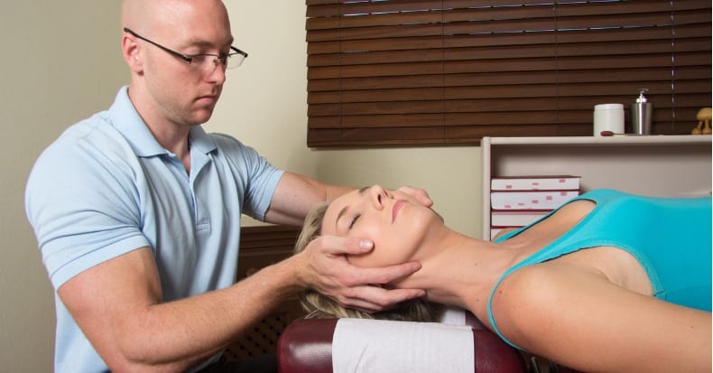
    Chiropractic Applications

    Janet Travell, MD, is one of the most recognized physicians of modern history. Her notoriety is attributed primarily to two events:

    • Myofascial problems and its consequent symptoms are nearly a universal human experience. Dr. Travell was an early pioneer in understanding the science, pathophysiology, and treatment of myofascial problems. She has been referred to as “the mother of myofascial trigger point knowledge”(1).
    • President John F. Kennedy suffered from a notorious bad back. His back problems significantly worsened by his legendary heroism in August of 1943 during the sinking of his boat PT-109 in the Pacific during WWII. Injured (he was awarded the Purple Heart for the event) Kennedy twice swam for miles in the Pacific Ocean, towing an injured crewmember with a life jacket strap in his teeth. Kennedy’s back problems never fully recovered. In 1954, Kennedy underwent an attempted spinal fusion operation, and it went badly; it was his second spinal surgery for his persistent low back pain. He nearly died, and his recovery took 8 months. The following year, Kennedy came under the care of myofascial pain expert Janet Travell, MD. When Kennedy was elected president of the United States (taking office in 1961), he chose Dr. Travell to be his personal White House Physician. Dr. Travell was the first female physician to hold this prestigious office (2, 3, 4).

    Dr. Janet Travell was born in New York in 1901, the daughter of a physician. She died in 1997 at the age of 96. In 1926, she received her MD degree from the Cornell University Medical College in New York City, where she graduated at the head of her class. Interestingly, she was the first female to graduate from Cornell. In the first three decades of her professional career she practiced cardiology while teaching pharmacology at Cornell. Her interest in musculoskeletal pain came about as a consequence of the neck, trunk, shoulder, and arm pains her cardiac patients suffered (5).

    In 1942, 1946, and 1948, Dr. Travell and colleagues reported a series of clinical outcomes following trigger point treatment in cardiac patients (6, 7, 8). Her successful outcomes led her to believe that she was treating more than somatic pain; she believed that she was also influencing aspects of the cardiac pain itself: “in patients suffering from the pain of myocardial infarction convinced her and her colleagues that the treatment could stop both non-cardiac pain of muscle origin and true cardiac pain of coronary insufficiency.”(9) This understanding is important because if true, it would mean that trigger point therapy was influencing aspects of the sympathetic nervous system.

    In 2003, Dr. Travell’s colleague and co-author of their books, David Simons, MD, Myofascial pain and dysfunction, the trigger point manual, commented:

    Trigger point therapy “could suppress cardiac arrhythmias. On several occasions, I saw the conversion of atrial fibrillation to normal rhythm when vapocoolant spray was applied over the arrhythmia TrP on the lower-right anterior chest wall; the same effect could be achieved by trigger point pressure release applied to that TrP. For several reasons, it appears likely that many such unexpected influences, to and from TrPs, depend on modulation of the autonomic nervous system, in addition to modulation of the sensory nervous system (referred pain).”

    “The pain patterns of the pectoralis major and pectoralis minor muscles mimic the pain referral patterns of cardiac ischemia.”

    “Symptoms of angina in the absence of demonstrable cardiac disease should be considered as likely due to TrPs.”

    By 1952, Dr. Travell’s clinical interest and practice had moved away from cardiology and she became a specialist in musculoskeletal pain syndromes. With her growing positive reputation and expertise, Senator John F. Kennedy’s orthopedic surgeon asked Dr. Travell to look at his patient’s chronic, disabling back problems (5). Dr. Travell treated Senator Kennedy’s trigger points and addressed a number of mechanical issues that perpetuated these trigger points (discussed below).

    When Dr. Travell first saw Senator Kennedy in May of 1955, he was non-ambulatory. He had suffered from 2 devastating spinal surgeries, yet he continued to suffer from debilitating back spasm and left leg pain. Things were so bad that Senator Kennedy was “questioning his ability to continue his political career.”(10) Dr. Travell treated Senator Kennedy’s trigger points. His improvement was so impressive that Dr. Travell’s daughter wrote (5):

    “Senator Kennedy received so much relief of pain from my mother’s medical treatments that he had ‘new hope for a life free from crutches if not from backache’.”

    In 2003, James Bagg wrote this, pertaining to Dr. Travell (10):

    “Jack Kennedy saw a great many physicians over the course of his short life, but one of them, according to his brother Bobby, enabled Jack to become President of the United States.”

    Kennedy considered Travell to be a medical genius (5). When Kennedy was elected to the presidency of the United States, he asked Dr. Travell to be his personal White House Physician, becoming the first woman and one of the few civilians (non military) to hold that post. After President Kennedy’s assassination in 1963, his successor, President Lyndon B. Johnson, asked Dr. Travell to stay on in the White House as his physician. A year and a half later, she resigned to return to private practice and to the position of Associate Clinical Professor of Medicine at George Washington University. She became Emeritus Clinical Professor of Medicine 1970–1988, and Honorary Clinical Professor of Medicine from 1988 until her death in 1997. Dr. Travell remained professionally active until the end of her life, writing articles, giving lectures, and attending conferences.

    The enduring pinnacle of Dr. Travell’s work occurred during her years at George Washington University. She had teamed up with ex-Air Force Flight Surgeon David G. Simons, MD, to write the most authoritative text pertaining to myofascial pain syndromes and their relationship to trigger points. During their corroboration, Dr. Simons was Clinical Professor in the Department of Physical Medicine and Rehabilitation at the University of California, Irvine. This book is 713 pages in length and details anatomy, neurology, physiology, and treatment of trigger points and myofascial pain syndromes. It was published in 1983, and titled (12):

    Myofascial Pain and Dysfunction:
    The Trigger Point Manual

    A decade later, in 1992, Volume 2 of their book (607 pages) was released, and titled (13):

    Myofascial Pain and Dysfunction:
    The Trigger Point Manual:
    THE LOWER EXTREMITIES

     In 1999, two years after Dr. Travell’s death, the final volume of the text was released (1038 pages), and titled (14):

    Travell & Simons’Myofascial Pain and Dysfunction
    The Trigger Point Manual:
    Volume 1, Upper Half of Body

     The title page of this volume includes these words:

    “Dr. Janet Travell’s genius and medical insight identified in the first edition the clinical picture of individual myofascial pain syndromes and many perpetuating factors. In addition, we were most fortunate to have had the benefit of her advice in preparing some of this edition. She emphasized the importance of including a new chapter that covers the respiratory muscles and supplied unique pearls of clinical wisdom that sprinkle this revision.”

    An overly simplified statement of Dr. Travell’s work is that the trigger point was able to cause muscle shortening, and that muscle shortening would as a rule cause chronic myofascial pain. People so inflicted with these trigger points and subsequent myofascial pain syndromes were chronic because conventional medical treatment did not address the root cause of the problem, the trigger point. The patient’s symptoms and signs would only resolve if the trigger point was successfully addressed.

    Initially, Dr. Travell’s approach to the trigger point problem was to inject it with small amounts of procaine. Later she emphasized less invasive approaches such as the “spray and stretch” technique and “ischemic compression.”

    The spray and stretch technique involved the use of “vapocoolant” while stretching the involved muscle. This cold “neurological distraction” allowed enough stretching of the interdigitation of the contractile proteins of the muscle to abort the trigger point, resulting in long-term muscle relaxation and ending the chronic pain cycle.

    Ischemic compression on the trigger point (sustained pressure for a minute or two) would deprive the trigger point of the oxygen required to maintain the local contraction. The contracted fibers of the trigger point would “give up”, allowing the entire muscle to relax, again resulting in long-term muscle relaxation and ending the chronic pain cycle.

    These newer trigger point ending techniques were less invasive and quickly became very popular with non-allopathic health care providers, especially chiropractors. James Bagg noted (10):

    “Her [Travell’s] contributions are perhaps more widely known and appreciated by chiropractors and massage therapists than by her fellow physicians.”

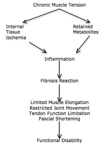
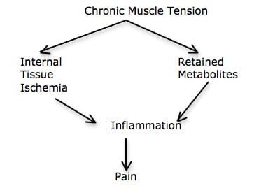
    Dr. Travell notes that not only are the trigger points generators of pain, but she agrees with the writings of Rene Cailliet, MD (18); both contend that a chronically contracted muscle will cut off its own blood supply, resulting in both internal ischemia and an accumulation of metabolites. Muscle ischemia and retained metabolites create an inflammatory reaction, altering the nociceptive threshold, generating pain. Release of the trigger point and the muscle tightness reverses this process.

    •••••••••

    The Contributions of C. Chan Gunn, MD

    1. Chan Gunn, MD, is a Clinical Professor, Multidisciplinary Pain Center at the University of Washington Medical School, Seattle, WA. In his 1996 book titled (11):

    The Gunn Approach to the Treatment of Chronic Pain:
    Intramuscular Stimulation for Myofascial Pain of Radiculopthic Origin

    In this book, Dr. Gunn added to Travell’s work on the understanding and treatment of trigger points and myofascial pain syndromes. In agreement with Travell he notes that myofascial pain syndromes cause musculoskeletal pain. He states:

    “A crucial ingredient of myofascial pain is muscle shortening from contractures. In fact, myofascial pain does not exist without [muscle] shortening.”

    “Chronic pain is never present without associated muscle shortening from contracture.”

    “Prolonged muscle shortening can not only cause pain in muscle, it also mechanically pulls on tendons, thereby straining them and distressing the joints they act on. The increased wear and tear in joints eventually leads to degenerative changes (e.g. osteoarthritis).”

    Muscle shortening can cause a large variety of pain syndromes by its relentless pull on various structures.

    Muscle shortening is the key to myofascial pain. “Stated differently, pain cannot exist in the absence of muscle shortening—no shortening, no pain.”

    “Muscle shortening is a fundamental feature of musculoskeletal pain.”

    “Muscle shortening is an inherent component of persistent musculoskeletal pain, and its release is central to treatment.”

    “An important source of pain in musculoskeletal pain syndromes is from muscle shortening that mechanically stresses muscle attachments, causing conditions such as bicipital tendonitis and lateral epicondylitis.”

    “Shortening in muscles acting across a joint can upset alignment, and can precipitate pain in the joint, i.e. arthralgia.”

    “Muscle shortening can eventually bring about joint degeneration—osteoarthritis.”

    “Shortening of paraspinal muscles acting across a disc space compresses the disc and can cause narrowing of the intervertebral foramina, indirectly irritating the nerve root.”

    “The increased mechanical tension that shortened muscles generates hastens wear and tear because it pulls on degraded collagen that provides the strength of ligaments, tendons, cartilage, and bone. Neuropathy [radiculopathy] expedites degeneration in weight-bearing and activity—stressed parts of the body, causing spondylosis, discogenic disease, and osteoarthritis among others.”

    The primary treatment goal is to “relieve shortening” in muscles that irritate the nerve root and perpetuates the pain.

    “Invariably, when muscle shortening is relieved, pain, whether in muscle, tendon, or joint, is alleviated.”

    Dr. Gunn’s contribution to the understanding of trigger points and myofascial pain syndromes are important. For example, it is known that the tissue primarily responsible for chronic low back pain is the annulus of the intervertebral disc (15). The tissue primarily responsible for chronic neck pain is the facet joint capsules (16, 17). Dr. Gunn explains how muscle shortening can place pain-producing mechanical stress on joints, capsules, tendons, etc., and how relieving muscle shortening reduces these stresses and their genesis of pain. Thus, trigger point/myofascial therapy not only alleviates muscle pain, but also reduces stress on other generators of pain as well.

    Dr. Gunn also adds to the arsenal of ways to effectively address the trigger point and shortened muscle. Much of his book describes the effectiveness of needle acupuncture. Apparently, insertion of the needle into the trigger point will mechanically release it, and in so doing relax the entire muscle.

    Dr. Gunn also talks about the value of low-level laser therapy to abort the trigger point. Physiologically, a muscle needs adenosine triphosphate (ATP) to contract. Ironically, a muscle also needs ATP to relax. As noted above, a contracted muscle compromises its own blood supply, reducing the availability of substrates required to make sufficient levels of ATP to relax. Low-level laser therapy works by improving the flow of electrons in the inner membrane of the mitochondria, sharply increasing levels of ATP. The increased ATP so generated allows contracted muscle fibers to relax, including the trigger point, the taught fibers, and the muscle as a whole. Dr. Gunn notes the following:

    “Low level laser therapy uses low energy lasers in order to achieve therapeutic effects.”

    “LLLT is safe to use: photo-energy emissions are low and treatment is painless, aseptic, and non-thermal.”

    “There are no known side effects. There has been no report of eye injury from low-level laser, although care must be taken not to stare into the probe.”

    “LLLT has anti-inflammatory and analgesic properties; it accelerates the wound healing process.”

    “LLLT is useful in myofascial pain and works well for conditions such as epicondylitis, TMJ dysfunction, pain in the neck, herpes simplex, herpes zoster and post-zoster neuralgia, rotator cuff syndrome (frozen shoulder), carpal tunnel syndrome, acute low back pain, sprain and post-traumatic swelling (hematoma).”

    “Laser therapy, with the benefit of painless healing, is widely used.”

    •••••••••

    The Contributions of David Hubbard, MD

    David R. Hubbard, MD, is from the Department of Neurosciences, University of California, San Diego (my alma mater, before chiropractic college). In 1993 he co-wrote an article in the journal Spinetitled (19):

    Myofascial Trigger Points Show Spontaneous Needle EMG Activity

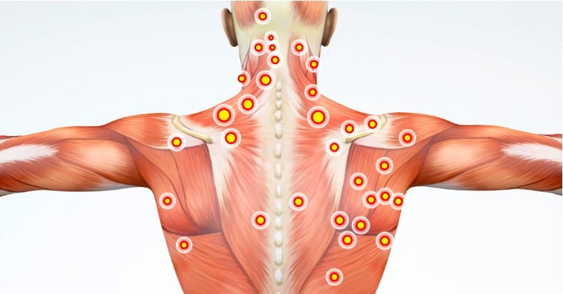
    Dr. Hubbard notes that trigger points are a common cause of chronic muscle pain. He credits Janet Travell, MD, as the person who first described the trigger pointin 1942, in the Journal of the American Medical Association (6). The purpose of his article was to determine the underlying pathophysiology of the trigger point.

    Dr. Hubbard and colleague inserted needle EMG leads into the trigger points of 62 people. On all subjects, they noted spontaneous contractual activity of the trigger point muscle. In some of the subjects they left the needle in place for as long as 50 consecutive minutes, noting a constant contraction of the muscle tissues. Needles inserted into non-trigger point muscle tissue showed no spontaneous electrical activity. If the needle was slightly moved away from the trigger point, in any direction, by as little as a millimeter, the spontaneous electrical activity immediately ceased.

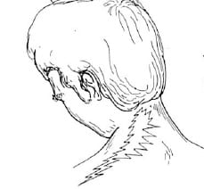
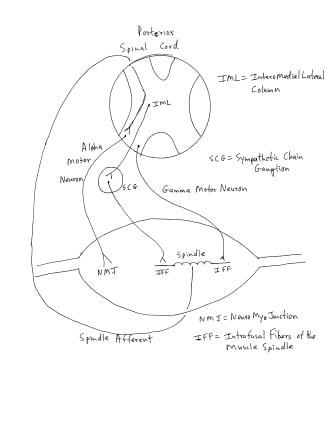
    Dr. Hubbard and colleague explain how the intrafusal muscle fiber of the muscle spindle is innervated by the sympathetic nervous system (see drawing). Segmental or regional increases in sympathetic activity would cause a contraction of the small intrafusal muscle of the muscle spindle, which increases the firing of the muscle spindle afferents. Increased muscle spindle afferents into the spinal cord communicate and excite the alpha motor neuron, causing the muscle to contract and shorten. This contraction increases muscle tone, reduces range of motion, and if prolonged, makes the muscle sore and painful. These authors believe that sympathetically stimulated muscle spindle intrafusal fiber contraction “causes an involuntary low-grade but symptomatic muscle tension.” [see drawing]

    In supporting evidence for their model, these authors note that in animal studies, sympathetic nerve stimulation causes an increase in tonic muscle tension. Cutting the sympathetic nerves, or administrating sympathetic nerve blocking drugs, immediately inhibits the increase in tonic muscle tension. Dr. Hubbard and colleague state:

    “Sympathetic activity explains the autonomic symptoms associated with trigger points and provides a mechanism by which local injury and nociception causes local tension, and by which emotional factors cause widespread tension and pain.”

    In short, these authors believe that the trigger point isthe sympathetically contracted intrafusal muscle fibers of the muscle spindle.

    It is well established that chiropractic spinal adjusting inhibits segmental/regional increases in sympathetic discharge (20, 21, 22, 23). This is a plausible explanation for the observed improvement in trigger points and myofascial problems following chiropractic spinal adjusting. Chiropractic adjusting, in part, inhibits sympathetic tone, relaxes the intrafusal muscle fibers of the muscle spindle, reduces spindle afferent discharge, reduces muscle tone, improves the range of motion, reducing inflammation and pain.

    •••••••••

    Perpetuating Factors Back To Dr. Travell

    Despite the biological plausibility of her trigger point model, Dr. Travell ran into difficult cases. As stated by her friend, colleague, and co-author Dr. David Simons, Dr. Travell “looked under every physical and medical stone imaginable until she found why that patient had failed to respond to treatment as expected.”(9) She discovered that patients often had one or more of “perpetuating factors.” These perpetuating factors were usually structural inadequacies. The two most common were:

    1) A difference in the length of the lower limbs.

    2) A long second metatarsal or a short first metatarsal (Morton’s Toe).

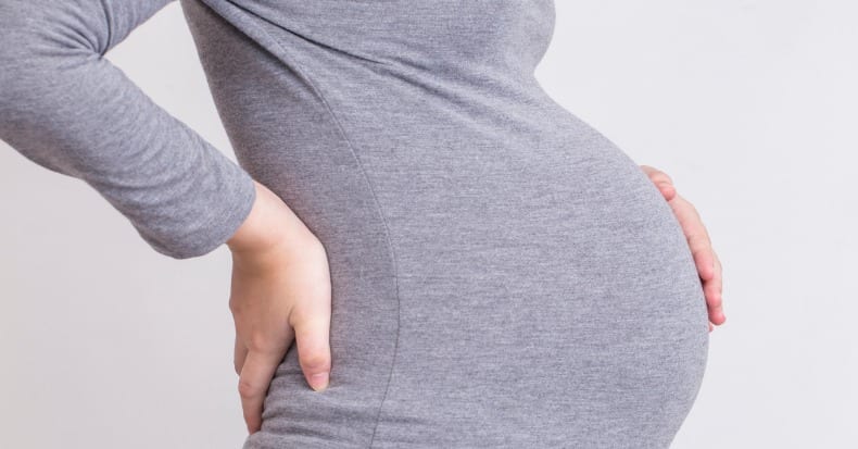
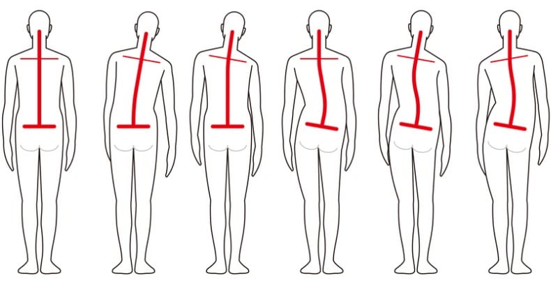
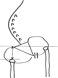
    It has been documented since 1946 that about 75% of people have legs of unequal lengths, and that about a third of people have leg length differences that can perpetuate trigger points. As a rule, the sacrum is lower on the side of the short leg (see drawing). The spinal column initially tilts towards the short leg, then compensates back to the midline as a consequence of chronic contraction of the quatratus lumborum muscle. According to Dr. Travell, the resulting trigger points in the quadratus lumborum muscle is a very common but frequently overlooked cause of chronic low back pain (13).

    The Morton’s Toe was described by American orthopedic surgeon Dudley J. Morton, MD, from Yale, in 1927 (25). Dr. Morton noted that people suffered from a variety of chronic pain syndromes when they had a long second metatarsal or a short first metatarsal (which he termed Morton’s Toe). This common anatomic variant would alter the normal weight-bearing function of the foot, causing a compensatory pronation. Travell observed that this would lower the pelvis on that side, creating the identical trigger points as a leg length inequality.

    Dr. Travell notes that the solution is a proper heal lift and an shoe orthotic that re-establishes foot mechanics and weight-bearing, usually by correcting the pronation. She notes that other causes of foot pronation can and should be similarly addressed with an orthotic.

    Dr. Travell also “discovered that one of [Senator] Kennedy’s legs was shorter than the other and made heel lifts for all of his left shoes to counter that additional source of stress on his back.” “Dr. Travell had a workbench in her office and made lifts for both patients and family members. ‘One of the first things I did for him [Kennedy] was to institute a heel lift—a correction for the difference in leg length’.”(10)

    References

    • pain-education.com/dr-travell.html‎; A Tribute to Dr. Janet Travell; accessed December 11, 2013.
    • Lacayo R; How Sick Was J.F.K.?; TIME; November24, 2002.
    • Altman LK, Purdum TS; In J.F.K. File, Hidden Illness, Pain and Pills; The New York Times; November 17, 2002.
    • Dallek R; The Medical Ordeals of JFK; The Atlantic; December 2002.
    • Wilson V; Janet G. Travell, MD: A Daughter’s Recollection; Texas Heart Institute Journal; 2003; Vol. 30; No. 1; pp. 8–12.
    • Travell J, Rinzler S, Herman M; Pain and disability of the Shoulder and Arm: Treatment by Intramuscular Infiltration with Procaine Hydrochloride; Journal of the American Medical Association; October 10,1942; Vol. 120; No. 6; pp. 417-422.
    • Travell J, Rinzler SH; Relief of cardiac pain by local block of somatic trigger areas. Proc Soc Exp Biol Med 1946;63:480–2.
    • Rinzler SH, Travell JG. Therapy directed at the somatic component of cardiac pain. Am Heart J 1948;35:248–68.
    • Simons DG; Cardiology and Myofascial Trigger Points: Janet G. Travell’s Contribution; Texas Heart Institute Journal; 2003; 30; No. 1; pp. 3–7.
    • Bagg JE; The President’s Physician; Texas Heart Institute Journal; 2003; 30; No. 1pp. 1–2.
    • Gunn CC; The Gunn Approach to the Treatment of Chronic Pain:Intramuscular Stimulation for Myofascial Pain of Radiculopthic Origin; Churchill Livingston, 1996.
    • Travell J, Simons D; Myofascial pain and dysfunction, the trigger point manual; New York: Williams & Wilkins, 1983.
    • Travell J, Simons D; Myofascial pain and dysfunction, the trigger point manual: THE LOWER EXTREMITIES; New York: Williams & Wilkins, 1992.
    • Simons D, Travell J; Travell & Simons’,Myofascial pain and dysfunction, the trigger point manual: Volume 1, Upper Half of Body; Baltimore: Williams & Wilkins, 1999.
    • Kuslich S, Ulstrom C, Michael C; The Tissue Origin of Low Back Pain and Sciatica: A Report of Pain Response to Tissue Stimulation During Operations on the Lumbar Spine Using Local Anesthesia; Orthopedic Clinics of North America, Vol. 22, No. 2, April 1991, pp. 181-187.
    • Bogduk N, Aprill C; On the nature of neck pain, discography and cervical zygapophysial joint blocks; Pain; August 1993;54(2):213-7.
    • Bogduk N; On Cervical Zygapophysial Joint Pain After Whiplash; Spine; December 1, 2011; Volume 36, Number 25S, pp S194–S199.
    • Cailliet R; Soft Tissue Pain and Disability; 3rdEdition; F A Davis Company, 1996.
    • Hubbard DR, Berkoff GM; Myofascial Trigger Points Show Spontaneous Needle EMG Activity; Spine; October 1993; Vol. 18; No. 13; pp. 1803-1807.
    • Korr IM; The spinal cord as organizer of disease processes: III. Hyperactivity of sympathetic innervation as a common factor in disease; December 1979;79(4):232-237.
    • Budgell B, Hirano F; Innocuous mechanical stimulation of the neck and alterations in heart-rate variability in healthy young adults; Autonomic Neuroscience; 2001 Aug 13;91(1-2):96-99.
    • Koch LE, Koch H, Graumann-Brunt S, Stolle D, Ramirez JM; Saternus KS; Heart rate changes in response to mild mechanical irritation of the high cervical spinal cord region in infants; Forensic Science International; Volume 128, Issue 3, August 28, 2002, Pages 168-176.
    • Ogura T, Tashiro T, Masud M, et. al; Cerebral metabolic changes in men after chiropractic spinal manipulation for neck pain; Alternative Therapies Health Medicine; Nov-Dec 2011;Vol. 17; No. 6; pp. 12-7.
    • Rush WA, Steiner HA; A Study of Lower Extremity Length Inequality; American Journal of Roentgenology and Radium Therapy; Vol. 51, No. 5, November 1946, 616-623.
    • Morton DJ; METATARSUS ATAVICUS: The Identification of a Distinctive Type of FootDisorder;J Bone Joint Surg Am, 1927 Jul 01;9(3):531-544.
  • Chiropractic Adjusting for Cervical Disc Herniation and Compressive Neuropathology

    Chiropractic Adjusting for Cervical Disc Herniation and Compressive Neuropathology

    The most common cause of cervical nerve root compression (compressive neuropathology) is degenerative joint disease with stenosis and narrowing of the intervertebral foramen narrowing. The second most common cause is cervical disc herniation (1).

    Cervical disc herniations with compressive neuropathology are commonly seen in asymptomatic populations (2, 3, 4). In a study published in 1996, 63% of asymptomatic men who were older than 40 years of age had protruding disks in the cervical spine (2). A study published in 1998 found demonstrable spinal cord compression secondary to cervical disc protrusion in 7.6% of asymptomatic subjects over the age of 50 years (3). However, it is rare for individuals to be asymptomatic with extruded disk herniations and spinal cord compression (4). This would mean that every day thousands of chiropractors are treating hundreds of thousands of patients with asymptomatic cervical spine disk herniations. It must be assumed that in a large percentage of these patients the chiropractor is performing high-velocity low-amplitude spinal adjusting at or adjacent to the level of the disk herniation.

    Additionally, chiropractors routinely diagnose and treat cervical disk herniations on patients using a variety of approaches, procedures and techniques. Often, the chiropractor will utilize high-velocity low-amplitude spinal adjusting. Considering this, it is important to note that chiropractic spinal adjusting is extremely safe. The largest prospective study do date assessing the safety of chiropractic spinal manipulation to the cervical spine was published in Spine, October 2007, and titled (5):

    Safety of Chiropractic Manipulation of the Cervical Spine
    A Prospective National Survey

    This study assessed the risk of serious and relatively minor adverse events following chiropractic manipulation of the cervical spine by a sample of United Kingdom chiropractors. The authors studied treatment outcomes from 19,722 patients from 377 chiropractors that involved 50,276 cervical spine manipulations. Manipulation was defined as the application of a high-velocity/low-amplitude or mechanically assisted thrust to the cervical spine. A serious adverse event was defined as referred to hospital and/or severe onset/worsening of symptoms immediately after treatment and/or resulted in persistent or significant disability/incapacity, and minor adverse events reported by patients as a worsening of presenting symptoms or onset of new symptoms, were recorded immediately, and up to 7 days, after treatment. The authors concluded:

    “Safety of treatment interventions is best established with prospective surveys, and this study is unique in that it is the only prospective survey on such a large scale specifically estimating serious adverse events following cervical spine manipulation.”

    “There were no reports of serious adverse events.”

    “Although minor side effects following cervical spine manipulation were relatively common, the risk of a serious adverse event, immediately or up to 7 days after treatment, was low to very low.”

    “No significant adverse event was reported by the chiropractors using the definition criteria.”

    “The risk rates described in this study compare favorably to those linked to drugs routinely prescribed for musculoskeletal conditions in general practice.”

    “The risks reported here are also lower than those reported for acupuncture, which were described as a very safe intervention in the hands of a competent practitioner.”

    “Although minor side effects were found to be relatively common, the risk of a serious adverse event, immediately and up to 7 days after treatment, was estimated to be low to very low in these consultations.”

    “On this basis, this survey provides evidence that cervical spine manipulation is a relatively safe procedure when administered by registered United Kingdom chiropractors.”

    Epidemiological studies show that the most common cervical disk herniations are C5-C6 (affecting the C6 nerve root) and the C6-C7 (affecting the C7 nerve root) levels (6). [Remember, since the C1 nerve root exits between the occiput and the atlas, there are 8 cervical nerve roots. This is why the C6 nerve roots exits between C5-C6 vertebrae, and the C7 nerve root exits between C6-C7 vertebrae].

    •••••••••

    Diagnosing Cervical Disk Herniation with Compressive Nerve Root Pathology Clinical Signs and Symptoms Diagnostic Imaging

    Acknowledging that cervical disk herniation can be asymptomatic, even in the presence of apparent imaging of compressive nerve root neuropathology, classic clinical presentation would include combinations of the followings:

    • Neck pain.
    • Arm pain, usually extending below the elbow.
    • Alterations of superficial sensation in a dermatomal pattern. This can be either hypoesthesia or hyper I often find that in acute situations there is hyperesthesia. In the subacute and/or chronic situation, there is hypoesthesia. According to Stanley Hoppenfeld, MD [Associate Clinical Professor of Orthopaedic Surgery Albert Einstein College of Medicine, New York] (7):
    • C5 nerve root would involve the lateral arm.
    • C6 nerve root would involve the lateral forearm, thumb, and 2nd
    • C7 nerve root would involve the 3rd (middle) phalange.
    • C8 nerve root would involve the 4th (ring) and 5th (little) phalanges.
    • Reduced segmental sensory-motor (deep tendon) reflex (7):
    • C5 nerve root: biceps tendon
    • C6 nerve root: brachioradialis tendon
    • C7 nerve root: triceps tendon
    • C8 nerve root: none
    • Myotomal muscle weakness is the weakness of a muscle associated primarily with a single nerve root. Again, as established by Stanley Hoppenfeld, MD, (7):
    • C5 nerve root: the deltoid muscle; arm abduction
    • C5 nerve root: the biceps muscle, elbow flexion
    • C6 nerve root: wrist extension
    • C7 nerve root: triceps muscle, elbow extension
    • C7 nerve root: wrist flexion, finger extension
    • C8 nerve root: finger flexion
    Shoulder Abduction C5 Deltoid
    Elbow Flexion C5 Biceps
    Elbow Extension C7 Triceps
    Wrist Extension C6 Wrist Extensors
    Wrist Flexion C7 Wrist Flexors
    Finger Extension C7 Finger Extensors
    Finger Flexion C8 Finger Flexors
    • Positive extremity pain exacerbation with cervical spine compression:
      • Neutral position
      • Lateral flexed position
      • Spurling’s test (lateral flexion with ipsilateral rotation [from (13)]):
    • Positive extremity pain with shoulder depression (shoulder depression test) (9).
    • Pain relief with cervical spine distraction.
    • Increased arm pain with the upper limb tension test (arm abducted 90°, elbow flexed 90°, fingers and wrist extended; followed by slowly extending the elbow).
    • Aggravation of neck and/or arm pain with an increase in intrathecal pressure: Valsalva test, sneezing, coughing, and straining at stool.
    • Diagnostic imaging findings, typically an MRI, showing compressive neuropathology at the level consistent with the other motor and sensory findings.

    •••••••••

    Studies on the Non-Pharmacological, Non-Surgical Management of Cervical Disk Herniation

    •••••••••

    In 1996, physicians (and brothers) Joel and Jeffery Saal, published a study in the journal Spine and titled (10):

    Non-operative Management of Herniated Cervical
    Intervertebral Disc With Radiculopathy

    These authors note that frequently, patients with cervical disc herniation and neurologic loss or radiculopathy, are treated surgically if they have symptoms that persists after rest or minimal intervention. They state:

    “For non-validated reasons, cervical disc extrusions have been frequently considered a definite indication for surgery.”

    Consequently, they evaluated the clinical outcomes in 26 patients with cervical herniated nucleus pulposus and radiculopathy who were treated with an aggressive physical rehabilitation program. These patients were followed for longer than 1 year.

    Inclusion criteria for this study included a focal cervical disc protrusion of less than 4 mm identified on magnetic resonance imaging and a major complaint of extremity pain compatible with cervical radiculopathy. Exclusion criteria included severe central canal stenosis and/or symptomatic cervical myelopathy.

    All patients had a predominance of radicular upper extremity pain as their chief complaint. All patients had a magnetic resonance imaging (MRI) showing a cervical herniated nucleus pulposus that extended at least 4 mm from the margin of the parent disc space. Twenty of twenty-six (20/26) patients had extruded cervical discs. Six of the twenty-six (6/26) patients had contained disc herniations.

    The rehabilitation program management consisted of traction, specific physical therapeutic exercise, and patient education. All patients were treated with ice, relative rest, a hard cervical collar worn for up to 2 weeks in a position to maximize arm pain reduction (all patients), manual and mechanical traction in physical therapy, followed by home cervical traction, and progressive strengthening exercises of the shoulder girdle and chest with training in postural control and body mechanics training. The duration of this portion of the program was 3 months, at which time the patient was discharged to an independent exercise program. The non-operative treatment in the patients in the present study averaged 9 months. Forceful joint manipulation was not used.

    Twenty-four patients (92%) were successfully treated without surgery.

    Twenty patients achieved a good or excellent outcome, and of these 19 had disc extrusions. Two patients underwent cervical spine surgery. Twenty-one patients returned to the same job. High patient satisfaction with non-operative care was achieved on outcome analysis.

    The authors concluded:

    “Many cervical disc herniations can be successfully managed with aggressive nonsurgical treatment.”

    “A small percentage of patients with cervical herniated nucleus pulposus do require surgery for radiculopathy. However, the majority can be treated successfully with a carefully applied and progressive non-operative program.”

    “The presence of radicular neurologic loss or nuclear extrusion should not be used solely as the criterion for surgical intervention.”

    A couple of side observations by these authors include noting that inflammation of neural elements appears to play an important role in radiculopathy. Additionally, they state:

    “Reabsorption of extruded disc material itself probably occurs in the cervical HNP as it does in the lumbar disc HNP.” Because of this reabsorption, “an extruded disc actually may have a more favorable non-operative prognosis than contained disc pathology.”

    The most important point made by this article is that herniated cervical discs, even extrusion of the cervical disc, with radiculopathy (motor and sensory signs), can be successfully conservatively managed by a regime that consists primarily of exercise, traction, and mobilization. Another important point is that herniated cervical discs, even extrusion of the cervical disc, with radiculopathy (motor and sensory signs), rarely require surgery, even if they have significant extremity weakness or severe pain longer than 8 weeks. Most importantly, herniated cervical discs, even extrusion of the cervical disc, with radiculopathy (motor and sensory signs), should always be conservatively managed before surgery is warranted.

    •••••••••

    In 1999, physician Brian Nelson and colleagues published a study in the journal Archives of Physical Medicine and Rehabilitation titled (11):

    Can spinal surgery be prevented by aggressive strengthening exercises?

    A prospective study of cervical and lumbar patients

    All 46 of the patients in this study had been recommended for spinal surgery. The objective of this study was to assess if an aggressive strengthening program could help these patients to avoid the surgery. Nine of these patients were diagnosed with “cervical disc syndrome” [distinct from degenerative cervical disc disease].

    The aggressive strengthening program was 10 weeks in duration. It consisted of intensive, progressive resistance exercise of the isolated lumbar or cervical spine. The exercises were continued until fatigue failure, and patients were encouraged to work through their pain. “An important point is that the training was quite vigorous and did not stop because of pain exacerbation.” Patients were encouraged to be vigorous. They were taught, “hurt does not necessarily mean harm.” The average follow-up was at 16 months following discharge.

    None of the cervical patients underwent surgery in the follow-up period.

    “The study is valuable because it shows that a large number of surgical candidates at a private practice clinic can avoid surgery over an extended period. Further, there were no significant complications or negative consequences associated with delaying surgery while patients participated in an aggressive strengthening program. Occasional exacerbations occurred, but these were self limited and did not prevent rehabilitation from continuing.”

    “Surgical candidates are often considered more ‘fragile’ than non-surgical patients and are more often guided toward inactivity to protect the spine. Many have been told to remain inactive based on MRI scans. They develop a keen sense of fear when it comes to spinal motion. Spinal pain patients become expert at substituting pelvic or thoracic movement for lumbar or cervical motion, respectively. In this way they protect the injured body part from meaningful exercise.”

    “Substitution protects the lumbar or cervical spine from normal movement. Without motion the disc deteriorates, disc pH decreases, joints stiffen, ligaments shorten, bone density decreases, and muscles become deconditioned. Recent evidence suggests that a damaged disc becomes more acidic and that reduced pH is a mediator of spinal pain. The adult disc is an avascular structure that depends on diffusion for its nutrition. Diffusion is facilitated by a pumping action through spinal motion. Lack of motion, however, hinders diffusion. In the aggressive strengthening program in this study, patients were not allowed to substitute. The cervical and/or lumbar spine was isolated in such a way that substitution was impossible. Exercise therefore facilitated fluid exchange in the disk, which may account for the subjective improvement.”

    “The significance of this study is that many patients were spared surgery during the study period even though surgery had been recommended. The findings show that a percentage of spinal patients can avoid surgery by completing an aggressive strengthening program and that even patients recommended for spinal surgery can tolerate intensive, specific exercise.”

    Although this study did not involve joint adjusting, there emerged an important physiological concept for chiropractors who are managing disc herniation patients:

    The pain of disc herniation and radiculopathy may be more due to changes in chemistry (inflammation, acidity) than to the actual compression. Understandably, such patients reduce the motion of the injured joints to avoid increased pain perception. However, reduced motion compromises tissue integrity and enhances the pathology. Without motion, the disc further deteriorates because of lack of nutrition though diffusion mechanisms, becoming more acidic and more painful.

    Theoretically, any treatment that restores motion to the motor segment would improve diffusion, improve disc chemistry, and reduce pain while improving function. In this study, intense isolated spinal exercises were able to accomplish this goal. Is there another approach to restoring spinal biomechanical motion that is both effective and safe?

    •••••••••

    Cynthia Peterson, DC, and colleagues, from the University of

    Zurich, Switzerland, published an important study last month (October 2013) pertaining to the use of chiropractic High-Velocity Low-Amplitude Spinal Manipulation in patients with cervical disc herniations. Their article was published in the Journal of Manipulative and Therapeutics, and titled (12):

    Outcomes From Magnetic Resonance Imaging:
    Confirmed Symptomatic Cervical Disk Herniation Patients Treated With High-Velocity, Low-Amplitude Spinal Manipulation Therapy:
    A Prospective Cohort Study With 3-Month Follow-Up

    Dr. Peterson is a Professor, Department of Chiropractic Medicine, Faculty of Medicine, Orthopedic University Hospital Balgrist, University of

    Zurich, Switzerland. The purpose of her study was to investigate outcomes of patients with cervical radiculopathy from cervical disk herniation (CDH) who were treated with spinal manipulative therapy.

    This study used 50 patients with a mean age of 44 years. Chiropractic treatment frequency was 3 to 5 times per week for the first 2 to 4 weeks and then 1 to 3 times per week thereafter until the patient was asymptomatic. Patients were evaluated at baseline, 2-weeks, 1-month, and 3-months.

    The patients in this study had neck pain and moderate to severe arm pain in a dermatomal pattern with sensory, motor, or reflex changes corresponding to the involved nerve root. Patients also had at least one of the following positive orthopedic tests for cervical radiculopathy:

    • Positive upper limb tension test
    • Positive cervical distraction test
    • Positive Spurling’s test
    • Positive cervical rotation test at less than 60°

    Finally, all of these patients had a magnetic resonance imaging–proven cervical disc herniation at the corresponding spinal segment.

    The measurement outcomes used to assess these patients included:

    • The Numeric rating scales (NRS) for pain where 0 is no pain and 10 is the worst pain imaginable for both the neck and the arm pain separately.
    • The Neck Disability Index (NDI).

    The chiropractic spinal adjustment given to these patients was described as follows:

    “High-velocity, low-amplitude spinal manipulations were administered by experienced doctors of chiropractic.”

    “The treatment procedure was a standardized, single, high-velocity, low-amplitude cervical manipulation with rotation to the opposite side and lateral flexion to the same side of the affected arm.”

    “The chiropractor stood on the affected side of the supine patient’s neck, with an index contact on the articular pillar of the most symptomatic vertebral motion segment on the side of the patient’s complaint and at the spinal level clinically assessed to correspond with the MRI findings.”

    “Rotation to the opposite and lateral flexion to the ipsilateral side was used to take out skin and joint slack.”

    “Once the patient was positioned, a high-velocity, low-amplitude thrust was applied, with the goal of moving the affected segment and producing an audible release.” An audible release was achieved in most case.

    “In the rare case where an audible release did not occur during the procedure, the chiropractor might repeat the manipulation up to 2 additional times.”

    “When a patient reported bilateral neck and/or arm pain (extremely rare), the procedure could be reproduced on the opposite side as well.”

    The measured outcomes for these patients were noted as follows:

    “By 2 weeks after the first treatment, 55.3% of all patients reported that they were significantly improved and none reported being worse.”

    “At 1 month, 68.9% were significantly improved.”

    By 3 months 85.7% were significantly improved with no patients being worse.

    “Statistically significant decreases in neck pain, arm pain, and NDI scores were noted at 1 and 3 months compared with baseline scores.”

    For the subacute/chronic patients, the mean duration of symptoms was 299 days. At 3 months, 76.2% of these patients reported clinically relevant improvement with no patients reporting that they were worse.These authors make these following comments:

    “Most patients in this study, including subacute/chronic patients, with symptomatic magnetic resonance imaging–confirmed cervical disc herniation treated with spinal manipulative therapy, reported significant improvement with no adverse events.”

    “Most patients in this study with MRI-proven symptomatic cervical disc herniations who were treated with high-velocity, low-amplitude spinal manipulation reported clinically significant improvement at all time points, particularly at 3 months.”

    “It is important to point out that even the subacute/chronic patients in this study with symptoms lasting longer than 4 weeks (mean duration, 298.73 days) reported high levels of clinically significant improvement. This is clinically important as the chronic patients are the ones who are usually the most costly in terms of health care use and quality-of-life disruption.”

    Although subacute/chronic patients responded extremely well in this study, overall acute patients showed faster and greater improvement.

    •••••••••

    Cervical disc herniations are commonplace in asymptomatic populations. All chiropractors occasionally see patients with cervical disc herniation and clinically correlated compressive neuropathology. Effective and safe management of these patients includes traction, mobilization, stabilization exercises, aggressive strengthening exercise program, and spinal adjusting at the level and side of the herniation. These studies suggest that the patient’s pain and disability are linked to local chemical changes, which themselves are linked to fear avoidance and biomechanical reductions of motion. Consequently, clinical improvement is dependent upon the restoration of movement, and specific chiropractic adjustments are safe and effective in this regard.

    References

    • Radhakrishnan K; Litchy WJ; O’Fallon WM; Kurland LT; Kurland LT; Epidemiology of cervical radiculopathy; A population-based study from Rochester, Minnesota, 1976 through 1990; Brain 1994;117; pp. 325-35.
    • Healy JF, Healy BB, Wong WHM, Olson EM; Cervical and lumbar MRI in asymptomatic older male lifelong athletes: frequency of degenerative findings; Journal of Computer Assisted Tomography;1996; Vol. 20; pp. 107-112.
    • Matsumoto M, Fujimura Y, Suzuki N, et al; MRI of cervical intervertebral discs in asymptomatic subjects; Journal of Bone and Joint Surgery (British); 1998; Vol. 80-B; pp. 19-24.
    • Ernst CW, Stadnik TW, Peeters E, Breucq C, Osteaux MJC; Prevalence of annular tears and disc herniations on MR images of the cervical spine in symptom free volunteers; European Journal of Radiology; 2005; Vol. 55; pp. 409-414.
    • Thiel HW, Bolton JE, Docherty S, Portlock JC. Safety of chiropractic manipulation of the cervical spine. A prospective national survey. Spine; 2007; Vol. 32; pp. 2375-2378.
    • Radhakrishnan K, Litchy WJ, O’Fallon WM, Kurland LT, Kurland LT. Epidemiology of cervical radiculopathy. A population-based study from Rochester, Minnesota, 1976 through 1990. Brain; 1994; Vol. 117; pp. 325-335.
    • Hoppenfeld S; Orthopaedic Neurology: A Diagnostic Guide to Neurologic Levels; Lippincott, 1977.
    • White AA, Panjabi MM; Clinical Biomechanics of the Spine; Lippincott; 1990.
    • Jackson R; The Cervical Syndrome; Thomas; 1978.
    • Saal, Joel S. MD; Saal, Jeffrey A. MD; Yurth, Elizabeth F. MD; Nonoperative Management of Herniated Cervical Intervertebral Disc With Radiculopathy; Spine; Volume 21(16) August 15, 1996, pp. 1877-1883.
    • Brian W Nelson, David M Carpenter, Thomas E Dreisinger, Michelle Mitchell, Charles E Kelly, Joseph A Wegner; Can spinal surgery be prevented by aggressive strengthening exercises? A prospective study of cervical and lumbar patients; Archives of Physical Medicine and Rehabilitation; January 1, 1999; Vol. 80; No. 1; pp. 20-25.
    • Peterson CK; Schmid C; Leemann S; Anklin B; Humphreys BK; Outcomes From Magnetic Resonance Imaging: Confirmed Symptomatic Cervical Disk Herniation Patients Treated With High-Velocity, Low-Amplitude Spinal Manipulation Therapy: A Prospective Cohort Study With 3-Month Follow-Up; Journal of Manipulative and Therapeutics; October 2013; Vol. 36; pp. 461-467.
    • White AA, Panjabi MM; Clinical Biomechanics of the Spine; 2nd Edition; Lippincott; 1990; p. 410.


  • Radiography, Chiropractors and X-rays, Pathology, Degeneration, Anomalies, and Biomechanics

    Radiography, Chiropractors and X-rays, Pathology, Degeneration, Anomalies, and Biomechanics

    The birth of chiropractic as a profession occurred in the year 1895 when Daniel David Palmer (1845-1913) adjusted the spine of a deaf man, William Harvey Lillard. Daniel David Palmer established the Palmer School in Davenport, Iowa, in 1896.

    Wilhelm Conrad Rontgen was a German physicist (1845-1923). In 1895, at age 50, he produced and detected x-rays (Rontgen rays). For that achievement, he was awarded the very first Nobel Prize in Physics in 1901. A few weeks after his discovery, he produced the first “x-ray picture” by applying this new technology to the bones of his wife’s hand. The following month he published the first paper on his new discovery, titled “On a New Kind of Rays.” This is considered to be the birth of diagnostic radiology.

    Bartlett Joshua Palmer (1882-1961) was the son of Daniel David Palmer. Bartlett Joshua Palmer took over the Palmer School in 1906. In 1910, Bartlett Joshua (BJ) Palmer began to apply x-rays for taking images of the spine. These were some of the first spinal x-rays ever taken. BJ Palmer called the exposing of x-rays of the spine spinography (1).

    Chiropractic and X-rays were born in the same year, 1895, by 50-year-old men who were born in 1845. The new technology of x-rays was quickly embraced by chiropractic, primarily for the purpose of imaging the spine. The relationship between chiropractic and x-rays continues through today.

    •••••••••

    Today’s chiropractor has extensive undergraduate education in radiology, and post-graduate certification in radiology is available. Most field practitioner chiropractors frequently attend continuing education classes in radiology. Today, chiropractors take x-rays on patients for a variety of reasons:

    • Pathology: This includes entities such as fracture, infection, malignancy, and benign tumors. In my clinical practice, I have taken x-rays on patients and diagnosed such entities as rib fractures, dens fracture, Hangman’s fracture, Jefferson’s fracture, uncinate process fracture, spinous/transverse process fracture, vertebral body fracture, cervical spine pillar fracture, spondylolysis with spondylolisthesis; osteogenic sarcoma, multiple myeloma, a few metastatic cancers; osteoid osteoma, giant cell tumor; osteomyelitis; etc.
    • Metabolic: This includes diagnosis such as rheumatoid arthritis, ankylosing spondylitis, Otto’s pelvis.
    • Developmental: Examples would include slipped capital femoral epiphysis, congenital hip dislocation, Legg-Calves-Perth’s disease, etc.
    • Degeneration: This would include disc disease, facet arthrosis, spondylosis, central canal stenosis, lateral recess stenosis, etc.
    • Anomalies: This would include block vertebrae, hemi vertebrae, demi vertebrae, Klippel-Feil syndrome, cervical ribs, os odontoideum, lumbosacral transitional segments (sacralization, lumbarization), facet tropism, dysplasia, agenesis, spina bifida, etc.
    • Biomechanics: This would include segmental malpositions, postural distortions, leg length inequality, scoliosis, ligamentous instabilities (stress radiography), etc. Many chiropractors base their adjustive line-of-drive on spinal biomechanical measurements.

    •••••••••

    In 1983, Stephen Kovach and Eldon Huslig published a study in the Journal of Manipulative and Physiological Therapeutics, titled (2):

    Prevalence of diagnoses on the basis of radiographic evaluation of chiropractic cases

    In this study they show the results of a review of all the radiographic examinations performed at the National College of Chiropractic Clinic during the 1982 calendar year. They show how these radiographs helped in the diagnosis of musculoskeletal, cardiopulmonary, or abdominal syndromes. They state:

    “The use of plain film radiography has long been a staple of the chiropractic profession. Radiographic examinations are a valuable tool in the chiropractic diagnosis of a patient’s condition.”

    •••••••••

    The following year, 1984, Kovach and Huslig published another study in the Journal of Manipulative and Physiological Therapeutics, titled (3):

    Shoulder pain and Pancoast tumor: A diagnostic dilemma

    In this study, the authors describe a case of shoulder pain radiating into the arm and ulnar side of the hand. Cervical spine radiographs were exposed. A careful evaluation showed a Pancoast tumor in the apical region of the lung. The Pancoast tumor is a malignant tumor. The authors note that radiography was necessary to acquire an accurate diagnosis and an appropriate referral.

    •••••••••

    In 1992, Owens published a study in the Journal of Manipulative and Physiological Therapeutics, titled (4):

    Line drawing analyses of static cervical X-ray used in chiropractic

    In this review article, Owens identifies the chiropractic techniques used to assess the structure of the cervical spine as seen on the static cervical radiograph. His data was accumulated from a search of MEDLINE using key words “radiograph and X ray in combination with cervical spine (vertebrae)”; the Chiropractic Research Archives Collection; indexes published in the Journal of Chiropractic Research; conference proceedings from the Annual Biomechanics Conference of the Spine, FCER sponsored conferences and the Annual Upper Cervical Spine Conference; references identified from bibliographies of pertinent articles; and a telephone poll of radiography/technique instructors at chiropractic colleges. He specifically looked at techniques that quantitatively assess relative alignment of skeletal structures or distortion of the spinal column, and the techniques were assessed for their reliability and validity.

    “Reliability studies exist showing that inter- and intra-examiner reliability are sufficient to measure lateral and rotational displacements of C1 to within +/- 1 degree. This amount of error allows objective analysis of upper cervical x-rays to detect changes in the angular positional relationships of radiographic images on the order of those already seen clinically. Methods of cervical analysis that use relative angular measures of skeletal positioning are best able to control the effects of radiographic distortion.”

    This study supports the utilization of upper cervical spine radiographs to determine the measureable biomechanics of atlas lateral and rotational displacements.

    •••••••••

    In 1995, John Taylor and colleagues from the Department of Radiology, University of California, Medical Center, San Diego, published a study in the journal Spine, titled (5):

    Interpretation of abnormal lumbosacral spine radiographs:

    A test comparing students, clinicians, radiology residents, and radiologists in medicine and chiropractic

    This study was a controlled comparison of radiographic interpretive performance based on training and experience by these different disciplines and students. The authors tested their abilities to interpret abnormal plain film radiographs of the lumbosacral spine and pelvis. This study is the first to compare radiographic interpretations of students, clinicians, radiology residents, and radiologists.

    The authors used 19 sets of radiographs with clinically important radiographic findings and had them interpreted by 496 volunteers, as follows:

    • 183 chiropractic students
    • 66 medical clinicians
      • 12 general practice physicians
      • 25 orthopedic surgeons
      • 21 orthopedic residents
      • 8 rheumatologists
    • 55 chiropractic radiologists
    • 48 general medical radiologists
    • 46 chiropractic clinicians
    • 36 skeletal radiologists and fellows
    • 27 medical radiology residents
    • medical students
    • 13 chiropractic radiology residents

    Percentage of Correct Diagnosis by Group

    Group Percent Accuracy
    Chiropractic radiologists 71.00%
    Skeletal radiologists and fellows 70.18%
    Chiropractic radiology residents 61.54%
    General medical radiologists 51.64%
    Medical radiology residents 44.64%
    Medical clinicians 31.26%
    Chiropractic clinicians 28.38%
    Chiropractic students 20.45%
    Medical students 5.74%

    The authors discovered that in the interpretation of abnormal plain film radiographs of the lumbosacral spine and pelvis, significant differences were found among professional groups. Yet, the study also clearly showed that chiropractic radiologists, chiropractic radiology residents, chiropractic clinicians, and chiropractic students have comparative ability to interpret pathology of lumbar spine/pelvis radiographs as do their medical counterpart group.

    •••••••••

    In 1997, Assendelft and colleagues from the Institute for Research in Extramural Medicine, Amsterdam, published a study in the journal Spine, titled (6):

    Reliability of lumbar spine radiograph reading by chiropractors

    This study investigated the variability in the interpretation of lumbar spine radiographs by chiropractors working in private practice. The authors used 4 chiropractors to read 100-blinded sets of standard, upright antero-posterior and lateral lumbar radiographs.

    The authors found that the interobserver and intraobserver agreement to be “fair to good,” which they judged to be “acceptable.”

    •••••••••

    Also in 1997, Harger and colleagues from the Department of Radiology, Western States Chiropractic College, Portland, Oregon, published a study in the Journal of Manipulative and Physiological Therapeutics, titled (7):

    Chiropractic radiologists:
    A survey of chiropractors’ attitudes and patterns of use

    The objective of this study was to assess the chiropractic use of radiography, referral patterns to both medical and chiropractic radiologists and attitudes toward radiologists by 197 practicing U.S. chiropractors. Seventy-four percent of the respondents have radiographic facilities in their offices. The results gave the following reasons for taking radiographs:

    71% To screen for contraindication to chiropractic care.

    63% To assess existence of pathological conditions.

    51% To observe/measure altered biomechanics and posture.

    27% For medicolegal protection.

    84% of the participating chiropractors refer to medical radiologists and/or to chiropractic radiologists for a formal interpretation of their radiographs.

    85% of the participating chiropractors felt that chiropractic radiologists are as well qualified as medical radiologists in interpreting their radiographs.

    •••••••••

    The Chiropractic Biophysics, Inc., group prides itself on precision radiographic postural analysis of patients, both prior to and after following a protocol of structural rehabilitation. Starting in the 1990s they performed a series of studies whose objective was to establish the reliability and validity of their postural radiological measurements. Their blinded radiological analysis was assessed with the help of the Department of Statistics, Temple University, Philadelphia, PA. They were able to prove both the reliability and validity of their x-ray measurement systems for postural analysis (8, 9, 10, 11, 12).

    •••••••••

    In 2002, an important study was published in the journal Spine, titled (13):

    Reliability and validity of lumbosacral spine radiograph reading by chiropractors, chiropractic radiologists, and medical radiologists

    The authors were from the Department of Radiology, Medical Center Alkmaar, Alkmaar, The Netherlands. Their design was a cross-sectional diagnostic study. Their objective was to determine and compare the reliability and validity of contraindications to chiropractic treatment (infections, malignancies, inflammatory spondylitis, and spondylolysis/spondylolisthesis) detected by chiropractors, chiropractic radiologists, and medical radiologists on plain lumbosacral radiographs.

    The authors acknowledge that plain radiography of the spine is an established part of chiropractic practice, but that few studies have assessed the ability of chiropractors to read plain radiographs.

    Five chiropractors, three chiropractic radiologists and five medical radiologists read a set of 300 blinded lumbosacral radiographs, 50 of which showed an abnormality, in two sessions. The results were expressed in terms of reliability and validity.

    The results were such that the authors concluded that the small differences between the groups were of “little clinical relevance.” The authors stated:

    “All the professional groups could adequately detect contraindications to chiropractic treatment on radiographs. For this indication, there is no reason to restrict interpretation of radiographs to medical radiologists. Good professional relationships between the professions are recommended to facilitate interprofessional consultation in case of doubt by the chiropractors.”

    •••••••••

    Percentages change over the years. Presented evidence suggests that about 75% of chiropractors have x-ray available to them in their clinics. Additional chiropractors (myself included) that do not have x-ray available in their clinics have the ability to readily obtain x-rays. I do not have an x-ray machine in my office, but I refer patients for x-rays to the chiropractor who practices across the parking lot from my office.

    The decision to take x-rays by some chiropractors is based upon specific aspects of individual case history or the presence of certain red flags. Yet, it appears that there is a large group of chiropractors, perhaps as high as 50% that take x-rays primarily for spinal biomechanical analysis reasons. These chiropractors often pride themselves on the accuracy of their exposure parameters and of their roentgenometric line analysis and measurements. Often, the chiropractor’s line-of-drive (adjustment) designed to correct the biomechanical distortion is based upon these biomechanical analysis. For example:

    In 1992, an article published in the journal Manual Medicine by German physician Heiner Biedermann, MD, titled (14):

    Kinematic Imbalances Due To Suboccipital Strain In Newborns

    Dr. Biedermann is from the Surgical Department of the University of Witten-Herdecke, Germany. In this article he discusses the importance of the biomechanics of upper cervical spine in infants, especially as related to birth delivery stress. He details both the analysis and manual correction of these biomechanical problems. In the article he states [Biedermann Group]:

    On these babies, “an A-P radiograph of the upper cervical spine is imperative.”

    “The radiological evaluation helps to find malformations and aids in determining the direction of the manipulation.”

    “In most cases the direction of the manipulation is determined by radiological findings (85%).”

    “Selection of the direction of the treatment without x-ray seems the most plausible cause of the less encouraging results of some colleagues.”

    The author determines the direction of the manipulation with an “exact evaluation of the lateral displacement of atlas and/or axis against the occiput.”

    In 2007, a study from Rush University Hypertension Center in Chicago was published in the Journal of Human Hypertension, and titled (15):

    Atlas vertebra realignment and achievement of arterial pressure goal in hypertensive patients

    Once again, the authors used a careful and precise radiological analysis to determine the line-of-drive for manual correction of aberrant biomechanics of the upper cervical spine. The article includes these concepts [Dickholtz Group]:

    Pre-Alignment Craniocervical Radiographs: Three X-ray views to measure in three dimensions the Atlas misalignments in precise degree of orientation.

    On the radiographs, the “clinician locates physiological landmarks to mark pencil lines at ‘vertical axis’ (perfect Atlas alignment) and the center of the skull on the Nasium view, then uses a protractor to measure physiologic angles that deviate from them, view by view, in order to visualize, in three dimensions, the Atlas misalignment’s degree and orientation.”

    “The hypothesis behind the Atlas adjustment suggests that misalignment involves either a displacement from an optimally centered location or a rotation away from an optimally angular orientation (as determined by X-ray).”

    Interestingly, both the Biedermann Group (14) and the Dickholtz Group (15) insist that optimum clinical outcomes on their patient populations require careful exposure, analysis, and specific line-of-drive spinal adjusting based upon the x-ray analysis. Biedermann is a medical physician. Dickholtz is a chiropractor.

    Chiropractors that expose x-rays on patients for biomechanical analysis also read the film for pathology and anomalies. Evidence suggests that chiropractors have fair to good, but not excellent, accuracy in reading x-rays for pathology. It is clear that both chiropractic or medical radiologists have significantly improved accuracy in pathology reading of x-rays. This suggests that chiropractors should often have questionable findings read by a certified radiologist. Even so, certified radiology reads only achieved an accuracy level of about 70%. Field practitioners who send their x-ray out to a radiologist for interpretation usually assume a 100% accuracy of the reading, but evidence indicates that is overly optimistic. This should be understood by field practitioners.

    An apparent universal problem with the human brain is “inattentional blindness,” which is defined as “people often miss the occurrence of an unexpected yet salient event if they are engaged in a different task.” Many examples of this “inattentional blindness” phenomenon are found on the internet’s YOUTUBE, usually found under the words “invisible gorilla.” Interestingly, this “inattentional blindness” has recently (September 2013) been applied to the reading of radiology. In the journal Psychological Science published a study titled (16):

    The invisible gorilla strikes again:
    Sustained inattentional blindness in expert observers

    The authors are from the Visual Attention Lab, Harvard Medical School, and Brigham and Women’s Hospital, Boston, Massachusetts. They note:

    “Researchers have shown that people often miss the occurrence of an unexpected yet salient event if they are engaged in a different task, a phenomenon known as inattentional blindness.

    However, demonstrations of inattentional blindness have typically involved naive observers engaged in an unfamiliar task. What about expert searchers who have spent years honing their ability to detect small abnormalities in specific types of images?

    We asked 24 radiologists to perform a familiar lung-nodule detection task. A gorilla, 48 times the size of the average nodule, was inserted in the last case that was presented. Eighty-three percent of the radiologists did not see the gorilla. Eye tracking revealed that the majority of those who missed the gorilla looked directly at its location.

    Thus, even expert searchers, operating in their domain of expertise, are vulnerable to inattentional blindness.”

    Perhaps this explains the 70% accuracy rate in reading x-rays for pathology by trained radiology experts. It’s a phenomenon that all clinicians should be aware of.

    References

    1. Martin SC (1993); Chiropractic and the social context of medical technology; Technology and Culture, Vol. 34, No. 4; pp. 808–34.
    2. Kovach SG; Huslig EL; Prevalence of diagnoses on the basis of radiographic evaluation of chiropractic cases; Journal of Manipulative and Physiological Therapeutics; December 1983; Vol. 6, No. 4; pp. 197-201.
    3. Kovach SG; Huslig EL; Shoulder pain and Pancoast tumor: A diagnostic dilemma; Journal of Manipulative and Physiological Therapeutics; March 1984; Vol. 7, No. 1; pp. 25-31.
    4. Owens EF; Line drawing analyses of static cervical X ray used in chiropractic; Journal of Manipulative and Physiological Therapeutics; September 1992; Vol. 15; No. 7; pp. 442-449
    5. Taylor JA; Clopton P; Bosch E; MillerKA; Marcelis S; Interpretation of abnormal lumbosacral spine radiographs. A test comparing students, clinicians, radiology residents, and radiologists in medicine and chiropractic; Spine; May 15, 1995; Vol. 20; No. 5; pp. 1147-1153.
    6. Assendelft WJ, Bouter LM, Knipschild PG, Wilmink JT; Reliability of lumbar spine radiograph reading by chiropractors; Spine; June 1, 1997; Vol. 22; No. 11; pp. 1235-1241.
    7. Harger BL, Taylor JA, Haas M; Nyiendo J; Chiropractic radiologists: A survey of chiropractors’ attitudes and patterns of use; Journal of Manipulative and Physiological Therapeutics; June 1997; Vol. 20; No. 5; pp. 311-314.
    8. Troyanovich SJ, Harrison DE, Harrison DD, Holland B, Janik TJ; Further analysis of the reliability of the posterior tangent lateral lumbar radiographic mensuration procedure: concurrent validity of computer-aided X-ray digitization; Journal of Manipulative and Physiological Therapeutics; September 1998; Vol. 21; No. 7; pp. 460-467.
    9. Troyanovich SJ, Harrison SO, Harrison DD, Harrison DE, Payne MR, Janik TJ, Holland B; Chiropractic biophysics digitized radiographic mensuration analysis of the anteroposterior lumbopelvic view: a reliability study; Journal of Manipulative and Physiological Therapeutics; June 1999; Vol. 22; No. 5; pp. 309-315.
    10. Troyanovich SJ, Harrison DE, Harrison DD, Holland B, Janik TJ; Chiropractic biophysics digitized radiographic mensuration analysis of the anteroposterior cervicothoracic view: a reliability study; Journal of Manipulative and Physiological Therapeutics; September 2000; Vol. 23; No. 7; pp. 476-482.
    11. Harrison DE, Holland B, Harrison DD, Janik TJ; Further reliability analysis of the Harrison radiographic line-drawing methods: crossed ICCs for lateral posterior tangents and modified Risser-Ferguson method on AP views; Journal of Manipulative and Physiological Therapeutics; February 2002; Vol. 25; No. 2; pp. 93-98.
    12. Harrison DE, Harrison DD, Colloca CJ, Betz J, Janik TJ, Holland B; Repeatability over time of posture, radiograph positioning, and radiograph line drawing: an analysis of six control groups; Journal of Manipulative and Physiological Therapeutics; February 2003; Vol. 26; No. 2; pp. 87-98.
    13. de Zoere A, Assendelft WJ, Algra PR, Oberman WR, Vanderschueren GM, Bezemer PD; Reliability and validity of lumbosacral spine radiograph reading by chiropractors, chiropractic radiologists, and medical radiologists; Spine; September 1, 2002; Vol. 27; No. 17; pp. 1926-1933.
    14. Biedermann H; Kinematic Imbalances Due To Suboccipital Strain In Newborns; Journal of Manual Medicine; June (No. 6) 1992, pp. 151-156.
    15. Bakris G, DickholtzSr M, Meyer PM, Kravitz G, Avery E, Miller M, Brown J, Woodfield C, Bell B; Atlas vertebra realignment and achievement of arterial pressure goal in hypertensive patients: a pilot study; Journal of Human Hypertension; March 2,2007 (advanced e-publication).
    16. Drew T, Vo ML, Wolfe JM; The invisible gorilla strikes again: sustained inattentional blindness in expert observers; Psychological Science; September 1, 2013; Vol. 24; No. 9; pp. 1848-1853.
  • FACT OR FICTION?

    FACT OR FICTION?

    Do 90% of Patients with an Acute Episode of Low Back Pain Resolve within 2 Months, with or without Treatment?

    This month we are going to discuss the commonly held clinical thought and apparently well documented fact that 90% of all acute low back pain episodes ultimately self-resolve within a 60 day period.

    It would appear that this commonly held “fact” in spite of widespread “documentation” and acceptance may not be “factual” after all.

    In 1978, the book Clinical Biomechanics of the Spine was published by J.B. Lippincott Company with them releasing the second edition in 1990. The authors were the well credentialed: Augustus A. White, MD, DMed Sci; Professor of Orthopedic Surgery at Harvard Medical School; Orthopedic Surgeon-in-Chief at Beth Israel Hospital in Boston along with Manohar M. Panjabi, PhD; Professor of Orthopedics and Rehabilitation and Mechanical Engineering; Director of Biomechanics Research; Yale University School of Medicine

    Widely read and regarded by many as an authoritative text, perhaps the most authoritative text on spinal clinical biomechanics of its time. An important comment was made on page 424 of this text that has not only been widely accepted as truth but has significantly helped in shaping the landscape of low back treatment over the last 29 years:

    “There are few diseases [low back pain] in which one is assured improvement of 70% of the patients 3 weeks and 90% of the patients in two months, regardless of the type of treatment employed.” Therefore, “it is possible to build an argument for withholding treatment.”

    TWO Critical Questions and Revelation About Commonly Held Thoughts Regarding Low Back Pain And It’s Treatment…

    Considering that dependable science rarely if ever is wholly based upon ONE single reference or ONE single opinion, the wide reaching impact of this single statement, begs TWO IMPORTANT questions…

    FIRSTLY, From WHERE is this statement derived? (a statement that is both so readily printed in and quoted from a most authoritative reference text)

    The highly regarded and respected White and Panjabi give a single reference: Alf Nachemson, MD; The Lumbar Spine, An Orthopedic Challenge; SPINE; Volume 1, Number 1; March 1976; Pages 59-71

    Both White and Panjabi with as much credibility in the musculoskeletal field as ANY researchers of the last 100 years, had this to say about the reference to Nachemson:

    An outstanding, well-written review of all aspects of the state of knowledge in 1976.”

    Nachemson’s exact quote they were referring to, in SPINE, 1976, is as follows:

    “Irrespective of treatment given, 70% of [back pain] patients get well within 3 weeks, 90% within 2 months.”

    This 1976 quote by Nachemson is essentially identical in percentages, time, concept and language as used by White and Panjabi in 1990.

    This concept of the natural history of an episode of back pain is readily expressed by some of the fields truly elite individuals (Nachemson in SPINE, White & Panjabi in Clinical Biomechanics of the Spine) in some of the most highly regarded publications.

    SECONDLY: Where EXACTLY is this statement printed in SPINE derived from? Our source, Nachemson gives us two very specific references:

    REFERENCE #1: A St. J Dixon; Progress and Problems in Back Pain Research; Rheumatology and Rehabilitation; Volume 12, Number 4; November 1973; Pages 165-175

    However there’s a slight turn this seemingly very straight forward story, surprisingly the Dixon reference is not a study at all. Rather Nachemson’s reference is…

    “From a paper read at the Annual Meeting of the British Association for Rheumatology and Rehabilitation, London, March 1973.” (p. 165)

    The first two sentences of the article are as follows:

    “It is a great honor to be invited to talk to my own Medical School, but I am not noted for my contribution to back pain research nor for my startling observations into the biochemistry of the human intervertebral disc. My only contribution has been to show that patients with non-specific back pain more often do better in a rabbit-wool body belt than in a rigid spinal corset which they are more frequently prescribed.” (p. 165)

    Following Dixon’s self depricating comments he spends the bulk of this article commenting on current and future directions for back pain research. The end of the article transcribes an informal question and answer session between the author and the audience. Dixon’s discussion includes comments such as: “Discs contain no pain nerve endings, so cannot hurt.” (p. 170)

    Needless to say, not only was Dixon NOT an expert in the field of low back pain as he openly admits but in addition much has changed in the years since he delivered this speech in 1973, as all of us now know it is well and firmly established that the disc IS IN FACT innervated (1, 2) and is a very common (if not the most common) producer of chronic low back pain (3, 4, 5, 6, 7).

    It is quite clear, from Dixon’s own opening admission, he is not an expert on back pain, nor is he a back pain researcher of any order.

    To seemingly add to the confusion, Dixon does reference the following statistics:

    “Of those who seek advice [for back pain] from their family doctors, 44% are better in one week irrespective of treatment and 86% are better in one month. Only 14% drag on longer than this. It takes little imagination to see that any treatment for acute back pain will have a high proportion of rapid successes. Manipulation, whether by osteopaths, chiropractors, registered medical practitioners, or physiotherapists, has to be judged against this background.”

    Note the following table:

    White & Panjabi

    Clinical Biomechanics of the Spine

    1990

    Nachemson

    SPINE

    1976

    Dixon

    RHEUMATOLOGY and REHABILITATION

    1973

    70% improvement in 3 weeks

    70% well within 3 weeks

    44% better in 1 week

    90% improvement in 2 months

    90% well within 2 months

    86% better in 1 month

    Reference

    Nachemson

    Reference

    Dixon

    Reference

    Fry


    This investigation of the literature has turned into solving an unexpected mystery of sorts…

    Here we have two very well decorated and authoritative authors (White & Panbjabi, Nachemson) in two extremely authoritative publications (Clinical Biomechanics of the Spine, SPINE, respectively), basing the natural history of back pain upon a lecture given by an individual (Dixon) who self admits he is no authority or researcher in spinal problems or back pain. Remarkable as it may seem, Nachemson’s poor reference of Dixon has laid the powerful groundwork for a widely held fact that ultimately appears to be little more than one mans off handed observation.

    When you actually carefully examine the numbers…

    Dixon states that 44% of the patients are “better” in 1 week.

    Nachemson states that 70% of the patients are “well” within 3 weeks.

    Not only are the numbers and time frames substantially different, the word “better” used by Dixon can imply any degree of improvement, while the word “well” used by Nachemson implies that the issue has completely resolved.

    As if this entire line of discussion wasn’t convoluted enough… the 86% number used by Dixon in 1 month became 90% within 2 months by Nachemson.

    It would seem apparent that the editors of SPINE in 1976 did not check or read the Dixon reference quoted by Nachemson, nor did White & Panjabi when they referenced him and quoted his ERROR exactly.

    Of equal importance to note, the journal used by Dixon; Rheumatology and Rehabilitation, Volume 12, Number 4, 1973, is not indexed at all by PubMed.

    Considering that PubMed searches the indexed articles in the National Library of Medicine in this authors opinion this makes Dixon’s comments and conclusions drawn from them even more suspect.

    When this lack of indexing is observed you suddenly realize that this often quoted article by Dixon;

    1. cannot easily be found by doing a PubMed data base search, by using either the journal (Rheumatology and Rehabilitation) or the author (Dixon).
    2. Because this article is difficult to find, and because those who reference it do so remarkably poorly, it suggests that few who reference Dixon are actually reading his article.
    3. It appears more likely that an individual (Nachemson) read the article by Dixon, misquoted him, proceeded to publish the error, and others are reading the error from an otherwise credible Nachemson and repeating it verbatim.
    4. Additionally, the Nachemson article is in the very first issue of SPINE, Volume 1, Number 1, March 1976. In 1976, SPINE was also not indexed in PubMed, and one cannot find this article by Nachemson by searching PubMed using either the journal (SPINE) nor the author (Nachemson). SPINE was not indexed by PubMed until 2 years later, beginning in March of 1978.

    As I stated earlier, Nachemson used two references, the reference other than Dixon: The SECOND REFERENCE was, Penntti M. Rissanen; The Surgical Anatomy and Pathology of the Supraspinous and Interspinous Ligaments of the Lumbar Spine With Special Reference to Ligament Ruputres; ACTA ORTHOPAEDICA SCANDINAVICA; Supplement Number 46;1960; Pages 1-100

    This second reference by Nachemson for comparison sake is 100 PAGES LONG. As the title suggests, this article is not related to the topic of the natural history of back pain.

    As a matter of fact at no place in the article is there any discussion of the natural history of back pain in any way shape or form.

    There are no numbers related to the percentages and time frame for back pain improvement, becoming better, or becoming well.

    The Rissanen article is a study of 306 cadavers evaluating ligament histology, fatty degeneration as a function of age, and incidence of adult rupture of the interspinous ligaments.

    Considering these facts, in discussing the natural history of low back pain, the referencing of White & Panjabi in Clinical Biomechanics of the Spine, Nachemson in SPINE 1976, or Dixon in Rheumatology and Rehabilitation 1973 is completely inappropriate.

    These Clear Observations Show That The Natural History Of Low Back Pain Statistics Used In These References Are The Erroneous Quoting Of A Non-Existent Study From A Non-Expert On The Topic That Was Published In A Non-Pubmed Indexed Journal. Yet, Sadly, Dixon In Particular, Continues To Be Referenced On The Topic Of The Natural History Of Low Back Pain.

    Although Dixon is the most often end reference of the natural history of back pain, a review of Dixon’s article finds that Dixon actually quotes another article as well…J Fry; Advisory Services Colloquia; “Back Pain and Soft Tissue Rheumatism”; Advisory Services (Clinical & General) Ltd., London; Number 1; 1972; Page 8

    A COLLOQUIA is “a gathering of scholars to discuss a given topic over a period of a few hours to a few days.” Thirteen individuals took part in this in this colloquium. Dr. J Fry, MD, is listed as a general practitioner from London. Dr. Fry’s published contribution to this colloquium includes the following:

    In an average [general practitioner] practice each year 125 patients could be expected for soft tissue rheumatism or acute back pain.

    “Of these 125 patients, 50 would be likely to be suffering from acute back pain and 25 from acute neck pain.”

    “44% of the patients with acute low back pain lost their symptoms in less than one week and 82% in less than 4 weeks.”

    Dr. Fry makes it abundantly clear that these numbers are from a retrospective review of his general practitioner practice of acute low back pain patients.

    Dr. Fry provides no information regarding how he evaluated his patients and their progress or lack there of. Equally he fails to discuss how many patients he used to establish these statistics.

    Consequently and shockingly these statistics by Dr. Fry are rendered completely meaningless and should under no circumstances be referenced as authoritative in any way shape or form on the natural history of low back pain.

    In addition, in the same short section by Dr. Fry in this colloquium it is stated:

    “It was agreed that it was the patients whose symptoms did not rapidly clear up who often formed part of the osteopath’s clientele.”

    One interpretation of this comment is that osteopaths (and chiropractors as well) are more likely to treat patients who are chronic, not acute; patients who did not respond to symptomatic general practitioner medical management; patients who are more difficult to manage and resolve.

    A more recent group of researchers, led by professor Peter Croft (published in the British Medical Journal) actually took the time to evaluate the statistics on the natural history of low back pain that are frequently attributed to Dixon, and they unequivocally show Dixon’s statistics to be false.

    Once again, in spite of their conclusions they too misquote Dixon, it is absolutely clear that the Croft group did NOT actually READ the Dixon article.

    Here is the review of the Croft Group article, the results speaks for themselves:

    Outcome of low back pain in general practice: a prospective study; British Medical Journal; May 2, 1998; Vol. 316, pp. 1356-1359; Peter R Croft, Gary J Macfarlane, Ann C Papageorgiou, Elaine Thomas, Alan J Silman; KEY MESSAGES FROM AUTHORS:

    1)It is widely believed that 90% of episodes of low back pain seen in general practice resolve within one month.

    2)While 90% of subjects consulting general practice with low back pain ceased to consult about the symptoms within three months, most still had substantial low back pain and related disability.

    3)Only 25% of the patients who consulted about low back pain had fully recovered 12 months later.

    KEY POINTS FROM THIS ARTICLE INCLUDE:

    1)This prospective study of 463 patients with an acute episode of low back pain agrees with numerous other studies that indicate that approximately 90% of such patients will stop consulting their doctor about their back within three months. In this study the number was actually 92%.

    2)However, this study is adamant that NOT seeing a doctor for a back problem does NOT mean that the back problem has resolved. This study showed that 75% of the patients with a new episode of low back pain have continued pain and disability a year later, even though most are not continuing to go to the doctor.

    3)The belief that “90% of episodes of low back pain seen in general practice resolve within one month” is false, and based primarily upon one flawed study published in 1973 by Dixon. [As noted above, Dixon is NOT a study, and should not be referred to as such.]

    4)It is generally believed that most low back pain episodes will be “short lived and that ’80-90% of attacks of low back pain recover in about six weeks, irrespective of the administration or type of treatment.’” This belief is untrue, false.

    5)Many patients seeing their general practitioner for the first time with an episode of back pain will still have pain or disability 12 months later but not be consulting their doctor about it. [Very Important]

    6)Low back pain should be viewed as a chronic problem with an untidy pattern of grumbling symptoms and periods of relative freedom from pain and disability interspersed with acute episodes, exacerbations, and recurrences.

    7)90% of episodes of low back pain DO NOT end in complete recovery within a few months.

    Important quotes from this article include:

    “It is generally believed that most of these episodes [of low back pain] will be short lived and that ’80-90% of attacks of low back pain recover in about six weeks, irrespective of the administration or type of treatment.’”

    These authors “investigated the claim that 90% of episodes [of low back pain] resolve within a month.”

    “By three months after the [initial] consultation with their general practitioner, only a minority of patients with low back pain had recovered.”

    “There was little increase in the proportion who reported recovery by 12 months, emphasizing the recurrent and persistent nature of this [low back pain] problem.”

    “The findings of our interview study are in sharp contrast to the frequently repeated assumption that 90% of episodes of low back pain seen in primary care will have resolved within a month.”

    “However, the results of our consultation figures are consistent with the interpretation that 90% of patients presenting in primary care with an episode of low back pain will have stopped consulting about this problem within three months of their initial visit. The original article to which the statement of ‘90% recovery’ can be traced drew on a record review in one general practice.” [Dixon AStJ. Progress and problems in back pain research. Rheumatol Rehabil 1973; 12(4): 165-175.]

    “The inference that the patients have completely recovered [becausethey have stopped going to the doctor] is clearly not supported by our data.”

    “We should stop characterising low back pain in terms of a multiplicity of acute problems, most of which get better, and a small number of chronic long term problems. Low back pain should be viewed as a chronic problem with an untidy pattern of grumbling symptoms and periods of relative freedom from pain and disability interspersed with acute episodes, exacerbations, and recurrences. This takes account of two consistent observations about low back pain: firstly, a previous episode of low back pain is the strongest risk factor for a new episode, and, secondly, by the age of 30 years almost half the population will have experienced a substantive episode of low back pain. These figures simply do not fit with claims that 90% of episodes of low back pain end in complete recovery.”

    In conclusion and in this authors opinion it is time for all credible publications to stop referencing White and Panjabi in Clinical Biomechanics of the Spine 1990, Nachemson in Spine 1976, and Dixon in Rheumatology and Rehabilitation 1973, as to the natural history of low back pain.

    The 90% “recovery” rate as the natural history of low back pain in these references is absolutely and unequivocally erroneous and the byproduct of misquoting of a non-existent study from a non-expert on the topic that was published in a non-PubMed indexed journal.

    In addition, the 90% recovery myth has subsequently been PROVEN to be false. It is no wonder consistent success in conservative management of these types of cases has remained elusive. Quite possibly getting high QUALITY studies properly referenced will make for a more optimistic future in the conservative treatment of these cases

    REFERENCES

    1)Bogduk N, Tynan W, Wilson AS. The nerve supply to the human lumbar intervertebral discs. J Anat. 1981 Jan;132(Pt 1):39-56.

    2)Bogduk N. The innervation of the lumbar spine. Spine. 1983 Apr;8(3):286-93.

    3)Kuslich SD, Ulstrom CL, Michael CJ. The tissue origin of low back pain and sciatica: a report of pain response to tissue stimulation during operations on the lumbar spine using local anesthesia. Orthop Clin North Am. 1991 Apr;22(2):181-7.

    4)Freemont AJ, Peacock TE, Goupille P, Hoyland JA, O’Brien J, Jayson MI.

    Nerve ingrowth into diseased intervertebral disc in chronic back pain. Lancet. 1997 Jul 19;350(9072):178-81.

    5)Coppes MH, Marani E, Thomeer RT, Groen GJ. Innervation of “painful” lumbar discs. Spine. 1997 Oct 15;22(20):2342-9.

    6)Freemont AJ, Watkins A, Le Maitre C, Baird P, Jeziorska M, Knight MT, Ross ER, O’Brien JP, Hoyland JA. Nerve growth factor expression and innervation of the painful intervertebral disc. J Pathol. 2002 Jul;197(3):286-92.

    7)Peng B, Wu W, Hou S, Li P, Zhang C, Yang Y. The pathogenesis of discogenic low back pain. J Bone Joint Surg Br. 2005 Jan;87(1):62-7.

    Historic Article Review

    In recent years the media and certain factions of the health care industry have brought the discussion of the safety and or risk of analgesic drugs to the public consciousness.

    Certainly patients appear to have more questions regarding their medications than ever before.

    And especially considering the considerable part these drugs tend to play in conservative management of many musculo-skeletal conditions it seems appropriate to evaluate this topic at least in part.

    In this column this month I’ll be looking at an article entitled:

    Risk of Kidney Failure Associated with the Use of Acetaminophen, Aspirin, and Nonsteroidal Antiinflammatory Drugs

    It was originally published in the New Eng J Med, Number 25, Volume 331:1675-1679,
    December 22, 1994 by Thomas V. Perneger, Paul K. Whelton, and Michael J. Klag

    In the abstract the authors describe as the background of the piece as the concept that people who take analgesic drugs frequently may be at increased risk of end-stage renal disease (ESRD), but the extent of the risk remains unclear.

    Determining this risk appears to have been the desired outcome of the study.

    The authors proceeded to study 716 patients treated specifically for ESRD and 361 other subjects used as controls who were of similar age. The authors also note that all participants were from Maryland, Virginia, West Virginia and Washington, DC.

    All of those participating in the study were interviewed by telephone regarding past use of these medications containing acetaminophen, aspirin, and other nonsteroidal antiinflammatory drugs (NSAIDs).

    Each analgesic drug was evaluated for average use per patient per year (in pills per year) and the accumulated intake over time as well (in total pills).

    These findings then were examined in an effort to determine their association with ESRD if any…

    The results appeared to be significant in that acetaminophen usage did in fact have a correlation with an increased risk of ESRD based upon dosage.

    The study participants who used an average of 0-104 pills per year was used as a reference marker.

    For the subjects who took 105-365 pills on average per year their odds ratio for contracting ESRD was 1.4.

    Those taking 366 or more pills per year after adjustments for race, sex, age and intake of OTHER analgesic drugs, their odds ratio for contracting ESRD was 2.1.

    When lifetime consumption by these subjects was evaluated those who had taken less than 1000 pills containing acetaminophen in their lifetime were used as the reference group.

    For those who had taken 1000-4999 pills cumulatively in their lifetime their odds ration for contracting ESRD was 2.0 and 2.4 for those at 5000 or more pills lifetime.

    Clearly when all is said and done, the authors did in fact demonstrate at least some relationship between ESRD and acetaminophen usage… approximately an 8-10% overall increase in incidence was attributable to acetaminophen use.

    Aside from acetaminophen as I said earlier, NSAIDs as a group and asprin were also evaluated using similar criteria…

    The cumulative dose of 5000 or more pills over a lifetime was in fact associated with an increased odds ratio of ESRD of a sizeable 8.8. However aspirin was NOT shown to have a relationship to the increased insidence of ESRD.

    The authors Perneger, Whelton and Klag’s final conclusion was simply that those who often take acetaminophen or NSAIDs have an increased risk of ESRD, but not those who often take aspirin.

    Perneger, Whelton and Klag’s article was also accompanied by an editorial entitled “Drug-Induced End-Stage Renal Disease” which was also published in NEJM, Volume 331, Number 25:1711-1712. December 22, 1994

    The commentary from the follow up editorial piece was as follows…

    “The advertisement and sale of analgesic drugs correlate better with the geographic distribution of analgesic-associated nephropathy than do any other factors, with high rates of both in Switzerland, Belgium, Austria, and the southeastern United States.”

    “The incidence of analgesic nephropathy has been dramatically reduced in Sweden and Australia, mainly because the over-the-counter sale of combination analgesics has been prohibited.”

    Reduced consumption of acetaminophen could lower the incidence of ESRD by 8 to 10 percent with a range of 2 to 20 percent.

    In summary and conclusion these two articles show us several KEY POINTS, each of which I’ve noted below…

    (1) In the 1950s, analgesics containing phenacetin was shown to damage the kidney, and withdrawn from the market.

    (2) Acetaminophen (Tylenol) is a metabolite of phenacetin.

    (3) Light pain drug use was defined as 0 to 104 pills per year, or 0 to 2 pills per week.

    (4) Moderate pain drug use was defined 105 to 365 pills per year, or up to 1 pill per day.

    (5) Heavy pain drug use was defined 366 or more pills per year, or more than 1 pill per day.

    (6) Low cumulative pain drug intake was defined as 0 to 999 pills.

    (7) Medium cumulative pain drug intake was defined as 1000 to 4999 pills.

    (8) High cumulative pain drug intake was defined as 5000 or more pills.

    (9) Those taking 105 to 365 acetaminophen pills per year had increased ESRD by 1.4. (40%)

    (10) Those taking 366 or more acetaminophen pills per year had increased ESRD by 2.1. (110%)

    (11) Taking 1000 to 4999 acetaminophen pills in their lifetime increased ESRD by 2.0. (100%)

    (12) Taking 5000 or more acetaminophen pills in their lifetime increased ESRD by 2.4. (140%)

    (13) 8 to 10 percent of the overall incidence of ESRD is attributable to acetaminophen use.

    (14) A cumulative dose of 5000 or more pills containing NSAIDs increased the odds of ESRD 8.8. (780%)

    (15) This study probably underestimated the risk because it is difficult to track over-the–counter drug use.

    (16) Countries, including the USA with the highest advertisement and sale of analgesic drugs have the highest incidence of analgesic-associated nephropathy.

    (17) The incidence of analgesic nephropathy is lowest where the sale of over-the-counter analgesics is prohibited.

  • Low Back Pain in Pregnancy and Chiropractic

    Low Back Pain in Pregnancy and Chiropractic

    The utilization of chiropractic spinal adjusting for the management of low back pain is increasingly less and less controversial. As an example, in December of 2011, the journal Alternative Therapies Health Medicine, published a study titled (1):

    Cerebral metabolic changes in men after chiropractic spinal manipulation for neck pain

    This study has 9 authors who are from the Division of Cyclotron Nuclear Medicine, Tohoku University (Graduate School of Medicine), Sendai, Japan. Eight of the 9 authors are credentialed with the degrees MD and PhD. The ninth author (the study’s primary author, Ogura) is credentialed DC, PhD. The beginning of this article makes these statements:

    “Chiropractic spinal manipulation is an alternative treatment for back pain.”

    “Research on chiropractic spinal manipulation has been extensively performed worldwide, and its efficacy on musculoskeletal symptoms has been well documented.”

    The purpose of this article is to look at some of the evidence for the utilization of chiropractic spinal adjusting in the management of low back and/or pelvic pain during pregnancy.

    Low back and/or pelvic pain in pregnancy is extremely common. A study published in the journal Spine in 1996 indicated that 76% of women reported back pain at some time during pregnancy (2). In a study published in the Australian and New Zealand Journal of Obstetrics and Gynaecology in 2002 indicated that 35.5% of pregnant women recall having moderate to severe back pain during pregnancy, and 68% of those women “continued to experience recurring low back pain with a self reported reduction in their health (3).” Interestingly, these authors listed the 4 most commonly used treatments for this pregnancy back pain as bed rest, pain killing medication, physiotherapy, and chiropractic.

    A study published in the journal Obstetrics and Gynecology in 2004 indicated that 68.5% of women reported experiencing low back pain during their current pregnancy (4). This low back pain caused sleep disturbances in 58% and impaired daily living in 57% of the women. The average pain was moderate in severity. Nearly 30% of respondents stopped performing at least one daily activity because of pain and reported that pain also impaired their performance of other routine tasks. In another study published in the journal Spine in 2005, the prevalence of low back/pelvic during pregnancy was 72% (5). “Low back pain during pregnancy is a common problem that causes hardship in this [pregnant women] population.” (4)

    Pregnancy low back and/or pelvic pain is prolonged in a significant number of women after giving birth. A study published in the European Spine Journal in 2002 showed that 20% of all women with back pain during pregnancy continued to have back pain 3 years later (6).

    •••••••••

    There are a limited number of studies assessing spinal adjusting (manipulation) for women with pregnancy back and/or pelvic pain. Yet, as noted above, it is one of the most commonly used treatments for pregnancy low back and/or pelvic pain (3). In a study published in the Journal of Alternative and Complementary Medicine in 2005, researchers from Yale University School of Medicine found that both providers of prenatal health care and pregnant women are likely to use complementary and alternative medicine (CAM) treatments for pregnancy-induced low back pain (7). In this study, the most common CAM therapies recommended for LBP in pregnancy by the providers of prenatal health care were massage (61.4%), acupuncture (44.6%), relaxation (42.6%), yoga (40.6%), and chiropractic (36.6%).

    Accessing the United States National Library of Medicine with the search engine pubmed.gov (www.pubmed.gov), I found these studies using the following key words and results (9/10/13):

    • “pregnancy low back pain and chiropractic” – 13 articles
    • “low back pain AND pregnancy AND manipulation” – 17 articles

    •••••••••

    In both of these searches, the first (most recent) study listed was the same. It was published in the journal Canadian Family Physician last month (August 2013), and titled (8):

    Optimizing pain relief during pregnancy using manual therapy

    The authors begin by proposing a question:

    Many of my pregnant patients have muscle and joint aches, and are reluctant to use analgesics. What is known about chiropractic care during pregnancy?

    This article contains no primary research. The authors then proceed to review a limited number of published studies showing the benefits of massage, acupuncture, exercise, and chiropractic spinal adjusting in pregnant women with low back pain, pelvic pain, and sacroiliac joint pain. They note that musculoskeletal pain is extremely common among pregnant women, with approximately 20% of pregnant women experiencing pelvic girdle pain, and 50%-85% experiencing low back pain. They attribute pregnancy musculoskeletal pain to an anterior shift in a woman’s center of mass in the 2nd and 3rd trimesters of pregnancy, arguing that therefore the root cause of much pelvic pain during pregnancy is mechanical (stemming from the low back or sacral joints) and not hormonal. The authors make these statements:

    “Owing to fears of the potential effects of medications, many women are unsure of what to do about low back and pelvic pain during pregnancy.”

    “Increasing recent evidence attests to the effectiveness and safety of treating [pregnancy low back and pelvic girdle] pain using manual therapy.”

    “Chiropractors, as primary health care professionals, have the ability to identify and diagnose mechanical problems and to alleviate many cases of undue stress or anxiety. Chiropractic doctors are also trained to understand when symptoms are indicative of something more ominous and to refer the patient to the appropriate professional if a non-mechanical issue is suspected.”

    “Massage therapy and chiropractic care, including spinal manipulation, are highly safe and effective evidence-based options for pregnant women suffering from mechanical low back and pelvic pain.”

    “Common sacroiliac joint dysfunction can often cause substantial pelvic pain and can be relieved with a simple adjustment by a chiropractor in minutes.”

    “Women experiencing musculoskeletal pain related to pregnancy can greatly benefit from manual therapies, including spinal manipulation, acupuncture, and massage therapy.”

    It is noted that in addition to spinal adjusting, chiropractors can prescribe exercise and stretches specific to their patients’ needs. Also, some chiropractors are trained in and practice acupuncture.

    The authors review a 2008 study published in the journal Midwifery Today / International Midwife titled (9):

    Chiropractic Evaluation and Management of the Pregnant Patient:
    An Update from Recent Literature

    In this study the authors note that many chiropractors work closely with midwives. Fifty-seven percent of midwives recommended complementary and alternative therapies, with chiropractic care being the third most popular choice.

    The authors also cite a 2002 study from the journal Spine, titled (10):

    Patients Using Chiropractors in North America:
    Who are They, and Why are They in Chiropractic Care?

    In this study, it was found that “87% of patients demonstrate a high level of satisfaction with their chiropractic experience.”

    •••••••••

    A study published in the Journal of Manipulative and Physiological Therapeutics in 1991 showed that there was a statistically significant reduction of degree of back pain during labor in women who received spinal manipulative therapy during pregnancy (12).

    •••••••••

    A study published in the Journal of Midwifery and Woman’s Health in 2006, retrospectively evaluated chiropractic spinal manipulation for low back pain of pregnancy (13). The 17 cases that met the inclusion criteria experienced significant improvement in pain attributed to chiropractic care. No adverse effects were reported in any of the 17 cases. The results suggest that chiropractic treatment was safe in these cases and support the hypothesis that it may be effective for reducing pain intensity.

    •••••••••

    A study published in the Journal of Manipulative and Physiological Therapeutics in 2009 documented the outcome of pregnancy-related lumbopelvic pain treated with chiropractic care in a prospective observational cohort study (14). Seventy-three percent of the patients reported their improvement as either “excellent” or “good.” These patients showed clinically significant improvements in pain and disability.

    •••••••••

    A randomized controlled trial study published in the American Journal of Obstetrics & Gynecology in 2010 evaluated the efficacy of spinal manipulative treatment of back pain and related symptoms during pregnancy (15). The authors were from the University of North Texas Health Science Center in Fort Worth. The 144 subjects in the study were in the third trimester of pregnancy. They were randomized into 3 groups: usual obstetric care and manipulative treatment; usual obstetric care and sham ultrasound treatment (placebo); and usual obstetric care only. Outcomes are summarized in the chart below:

    Results of 144 Patients With Pregnancy Low Back Pain

    Group Outcome
    Usual Obstetric Care
    Only
    Back Pain Increased
    Usual Obstetric Care
    +
    Sham Ultrasound
     

    Back Pain Unchanged

    Usual Obstetric Care
    +
    Spinal Manipulation
     

    Back Pain Decreased

    The authors concluded that spinal “manipulative treatment slows or halts the deterioration of back-specific functioning during the third trimester of pregnancy.”

    •••••••••

    A study published in the journal Chiropractic & Manual Therapies in 2012, assessed the quality of the treatment experience of patients with low back pain during pregnancy and their chiropractors (16). The study abstract states:

    “Chiropractors regularly treat pregnant patients for low back pain during their pregnancy. An increasing amount of literature on this topic supports this form of treatment; however the experience of the pregnant patient with low back pain and their chiropractor has not yet been explored. The objective of this study is to explore the experience of chiropractic treatment for pregnant women with low back pain, and their chiropractors.”

    The study participants consisted of 11 pregnant patients in their second or third trimester with low back pain, and their 12 chiropractors. The interviews consisted of 10 open-ended questions for patients, and eight open-ended questions for chiropractors, asking about their treatment experience or impressions of treating pregnant patients with LBP.

    The authors note that the structural, postural, or hormonal changes that occur during pregnancy may lead to low back pain. Chiropractic care may include spinal manipulative therapy (SMT), mobilizations and soft tissue therapy, as well as exercise prescription. “Previous studies and systematic reviews of the literature have indicated the relative safety and effectiveness of chiropractic treatment for low back pain during pregnancy.”

    Nearly all of the chiropractors indicated that chiropractic treatment was effective in relieving the low back pain of pregnant patients.

    “The chiropractors generally emphasized communicating well with their patients as part of providing care and outlining the outcomes to expect. A few of the chiropractors placed great emphasis on patient education about pregnancy, particularly with respect to the changes that are taking place in the patient’s body as they are going through their pregnancy, or the various reasons behind their LBP symptoms.”

    Chiropractors were generally open to referring their patients to other professionals, including encouragement to see their midwife or OB-GYN.

    The chiropractors in this study tailored each patient’s treatment plan to the specific needs and particular timing in the patient’s pregnancy. “The chiropractors in this study demonstrated concern regarding patient safety and were vigilant in evaluating for the presence of any contraindications to spinal manipulation.”

    Exercise prescription appeared to be an important component of the treatment program in this study. Most of the chiropractors advised specific stretches or exercises for their patients.

    The comments of the patients and chiropractors lend support to reports in the literature of positive outcomes of chiropractic care for low back pain during pregnancy. In this study it appeared that these women benefited from chiropractic treatment, including spinal manipulation, soft tissue therapy and exercise therapy. No adverse events were reported by the pregnant patients or their chiropractors in response to the spinal manipulation received. The article concluded with:

    Chiropractors approach pregnant patients with low back pain from a patient-centered standpoint, and the pregnant patients interviewed in this study who sought chiropractic care appeared to find this approach helpful for managing their back pain symptoms.”

    •••••••••

    An important study was published earlier this year (April 2013), also in the American Journal of Obstetrics & Gynecology, and titled (17):

    A randomized controlled trial comparing a multimodal intervention and standard obstetrics care for low back and pelvic pain in pregnancy

    These authors examined whether a multimodal approach of musculoskeletal and obstetric management (MOM) was superior to standard obstetric care to reduce pain, impairment, and disability in the antepartum period. The MOM care was provided and directed by chiropractors. It included manual therapy, stabilization exercises, and patient education. The standard obstetric care (STOB) consisted of combinations of these options: rest, aerobic exercise, heating pad for up to 10 minutes, use of acetaminophen for mild pain, or narcotics for discomfort unrelieved by other measures. Both the MOM and STOB groups received routine obstetric care.

    This was a prospective study that randomized 169 women to the two groups. Baseline evaluation occurred at 24-28 weeks’ gestation, with follow-up at 33 weeks’ gestation. The authors evaluated these patient populations with 3 subjective questionnaires and 4 physical tests to quantify pain, disability, and physical function.

    The authors note that women commonly experience low back pain during pregnancy. Up to 40% of women who have recently given birth have musculoskeletal pain for 18 months after delivery. Ninety-four percent of women who experienced low back pain (LBP) in pregnancy have recurrent symptoms with subsequent pregnancy.

    The MOM group had weekly visits with a chiropractor that provided education, manual therapy, and stabilization exercises. The goal of manual therapy was to restore joint motion and reduce muscle tension. Hypomobile joints were treated with routine joint mobilization to help restore proper range of motion.

    The stabilization exercises were targeted to strengthen the muscles that support the low back and pelvis. The chiropractic provider individualized the home exercises used for each patient. “Patients were instructed to perform their home exercise program twice daily.”

    The MOM group “demonstrated significant reductions” in both pain and disability from baseline to follow-up evaluation. “The group that received standard obstetric care demonstrated no significant improvements.” Additionally, the MOM group reported significantly less trouble sleeping at 33 weeks’ gestation than the STOB group.

    Important comments from these authors include:

    A multimodal approach to low back and pelvic pain in mid pregnancy benefits patients more than standard obstetric care.”

    “We conclude that a multimodal approach to musculoskeletal LBP/PP that is instituted in the late second and early third trimesters of pregnancy benefits patients above and beyond standard obstetrics provider care.”

    “We have shown that a combination of manual therapy, exercise, and patient education reduces pain and disability when applied at 24-33 weeks’ gestation. The benefits derived are both subjective and objective. Patients perceived less pain and disability and an overall global improvement in daily activities. Their physical examinations revealed improved range of motion, stability, and less irritation at the lumbar and pelvic joints.”

    “Chiropractic interventions and education, meshed with standard prenatal practice, led to an improvement in the MOM group that were not observed in the STOB group between 24 and 33 weeks’ gestation.”

    “These results suggest that the multimodal approach in the MOM treatment in pregnancy reduces pain and discomfort, while improving the quality of daily activities for pregnant women who experience LBP/PP.”

    This article supports that pregnant women with low back and/or pelvic pain would benefit from the inclusion of weekly chiropractic care that includes manipulation, mobilization, and patient-specific home exercise coaching, along with the standard management of obstetric care.

    •••••••••

    Perhaps the most interesting article pertaining to pregnancy and spinal manipulation was published in 2012 in the Journal of the American Osteopathic Association (18). The author, Dr. John M. Lavelle, DO, is Chief Resident, Department of Physical Medicine and Rehabilitation, Boston University Medical Center. The article is titled:

    Osteopathic Manipulative Treatment in Pregnant Women

    Dr. Lavelle notes that in the body structure and function are interrelated. “Our body systems depend upon one another to function, and that maintenance of the body in its proper alignment improves the body’s function and its ability to maintain health.” During pregnancy, the maternal body undergoes various structural changes to accommodate the growing fetus. As the pelvis begins to tilt anteriorly, the lumbar lordosis increases, placing increased stretch on the back extensor muscles and on the sacroiliac joints, leading to increased low back and pelvic pain. As the fetus descends into the pelvis, there is increased pressure on the lumbosacral plexus, which can induce sciatic pain and paresthesia in the leg.

    Although Dr. Lavelle acknowledges that maternal structural changes in pregnancy increase the risk of low back pain, he also discusses how such changes may also influence whole body homeostasis and wellness. He states:

    “The addition of [spinal manipulation] to the standard care of pregnant women has been hypothesized to enhance homeostasis and improve quality of life as the body adapts to these changes.”

    “[Spinal manipulation] can ease pain in pregnant women by eliminating somatic dysfunction and maintaining proper structure.”

    “The application of [spinal manipulation] may improve and optimize physiologic function, which can alleviate somatic dysfunctions and improve quality of life for pregnant women.

    Dr. Lavelle cites two older studies that indicate spinal manipulation to the lumbar spine during pregnancy may alter (shorten) the duration of labor (19, 20). The first study involving 223 pregnant women showed a decrease in the duration of labor in pregnant women who received spinal manipulation compared with pregnant women who did not receive spinal manipulation (19):

    Average Duration of Labor for 223 Women

    First Pregnancy Second + Pregnancy
    YES spinal manipulation 9 hrs. 54 mins. 6 hrs. 19 mins.
    NO spinal manipulation 21 hrs. 6 mins. 11 hrs. 41 mins.

    The second study involving 100 pregnant women also showed a decrease in labor time in women who received spinal manipulation to the lumbar vertebrae compared with those who did not (20):

    Average Duration of Labor for 100 Women

    First Pregnancy Second + Pregnancy
    YES spinal manipulation 9 hrs. 20 mins. 5 hrs.
    NO spinal manipulation 15 hrs. 9 hrs.

    These studies suggest that by manipulating the lumbar spine, it is possible to affect the pelvic viscera and possibly induce uterine contractions. Dr. Lavelle notes that there is a “statistically significant reduction of degree of back pain during labor in women who received spinal manipulative therapy during pregnancy.”

    Dr. Lavelle cites a 1982 study of 500 pregnant women that showed manipulation to the lumbar spine not only decreased pain during labor, but also “reduced the need for major narcotic pain medication.” (21)

    Dr. Lavelle cites a 1991 study that showed 91% of pregnant patients with sacroiliac dysfunction had alleviation of their low back pain after receiving manipulation (22).

    In summary, Dr. Lavelle makes these points:

    “The data reviewed support the theory that [spinal] manipulation can have a beneficial effect on back pain in pregnancy.”

    Many physical discomforts of pregnancy can be alleviated by using spinal manipulation “at each patient encounter through the duration of pregnancy.”

    “Manipulative treatment can alleviate musculoskeletal complaints that arise during pregnancy.”

    “Treatment of somatic dysfunction in pregnant women can enhance homeostasis and improve comfort and quality of life as the maternal body adapts to the physiologic and structural changes of pregnancy.”

    “[Spinal manipulation] in pregnant women alleviates somatic dysfunctions and improves their quality of life.”

    CONCLUDING COMMENTS

    There is increasing evidence from good journals, including studies that are randomized clinical trials, for the following key points:

    • Most pregnant women experience low back and/or pelvic during pregnancy, especially in the 2nd and 3rd trimesters as a consequence of structural changes in the woman’s body.
    • Pregnant women with low back and/or pelvic pain tend to respond well when they add chiropractic care to their standard obstetrical care. The chiropractic care is often a combination of spinal manipulation, mobilization, and home exercise instruction.
    • Chiropractic care during pregnancy may reduce labor time.
    • Chiropractic care during pregnancy may have other health benefits as a consequence of the influence on whole body homeostasis.
    • Chiropractic care of the pregnant woman appears to be quite safe for both the mother and the fetus.

    References

    1. Ogura T, Tashiro M, Masud M, Watanuki s, Shibuya K, Yamaguchi K, Itoh M, Fukuda H, Yanaik; Cerebral metabolic changes in men after chiropractic spinal manipulation for neck pain; Alternative Therapies Health Medicine Nov-Dec 2011;Vol. 17; No. 6; pp. 12-17.
    2. Kristiansson P, Svarsudd K, von Schoultz B. Back pain during pregnancy: a prospective study. Spine 1996;21:702-9.
    3. Stapleton DB, MacLennan AH, Kristiansson P. The prevalence of recalled low back pain during and after pregnancy: a south Australian population survey. Aust N Z J Obstet Gynaecol 2002;42:482-5.
    4. Wang SM, Dezinno P, Maranets I, Berman MR, Caldwell-Andrews AA, Kain ZN. Low back pain during pregnancy: prevalence, risk factors and outcomes. Obstet Gynecol 2004;104: 65-70.
    5. Mogren IM, Pohjanen AI. Low back pain and pelvic pain during pregnancy: prevalence and risk factors. Spine 2005;30:983-91.
    6. Noren L, Ostgaard S, Johansson G, Ostgaard HC. Lumbar back and posterior pelvic pain during pregnancy: a 3-year follow-up. Eur Spine J 2002;11:267-71.
    7. Wang SM, DeZinno P, Fermo L, Williams K, Caldwell-Andrews AA, Kain ZN: Complementary and alternative medicine for low-back pain in pregnancy: a cross-sectional survey. 
J Altern Complement Med 2005, 11:459-64.
    8. Oswald C, Higgins CC, Assimakopoulos D; Optimizing pain relief during pregnancy using manual therapy; Canadian Family Physician; August 2013; Vol. 59; No. 8; pp. 841-842.
    9. Zerdecki L, Passmore S. Chiropractic evaluation and management of the pregnant patient: an update from recent literature. Midwifery Today Int Midwife; 2008;(87):28-9, 67-8.
    10. Coulter ID, Hurwitz EL, Adams AH, Genovese BJ, Hays R, Shekelle PG. Patients using chiropractors in North America: who are they, and why are they in chiropractic care? Spine; Feb 1, 2002, Vol. 27; No. 3, pp. 291-296.
    11. Intentionally left blank.
    12. Diakow PR, Gadsby TA, Gadsby JB, Gleddie JG, Leprich DJ, Scales AM. Back pain during pregnancy and labor. J Manipulative Physiol Ther. 1991;14(2):116-118.
    13. Lisi AJ. Chiropractic spinal manipulation for low back pain of pregnancy: a retrospective case series. J Midwifery Women’s Health 2006; 51:7-10.
    14. Murphy DR, Hurwitz EL, McGovern EE. Outcome of pregnancy-related lumbopelvic pain treated according to a diagnosis-based decision rule: a prospective observational cohort study. J Manipulative Physiol Ther 2009;32:616-24.
    15. Licciardone JC, Buchanan S, Hensel K, King HH, Fulda KG, Stoll ST. Osteopathic manipulative treatment of back pain and related symptoms during pregnancy: a randomized controlled trial. Am J Obstet Gynecol 2010; 202:43.e1–8.
    16. Sadr S, Pourkiani-Allah-Abad N, Stuber KJ; The treatment experience of patients with low back pain during pregnancy and their chiropractors: a qualitative study; Chiropractic & Manual Therapies; 2012 Oct 9;20(1):32.
    17. George JW, DC; Skaggs CD, DC; Thompson PA, Nelson DM, Gavard JA, Gilad A. Gross GA; A randomized controlled trial comparing a multimodal intervention and standard obstetrics care for low back and pelvic pain in pregnancy; American Journal of Obstetrics & Gynecology; April 2013;208:295.e1-7.
    18. Lavelle JM; Osteopathic Manipulative Treatment in Pregnant Women; Journal of the American Osteopathic Association; June 2012; Vol. 112; No. 6; pp. 343-346.
    19. Whiting LM. Can the length of labor be shortened by osteopathic treatment? J Am Osteopath Assoc. 1911;11:917-921.
    20. Hart LM. Obstetrical practice. J Am Osteopath Assoc. 1918:609-614.
    21. Guthrie RA, Martin RH. Effect of pressure applied to the upper thoracic (placebo) versus lumbar areas (osteopathic manipulative treatment) for inhibition of lumbar myalgia during labor. J Am Osteopath Assoc. 1982;82(4):247-251.
    22. Daly JM, Frame PS, Rapoza PA. Sacroiliac subluxation: a common, treatable cause of low-back pain in pregnancy. Fam Pract Res J. 1991;11(2):149-159.
  • Fructose

    Fructose

    The Misunderstanding of an Important Player in Obesity, Arthritis, and Pain Syndromes

    Scientific publications have noted for nearly seven decades that obesity increases the risk of osteoarthritis. A recent search (08/08/2013) of the US National Library of Medicine using the PubMed search engine (www.pubmed.gov) with the words “obesity AND osteoarthritis” revealed 1,310 studies. These studies spanned between 1946 and August 2013. The link between obesity and osteoarthritis is established. Greg Crister notes (4):

    “A pound of extra body weight places from two to four pounds of extra stress on the knees and hips.”

    “In the arthritic knee, which takes the majority of the pounding, that stress causes the cartilage to wear away, letting the exposed bone surfaces grind against one another. That brings even more swelling, pain, and difficulty in moving about in general.”

    It is clear to both the public and to public health officials that there has been a significant increase in obesity rates in the United States over the past several decades. In 2012, official tabulations indicated that 69% of American adults are overweight or obese (1). Authorities predicted that these obesity rates would nearly double by 2030 (2). In our heaviest state, Mississippi, it is estimated that by 2030, 67% of adults will be obese (2). Strikingly, severe obesity rates (being more than 100 pounds overweight) are skyrocketing (3). In the United States, obesity is so prevalent that it has become “normal.”

    •••••••••

    There are two categories of sugar: monosaccharaides and disaccharides. The monosaccharaides include:

    Glucose: Glucose does not taste sweet.
    Glucose triggers the production of insulin by the pancreas and its release into the blood stream. The greater the glucose in a food, the greater the production and release of insulin. This is known as the glycemic index.

    Fructose: Fructose tastes very sweet, and therefore people like to consume foods with higher amounts of fructose.

    Fructose does not trigger the production and release of insulin.

    Galactose: Galactose, like glucose does not taste sweet.

    In nature, these three monosaccharaides do not exist alone. Rather, they are always bonded to another monosaccharaide to create a disaccharide.

    The disaccharides include:

    Sucrose :Glucose-Fructose; Sucrose is abundant in nature. Sucrose can be refined to produce common white table sugar.

    Lactose: Glucose-Galactose; Lactose is found in dairy products.

    Maltose: Glucose-Glucose; Maltose is found in beer.

    The sweet part of sucrose (common table sugar), is not the glucose, it’s the fructose. If the percentage of fructose is increased in a food choice, the product becomes sweeter. In his 2013 book Salt, Sugar, Fat (5), Pulitzer Prize winner Michael Moss notes that the taste of sweetness is innate, it is present at birth. Humans crave that taste of sweet. Newborn babies love the taste of sweet and will consume as much sweetness as they can. “Our bodies are hard-wired for sweets.” Sweetness “makes the taste of food and drink irresistible.” Sweetness is “so powerful, it could compel us to eat more than we should and thus do harm to our health.” Brain scans show that sweetness lights up our brain’s pleasure centers, creating “bliss.”

    •••••••••

    In 2004, science writer Greg Critser wrote a book titled Fat Land: How Americans Became the Fattest People in the World (4). Although obesity is multifactorial, Critser points directly at fructose as being the central player in the recent escalation of the obesity epidemic.

    In 1971, food scientists in Japan found a way to economically produce a sweetener cheaper than sugar (sucrose). It was produced from corn, and they called it high-fructose corn syrup, or HFCS. This HFCS was 55% fructose and 45% glucose. Its higher percentage of fructose made it sweeter than sugar (sucrose). For food manufactures HFCS was golden. Its increased sweetness created more craving, more pleasure, more consumption, and it was cheaper than sugar (sucrose).

    By the 1980s, HFCS had become the dominant sweetener in the US society. It was found in nearly all sodas, other sweetened drinks, sweet snacks, baked goods, and candies. Drink manufactures were particularly excited about HFCS because its liquid form was easily homogenized into their products.

    Importantly, HFCS is metabolized differently than sugar (sucrose). HFCS arrives almost completely intact at the liver, where it is converted into triglycerides. These triglycerides alter the function of the insulin receptor on the cell membrane, thus causing the cell to become insulin resistance. When the cell is insulin resistant, these fructose-derived triglycerides are stored (as fat) rather than being burned to produce the energy required to run the human body.

    •••••••••

    A growing number of contemporary obesity experts are questioning the status quo of “calorie in—calorie out” cause and solution to the escalating obesity epidemic. Citing the laws of thermodynamics, “calorie in—calorie out” proponents argue that obesity is the consequence of more calories going into our bodies (gluttony) than are burned during activity (sloth). The adage of these proponents is that one is obese because they eat too much and exercise too little.

    The newer alternative theory is that some consumed calories cannot be burned to supply the energy that physical activities require because they are metabolically stored as fat, expanding our weight and our waistlines; and also depriving our metabolism machinery of the energy required to fuel our activities. Additionally, under specific physiological conditions, these stored fat calories cannot be accessed as a source of energy, rendering one tired and unable to perform physical activity. In other words, one is fat because consumed calories are stored as fat, and these fat-stored calories are inaccessible to supply the fuel required to perform physical activities.

    Champions of this newer alternative theory include:

    Gary Taubes
    Educated at Harvard and Stanford, Taubes is currently a Fellow at the University of California, Berkeley. His recent writings on the obesity topic include Good Calories, Bad Calories (2008) (6), Why We Get Fat (2011) (7), and the cover article in the political magazine Newsweek (5/14/12) (8). A recent search (08/014/2013) of the US National Library of Medicine using the PubMed search engine (www.pubmed.gov) finds 124 citations by Gary Taubes.

    Richard J Johnson, MD
    Dr. Johnson is a physician, Professor of Medicine (University of Colorado, Denver), and a clinical scientist. He has published more than 500 papers. His recent books include The Sugar Fix (2008) (9) and The Fat Switch (2012)(10).

    Robert H. Lustig, MD
    Dr. Lustig is a pediatric endocrinologist at the University of California, San Francisco, where he is also a Professor of Clinical Pediatrics. His most recent book is Fat Chance, Beating the Odds Against Sugar, Processed Food, Obesity, and Disease, (2013) (11). A recent search (08/014/2013) of the US National Library of Medicine using the PubMed search engine (www.pubmed.gov) finds 105 citations by Dr. Lustig.

    The physiological mechanisms for obesity expressed by these authors overlap considerably. Their common ground pertaining to obesity are:

    • Glucose consumption causes the pancreas to produce and release insulin, elevating blood insulin levels.
    • Elevated blood insulin levels causes caloric storage, primarily in our fat cells. This makes us obese.
    • Elevated blood insulin levels makes it nearly impossible to burn the stored fat as a source of energy. This makes us tired.
    • The more glucose we consume, the more insulin is produced and released. The more insulin our cells are exposed to the more resistance they become to the effects of insulin. This is called insulin resistance.
    • When our cells become insulin resistant, the pancreas responds by producing more and more insulin. This further enhances energy storage in our fat cells and renders us more tired. Thus a deleterious positive feedback loop is created.
    • Fructose, unlike glucose, is metabolized as if it were a fat, and not as if it were a carbohydrate. Consequently, fructose is converted into triglycerides in the liver. Fructose elevates blood triglyceride levels.
    • Elevated triglyceride levels enhances insulin resistance, adding to the deleterious positive feedback loop: more fat (triglyceride) storage, less energy, more tired, more insulin resistance, leading to more insulin production and release.
    • The real key to obesity is leptin. Leptin is a hormone released by fat cells. Leptin turns off the hunger and craving signal in the brain.
    • Without proper leptin function and signaling, we become hungry and crave things. No matter how determined and strong we are, it is inevitable that a hungry person who is craving things will eat them, and our diet will fail.
    • THE KEY: FRUCTOSE CAUSES LEPTIN RESISTANCE.
    • Fructose also increases the production of uric acid. Uric acid damages the mitochondria, reducing ATP energy production, making us hungry and also triggering the foraging (going to the refrigerator) response.
    • When we become hungry and have cravings, we will eat. This is such a strong innate biological drive that it is inevitable.
    • Food scientists, using functional MRI scans and position emission tomography (PET) scans, have proven that hunger and cravings are best satisfied by consuming a combination of fat, salt and sugar. This is known as the “Bliss Point.” The sugar, a combination of glucose-fructose, perpetuates the deleterious feedback loop (14, 5).
    • To satisfy hunger and cravings, the primary fat consumed in modern times are omega-6 vegetable oils (like subsidized corn and soybean oils). In 1909 Americans consumed 2 lbs. of omega-6 vegetable oils per year; in modern times it is about 25 lbs. per year (15).
    • Omega-6 vegetable oils are precursors to inflammatory eicosanoid hormones, like prostaglandin-E2 (PGE2). Inflammatory eicosanoid hormone PGE2 drives pain and degenerative joint disease (16, 17).
    • The speed and efficiency for the conversion of omega-6 vegetable oils into inflammatory PGE2 is regulated by a gatekeeper enzyme called delta-5-desaturase (D5D) (16).
    • Importantly, the gatekeeper enzyme delta-5-desaturase (D5D) is controlled by insulin. This means that elevated insulin, from glucose consumption and/or from insulin resistance upregulates the D5D enzyme, accelerating the cascade of inflammation, pain, and arthritis (15, 16).

    •••••••••

    Science writer Gary Taubes presents compelling evidence that obesity is linked to insulin. The more insulin produced in response to a consumed food (insulin response), the fatter the person becomes. The insulin response to a consumed food is called the Glycemic Index. Taubes notes:

    • Fat cells become fatter when more fat enters the cell than leaves the cell.
    • The “door” that lets fat into the cell is controlled by an enzyme called lipoprotein lipase.
    • The “door” that lets fat leave the cell is controlled by an enzyme called hormone sensitive lipase.
    • Both enzymes (lipoprotein lipase and hormone sensitive lipase) are controlled by insulin.
    • Insulin up-regulates the production of lipoprotein lipase. This opens the “door” to the fat cell and more fat enters the cell. The fat cell becomes fatter.
    • Insulin down-regulates the production of hormone sensitive lipase. This means that the “door” that lets fat out of the fat cell is closed, and the fat cannot escape.
    • The fat cell can only hold so much fat. When capacity is reached, the cell responds by dividing, doubling the number of fat cells.

    As biochemist Dr. Barry Sears says (12):

    “It is excess insulin that makes you fat and keeps you fat.”

    A few hundred years ago the typical American consumed 5 pounds of sugar. Today average sugar consumption is 160 pounds per American per year (13).

    •••••••••

    Although Richard Johnson, MD, is a physician (Professor of Medicine, University of Colorado, Denver), he approaches obesity from the perspective of a biological anthropologist. In his 2012 book The Fat Switch (10), Dr. Johnson reviews the common denominators in a number of different hibernating animals that must become fat to ensure survivability prior to their long rest, and compares them to the human obesity experience. He concludes that the “fat switch” is fructose. Dr. Johnson makes these points:

    • Satiety is a brain signal that tells us when we are full (no longer hungry). Obesity is subsequent to an underlying mechanism that alters the regulation of satiety.
    • The key player in the satiety response is leptin. Leptin is a hormone released from fat cells that tells the brain to stop eating.
    • Fructose causes leptin resistance. This means that fructose causes the satiety, fullness, not hungry signal to not work. As a result, one will eat more calories than one needs.

    Dr. Johnson argues that fructose is the “fat switch” not only as a consequence of its calories, but also because of its metabolic effects that cause leptin resistance and its triglyceride production that causes insulin resistance. He states:

    “Fructose is distinct from other foods in that it activates the fat switch that preferentially shunts the food we eat into fat. By activating the switch, obesity results from both an increase in food intake and a decrease in physical activity. The law of thermodynamics holds. However, unlike classical thinking, obesity occurring in response to fructose is not due to the animal choosing to eat too much or deciding to exercise too little. Rather, the switch is signaling the animal to eat too much and to exercise too little.”

    •••••••••

    Dr. Robert Lustig presents a unique perspective. As a pediatric endocrinologist, he notes that when pituitary gland tumors are successfully destroyed, often the child will gain significant weight, even when placed on a severely calorie restricted diet. Destruction of the pituitary gland influences the pancreas in such a manner that it produces massive quantities of insulin, triggering the fat storage mechanism. Administration of a drug that reduces the pancreatic production of insulin to normal levels will quickly cause significant weight loss, even though the calorie consumption remained unchanged.

    Dr. Lustig notes that the villain of his story is sugar (sucrose, half glucose and half fructose), stating it’s a “substance that now permeates nearly all food and drink worldwide.” He further states:

    “Every successful diet in history restricts sugar. Sugar is, bar none, the most successful food additive known to man. When the food industry adds it for ‘palatability,’ we buy more. And because it’s cheap, some version of sugar appears in virtually every processed foodstuff now manufactured in the world. Sugar, and specifically fructose, is the Lex Luthor of this story.”

    “To blame obesity on the obese is the easy answer, but it is the wrong answer. The current formulation of gluttony and sloth, diet and exercise, while accepted by virtually everyone, is based on faulty premises and myths that have taken hold in the world’s consciousness.”

    “Our consumption of fructose has doubled in the past thirty years and has increased sixfold in the last century.” “If it’s sweet, and it’s caloric, it’s fructose. Period.”

    “There is no energy storage without insulin.” “Insulin makes fat—the more insulin, the more fat. And there it sits…and sits…as long as there is insulin around.”

    Leptin resistance. This is the key to the obesity epidemic.” “Deciphering leptin resistance is the ‘holy grail’ of obesity.” Fructose causes leptin resistance.

    “The fructose component [of sugar] is a toxin by itself.” “Every single disease or condition of the metabolic syndrome is driven by fructose, including hypertension, through increases in uric acid; high triglycerides and insulin resistance, through synthesis of fat in the liver; diabetes, via increased liver glucose production combined with insulin resistance; accelerated aging, due to damage to lipids and protein; likely cancer, due to DNA damage, high insulin levels, and the fact that some cancers seem to use fructose preferentially for energy; and likely dementia, through insulin resistance in the brain.”

    •••••••••

    Dr. Johnson notes that the deleterious effects of fructose are somewhat mitigated by the presence of vitamin C. Dr. Lustig notes that the deleterious effects of fructose are somewhat mitigated by the presence of fiber. Hence, both men claim that eating fruit, like an orange or apple, is acceptable. Although they contain fructose, they also contain both vitamin C and fiber. However, both men insist that drinking real fruit juice, like orange or apple juice, is unacceptable because of the lack of fiber. And both men are extremely negative on drinking sodas or eating any processed foods that are sweetened with HFCS because of the lack of both fiber and vitamin C.

    SUMMARY

    The sweet taste sensation is innate from birth. The primary driver of sweet is fructose. The main problem with fructose is that it leads to leptin resistance and consequently to lack of satiety. No matter how strong-willed a person is, the hunger signal is stronger and will eventually prevail. When one eats, the “bliss point” is best achieved by consuming combinations of salt, sugar and fat, perpetuating addiction, insulin resistance, and lack of satiety, creating a feedback loop.

    To lose weight and to maintain health, do not consume fructose without its naturally accompanying fiber and vitamin C. This means one can consume fruit but not fruit juice. Do not consume fructose without both vitamin C and fiber. This means sucrose is to be avoided because it is both glucose and fructose. This especially means do not consume anything that is sweetened with HFCS, especially sodas.

    Even modest loss of weight significantly helps in the management of our musculoskeletal patients.


    ••••


    References

    1. Hellmich N; Obesity rate may hit 42% by 2030; USA Today; May 8, 2012.
    2. Hellmich N; State obesity rates could soar by 2030; USA Today; September 19, 2012.
    3. Hellmich N; Percentage of severely obese adults skyrockets; USA Today; October 2, 2012.
    4. Critser G; Fat Land: How Americans Became the Fattest People in the World; Mariner Books, 2004.
    5. Moss M; Salt, Sugar, Fat: How the Food Giants Hooked Us, Random House, 2013.
    6. Taubes G; Good Calories, Bad Calories: Challenging the Conventional Wisdom on Diet, Weight Control, and Disease; Anchor Books; 2008.
    7. Taubes G; Why We Get Fat and What to do About It; Anchor Books; 2011.
    8. Taubes G; “The new obesity campaigns have it all wrong”; Newsweek; 5/14/12; pp. 32-36.
    9. Johnson RJ, Gower T; The Sugar Fix, Rodale, 2008.
    10. Johnson RJ; The Fat Switch, Learn What Causes Obesity and Simple Methods to Fight It, Mercola.com, 2012.
    11. Lustig RH; Fat Chance, Beating the Odds Against Sugar, Processed Food, Obesity, and Disease, Hudson Street Press, 2013.
    12. Sears B; Toxic Fat: Even Good Fat Turns Bad; Thomas Nelson; 2008.
    13. O’Connell J; Sugar Nation: The Hidden Truth Behind America’s Deadliest Habit and the Simple Way to Beat It; Hyperion; 2010.
    14. Kessler D; The End of Overeating, Taking Control of the Insatiable American Appetite; Rodale; 2009.
    15. Edited by Mark Boswell and B. Eliot Cole; American Academy of Pain Management; Weiner’s Pain Management; A Practical Guide for Clinicians;Seventh Edition, 2006, pp. 584-585.
    16. Maroon JC, Bost JW; Omega-3 Fatty acids (fish oil) as an anti-inflammatory: an alternative to nonsteroidal anti-inflammatory drugs for discogenic pain; Surgical Neurology ; 65 (April 2006); pp. 326– 331.
    17. Cleland LG, James MJ; Omega‑3 fatty acids and synovitis in osteoarthritic knees; Nature Reviews Rheumatology; April 2012 [epub].
  • Physical Trauma and Multiple Sclerosis

    Physical Trauma and Multiple Sclerosis

    Evidence, Pathophysiology, Management

    At this moment I am co-managing two patients with multiple sclerosis. Both patients have a history of significant cervical spine/head trauma that occurred within a year of initiation of their multiple sclerosis symptoms. The primary symptoms that brought them to my care is spinal stiffness and pain. Both are experiencing significant improvement with spinal adjusting.

    The concept of physical trauma causing or exacerbating the signs and symptoms of multiple sclerosis remains controversial (1). My initial introduction to the concept of physical trauma being potentially related to multiple sclerosis occurred in the mid-1980s. A local neurologist referred me a patient that he described as suffering from “trauma-related” multiple sclerosis. The patient, a 26 year old female, did well under our management. Her legal case went to trial, and both myself and the referring neurologist testified in court about the patient’s clinical status, prognosis, and causation. We lost the trial, primarily on causation issues. Since then, new information regarding multiple sclerosis and physical trauma has been published, some of which is reviewed here:

    ••••

    In 1994, neurologist Charles Poser, MD, from the Department of Neurology at Harvard’s Medical School, published a study in the journal Clinical Neurology and Neurosurgery, titled (2):

    The Role of Trauma in the Pathogenesis of Multiple Sclerosis:
    A Review

    The abstract from this article by Dr. Poser states:

    The suggestion that an alteration of the blood-brain barrier (BBB) is an obligatory step in the pathogenesis of the multiple sclerosis (MS) lesion has been amply confirmed by innumerable magnetic resonance scans. There also exists a large body of clinical, neuropathologic, neuropsychologic, radiologic and experimental evidence that shows that trauma, in particular mild concussive injury to the head, neck or upper back, thus impinging on the brain and spinal cord, may result in an increase in BBB permeability. It is only logical therefore to infer that when such mild trauma to those parts of the body affects MS patients, the resulting alteration of the BBB leads to the formation of new lesions or the enlargement and activation of old ones. In such situations trauma acts as a facilitator of the postulated but still not fully understood pathogenetic mechanism of lesion formation. Because of the extremely poor correlation between site and size of the lesions and clinical manifestations of MS, one cannot expect that every episode of trauma will result in the appearance of new symptoms in an hitherto asymptomatic individual, or the recurrence of old symptoms in an MS patient. It is inappropriate to attempt to prove or disprove a causal relationship between physical trauma and MS exacerbations or clinical onset by means of epidemiologic studies. The unpredictability and variability of the clinical manifestations of the disease, the differences in the genetic and immunologic backgrounds of individuals, as well as in their degree of clinical and pathologic involvement and level of activity, render such investigations pointless.

    ••••

    The November 16, 1996 issue of the British Medical Journal published a report titled (3):

    Multiple Sclerosis Linked With Trauma in Court Case

    The report notes that a former policeman was awarded $820,875.00 in damages by a court which accepted that he developed multiple sclerosis (MS) after sustaining whiplash injures in a road motor vehicle accident. The 49 year old man developed symptoms within a week of injuring his neck during a crash and overturn motor vehicle accident.

    Neurologists who gave evidence on behalf of the officer told the court that they had seen people in which symptoms of MS developed within weeks of suffering whiplash injuries. One professor of neurology emphasized that such injuries could not cause MS by themselves but could bring on the condition in already susceptible individuals. He noted that the patient “might well have lived a normal life but for the injuries he sustained.” Dr. Charles Poser of the Harvard Medical School, said: “there were hundreds of such cases, too many to be caused by chance.”

    The Judge (Lord) “accepted that the historical, anecdotal and experimental evidence supported the proposition that a causative factor in some cases” of MS, noting that the medical witnesses “had all themselves seen cases where they had accepted that the onset or recurrences of symptoms had been brought about by trauma, especially whiplash injury. In my opinion, these circumstances are far too strong to be put down to mere chance.”

    At the time, this controversial judgment was expected to give rise to further legal action from patients with multiple sclerosis who suffered injuries before the onset of their symptoms.

    ••••

    In July 2000, Harvard neurologist Charles Poser, MD, publishes another study pertaining to physical trauma and miultiple sclerosis. Published in the journal Archives of Neurology in a section titled Controversies in Neurology, and titled (4):

    Trauma to the Central Nervous System May Result in Formation or Enlargement of Multiple Sclerosis Plaques

    This is a review article with 44 references. In this article, Dr. Poser proposes that in some patients with multiple sclerosis (MS), trauma may act as a trigger for the appearance of new or recurrent symptoms. He notes that only trauma affecting the head, neck, or upper back, affecting the brain and/or spinal cord, can be considered significant. This premise is based on the two considerations:

    • An alteration of the blood-brain barrier (BBB) is a necessary step in the pathogenesis of the MS lesion.
    • Trauma to the central nervous system (CNS) can result in a breach of the BBB.

    Dr. Poser notes that the fact that an alteration of the BBB is an important step in the formation of the MS lesion has been demonstrated many times by serial magnetic resonance imaging (MRI) studies and from positron emission tomography (PET) studies. The alteration in the permeability of the BBB for the development of MS is most frequently triggered by an inflammatory immunological phenomenon.

    Dr. Poser argues that:

    • Repeated episodes of asymptomatic breakdowns of the BBB eventually lead to demyelination and symptomatic relapse.
    • Without BBB penetration, myelin injury would not occur.
    • Multiple sclerosis is explainable as a focal breakdown of the BBB.
    • Breakdown of the BBB is an early if not first step in multiple sclerosis plaque generation.

    Dr. Poser points out that for many years, trivial head injuries such as concussion were considered to result only in physiological disruption of neural function without anatomical changes. However, both spontaneous and experimental concussive CNS injury can cause diffuse microscopic lesions of blood vessel walls that often escape notice on superficial examination of the brain.

    Pertaining to whiplash trauma, Dr. Poser states:

    “The effects of minor trauma on the CNS assume great importance because whiplash injury is a frequent result of minor vehicular accidents, in particular, the rarely mentioned minor rear-end collision.”

    “Direct impact to the head is not necessary for brain injury; whiplash injuries can cause brain damage.”

    “The degree of associated bone and soft tissue injury has no bearing on the extent of the spinal cord injury or neurologic deficit.”

    The fact that CNS trauma affects the deep cerebral white matter has been demonstrated by MRI, even in patients who had experienced mild head injuries. Such white matter changes indicate alteration of the BBB. Research on monkeys has shown that a blow to the occipital area altered the BBB in the medulla and in the cervical spinal cord.

    Other experiments have shown small cerebral vessels and capillary damage in animals sustaining mechanical brain injury “too slight to produce microscopic intraparenchymal hemorrhage or other neuropathological changes but strong enough to provoke a physiological concussive response.”

    Others have shown a “significant increase in BBB permeability in the brains of animals subjected to whiplash injury that had no head trauma.”

    Research has “implicated trauma as a pathogenetic factor in the formation of MS lesions in the cervical spinal cord in cases studied postmortem, as a result of the normal stretching of the tethered cord during flexion and extension of the neck, especially in the presence of cervical spondylosis.” Others blame “repeated episodes of trauma to the cervical cord to explain the frequent clinical association between cervical spondylosis and MS. This relationship has now been well documented by MRI in many patients with MS, revealing a close anatomical correspondence between compression of the cervical spinal cord by spondylosis or herniated discs, even in the absence of external trauma, and intraspinal plaques at the same level. Such MRI changes in the spinal cord have not been seen in patients with the same degree of spinal stenosis who do not have MS.” “Magnetic resonance images of the neck in voluntary flexion show how traumatic hyperflexion-extension augments the degree of impingement on the spinal cord, while it cannot fully reflect the much more severe impact during the actual whiplash injury.”

    Other multiple sclerosis experts have recognized the potential role played by trauma in the alteration of the BBB in multiple sclerosis, noting:

    “Although there are many potential reasons for the BBB break, a simple model of traumatic damage could account for the commoner sites of lesions being in the highly mobile optic nerve and cervical cord, especially when tethered by the dentate ligaments.”

    “Any mechanism which physically destroys the components of the BBB will render the CNS open to the cellular and molecular constituents of the blood. This causes inflammatory participants to be rapidly delivered to the site of injury in a gross, nonspecific fashion.”

    Dr. Poser notes that trauma-induced alteration of the BBB does not always result in demyelination in patients with multiple sclerosis, but that there is biological plausability to link physical trauma to multiple sclerosis, “based on clinical, neuropathological, radiological, and experimental evidence.”

    ••••

    The following year, in November of 2001, Abhijit Chaudhuri, PhD, and Peter Behan, DSc, from the Department of Neurology at the University of Glasgow published an article in the European Journal of Neurology titled (5):

    Acute cervical hyperextension-hyperflexion injury may precipitate and/or exacerbate symptomatic multiple sclerosis

    The authors reported on 39 cases in which definite multiple sclerosis (MS) was precipitated or exacerbated by specific hyperextension-hyperflexion cervical cord trauma. The worsening or onset of the symptoms bore a striking temporal relationship to the focal injury. They state:

    “Our data suggests that central nervous system (CNS)-specific acute physical trauma such as cervical cord hyperextension-hyperflexion injury may aggravate latent clinical symptoms in multiple scelerosis.”

    “The deterioration of multiple sclerosis bore no direct relationship with the severity of neck injury.”

    These authors discuss the possible pathogenic mechanisms of focal CNS-specific trauma aggravating the course of asymptomatic or benign multiple sclerosis. Their discussion includes that axonal pathology from focal trauma can increase nitric oxide in the brain, leading to demyelinating injury.

    These authors agree with Dr. Poser in that physical trauma is linked to multiple sclerosis as a consequence of disruption of the blood brain barrier. The critical role of changes in the BBB influencing the clinical course of MS has been evident since 1950. They note evidence for massive breakdown of the BBB of the cord and of the brain follows experimental induction of whiplash injuries in monkeys. There are many studies showing pathologically verified new MS plaques surrounding the specific areas where the BBB had broken down. Therefore, any external factor that can influence the integrity of the BBB of an individual will increase chances to develop MS and have the potential to trigger the disease symptoms. They state:

    “Breakdown in the blood-brain barrier (BBB) is an early and obligatory event in the development of acute multiple sclerosis lesions.”

    “Research has clearly established that an abnormal BBB plays a critical role in the initiation and progression of demyelination.”

    “Our hypothesis is that acute hyperextension-hyperflexion injuries of the neck will at the very least produce a local breakdown of the BBB.”

    They reitirate that hyperextension-hyperflexion injuries to the cervical spinal cord have been shown to cause a severe disruption of the BBB both locally and generally. In this study they documented 39 patients who developed symptomatic multiple sclerosis or in whom a stable disease with minimal disability was converted to a rapidly progressive form within some days to weeks after an acute hyperextension-hyperflexion injury to the cervical spinal cord.

    Of the 39 cases, 24 were of new onset. These cases had no previous history of neurological symptoms and were previously in excellent health. The onset of symptoms occurred within 12 hours to 12 weeks post-trauma with a peak between 2 and 3 weeks.

    In the other 15 cases with pre-injury mild MS, their condition rapidly accelerated to a progressive form following their injury. The worsening of their MS occurred between 1 and 12 weeks post-trauma with a peak at 1-2 weeks.

    In both groups, there was no correlation between the severity of injury and the subsequent deterioration of MS symptoms. There were no cervical vertebral fractures, dislocations or spinal cord compressions.

    These authors cite 9 studies (1946, 1950, 1957, 1964, 1966, 1975, 1975, 1988, 1991, 1992) that support that specific CNS trauma may precipitate or aggravate MS. The proposed mechanism in these studies includes:

    • specific focal trauma
    • co-existence of cervical spondylitic myelopathy
    • mechanical stresses communicated to the cord via the denticulate ligaments during flexion of the spine
    • repetitive stresses that cause breakdown of the BBB

    Important comments made by these authors in this study include:

    “The role of physical trauma on MS has been debated for a number of years.”

    “It is important to stress that the trauma was of a uniform type in all cases, i.e. an acute hyperextension-hyperflexion focal injury to the cervical spinal cord.” The majority of cases occurred in motor car accidents.

    “The severity of the soft tissue injury was mild to moderate in the vast majority of the cases.”

    “The cervical region is the commonest site of spinal cord involvement in MS and spinal cord atrophy provides the best correlate of the degree of disability. Thus, it would only seem logical that rapid progression of disability was a direct consequence of the cervical cord disease in our cases.”

    CNS-specific focal trauma has a role in “precipitating the symptoms of undeclared MS and adversely affecting the course of benign MS.”

    Cervical cord hyperextension-hyperflexion injury is likely to unmask or worsen the natural course of MS in a subgroup of affected patients.

    ••••

    The largest study I am aware of in assessing the relationship between physical trauma and multiple sclerosis was published in the Journal of Neurotrauma in 2012, and titled (6):

    Increased Risk of Multiple Sclerosis After Traumatic Brain Injury:
    A Nationwide Population-based Study

    The aim of this study was to investigate the risk for MS following a traumatic brain injury (TBI) using a large-scale cohort study using data from the National Health Insurance Research Database. A total of 72,765 patients with TBI were identified for the study cohort, and 218,295 randomly selected subjects were matched and used as controls. Each group was followed for a period of 6 years.

    Patients with TBI had a 97% increased risk of developing MS as compared to the control group. They concluded:

    “Our study concludes that patients with TBI are at higher risk for subsequent MS over a 6-year follow-up period.”

    ••••

    Adjunct Management Suggestions

    I use a number of adjunct approaches in patients with multiple sclerosis. They include:

    • Paleo diet
    • Vitamin D
    • Omega-3s
    • Resveratrol
    • Curcumin
    • Sodium restriction

    Paleo Diet

    Physician Terry Wahls, MD, had an active, productive life, until she was stricken with mutiple sclerosis. Being cared for at the prestegious Cleveland Clinic, her signs and symptoms steaidly worsened. Dr. Wahls claims her low point occurred when her hospital staff privlidges were revoked. Realizing that what she was doing was not working, she decided to take charge of her own health and do her own due deligence. She accessed the appropriate literature using the PubMed search engine of the National Library of Medicine. Dr. Wahls radically changed her eating habits, and went “Paleo.” Subsequently, her signs and symptoms began to steadily improve, and her vitality was restored.

    Dr. Wahls now lectures on her recovery approach, and she has become viral on the internet. We have all of our multiple sclerosis patients watch the Terry Wahls YOUTUBE video that is 17m 47s in length, titled Minding Your Mitochondria (7).

    Vitamin D

    It has been known for decades that low levels of the active form of vitamin D are associated with increased incidence of multiple sclerosis. Using the words “vitamin D” AND “multiple sclerosis” in the PubMed search engine of the National Library of Medicine finds 645 articles ( as of 7/11/13), the oldest being dated in 1986.

    For our multiple sclerosis patients, we target 50-70 ng/mL of 25(OH) vitamin D. We use the dosing recommendations of Pizzorno (8):

    The loading dose of supplemental vitamin D3 should be about 20,000 IU/day for 3 – 6 months with a maintenance dose of 5,000 IU/day. Those taking this amount of supplemental vitamin D3 should periodically have their serum 25(OH)D3 levels measured. Prolonged intake of 10,000 IU of supplemental vitamin D3 “is likely to pose no risk of adverse effects in almost all individuals.”

    Omega-3s

    There is potential for omega-3 fatty acids to improve the signs and symptoms of multiple sclerosis. This is probably linked to an atenuated response of some aspects of the autoimmunity cascade. Using the words “omega-3 and multiple sclerosis” in the PubMed search engine of the National Library of Medicine finds 56 articles (as of 7/11/13). Reading the abstracts shows varying cliniical outcomes.

    Of interest is the case of Randal McCloy. On January 2, 2006, in Upshur County, West Virginia, there was an explosion in the Sago Coal Mine. The blast trapped 13 miners for nearly two days; only one miner survived. The lone survivor was Randal McCloy, age 26. He was found practically dead, unconscious and suffering from carbon monoxide posioning. The carbon monoxide had stripped the protective myelin sheath from most of his brain’s neurons (multiple sclerosis pathology also involves demyleniation). His prognosis for survival was extremely grim, let alone prospects for recovery. McCloy’s neurosurgeon started to enternally feed him a daily dose of 15,000 milligrams (mg) docosahexaenoic acid (DHA) and eicosapentaenoic acid (EPA) fish oil. Soon, his brain electrical activity returned and he emerged from his coma, gradually regaining his ability to walk, talk, and see. Apparently the omega-3s helped rebuild the damaged gray and white matter of his brain. McCloy is the longest exposure to carbon monoxide poisoning to have survived. McCloy is now married, has children, and is largely functioning normally (9).

    In accordance with physician/geneticists Artemis Simopoulos, MD, we target an omega-6/omega-3 ratio of 2/1 (10). For most, this will require supplementation with about 3000 mg of EPA + DHA daily.

    Resveratrol

    There is evidence that multiple sclerosis is an autoimmune disease, driven by thymus helper 17 cells (Th17). There is evidence that this autoimmune response in multiple sclerosis patients can be improved with supplementation of the “potent anti-inflammatory compound” resveratrol (11). Consequently, we recommend that all of our multiple sclerosis patients supplement with 100 mg of resveratrol per day.

    Curcumin

    There is also evidence that the multiple sclerosis autoimmune thymus helper 17 cells (Th17) response in multiple sclerosis patients can be improved with supplementation of the “outstanding anti-inflammation and neuroprotective” compound curcumin (12). Consequently, we recommend that all of our multiple sclerosis patients supplement with 200 mg of curcumin per day.

    Sodium Restriction

    Earlier this year (4/25/13), researchers from the Yale School of Medicine, Departments of Neurology and Immunobiology, published an interesting and important study in the journal Nature, titled (13):

    Sodium chloride drives autoimmune disease
    by the induction of pathogenic TH17 cells

    These authors state that thymus helper 17 cells (Th17) play a key role in autoimmune diseases, specifically emphasizing multiple sclerosis. The authors use a number of sophisticated and detailed experiments on human and murine cell cultures and on mice to establish their findings and conclusions, stating:

    “There has been a marked increase in the incidence of autoimmune diseases in the past half-century.”

    “Diet has long been postulated as a potential environmental risk factor for this increasing incidence of autoimmune diseases in developed countries over recent decades. One such dietary factor, which rapidly changed along with the Western diet and increased consumption of processed foods or ‘fast foods’, is salt (NaCl).”

    “The salt content in processed foods can be more than 100 times higher in comparison to similar home-made meals.”

    “Increased dietary salt intake might represent an environmental risk factor for the development of autoimmune diseases through the induction of pathogenic TH17 cells.”

    These authors describe how increased NaCl concentrations specifically promote the generation of a highly pathogenic TH17 cell type. Even modest increases in NaCl concentration stimulate an almost logarithmic in vitro induction of Th17. Salt restriction protocol should be used in all cases of multiple sclerosis. Excess salt in the diet is a potential risk factor for all autoimmune diseases.

    Logistically, high dietary salt levels are very problematic. Salt (NaCl) is added to nearly all processed, fast, convenience, and tasty foods because it adds to their addiction factor. This is very well documented in the books The End of Overeating by former FDA Commissioner David Kessler, MD (14), and Salt, Sugar, Fat by Pulitizer Prize winner Michael Moss (15). In population based studies, 7% of salt consumption is naturally found in food, 10% of salt consumption is from the salt shaker; and about 83% of consumed salt is pre-added to processed foods by the food packaging manufacturer.

    We put all of our autoimmune patients, including those with multiple sclerosis, on very low sodium diets. This necessitates strict avoidance of pre-packaged/processed foods. I remind the reader to view the Terry Wahls, MD YOU TUBE (Paleo Diet).

    ••••

    An Interesting Chiropractic Connection

    Adding recently to the biological plausability of physical trauma contributing to the development/exacerbation of multiple sclerosis is the primary research of Raymond V. Damadian and David Chu. Their article was published in the journal Physiological Chemistry and Physics and Medical NMR in September of 2011, and is titled (16):

    The Possible Role of Cranio-Cervical Trauma and Abnormal CSF Hydrodynamics in the Genesis of Multiple Sclerosis

    In their study, 8 multiple sclerosis patients and 7 normal volunteers were MRI scanned to visualize cerebral spinal fluid (CSF) flow patterns within the cranial vault and spinal canal. Seven of 8 MS patients in this study had a history of serious prior cervical trauma which resulted in significant cervical pathology. The MRI revealed “significant obstructions to CSF flow were present in all MS patients. The obstructions are believed to be responsible for ‘leakages’ of CSF from the ventricles into the surrounding brain parenchyma. These ‘leakages’ can be the source of the MS lesions in the brain that give rise to MS symptomatology.” All MS patients exhibited CSF flow interruptions or obstructions somewhere in the cervical spinal canal, corresponding to the location and extent of their cervical spine pathology. Normal examinees did not display these flow obstructions.

    These authors suggest that trauma induced blockages to CSF flow increases CSF pressure, driving out protiens that becomed antigenic, initiating the multiple sclerosis autoimmune response. They state:

    “All seven patients had distinct cervical anatomic pathology on their current MR images that corresponded with their trauma histories, thereby establishing that the historical trauma events contributed directly to their permanent pathologies of the cervical spine and that their cervical trauma histories were not inmaterial.”

    “The abnormal CSF flow dynamics found in the MS patients of this study corresponded to the MR cervical pathology that was visualized.”

    “The findings raise the possibility that interventions might be considered to restore normal intracranial CSF flow dynamics and intracranial pressure.”

    “Victims of Motor Vehicle Whiplash injuries with persisting symptoms, e.g., headache, neck pain, should be scanned by MRI to assure that their CSF hydrodynamics and cervical anatomy (C1-C7) are normal. Should their CSF hydrodynamics prove abnormal, they should be monitored by UPRIGHT(R) MRI to assure they are restoring to normal over time, or ultimately decompressed by expansion stenting or cervical realignment if they are not.”

    “In conclusion, the results of our investigation suggest that Multiple Sclerosis may be biomechanical in origin wherein traumatic injuries to the cervical spine result in cervical pathologies that impede the normal circulation of CSF to and from the brain.” “The obstruction to CSF outflow would result in an increase in ventricular CSF pressure (ICP) which in turn could result in ‘leakage’ of cerebrospinal fluid and its content antigenic proteins (e.g., tau proteins) into surrounding brain parenchyma. The attachment of antigenic proteins to surrounding brain nerve fibers would stimulate the antigen-antibody reactions that produce the axon demyelinations characteristic of multiple sclerosis.”

    This is an important article for chiropractors. These authors suggest that cervical spine trauma and malalignment obstructs the flow of cerebral spinal fluid. This obstruction of CSF flow increases intracranial pressure, causing cerebral spinal fluid to leak out, along with antigenic proteins. The immune system’s response to these antigenic proteins cause the demyelination of multiple sclerosis.

    These authors suggest that the improvement of spinal malalignment could improve cerebral spinal fluid flow, stopping the aforementioned cascade to MS. In fact, in one of the MS patients, the MRI found a malalignment of the atlas. This malalignment “was successfully treated” by a chiropractor using a specific upper cervical technique. “The patient’s symptoms, severe vertigo accompanied by vomiting when recumbent and stumbling from unequal leg length, ceased upon treatment.” Objective improvements in obstructed CSF fluid was also noted immediately following the chiropractic upper cervical adjustment.

    ••••

    All chiropractors have patients with multiple sclerosis. We usually treat these patients for musculoskeletal symptoms, primarily pain and stiffness. The adjuncts mentioned in this review have the potential to further enhance the clinical improvements of signs and symptons. Especially intriguing is the pathophysiology related to trauma, especially upper cervical spine trauma, and the response these patients have to specific upper cervical chiropractic approaches to realingment.

    REFERENCES

    • Warren SA, Olivo SA; Contreras JF, Turpin KV, Carroll LJ, Warren KG; Traumatic injury and multiple sclerosis: a systematic review and meta-analysis; Canadian Journal of Neruological Science; March 2013; Vol. 40; No. 2; pp. 168-76.
    • Poser CM; The role of trauma in the pathogenesis of multiple sclerosis: a review; Clinical Neurology and Neurosurgery; May 1994; Vol. 96; No. 2; pp. 103-110.
    • Christie B; Multiple sclerosis linked with trauma in court case; British Medical Journal, Vol. 313, November 16, 1996.
    • Poser CM; Trauma to the Central Nervous System May Result in Formation or Enlargement of Multiple Sclerosis Plaques; Controversies in Neurology; Archives of Neurology; July 2000; Vol. 57; No. 7; pp. 1074-77.
    • Chaudhuri A, Behan PO; Acute cervical hyperextension-hyperflexion injury may precipitate and/or exacerbate symptomatic multiple sclerosis; European Journal of Neurology; November 2001; Vol. 8; No. 6; pp. 59-64.
    • Kang JH, Lin HC; Increased risk of multiple sclerosis after traumatic brain injury: a nationwide population-based study; Journal of Neurotrauma; January 1, 2012; Vol. 29; No. 1; pp. 90-95.
    • Minding Your Mitochondria: Dr. Terry Wahls at TEDxIowaCity – YouTube
    • Pizzorno J; What We Have Learned About Vitamin D Dosing?; Integrative Medicine; 9, No. 1, Feb/Mar 2010.
    • Roberts L, Bailes J, Dedhia H, et al.; Surviving a mine explosion; J Am Coll Surg, 207 (2) (2008), pp. 276–283
    • Simopoulos AP; Evolutionary Aspects of Diet: The Omega-6/Omega-3 Ratio and the Brain; Molecular Neurobiology; October 2011; Vol. 44; No. 2; pp. 203-15.
    • Petro TM; Regulatory role of resveratrol on Th17 in autoimmune disease; Int Immunopharmacol; March 2011;Vol. 11; No. 3; pp. 310-18.
    • Xie L, Li XK, Takahara S; Curcumin has bright prospects for the treatment of multiple sclerosis; Int Immunopharmacol; March 2011; Vol. 11; No. 3); pp. 323-330.
    • Kleinewietfeldt M, Manzel A, Titze J, Kvakan H, Yosef N, Linker R, Muller D, Hafler D; Sodium chloride drives autoimmune disease by the induction of pathogenic TH17 cells; Nature — International Weekly Journal of Science; April 25, 2013; 496; No. 7446; pp. 518-522.
    • Kessler D; The End of Overeating, Taking Control of the Insatiable American Appetite, 2009.
    • Moss M; Salt, Sugar, Fat: How the Food Giants Hooked Us, Random House, 2013.
    • Damadian RV, Chu D; The Possible Role of Cranio-Cervical Trauma and Abnormal CSF Hydrodynamics in the Genesis of Multiple Sclerosis; Physiological Chemistry and Physics and Medical NMR; September 20, 2011; 41: 1–17.
  • Posture Concepts

    Posture Concepts

    Posture is important to health and physiology. Entire medical texts and chapters in medical texts are dedicated to posture and its influences (1,2). An excellent description of the importance of gravity and posture on physiology is found in the text by James Oschman, PhD, titled (Oschman):

    Energy Medicine, The Scientific Basis

    Dr. Oschman notes:

    Gravity is the most potent physical influence in any human life. “Gravity pervades our bodies and our environment and affects our every activity. All of the structures around us – our homes, furniture, buildings, machinery, plant, and animal, – and our own bodies, are designed to function in a world dominated by gravity.”

    “To introduce the therapeutic significance of gravity, we summarize the work of Joel E. Goldthwait and his colleagues at Harvard Medical School.” “A surgeon in Boston and founder of the orthopaedic clinic at the General Hospital, Goldthwait developed a successful therapeutic approach to chronic disorders. The aim of his therapies was to get his patients to sit, stand, and move with their bodies in a more appropriate relationship with the vertical. After years of treating patients with chronic problems, he concluded that many of these problems arise because parts of the body become misaligned with respect to the vertical, and organ functions therefore become compromised.”

    “Goldthwait’s therapeutic approach was based in part on observations made while performing surgery on such patients. He noticed that abdominal nerves and blood vessels are under tension in individuals whose bodies are out of alignment. He also reported ‘stretching and kinking’ of the cerebral arteries and veins in those whose necks were bent. Various cardiac problems were correlated with ‘faulty body mechanics’ that distorted the chest cavity in a way that impaired circulatory efficiency. Goldthwait also documented with X-rays a build-up of calcium deposits around the vertebrae of individuals with chronic arthritis, and observed that these deposits can diminish when the individual acquires a more vertical stance. His therapeutic approach corrected many difficult problems without the use of drugs. He viewed the human body from a mechanical engineering perspective, in which alignment of parts is essential to reduce wear and stress. He pleaded with physicians to recognize and correct misalignments to prevent long-term harmful effects.”

    “The basic principle of gravitational biology is known to any child who plays with blocks. The center of gravity of each block must be vertically above the center of gravity of the one below, to have a stable, balanced arrangement. If the center of gravity of one block lies outside of the gravity line, stability is compromised.”

    “Likewise, there is only one stable, strain-free arrangement of the parts of the human body. Any variation from this orientation will require corresponding compensations in other parts of the support system.”

    “Misalignment of any part will affect the whole system, and that restoration of verticality is a way to address a wide variety of clinical problems.”

    Dr. Goldthwait pleaded for everyone to pay more attention to the ways they hold and move their bodies in relation to the gravity field. However, medicine was being swept away in the tide of drug-based medicine that continues today.

    ••••

    Upright posture is a first-class lever mechanical system, such as a teeter-totter or seesaw (4, 5). Increased weight on one side of the fulcrum requires a proportionate contraction of the muscles on the opposite side of the fulcrum in order to maintain upright posture. This counterbalance contraction of the muscles is both fatiguing to the muscle and increases the compressive loads on the fulcrum tissues.


    As an example, Rene Cailliet, MD, uses an example where a patient has unbalanced forward head posture (6). Dr. Cailliet assigns the head a weight of 10 lbs. and displaces the head’s center of gravity forward by 3 inches. The required counter balancing muscle contraction on the opposite side of the fulcrum (the vertebrae) would be 30 lbs. (10 lbs. X 3 inches):

    The fulcrum of a first class lever is the place where the force is the greatest. In the spine, the fulcrum of the first class lever is the vertebra. Approximately 60% of weight is born by the vertebral body/intervertebral disc complex, and the other 40% is shared between the two facet joints. This means that when the first class lever of upright posture is altered, for any reason, there is an increased mechanical load born by the fulcrum, i.e. the spinal intervertebral discs and facet joints. Such increased mechanical loads accelerate degenerative joint disease (4, 7). In their 1990 book Clinical Biomechanics of the Spine (5), White and Panjabi state:

    “The load on the discs is a combined result of the object weight, the upper body weight, the back muscle forces, and their respective lever arms to the disc center.”

    The constant muscle contraction required to balance postural distortions creates muscle fatigue and myofascial pain syndromes. Rene Cailliet, MD states “This increase [in muscle tension] not only is fatiguing, but acts as a compressive force on the soft tissues, including the disk.” (4).

    Dr. Cailliet explains how the constant contraction in the counterbalancing muscles creates a cascade that leads to muscle fatigue, inflammation, fibrosis, and eventually to chronic musculoskeletal pain syndromes (6):


    ••••

    Chiropractors view postural distortions as significantly adversely impacting a patient’s physiology. In the journal Forensic Science International, German physician LE Koch (8) and colleagues suggest that people should have chiropractic treatment if they have “asymmetries in the horizontal and sagittal plane of body posture and motion.”

    A number of recent studies have added to the evidence of the deleterious nature of postural distortions. I have reviewed a few of them below.

    ••••

    Cervical Lordosis / KyphosisIn 1978, Swedish neurosurgeon Alf Breig, MD authored a book (9) titled Adverse Mechanical Tension in the Central Nervous System. In this book, based upon his own surgical observations, Dr. Breig asserts that loss of cervical lordosis tethers the spinal cord. The spinal cord becomes ischemic and dysfunctional, affecting motor, sensory, and autonomic neurons. Dr. Breig demonstrated that cervical kyphosis produces flattening of the small feeding vessels to the spinal cord. “If the kyphotic deformity continues, there may be progression of myelomalacia and spinal cord atrophy. Patients with long-standing kyphotic deformities are at risk for progression of myelopathy with resultant permanent damage to the spinal cord.”

    ••••

    In 2009, researchers from the Department of Orthopaedics and Rehabilitation Medicine, Fukui University, Japan, published a study in the Journal of Neurosurgery: Spine titled (10):

    Cervical Spondylotic Myelopathy Associated with Kyphosis or Sagittal Sigmoid Alignment

    These authors assessed the records of 476 patients to determine the effects of kyphotic sagittal alignment of the cervical spine in terms of neurological morbidity. They specifically looked at the effects of sagittal kyphotic deformities or mechanical stress on the development of cervical spondylotic myelopathy.

    These authors concluded that cervical spine kyphotic deformity and mechanical stress may play an important role in neurological dysfunction. This was especially true when the kyphotic deformity exceeded 10 degrees. They state:

    “In patients with kyphotic deformities, the spinal cord shifts to the anterior portion of the spinal canal and abuts the posterior aspect of the vertebral bodies at the apex of the deformity. With the progression of kyphosis, the mechanical stress applied to the anterior aspect of the spinal cord eventually increases.”

    Importantly, 65.1% of the patients showed signs of spinal cord pathology at and around the level of the maximal kyphosis. This was best ascertained with the T2-weighted MRI image. These authors cite references and propose that cervical spine kyphosis distracts (tethers) the spinal cord, and the effects on the cord are maximal at the kyphotic apex, usually C4 and C5 levels. This causes disappearance of spinal cord evoked potentials in that area, causing injury to the anterior horn and the pyramidal tracts (both are muscle motor function). The authors state:

    “Loss of lordosis or kyphotic alignment of the cervical spine and spinal cord may contribute to the development of myelopathy, and in patients with cervical kyphotic deformity, the spinal cord could be compressed by tethering over the apical vertebra or intervertebral disc.”

    “We conclude that the [cervical spine] sagittal kyphotic deformity related to flexion mechanical stress may be a significant factor in the development of cervical spondylotic myelopathy.”

    This article emphasizes the adverseness of cervical spine postural kyphosis: accelerated spondylosis, spinal cord tethering and myelopathy.

    ••••

    In April of this year (2013), another article was published in the Journal of Neurosurgery: Spine, titled (11):

    Relationship between degree of focal kyphosis correction and neurological outcomes for patients undergoing cervical deformity correction surgery

    These authors performed a retrospective review of 36 patients with myelopathic symptoms who underwent cervical deformity correction surgery. They note:

    “The normal lordotic curvature of the cervical spine is critical to maintaining sagittal alignment and spinal balance.”

    “It is believed that the neurological symptoms seen in cervical kyphosis are a result of deformity-induced anatomical changes that apply pressure to the spinal cord and nerve roots.”

    “The reversal of normal cervical curvature, as seen in kyphosis, can occur through a variety of mechanisms and can lead to mechanical pain, neurological dysfunction, and functional disabilities.”

    “Kyphosis of the cervical spine can be a debilitating condition that leads to significant neurological dysfunction.”

    “Our study found that the degree of correction of focal kyphosis deformity correlates with improved neurological outcomes.”

    It is thought that the tension created from kyphotic deformity on the anterior spinal cord “results in compression on the regional blood supply and nerves.” The results of this study showed significantly improved neurological outcomes with better correction of focal kyphosis and with attainment of global lordosis.

    Although this is a surgical study, it has a number of chiropractic applications. Cervical kyphosis adversely affects spinal cord function, probably by contributing to anterior spinal cord ischemia and pressure on the spinal cord and nerve roots. Cervical kyphosis can cause mechanical pain, neurological dysfunction, and functional disabilities. Correction of cervical lordosis is desirable. The better the correction of focal cervical kyphosis and the better the achievement of global cervical lordosis, the better the improvement of neurological outcomes.

    ••••

    Thoracic Kyphosis

    The champion of the influence of the thoracic spine posture on health and physiology is physician Deborah Kado, MD. In 2004, Dr. Kado and colleagues published a study in the Journal of the American Geriatrics Society, titled (12):

    Hyperkyphotic Posture Predicts Mortality in Older Community-Dwelling Men and Women: A Prospective Study

    Hyperkyphosis, commonly known as the dowager’s hump, is frequently observed in older persons. The objective of these authors was to determine the association between hyperkyphotic thoracic posture and rate of mortality and cause-specific mortality in older persons. It was a prospective cohort study that included 1,353 participants. Kyphotic posture was measured as the number of 1.7-cm blocks that needed to be placed under the participant’s head to achieve a neutral head position when lying supine on a rigid radiology table. Individuals with hyperkyphosis cannot lie flat with their heads touching a flat surface unless they hyperextend their necks. Hyperkyphotic posture was defined as requiring one or more blocks under the occiput to achieve a neutral head position while lying supine.

    Study participants were followed for an average of 4.2 years, with mortality and cause of death confirmed using review of death certificates.

    Persons with hyperkyphotic posture had a 44% increased rate of mortality.

    “Hyperkyphotic posture was specifically associated with an increased rate of death due to atherosclerosis.” These authors note:

    “Hyperkyphosis is associated with restrictive pulmonary disease and poor physical function, suggesting that hyperkyphosis might be associated with other adverse health outcomes.”

    “With increasing kyphotic posture, there was a trend towards greater mortality.”

    “For deaths due to atherosclerosis, participants with hyperkyphotic posture had a significant 2.4 times greater rate of death.”

    “These study findings place new importance on the clinical finding of hyperkyphotic posture.”

    “It is possible that hyperkyphotic posture reflects an increased rate of physiological aging.”

    “The realization that hyperkyphosis is not synonymous with vertebral fractures may lead to interventions specifically targeted at improving posture, possibly resulting in reduced mortality rates.”

    Interestingly, hyperkyphosis is generally thought to be due to osteoporosis. However, this study did not support that thinking, Noting that hyperkyphosis is not primarily caused by osteoporosis. The authors note that interventions specifically targeted at improving hyperkyphotic posture could result in reduced mortality rates.

    ••••

    In 2009, Dr. Deborah Kado and colleagues published a follow-up study on thoracic kyphosis and mortality rates. This article appeared in the Annals of Internal Medicine, and is titled (13):

    Hyperkyphosis Predicts Mortality Independent of Vertebral
    Osteoporosis in Older Women

    The objective of this study was to determine whether thoracic hyperkyphosis predicts increased mortality. It was also a prospective cohort study, and it involved 610 women, aged 67 to 93 years. Their thoracic kyphosis was measured, and mortality was assessed an average of 13.5 years later.

    The authors concluded that each standard deviation increase in kyphosis carried a 14% increased risk for death.

    After adjustment for age and other predictors of mortality, including such osteoporosis-related factors as low bone density, moderate and severe prevalent vertebral fractures, and number of prevalent vertebral fractures, women with greater kyphosis were at increased risk for earlier death by 15%. These authors note:

    “In older women with previous vertebral fractures, increased kyphosis predicts increased risk for all-cause mortality independent of the extent and severity of the underlying spinal osteoporosis.”

    “Other large epidemiologic studies have demonstrated that kyphotic posture may be associated with worse health, including impaired pulmonary function, poor physical function, inferior quality of life, injurious falls, fractures, and death.”

    “Our previous work and that of others demonstrated an association between hyperkyphosis and compromised pulmonary function and pulmonary death.”

    “We postulate that the phenotype of hyperkyphosis is an easily assessable clinical marker of accelerated physiologic aging or frailty.”

    “Hyperkyphosis may be a good marker of at least 1 category of a frail phenotype.”

    “These results add to a growing literature that suggests that hyperkyphosis is a clinically important finding.”

    “Because it is readily observed and is associated with ill health in older persons, hyperkyphosis should be recognized as a geriatric syndrome—a ‘multifactorial health condition that occurs when the accumulated effect of impairments in multiple systems renders a person vulnerable to situational challenges.’”

    This study reinforces the concept that altered posture affects systemic health, quality of life, and mortality.

    ••••

    Lumbar Kyphosis and the Forward (Anterior) Gravity Line

    Two studies have found that when a person has lumbar kyphosis, the entire spinal column is pitched forward. The first study was published by physician Steven Glassman, MD, and colleagues, in the journal Spine in 2005, and titled (14):

    The Impact of Positive Sagittal Balance in Adult Spinal Deformity

    These authors measured the pain, systemic health, and disability status of 298 individuals and compared such measurements to a radiographic measurement of sagittal postural balance. A full-spine (36 inch) lateral x-ray was exposed. A plum line was dropped from the body of the C7 vertebrae and measured with respects to the articulating surface of L5 with the sacral base. All measures of health status showed significantly poorer scores as C7 plumb line deviation increased in the forward direction (anterior to the sacral base). The authors note:

    “Patients with relative kyphosis in the lumbar region had significantly more disability than patients with normal or lordotic lumbar sagittal measures.”

    “This study shows that although even mildly positive sagittal balance is somewhat detrimental, severity of symptoms increases in a linear fashion with progressive sagittal imbalance.”

    “The results also show that kyphosis is very poorly tolerated in the lumbar spine.”

    “There was clear evidence of increased pain and decreased function as the magnitude of positive [forward] sagittal balance increased.”

    “All measures of health status showed significantly poorer scores as C7 plumb line deviation increased [forward].”

    “This study shows that although even mildly positive [forward] sagittal balance is somewhat detrimental, severity of symptoms increases in a linear fashion with progressive [forward] sagittal imbalance.”

    This study is quite supportive of postural chiropractic techniques that stress the importance of the restoration of sagittal balance and restoration of lumbar lordosis.

    ••••

    The second study was published only a few days ago (May 2013). It was published in The Journals of Gerontology: Series A: Biological Sciences, and titled (15):

    Spinal Posture in the Sagittal Plane Is Associated With Future Dependence in Activities of Daily Living

    These authors noninvasively measured spinal postures in a community-based prospective cohort of older adults (804 participants: 338 men, 466 women, age range 65–94 years) to determine if any such postures were associated with the need for future assistance in Activities of Daily Living (ADL). They found that lumbar kyphosis pitched the body and head forward, and this postural distortion was significantly associated with the need for future assistance in the person’s activity of daily living. Specifically, when comparing postures in quartiles:

    Best v. second: 46% increased risk of need for ADL assistance

    Best v. third: 290% increased risk of need for ADL assistance

    Best v. fourth: 393% increased risk of need for ADL assistance

    These authors state:

    “Accumulated evidence shows how important spinal posture is for aged populations in maintaining independence in everyday life.”

    “Spinal posture changes with age, but accumulated evidence shows that continued good spinal posture is important in allowing the aged to maintain independent lives.”

    Larger anterior inclination of the trunk causes a greater decline in balance and gait skills. “Declines in balance and gait skills caused by inclination lead to falls and fractures, and that these negative outcomes in turn lead to dependence in ADL among elderly people.”

    “The gravity line moves further anterior as inclination of the trunk increases.” “Even mildly positive sagittal balance is somewhat detrimental, the decline in health status increases in a linear fashion with progressive sagittal imbalance.”

    Spinal “inclination is associated with future dependence in ADL among older adults and warrants wider attention.”

    The “results indicate that attention needs to be paid to inclination in spinal posture to identify elderly people at high risk of becoming dependent in ADL.”

    This article adds to the evidence that sagittal posture is very important in human physiology, pain, health, and predictive of the ability for one to take care of themselves as they age (ADL). The most egregious postural distortion is an anterior inclination. Forward posture is associated with a loss of lumbar lordosis, and this could be a site of therapeutic intervention.

    ••••

    For decades, biomechanically based chiropractors have recognized the adverseness of postural distortions. A number of chiropractic techniques are primarily concerned with assessing, preventing, and changing these (and other) postural distortions. There are several studies in the PubMed Database indicating that chiropractic can improve and even reverse cervical kyphosis. The procedures usually involve combinations of certain adjustments and extension traction.

    Postural distortions necessitate constant contraction of the counter-balancing musculature, resulting in fatigue and pain. The long-term consequences includes acceleration of degenerative joint disease as well as spinal cord tethering and increased risk for myelopathy.

    Chiropractic groups have researched many methods for improving postural distortions. These methods included certain adjusting methods, exercises, and traction protocols that involve cervical extension. These methods have proven to be successful (16, 17, 18, 19, 20, 21, 22, 23, 24).

    References

    1. Kendall HO, Kendall FP, Boynton DA; Posture and Pain, Williams and Wilkins, 1985.
    2. Mennell JM; “The Forward Head Syndrome” in The Musculoskeletal System, Differential Diagnosis from Symptoms and Physical Signs; Aspen; 1992.
    3. Oschman J; Energy Medicine, The Scientific Basis; Chruchill Livingstone; 2000.
    4. Cailliet R; Low Back Pain Syndrome, 4th edition, F A Davis Company, 1981.
    5. White AA, Panjabi MM; Clinical Biomechanics of the Spine, Second Edition, Lippincott, 1990.
    6. Cailliet R; Soft Tissue Pain and Disability; 3rd Edition; F A Davis Company, 1996.
    7. Garstang SV, Stitik SP; Osteoarthritis; Epidemiology, Risk Factors, and Pathophysiology; American Journal of Physical Medicine and Rehabilitation; November 2006, Vol. 85, No. 11, pp. S2-S11.
    8. Koch LE, Koch H, Graumann-Brunt S, Stolle D, Ramirez JM, Saternus KS; Heart rate changes in response to mild mechanical irritation of the high cervical spinal cord region in infants; Forensic Science International; Volume 128, Issue 3, August 28, 2002, pp. 168-176.
    9. Breig A; Adverse Mechanical Tension in the Central Nervous System; Almqvist and Wiksell; 1978.
    10. Kenzo Uchida, M.D., Ph.D., Hideaki Nakajima, M.D., Ph.D., Ryuichiro Sato, M.D., Ph.D., Takafum i Yayama, M.D., Ph.D., Erisa S. Mwaka, M.D., Shigeru Kobayashi, M.D., Ph.D., and Hisatoshi Baba, M.D., Ph.D. Cervical Spondylotic Myelopathy Associated with Kyphosis or Sagittal Sigmoid Alignment: Outcome after Anterior or Posterior Decompression; Journal of Neurosurgery: Spine; November 2009, Volume 11, pp. 521-528.
    11. Matthew Grosso, BS; Roy Hwang MD; Thomas Mroz MD; Edward Benzel MD; Michael Steinmetz MD; Relationship between degree of focal kyphosis correction and neurological outcomes for patients undergoing cervical deformity correction surgery; Journal of Neurosurgery: Spine; April 5, 2013 [epub].
    12. Deborah M. Kado, MD, MS, Mei-Hua Huang, DrPH, Arun S. Karlamangla, MD, PhD, Elizabeth Barrett-Connor, MD and Gail A. Greendale, MD; Hyperkyphotic Posture Predicts Mortality in Older Community-Dwelling Men and Women: A Prospective Study; Journal of the American Geriatrics Society; Volume 52Issue 10Page 1662 – October 2004.
    13. Deborah M. Kado, MD, MS; Li-Yung Lui, MA, MS; Kristine E. Ensrud, MD; Howard A. Fink, MD, MPH; Arun S. Karlamangla, PhD, MD; Steven R. Cummings, MD; Hyperkyphosis Predicts Mortality Independent of Vertebral Osteoporosis in Older Women; Annals of Internal Medicine; May 19, 2009; Volume 150; Number 10 W-121; pp. 681-687.
    14. Glassman, Steven D. MD; Bridwell, Keith MD; Dimar, John R. MD; Horton, William MD; Berven, Sigurd MD; Schwab, Frank MD; The Impact of Positive Sagittal Balance in Adult Spinal Deformity; Spine; Volume 30(18), September 15, 2005 pp. 2024-2029.
    15. K Kamitani, T Michikawa, S Iwasawa, N Eto, T Tanaka, T Takebayashi, Y Nishiwaki; Spinal Posture in the Sagittal Plane Is Associated With Future Dependence in Activities of Daily Living: A Community-Based Cohort Study of Older Adults in Japan; The Journals of Gerontology: Series A: Biological Sciences; May 2013; Vol. 68; No. 5.
    16. Leach RA. An evaluation of the effect of chiropractic manipulative therapy on hypolordosis of the cervical spine. J Manipulative Physiol Ther. 1983 Mar;6(1):17-23.
    17. Harrison DD, Jackson BL, Troyanovich S, Robertson G, de George D, Barker WF. The efficacy of cervical extension-compression traction combined with diversified manipulation and drop table adjustments in the rehabilitation of cervical lordosis: a pilot study.J Manipulative Physiol Ther. 1994 Sep;17(7):454-64.
    18. Troyanovich SJ, Harrison DE, Harrison DD. Structural rehabilitation of the spine and posture: rationale for treatment beyond the resolution of symptoms. J Manipulative Physiol Ther. 1998 Jan;21(1):37-50.
    19. Harrison DE, Harrison, DD, Haas JW. CBP Structural Rehabilitation of the Cervical Spine, 2002.
    20. Harrison DE, Cailliet R, Harrison DD, Janik TJ, Holland B. A new 3-point bending traction method for restoring cervical lordosis and cervical manipulation: a nonrandomized clinical controlled trial. Arch Phys Med Rehabil. 2002 Apr;83(4):447-53.
    21. Morningstar MW, Strauchman MN, Weeks DA. Spinal manipulation and anterior headweighting for the correction of forward head posture and cervical hypolordosis: A pilot study. J Chiropr Med. 2003 Spring;2(2):51-4.
    22. Harrison DE, Harrison DD, Betz JJ, Janik TJ, Holland B, Colloca CJ, Haas JW. Increasing the cervical lordosis with chiropractic biophysics seated combined extension-compression and transverse load cervical traction with cervical manipulation: nonrandomized clinical control trial. J Manipulative Physiol Ther. 2003 Mar-Apr;26(3):139-51.
    23. Ferrantelli JR, Harrison DE, Harrison DD, Stewart D. Conservative treatment of a patient with previously unresponsive whiplash-associated disorders using clinical biomechanics of posture rehabilitation methods. J Manipulative Physiol Ther. 2005 Mar-Apr;28(3):e1-8.
    24. Oakley PA, Harrison DD, Harrison DE, Haas JW. Evidence-based protocol for structural rehabilitation of the spine and posture: review of clinical biomechanics of posture (CBP) publications. J Can Chiropr Assoc. 200
  • Pran Manga and Low Back Pain

    Pran Manga and Low Back Pain

    Pran Magna graduated from the University of Toronto with a PhD in economics in 1970. Since 1977 he has been a Full Professor at the Telfer School of Management at the University of Ottawa, where he teaches ethics, health economics, and globalization. He is also the director of the Masters Program in Health Administration.

    ••••

    In 1993, the Ministry of Health, Government of Ontario, CANADA, commissioned Dr. Pran Manga and colleagues to evaluate the effectiveness and cost-effectiveness of chiropractic management of low-back pain. This project was undertaken because:

    • “The Government of Ontario is placing increasing emphasis on allocating public funds for services that are relatively more cost effective and appropriate.”
    • “The Government of Ontario is also keenly interested in reducing the incidence of work-related disability and injury and to improve the rehabilitation of disabled and injured workers.”
    • “The Ontario Worker’s Compensation Institute notes that low back pain is ubiquitous.”

    The result of these efforts is a 104-page publication titled (1):

    The Effectiveness and Cost-Effectiveness of Chiropractic Management of Low-Back Pain

    The last 15 pages of the book are a bibliography that contains approximately 400 citations. In the beginning of the book, Dr. Manga writes and signs a letter to:

    The Honourable Ruth Grier, Minister of Health, Ministry of Health, Government of Ontario, Toronto, Ontario, CAN. Pertaining to his booking regarding chiropractic and low back pain, his letter states:

    “We are very pleased to enclose our final report on the ‘The Effectiveness and Cost-Effectiveness of Chiropractic Management of Low-Back Pain’. It is rather voluminous testimony to the extensive and growing literature and clinical research in this area.”

    “In these times of severe fiscal constraints, we commend the Ministry of Health for funding much needed research in this increasingly important area, and we encourage further research pertinent to the consumer and provide surveys we recommend in the report. The potential for major gains in effectiveness and cost-savings is very significant. Our recommended reforms are all consistent with and promote the health care objectives of the Government of Ontario.”

    The EXECUTIVE SUMMARY of Dr. Manga’s book includes these points:

    [I added the bold emphasis]

    INTRODUCTION

    The serious fiscal crisis of all governments in Canada is compelling them to contain and reduce health care costs. It has brought a new and unprecedented emphasis on evidence-based allocation of resources, with an overriding objective of improving the cost-effectiveness of health care services.

    The area of low-back pain (LBP) offers governments and the private sector an excellent opportunity to attain the twin goals of greater cost-effectiveness and a major reduction in health care costs. Today LBP has become one of the most costly causes of illness and disability in Canada – a phenomenon which does not appear to be generally appreciated or understood in medical and government circles in Canada. Studies on the prevalence and incidence of LBP suggest that it is ubiquitous, probably the leading cause of disability and morbidity in middle-aged persons, and by far the most expensive source of workers’ compensation costs in Ontario – as indeed in most other jurisdictions.

    Much of the treatment of LBP appears to be inefficient. Evidence from Canada, the USA, the UK and elsewhere shows that there are conflicting methods of treatment, many with little – if any scientific evidence of effectiveness, and very high costs of treatment. Despite this, levels of disability from LBP are increasing.

    In the Province of Ontario, LBP is managed mostly by physicians and chiropractors, with physiotherapists also playing a significant role. While medical services are fully insured under Medicare, chiropractic care services are only partially covered. LBP patients incur the highest out-of-pocket expenses for chiropractic services. Virtually, no out-of-pocket expenses are incurred for medical treatment, with the exception of drugs, and out-of-pocket expenses incurred for physiotherapy services fall somewhere in between the two.

    Medical physicians, chiropractors, physiotherapists and an assortment of other professionals together offer about thirty-six therapeutic modalities for the treatment of LBP. In this study we focused principally on the effectiveness and cost effectiveness of chiropractic and medical management of LBP.

    FINDINGS

    1. Based on the evidence, particularly the most scientifically valid clinical studies, spinal manipulation applied by chiropractors is shown to be more effective than alternative treatments for LBP. Many medical therapies are of questionable validity or are clearly inadequate.
    2. There is no clinical or case-control study that demonstrates or even implies that chiropractic spinal manipulation is unsafe in the treatment of low-back pain. Some medical treatments are equally safe, but others are unsafe and generate iatrogenic complications for LBP patients. Our reading of the literature suggests that chiropractic manipulation is safer than medical management of low-back pain.
    3. While it is prudent to call for even further clinical evidence of the effectiveness and efficacy of chiropractic management of LBP, what the literature revealed to us is the much greater need for clinical evidence of the validity of medical management of LBP. Indeed, several existing medical therapies of LBP are generally contraindicated on the basis of the existing clinical trials. There is also some evidence in the literature to suggest that spinal manipulations are less safe and less effective when performed by non-chiropractic professionals.
    4. There is an overwhelming body of evidence indicating that chiropractic management of low-back pain is more cost-effective than medical management. We reviewed numerous studies that range from very persuasive to convincing in support of this conclusion. The lack of any convincing argument or evidence to the contrary must be noted and is significant to us in forming our conclusions and recommendations. The evidence includes studies showing lower chiropractic costs for the same diagnosis and episodic need for care.
    5. There would be highly significant cost savings if more management of LBP was transferred from medical physicians to chiropractors. Evidence from Canada and other countries suggests potential savings of many hundreds of millions annually. The literature clearly and consistently shows that the major savings from chiropractic management comes from fewer and lower costs of auxiliary services, much fewer hospitalizations, and a highly significant reduction in chronic problems, as well as in levels and duration of disability. Workers’ compensation studies report that injured workers with the same specific diagnosis of LBP returned to work much sooner when treated by chiropractic physicians than by medical physicians. This leads to very significant reductions in direct and indirect costs.
    6. There is good empirical evidence that patients are very satisfied with chiropractic management of LBP and considerably less satisfied with physician management. Patient satisfaction is an important health outcome indicator and adds further weight to the clinical and health economic results favoring chiropractic management of LBP.
    7. Despite official medical disapproval and economic disincentive to patients (higher private out-of-pocket cost), the use of chiropractic has grown steadily over the years. Chiropractors are now accepted as a legitimate healing profession by the public and an increasing number of medical physicians.

    RECOMMENDATIONS

    1. Current policy discourages the utilization of chiropractic services for the management of LBP. There should be a shift in policy to encourage and prefer chiropractic services for most patients with LBP.
    2. Chiropractic services should be fully insured under the Ontario Health Insurance Plan, removing the economic disincentive for patients and referring health providers. This one step will bring a shift from medical to chiropractic management that can be expected to lead to very significant savings in health care expenditure, and even larger savings if a more comprehensive view of the economic costs of low-back pain is taken.
    3. Chiropractic services should be fully integrated into the health care system. Because of the high incidence and cost of LBP, hospitals, managed health care groups (community health centers, comprehensive health organizations, and health service organizations) and long-term care facilities should employ chiropractors on a full-time and/or part-time basis. Additionally such organizations should be encouraged to refer patients to chiropractors.
    4. Chiropractors should be employed by tertiary hospitals in Ontario. Hospitals already employ chiropractic in the United States with good effect. Similar recommendations have been made recently by government inquiries in Australia and Sweden, and following government funded research in the U.K. and other countries. Unnecessary or failed surgery is not only costly but also represents low quality care. The opportunity for consultation, second opinion and wider treatment options are significant advantages we foresee from this initiative which has been employed with success in a clinical research setting at the University Hospital, Saskatoon.
    5. Hospital privileges should be extended to all chiropractors for the purposes of treatment of their own patients who have been hospitalized for other reasons, and for access to diagnostic facilities relevant to their scope of practice and patients’ needs.
    6. Chiropractors should have access to all pertinent patient records and tests from hospitals, physicians, and other health care professionals upon the consent of their patients. Access should be given upon the request of chiropractors or their patients.
    7. Since low-back pain is of such significant concern to workers’ compensation, chiropractors should be engaged at a senior level by Workers’ Compensation Boards to assess policy, procedures and treatment of workers with back injuries. This should be on an interdisciplinary basis with other professional, technical and managerial staff so that there is early development of more constructive relationships between chiropractors, physicians, physiotherapists and Board staff and consultants. A very good case can be made for making chiropractors the gatekeepers for management of low-back pain in the Workers’ Compensation System in Ontario.
    8. The government should make the requisite research funds and resources available for further clinical evaluation of chiropractic management of LBP, and for further socioeconomic and policy research concerning the management of LBP generally.
    1. Chiropractic education in Ontario should be in the multidisciplinary atmosphere of a university with appropriate public funding. Chiropractic is the only regulated health profession in Ontario without public funding for education at present, and it works against the best interests of the health care system for chiropractors to be educated in relative isolation from other health science students.
    2. Finally, the government should take all reasonable steps to actively encourage cooperation between providers, particularly the chiropractic, medical and physical therapy professions. Lack of cooperation has been a major factor in the current inefficient management of LBP. Better cooperation is important if the government is to capture the large potential savings in question and, it should be noted, is desired by an increasing number of individuals within each of the professions.

    This report by Dr. Manga and colleagues assesses the most appropriate use of available health care resources for the management of low back pain. The report overwhelmingly supported the efficacy, safety, scientific validity and cost-effectiveness of chiropractic for low back pain.

    ••••

    In 1998, Dr. Manga and colleague Doug Angus updated their original Manga Report with (2):

    Enhanced Chiropractic Coverage Under OHIP as a Means of Reducing Health Care Costs: Attaining Better Health Outcomes and Achieving Equitable Access to Health Services

    This document consists of 70 pages and 59 references. Their Executive Summary includes these Key Points:

    1. The deterrent effect of existing copayments or user fees for chiropractic care is now very high, and represents a major barrier to access for most Ontarians. Patients are steered away from chiropractic care to medical management which is free under OHIP.
    2. The OCA proposes improved access to chiropractic services through enhanced coverage under OHIP.
    3. Currently 4 out of 5 chiropractic patients have had their disorders for over 6 months and many have already had extensive medical diagnosis and treatment.
    4. Expenditure to improve access to chiropractic services, and the changed utilization patterns it produces, will lead to very substantial net savings in direct and indirect costs. Direct savings to Ontario’s health care system may be as much as $770 million, will very likely be $548 million, and will be at least $380 million. The corresponding savings in indirect costs – made up of the short and long term costs of disability – are $3.775 billion, $1.849 billion and $1.255 billion.
    5. Approximately 95% of chiropractic practice in Ontario involves the management of patients with neuromusculoskeletal disorders and injuries.Musculoskeletal disorders and injuries are the second and third most costly categories of health problems in economic burden of illness studies.Musculoskeletal disorders are also among the most important reasons for activity limitations and short-term disability. They rank first in prevalence in chronic health problems and first as a cause of long-term disability.Musculoskeletal disorders rank first as a reason for consultation with a health professional in Ontario, and rank second as a reason for the use of prescription and non-prescription drugs.

      There is considerable empirical support for the cost-effectiveness and the safety of chiropractic management of musculoskeletal disorders. This means that chiropractic care can bring about improved health outcomes at a lower cost.

    6. The Ministry of Health of Ontario should employ chiropractors on a salaried basis in hospitals, community health centers, and long-term care institutions.
    7. More workers with neuromusculoskeletal disorders covered by the Workers Safety and Insurance Board should be channeled to chiropractic care.
    8. Medical doctors and chiropractors are both substitutes for and complementary to each other in the management of neuromusculoskeletal conditions and injuries. Interprofessional relations between the two have improved over the years, and are evidenced by official pronouncements and greater inter-referral of patients between the two professions.

    This 1998 Mange Report concludes that NMS disorders and injuries are very prevalent and costly, causing a disproportionate amount of chronic illness and disability. Chiropractic and medical management of many NMS disorders and injuries are substitutes and yet complimentary. Chiropractic care is significantly more cost-effective than medical care within a common scope of practice. Chiropractic is safe and preferred by patients. Chiropractic is superior in both efficiency (producing health care services at the lowest costs) and effectiveness (getting the best health outcomes per dollar spent).

    Four out of five patients of chiropractors have had their problems for more than six months and have typically had medical and/or physiotherapy care before their visit to the chiropractor. The evidence is that chiropractic services should be frontline services for many NMS disorders rather than the default system of care when all else fails, as it seems to be for many patients.

    ••••

    In 2000, Dr. Manga published an article in the Journal of Manipulative and Physiological Therapeutics, titled (3):

    Economic case for the integration of chiropractic services into the health care system

    In this article Dr. Magna states:

    “The role and position of chiropractic care in the health care system must be transformed from being alternative and separate to alternative and mainstream. This transformation requires that chiropractic services become integrated in the many health care delivery organizations that collectively constitute the health care system. There is solid and impressive economic and related justification for the desired integration.”

    “Chiropractic care is a cost-effective alternative to the management of neuromusculoskeletal conditions by other professions. It is also safer and increasingly accepted by the public, as reflected in the growing use and high patient retention rates. There is much and repeated evidence that patients prefer chiropractic care over other forms of care for the more common musculoskeletal conditions.”

    “The public interest will be well served by this transformation.”

    “Musculoskeletal disorders and injuries are the second and third most costly categories of health problems in economic burden-of-illness studies. They rank first as a cause in the prevalence of chronic health problems and long-term disability and rank at the top for activity limitations and short-term disability. They rank first as a reason for consultation with a health professional and second as a reason for the use of prescription and nonprescription drugs.”

    “These conditions are more prevalent among the poor, lower- middle income groups, and the elderly, yet those are precisely the groups that make the least use of chiropractic care for reasons of inadequate insurance coverage.”

    “The integration of chiropractic care into the health care system should serve to reduce health care costs, improve accessibility to needed care, and improve health outcomes.”

    ••••

    In 2007, Dr. Manga was a co-author of a study also published in the Journal of Manipulative and Physiological Therapeutics, titled (4):

    Chiropractic care of musculoskeletal disorders in a unique population within Canadian community health centers

    In this study Dr. Magna and colleagues followed and assessed 259 patients with musculoskeletal complaints before and after a 12-week treatment period. Outcomes were assessed with:

    • Short Form-12 as a general health measure
    • The VAS pain scale
    • The Roland-Morris Questionnaire Disability Index
    • The Neck Disability Index

    This study was part of a project assessing the integration of chiropractic care into publicly funded Canadian community health centers. It investigated the effectiveness of chiropractic care in reducing pain and disability as well as improving general health status in a population of urban, low-income, and multiethnic patients with musculoskeletal complaints. The study findings and conclusions include:

    “Clinically important and statistically significant positive changes were observed for all outcomes [with chiropractic care].”

    “No adverse events were reported.”

    “Patients of low socioeconomic status face barriers to accessing chiropractic services. This study suggests that chiropractic care reduces pain and disability as well as improves general health status in patients with musculoskeletal conditions.”

    To restate, this study found that a 12-week treatment with chiropractic in patients with musculoskeletal complaints resulted in statistically significant improvements in pain, in neck and back disability, and in general health status.

    ••••

    In 2008, Dr. Manga was a co-author of another study published in the journal Explore (NY), [which is PubMed Indexed] titled (5):

    Developing integrative primary healthcare delivery:
    Adding a chiropractor to the team

    In this study Dr. Magna and colleagues sought to investigate the effect of integrating chiropractic on the attitudes of providers of healthcare teams. Two multidisciplinary healthcare teams at two community health centers in Ottawa, Ontario, CAN, participated in the study. The study lasted for 18 months.

    The health centers consisted of physicians, nurse practitioners, and degree-trained nurses. A chiropractor was introduced into each of the two healthcare teams.

    A quantitative questionnaire assessed providers’ opinions, experiences with collaboration, and perceptions of chiropractic care. Focus groups were used to encourage providers to communicate their experiences and perceptions of the integration and of chiropractic. The authors state:

    “Twelve providers were followed for the full 18 months of integration. The providers expressed increased willingness to trust the chiropractors in shared care. Questions regarding the legitimacy and effectiveness of chiropractic became increasingly positive by study end.”

    “This project has demonstrated the successful integration of chiropractors into primary healthcare teams.”

    This is one of the first and most unique assessments of the integration of traditional medical practice with chiropractic in an effort to create a workable and superior healthcare team. The positive outcome should be a model for continuing these efforts with the patient’s best welfare in mind.

    ••••

    As closing note, on October 29, 1993, the newspaper USA TODAY published a follow-up to the original Manga Report, stating:

    A study, “done by a team of Canadian health economists commissioned and funded by the Ontario Ministry of Health – is based on an extensive review of international medical literature, as well as interviews with researchers, practitioners and patients.”

    “The best clinical studies show that spinal manipulation by chiropractors is more effective, safer and more cost-effective than other treatments for low-back pain.”

    “Chiropractors could save [Ontario, CAN, Health Ministry] hundreds of millions of dollars a year.”

    “Many medical therapies [for low back pain] are of questionable validity or are inadequate, and some are unsafe and even cause complications.”

    There is “good empirical evidence patients are very satisfied” with chiropractors and “considerably less satisfied with physician management” of low back pain.

    “Many [patients] pay out-of-pocket for chiropractor bills not covered by insurance; they wouldn’t if it didn’t help.”

    Manga states “I did not talk to a single chiropractor or person working for them, and they [chiropractors] did not know the study was being done.”

    The Manga Report “seems so positive one can’t help but wonder: does he have some personal interest in giving chiropractors a boost? ‘My interest is serving the public interest,’ Magna responds. ‘It’s not personal, and not family-related. If anything, I have many (medical) doctor in my family.’”

    Magna “simply believes his findings. ‘The evidence is overpowering.’”

    Magna states “I think [medical] doctors do know chiropractors are better and more effective for back pain.”

    CONCLUDING REMARKS

    The Manga Reports and journal articles are incredibly supportive of the perspective shared by the doctor sending you this letter. A thorough review of international literature shows that chiropractic is significantly superior to other approaches in the treatment of low back pain.

    Chiropractic care for the relief of low back pain is effective, cost effective, extremely safe, has extremely high levels of patient satisfaction, and could save hundreds of millions of dollars in Canada and undoubtedly billions of dollars in the United States. Primary research shows that chiropractic care for low back pain provides not only symptom relief but also reduces disability.

    Primary research also shows that integrating chiropractic into established medical practices remains impressive and well accepted by other health care providers.

    References:

    1. Pran Manga, PhD; Doug Angus, MA; Costa Papadopoulos, MPH; William Swan, BA; The Effectiveness and Cost-Effectiveness of Chiropractic Management of Low-Back Pain; Ontario Ministry of Health; Kenilworth Publishing; 1993.
    2. Pran Manga, PhD; Doug Angus, MA; Enhanced Chiropractic Coverage Under OHIP as a Means of Reducing Health Care Costs: Attaining Better Health Outcomes and Achieving Equitable Access to Health Services; Ontario Ministry of Health; 1993.
    3. Manga P; Economic case for the integration of chiropractic services into the health care system; Journal of Manipulative and Physiological Therapeutics; February 2000; Vol. 23; No. 2; pp. 118-122.
    4. Garner MJ, Aker P, Balon J, Birmingham M, Moher D, Keenan D, Manga P; Chiropractic care of musculoskeletal disorders in a unique population within Canadian community health centers; Journal of Manipulative and Physiological Therapeutics; March-April 2007; Vol. 7; No. 3; pp. 165-170.
    5. Garner MJ, Birmingham M, Aker P, Moher D, Balon J, Keenan D, Manga P; Developing integrative primary healthcare delivery: Adding a chiropractor to the team; Explore (NY), Jan-Feb 2008; Vol. 4; No. 1; pp. 18-24.
  • Traumatic Brain Injury and Omega-3 Fatty Acids

    Traumatic Brain Injury and Omega-3 Fatty Acids

    BACKGROUND

    The most powerful agents in human physiology are a group of short-lived hormones called eicosanoids. Eicosanoids are derived from 20-carbon long fatty acids. The two main 20-carbon long fatty acids for eicosanoid production are arachidonic acid (AA) and eicosapentaenoic acid (EPA).

    Fatty acids are a group of connected carbons with a carboxyl group at one end (-COOH). Fatty acids are either saturated or unsaturated. Saturated fatty acids are saturated with hydrogen atoms (-H).

    Unsaturated fatty acids have one or more double bonds (C=C). To make a carbon-to-carbon double bond, two hydrogen atoms have to be eliminated, and therefore the fatty acid molecule is no longer saturated with hydrogen atoms. When the fatty acid molecule has more than one carbon-to-carbon double bond, it is referred to as a polyunsaturated fatty acid.

    A representative saturated fatty acid, stearic acid, is represented as follows:

    Its chemical formula is: 18:0

    The 18 means it is 18 carbons long.

    The :0 means that there are no carbon-to-carbon double bonds. This means it is a saturated fatty acid.

    The 20-carbon long fatty acid arachidonic acid is represented as follows:

    Its chemical formula is: 20:4n-6

    The 20 means it is 20 carbons long.

    The :4 means it has four carbon-to-carbon double bonds. This means it is a polyunsaturated fatty acid.

    The n-6 means the first of the four double bonds is located at the sixth carbon from the end methyl carbon (the carbon on the far left, which would be attached to three hydrogen atoms).

    The n-6 is pronounced omega-6. Using the Greek alphabet where omega means “the end,” n-6 means the first of the four double bonds is located at the sixth carbon from the end methyl group.

    The eicosanoid hormones that are derived from the omega-6 fatty acid arachidonic acid are inflammatory.

    ••••

    Omega-3 fatty acids come in three lengths:

    • Alpha-linolenic acid (ALA): 18:3n-3
      Its primary source is flax seed oil.
    • Eicosapentaenoic acid (EPA): 20:5n-3
      Its primary source is cold-water fatty fish and cold-water fatty fish oil supplements.
    • Docosahexaenoic acid (DHA): 22:6n-3
      Its primary source is also cold-water fatty fish and cold-water fatty fish oil supplements.

    The eicosanoid hormones that are derived from the omega-3 fatty acid eicosapentaenoic acid are anti-inflammatory.

    Docosahexaenoic acid is the most abundant and most important fatty acid in the brain.

    ••••

    On January 2, 2006, in Upshur County, West Virginia, USA, there was an explosion in the Sago Coal Mine. The blast trapped 13 miners for nearly two days; only one miner survived. The lone survivor was Randal L. McCloy, Jr., age 26. He was found practically dead, unconscious and suffering from carbon monoxide poisoning, a collapsed lung, brain hemorrhaging, edema, muscle injury, faulty liver and heart function, and almost no brain electrical activity. His initial prognosis for survival and recovery was grim, expecting permanent damage to his brain if he did survive. However, McCloy recovered almost fully.

    McCloy is the person with the longest exposure to carbon monoxide poisoning to have survived. His doctors predicted that if he did survive, he would be severely brain damaged since the carbon monoxide had stripped the protective myelin sheath from most of his brain’s neurons.

    McCloy’s neurosurgeon started to enternally feed him a daily dose of 15,000 milligrams (mg) docosahexaenoic acid (DHA) and eicosapentaenoic acid (EPA) fish oil (omega-3 fatty acids). Soon, his brain electrical activity returned and he emerged from his coma, gradually regaining his ability to walk, talk, and see. Apparently the omega-3s helped rebuild the damaged gray and white matter of his brain. McCloy is now married, has children, and is largely functioning normally.

    ••••

    The incidence of all-cause traumatic brain injuries in the United States is 1.6 to 3.8 million annually, with the reported number of sport- or recreation-related concussions increasing dramatically, especially in youth sports (3).

    Traumatic brain injury (TBI) is one of the most common causes of death and disability in United States. It is responsible for 220,000 hospitalizations, 52,000 deaths, and 80,000-90,000 patients suffering from permanent disability each year (1).

    The use of roadside bombs in Iraq and Afghanistan has propelled the incidence of concussion and other traumatic brain injuries to the highest levels ever encountered by the US military. The wars in Iraq and Afghanistan cause about 300 concussions per month (4).

    ••••

    In October 2011, Aiquo Wu and colleagues from the Department of Integrative Biology and Physiology, University of California at Los Angeles, published an article in the Journal of Neurotrauma titled (1):

    The salutary effects of DHA dietary supplementation on cognition, neuroplasticity, and membrane homeostasis after brain trauma

    These authors note that TBI results in long lasting consequences on the cognitive ability of patients. Even though over 30 major clinical trials have been performed, no efficient treatment for TBI has been found to date.

    The pathology of traumatic brain injury is characterized by neuronal membrane damage, oxidative stress, failure in the capacity of neurons to metabolize energy and sustain synaptic function, and likely resulting in cognitive and emotional disorders.

    These authors assessed the potential of the omega-3 fatty acid docosahexaenoic acid (DHA) to counteract the effects of traumatic brain injury on important aspects of neuronal function and cognition. Specifically, they investigated the healing capacity of DHA dietary supplementation when provided immediately after a traumatic brain injury in rats.

    DHA is a crucial omega-3 fatty acid abundant in the brain, is important for brain development and plasticity, and has been shown to support learning and memory and neurodegerative disorders such as Alzheimer’s disease. It is known that TBI causes degradation of membrane phospholipids. Synaptic membrane phospholipids are preferentially enriched in omega-3 fatty acid DHA. Increased dietary DHA content helps prevent the loss of DHA from membrane lipids.

    The overall results of this study emphasize the potential of dietary DHA to counteract broad and fundamental aspects of the TBI pathology that can preserve cognitive capacity. Not only was there a homeostatic effect of DHA dietary supplementation when provided immediately after TBI, but also suspicion that dietary supplementation of fish oil before brain injury can protect the brain from the deleterious effects of TBI on cognition and plasticity. These authors state:

    A short period of DHA supplementation “significantly counteracted the negative effects of the injury on cognitive function, neuronal signaling, and membrane homeostasis.”

    “These results indicate that DHA supplementation can provide the type of broad protection important for counteracting the effects of TBI.”

    “Given the role of DHA in membrane homeostasis and neuronal signaling,

    these findings implicate dietary DHA as a potential candidate for counteracting the adverse effects of TBI on synaptic plasticity and cognition.”

    “The increase in DHA content may help maintain membrane fluidity, thereby preserving cognitive function in TBI animals.”

    “Our findings suggest that supplementation of DHA may help the TBI brain preserve synaptic membrane integrity and fluidity, thereby enhancing membrane related cellular function and subsequent cognitive improvement.”

    “Our results demonstrate that DHA dietary supplementation applied immediately after TBI counteracts the related cognitive decay.”

    Traumatic brain injury often leaves the patient with serious long-tern cognitive and functional consequences. This article suggests that an important therapeutic intervention to markedly improve the clinical outcome is to immediately supplement the patient with fish oil (as it is high is DHA).

    ••••

    In August 2012, physician Michael Lewis, MD, and colleagues published an article in The American Journal of Emergency Medicine titled (2):

    Therapeutic use of omega-3 fatty acids in severe head trauma

    The authors of this study state:

    “We are aware of only one report where n-3FA were used, that being the survivor of the Sago Mine accident in January 2006 suffering from hypoxia and exposure to toxic gases, dehydration, and rhabdomyolysis.” (3)

    Recall, in the Sago Mine accident, the patient, Randall McCloy, was enternally give 15 grams of EPA + DHA fish oil per day.

    These authors present a case study stating:

    “To our knowledge, this is the first report of specific use of substantial amounts of omega-3 fatty acids following severe TBI.”

    A teenager sustained a severe TBI in a motor vehicle accident. His extrication was prolonged, he was in a coma, and he sustained a subdural hematoma that was surgically decompressed. The attending neurosurgeons believed the injuries were likely fatal. On day 10, (still in a coma), MRI showed significant subdural hemorrhage and diffuse axonal injury. His doctors believed him to be in a permanent vegetative state. A tracheotomy and percutaneous endoscopic gastrostomy tube were placed for custodial care and enteral feedings were started.

    On Day 11, 9,756 mg eicosapentaenoic acid (EPA) and 6,756 mg docosahexaenoic acid (DHA) per day were added to enteral feedings.

    On day 21, he was weaned off the ventilator and transported to a specialized rehabilitation institute 3 days later.

    Three months after the injury the patient attended his high school graduation to receive his diploma. Four months after the injury the patient was discharged to home.

    For the following year, the patient continued to take 16,500 mg of EPA + DHA fish oil supplements per day along with 6000 IU of vitamin D3; his improvement continued and he experienced no adverse effects.

    The primary management of severe traumatic brain injury (TBI) is often surgical or intensive care unit, with the goals of maintaining adequate oxygenation, controlling intracranial pressure, and ensuring proper cerebral perfusion pressure. “The secondary injury phase of TBI is a prolonged pathogenic process characterized by neuroinflammation, excitatory amino acids, free radicals, and ion imbalance. There are no approved therapies to directly address these underlying processes.”

    These authors present a case that was intentionally treated with substantial amounts of omega-3 fatty acids to provide the nutritional foundation for the brain to begin the healing process following severe TBI. They note that Omega-3 fatty acids “must be in place if the brain is to be given the opportunity to repair itself to the best possible extent.”

    The authors note that omega-3 fatty acids attenuate the deleterious inflammatory cascade that occurs following traumatic brain injury. They advocate that omega-3 administration is best if given early in the course of treatment, in the emergency department or sooner. Also, omega-3s may help improve clinical outcomes when administered before or following TBI, spinal cord injury, and brain ischemia.

    These authors state:

    “It is well recognized that omega-3 fatty acids are important for proper neurodevelopment and function.”

    “Average Western dietary intakes result in a deficiency of omega-3 fatty acids and an over-dominant intake of pro- inflammatory omega-6s.”

    “The ratio of omega-3:omega-6 fatty acids in the Western diet can be as low as 1:50. Such imbalance is reflected directly in the composition of neuron membrane phospholipids favoring inflammatory processes.”

    “Docosahexaenoic acid, in particular, promotes neuronal survival, neurogenesis, neurite development, neuronal cell migration, synaptogenesis, and modulation of inflammatory cascade.”

    Omega-3 fatty acids “significantly reduce the number of injured axons.”

    “When DHA was given within an hour of spinal cord injury, neuromotor function was maintained; but the effect was lost when treatment was delayed for 4 hours.”

    “Early nutritional intervention in TBI is underappreciated. Patients not fed within 5 and 7 days after TBI have a 2- and 4-fold increased likelihood of death, respectively.”

    Our experience suggests that aggressively adding substantial amounts of omega-3 fatty acids to optimize the nutritional foundation of severe TBI patients will significantly improve clinical outcomes. “An optimal nutritional foundation must be in place if the brain is to be given the best opportunity to repair itself.”

    ••••

    In November 2012, physician Joseph Maroon, MD, and colleagues published an article in the journal The Physician and Sportsmedicine titled (4):

    Postconcussion Syndrome:

    A Review of Pathophysiology and Potential Nonpharmacological Approaches to Treatment

    Dr. Maroon, from the University of Pittsburgh, is the neurosurgeon for the Pittsburgh Steelers football team.

    These authors note that as a consequence of the incidence of traumatic brain injury, there is a marked increase in post-concussion syndrome (PCS) and the associated cognitive, emotional, and memory disabilities associated with the condition. They also note that there have been no significant advancements in the understanding or treatment of PCS for decades. The current management of PCS mainly consists of rest, reduction of sensory inputs, and treating symptoms as needed.

    Most traumatic brain injuries (TBIs) are mild traumatic brain injuries (mTBIs) and are often referred to as concussions. These concussions can cause long-term disability.

    The short-term symptoms of concussion, such as immediate confusion and disorientation, usually resolve within minutes to several hours and are probably due to electrochemical changes.

    Other signs and symptoms of a cerebral concussion will spontaneously resolve within 2 to 7 days, including:

    • Head­ache
    • Nausea
    • Visual disturbance
    • Balance abnormalities
    • Fogginess
    • Poor concentration

    Up to 15% of concussion individuals “may experience prolonged and intractable physical, cognitive, emotional, and/or sleep disturbances that result in severe debilitation—the so-called post-concussion syndrome (PCS). These symptoms often result in significant disruption and even withdrawal from school, job, or military activity.”

    Post-concussion syndrome requires at least 3 symp­toms for a minimum of 4 weeks following a head injury, including:

    • Headache
    • Dizziness
    • Sleep problems
    • Psychological disturbances
    • Cognitive disturbances

    Management of PCS includes rest and reduction of sensory input from schoolwork, computers, and any processing of new information.

    These authors present evidence that indicates that traumatic brain injury pathology and the signs/symptoms associated with the PCS are linked to a chronic inflammatory cascade that involves omega-6 derived inflammatory eicosanoids, excitotoxins, and activation of the brain immune cells (microglia). Consequently, they advocate an anti-inflammatory approach be included in the management of both traumatic brain injury and post-concussive syndrome, noting:

    “Omega-3 essential fatty acids (EFAs), vitamin D3, curcumin, resveratrol and other poly­phenols, and magnesium have all been shown to clinically reduce inflammation, reduce microglial activation, affect excitotoxic cell signaling processes, and are used worldwide to treat inflammatory-related conditions.”

    The major components of omega-3 EFAs, eicosapentaenoic acid (EPA) and doco­sahexaenoic acid (DHA), can inhibit production of proin­flammatory eicosanoids.

    Docosahexaenoic acid (DHA) constitutes > 25% of brain phospholipids, “maintaining membrane integrity, fluidity, excitability, and function.”

    Oral supplementation of DHA for 30 days following TBI significantly decreases the number of swollen, disconnected, and injured axons, attenuates glutamate-induced neuronal injury and death.

    Omega-3 EFAs increase neuronal survival following brain injury by reduc­ing excitoxicity through inhibition of glutamate-induced neuronal toxicity.

    Therapeutic EPA+DHA dosing for TBI is a total of 1.5 to 5.0 g per day.

    These nutritional approaches for TBI and PCS attack the pathophysiology of the disorder, shortening its duration, not just ameliorating the symptoms.

    ••••

    In January 2013, physician John Wang, MD, and colleagues from the Department of Neurosurgery, University of Illinois College of Medicine, published an article in the Journal of Neurotrauma titled (5):

    Omega-3 Fatty Acids as a Putative Treatment for Traumatic Brain Injury

    These authors note that traumatic brain injury (TBI) is a global public health epidemic that may “cause motor and sensory deficits and lead to severe cognitive, emotional, and psychosocial impairment, crippling vital areas of higher functioning.”

    TBI is the “signature injury” in wounded soldiers in Iraq and Afghanistan. TBI may have devastating impact on athletes playing contact sports. Long-term health disorders associated with TBI include:

    1. Post-traumatic stress disorder (PTSD)
    2. Neurodegenerative diseases (Alzheimer’s disease or Parkinsonism)
    3. Neurocognitive deficits
    4. Psychosocial health problems (e.g., binge drinking, major depression, impairment of social functioning and ability to work, suicide)
    5. Epilepsy
    6. Pain
    7. Other alterations in personality or behavior

    There has been little progress in developing effective TBI interventions. Nutritional intervention may provide a unique opportunity to enhance the neuronal repair process after TBI. The two omega-3 fatty acids that are most promising for their neuro-restorative capacities in TBI are docosahexaenoic acid (DHA) and eicosapentaenoic acid (EPA). DHA comprises more than 50% of brain neuronal membrane phospholipids.

    In the brain, omega-3s, especially DHA are known to do the following:

    1. Increase the size of neurons
    2. Increase the complexity of dendritic arboritizations
    3. Increase the number of neurons
    4. Increase serotonin and acetylcholine receptors
    5. Improve attention, task performance, learning, exploratory, and memory
    6. Improved dopamine neurotransmission, increases dopamine receptors, which improves motivational behavior and emotional functions

    Case studies show great benefit on the use of omega-3s in the acute phase of severe head injury. These authors note:

    “Omega-3 fatty acids have long been known to play a restorative role in several pathways implicated in traumatic insult to the brain.”

    Omega-3 fatty acids are uniquely protective against degeneration induced by TBI, and they improve behavioral and cognitive outcomes in patients with TBI.

    DHA is neuroprotective, and supplementation significantly ameliorates secondary mechanisms of injury and reduces the number of damaged axons.

    “Omega-3 fatty acids mitigate the consequences of several key pathological pathways in TBI, such as mitochondrial malfunction, apoptotic cell death, glutamate-triggered excitotoxicity, and injury-induced oxidative stress and inflammation.”

    “Omega-3 fatty acids may play a critical role in the restoration of cellular energetics and repair of neuronal damage after TBI.”

    Omega-3 fatty acids in the brain appear be neuroprotective in TBI because they protect against inflammation, apoptosis, and oxidative stress mechanisms.

    ••••

    In March 2013, Ethika Tyagi and colleagues from the University of California, Los Angeles (UCLA) published an article in the journal Public Library of Science ONE titled (6):

    Vulnerability Imposed by Diet and Brain Trauma for Anxiety-Like Phenotype:
    Implications for Post-Traumatic Stress Disorders

    These authors note that post-traumatic stress disorder (PTSD) is a condition in which “individuals exposed to trauma develop high levels of anxiety and inability to cope with routine living conditions.” Mild traumatic brain injury (mTBI, cerebral concussion) is a risk factor for the development of psychiatric illness such as posttraumatic stress disorder (PTSD).

    These authors are critical of the typical Western diet primarily because it is loaded with excessive inflammatory omega-6 fatty acids and with excess sugars (refined carbohydrates). They note that this diet enhances the deleterious cascade of traumatic brain injury which would increase the probability, severity, and duration of PTSD in brain injured patients. They indicate that the Western diet is a typically maladaptive dietary habit, which lowers the threshold for neurological disorders in response to challenges, including mTBI. Adoption of unhealthy dietary habits is increasingly common in the modern society, and this may act as a vulnerability factor for neurological disorders.

    Using experiments on rats that included early life supplementation with omega-3 fatty acids, adult life brain trauma, the inflammatory Western diet and measuring markers of PTSD, these authors concluded that dietary DHA is protective and improves cognitive impairment following TBI. Specifically, they state:

    The balance between brain health and disease is likely dependent on “factors acquired particularly during early life.”

    “Dietary factors are surfacing as strong modulators of brain plasticity with the capacity to alter the course of brain disorders.”

    “Dietary DHA has been shown to protect against cognitive impairment following brain trauma.”

    “Consumption of DHA is below recommended levels while the consumption of high fat and high sugar is on the rise in the western society, and this hardship has been attributed to increased incidence of psychiatric disorders.”

    “We found that exposure to an n-3 diet during gestation and throughout maturation of the brain is crucial for building neural resilience during adulthood. The lack of dietary n-3s during brain maturation worsened the effects of transition to a Western diet and subsequent TBI on anxiety-like behavior and its molecular counterpart.” In TBI, the Western diet enhances inflammation in animals deprived of n-3s during development.

    “Our results showed that the effects of mTBI were more pronounced in the n-3 deficient animals switched to a Western diet, arguing about the critical role of dietary n-3s to protect against the development of anxiety like disorders after TBI.”

    “Our results emphasize the powerful action of diet during early life for determining later susceptibility to brain insults, involving elements associated with plasma membrane signaling, synaptic plasticity, and immune system.”

    “Given the increasing consumption of unhealthy diets in environments with high prevalence of brain trauma, diet may be a factor for predisposing towards the development of disorders like PTSD.”

    “The amount of n-3s during gestation and early life influences the vulnerability of the brain to future challenges (changes to Western diet or TBI) during adult life.”

    Key messages for this study include:

    • Omega-3 fatty acids, especially EPA and DHA, are critical for optimum brain function.
    • In utero and infant dietary omega-3s greatly influence adult brain physiology, including response to traumatic brain injury and PTSD.
    • Supplemental omega-3s are both preventative and therapeutic for TBI and PTSD.

    ••••

    The articles reviewed in this presentation are all recent, the oldest being 2011 and the most recent from last month (March 2013). In total they indicate:

    • Omega-3 fatty acids, especially EPA and DHA are crucially important for optimum brain function.
    • Early life and pre-injury omega-3 states influences the severity, duration, and recovery from traumatic brain injury, post-concussive symptoms, PTSD, and future neurodegenerative diseases.
    • Supplementation with omega-3 fatty acids is both safe and effective for patients who have suffered from traumatic brain injury and post-concussive symptoms and/or PTSD.
    • Individuals with severe traumatic brain injury, including traumatic coma, may benefit greatly from enternally given, high doses (15,000 – 16,500 gm/day) of EPA+DHA fish oil.

    REFERENCES

    1. Wu A, Ying Z, Gomez-Pinilla F;The salutary effects of DHA dietary supplementation on cognition, neuroplasticity, and membrane homeostasis after brain trauma; Journal of Neurotrauma; October 2011; Vol. 28; No. 10; pp. 2112-2122.
    2. Lewis M, Ghassemi P, Hibbeln J; Therapeutic use of omega-3 fatty acids in severe head trauma; The American Journal of Emergency Medicine August 3, 2012 [epub].
    3. Roberts L, Bailes J, Dedhia H, et al.; Surviving a mine explosion; J Am Coll Surg, 207 (2) (2008), pp. 276–283.
    4. Joseph C. Maroon, MD; Darren B. LePere, BS; Russell L. Blaylock, MD; Jeffrey W. Bost; Postconcussion Syndrome: A Review of Pathophysiology and Potential Nonpharmacological Approaches to Treatment; The Physician and Sportsmedicine; November 2012, Volume 40, Issue 4, pp. 73-87.
    5. Huan (John) Wang, MD; Hasadsri L MD, PhD; Wang BH MD; Lee JV PhD; Erdman JW PhD; Llano DA MD, PhD; Wszalek T PhD; Sharrock MF; Omega-3 Fatty Acids as a Putative Treatment for Traumatic Brain Injury; Journal of Neurotrauma; January 30, 2013 [epub].
    6. Ethika Tyagi, Rahul Agrawal, Yumei Zhuang, Catalina Abad, James A. Waschek, Fernando Gomez-Pinilla F; Vulnerability Imposed by Diet and Brain Trauma for Anxiety-Like Phenotype: Implications for Post-Traumatic Stress Disorders; Public Library of Science ONE; March 12, 2013; Volume 8; Issue 3; e57945.