Author: tmpl_admin

  • High Velocity Low Amplitude Thrust Manipulation of the Cervical Spine And Vertebral Artery Dissection Risk

    High Velocity Low Amplitude Thrust Manipulation of the Cervical Spine And Vertebral Artery Dissection Risk

    The public press and indexed journals have claimed that chiropractic adjustments to the cervical spine can injure the vertebral artery. Students in Chiropractic College are also taught about the vertebral artery injury risk. Specifically, in college, chiropractic students are taught about:

    • The anatomy of the cervical spine and of the vertebral artery.
    • The manipulative maneuvers that theoretically would have the greatest risk of injuring the vertebral artery.
    • The signs and symptoms of a person who is having vascular compromise of their vertebral artery.

    The anatomy of the cervical spine and of the vertebral artery

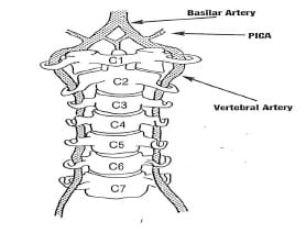
    The brain is supplied by blood from two arterial sources: the paired internal carotid arteries and the paired vertebral arteries. The blood supply to the brain from the carotid arteries is referred to as the anterior circulation to the brain. The blood supply to brain from the vertebral arteries is referred to as the posterior circulation to the brain. The vertebral arteries are noted in the above drawing (reference 1).

    The vertebral arteries are the main blood supply to the brain stem (where the cranial nerves originate), to the cerebellum (for coordination of movements) and to the occipital lobe of the brain (for vision). Consequently, interruption of blood flow through the vertebral arteries tends to give rise to signs and symptoms that are associated with these anatomical regions. These signs and symptoms are elaborated upon below.

    The vertebral arteries are exceptionally unique: they ascend to the brain through an opening, a foramen, in the transverse process of the cervical vertebrae. This opening is called the foramen transversarium. The foramen transversarium exists in the cervical vertebrae C6-C1. The vertebral arteries ascend in the foramen transversarium before entering the skull through the foramen magnum. After entering the skull, the paired vertebral arteries merge to become the singular basilar artery (drawing from reference 1).

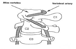
    The atlas-axis (C1-C2) vertebral articulation of the cervical spine is mechanically unique. It is designed for the function of rotational motion. When one maximally turns one’s head, approximately 55% of that motion occurs at the atlas-axis articulation. The vertebral artery in the foramen transversarium between the atlas and axis must accommodate this rotational motion. This places the vertebral artery at increased risk of tractional types of stress and potential injury as a consequence of a variety of upper cervical spine mechanical loads.

    The potential tractional injury to the vertebral artery is a dissection, and usually referred to as vertebral artery dissection, or VAD.

    Right cervical spine (head) rotation, showing the tension on the left vertebral artery between the atlas and axis (drawing from reference 1).

    The manipulative maneuvers that theoretically would have the greatest risk of injuring the vertebral artery

    When there is a vertebral artery vascular event, imaging and pathology have noted vertebral artery dissection. The most plausible explanation for the vertebral artery dissection is a stretching injury. As noted above, over 50% of cervical spine rotation occurs at a single articulation, Atlas (C1) – Axis (C2). As depicted in the drawing above, C1-C2 rotation would stretch the vertebral artery.

    Consequently, students in Chiropractic College are taught not to apply a rotational manipulative thrust of C1 on C2. Furthermore, biomechanical studies indicate that the vertebral artery will experience additional tractional stress if the upper cervical spine is in extension. It is emphasized to students in Chiropractic College to not use the combination of C1—C2 extension—rotation—thrust manipulation (adjustments).

    The signs and symptoms of a person who is having vascular compromise of their vertebral artery

    Signs and symptoms that would warn of a possible vertebral artery dissection with ischemia are often summarized as the 5 Ds And the 3 Ns (1):

    Dizziness (vertigo, light-headedness)

    Drop attacks

    Diplopia (or other visual problems)

    Dysarthria [Speech Disorder]

    Dysphagia [Difficult or Painful Swallowing]

    Ataxia of gait (Hemiparesis)

    Nausea (possibly with vomiting)

    Nystagmus

    Numbness (hemianesthesia)

    A history that would warn of a possible acute vertebral artery dissection with ischemia involves a sudden onset of severe head and/or neck pain, which is like no other pain the patient has previously suffered. This is especially important if the patient can isolate the pain to the suboccipital region (1).

    Background

    Cervical arterial dissection is one of the main causes of ischemic stroke in young adults. Cervical arterial dissections can be categorized as traumatic or spontaneous. The primary cause of posttraumatic cervical artery dissections is motor vehicle collisions (whiplash trauma) (2, 3).

    According to a review of the literature by Alan Terrett (1), a number of non-manipulative mechanical events have been linked to vertebral artery dissections. These mechanical events usually involve rotation and/or extension, and include:

    Childbirth
    By Surgeon or Anesthetist During Surgery
    Calisthenics, Athletics, Fitness Exercise
    Yoga
    Overhead Work, Painting a Wall
    Hanging Out the Washing
    Neck Extension during Radiography
    Neck Extension for a Bleeding Nose
    Turning the Head while Driving a Vehicle
    Tonic Clonic Convulsive Seizure
    Amusement Park Ride
    Protracted Dental Work
    Archery
    Sneezing/Nose Blowing/Coughing
    Wrestling
    Emergency Resuscitation
    Star Gazing, Watching Aircraft
    Sleeping Position
    Swimming
    Break Dancing
    Football
    Beauty Parlor Stroke, Sitting in a Barber’s Chair
    Tai Chi
    Sexual Intercourse

    In another article by Alan Terrett (4), the published literature was reviewed pertaining to the incidence of reported adverse events associated with chiropractic spinal adjusting (manipulation). Astonishingly, his results revealed that in many of the published adverse events ascribed to chiropractic manipulation, they were in fact, not associated with chiropractic in any manner. Apparently, the authors of the articles assumed “chiropractic” and “manipulation” were synonyms. When untrained laypersons or physicians performed a manipulation resulting in a reported adverse event, authors would claim that the manipulation was performed by a chiropractor. The list of discovered manipulators included:

    A Blind Masseur
    An Indian Barber
    A Wife
    A Kung-Fu Practitioner
    Self Manipulation

    Often the manipulation was performed by a medical doctor, an osteopath, a naturopath, or a physical therapist.

    Dr. Terrett concluded:

    “This study reveals that the words chiropractic and chiropractor commonly appear in the literature to describe spinal manipulative therapy, or practitioner of spinal manipulative therapy, in association with iatrogenic complications, regardless of the presence or absence of professional training of the practitioner involved.”

    “The words chiropractic and chiropractor have been incorrectly used in numerous publications dealing with spinal manipulative therapy injury by medical authors, respected medical journals and medical organizations.”

    “In many cases, this is not accidental; the authors had access to original reports that identified the practitioner involved as a non-chiropractor. The true incidence of such reporting cannot be determined.”

    “Such reporting adversely affects the reader’s opinion of chiropractic and chiropractors.”

    “It has been clearly demonstrated that the literature of medical organizations, medical authors and respected, peer-reviewed, indexed journals have, on numerous occasions, misrepresented the facts regarding the identity of a practitioner of manual therapy associated with patient injury.”

    “Such biased reporting must influence the perception of chiropractic held by the reader, especially when cases of death, tetraplegia and neurological deficit are incorrectly reported as having been caused by chiropractic.”

    “Because of the unwarranted negative opinion generated in medical readers and the lay public alike, erroneous reporting is likely to result in hesitancy to refer to and underutilization of a mode of health care delivery.”

    Following Dr. Terrett’s lead, other authors have watched the indexed literature and continue to note that when an untrained individual injures a patient using spinal manipulation, it is often reported as having been done by a chiropractor. Examples include:

    5) Weban A, Beck J, Raabe A, Dettmann E. Seifert V; Misuse of the terms chiropractic and chiropractor J Neurol Neurosurg Psychiatry. 2004 May; 75(5): 794.

    “I am attempting to clarify this important difference between a chiropractor and chiropractitioner because there is the potential to create confusion such that the injury reported by Beck et al could be incorrectly attributed to a chiropractor as compared with a chiropractitioner (lay medical practitioner). In fact, unfortunately, this has already happened. A brief review of Dr. Beck’s case report, by a BMJ editor, inappropriately uses the title ‘chiropractic causes CSF leak’ and suggests the care provider was a chiropractor.”

    6) Weban A; Inappropriate use of the title ‘chiropractor’ and term ‘chiropractic manipulation’ in the peer-reviewed biomedical literature. Chiropractic and Osteopathy; 2006 Aug 22;14:16.

    “The results of this year-long prospective review suggests that the words ‘chiropractor’ and ‘chiropractic manipulation’ are often used inappropriately by European biomedical researchers when reporting apparent associations between cervical spine manipulation and symptoms suggestive of traumatic injury. Furthermore, in those cases reported here, the spurious use of terminology seems to have passed through the peer-review process without correction. Additionally, these findings provide further preliminary evidence, beyond that already provided by Terrett, that the inappropriate use of the title ‘chiropractor’ and term ‘chiropractic manipulation’ may be a significant source of over-reporting of the link between the care provided by chiropractors and injury.”

    In 2002, Dr. Scott Haldeman from the Department of Neurology, University of California, Irvine, and colleagues, published a study titled (7):

    “Unpredictability of cerebrovascular ischemia associated with cervical spine manipulation therapy: a review of sixty-four cases after cervical spine manipulation”

    The study, published in Spine, was a retrospective review of 64 medicolegal records describing cerebrovascular ischemia after cervical spine manipulation. The authors note, that up to 2002, only about “117 cases of post-manipulation cerebrovascular ischemia have been reported in the English language literature.”

    The authors further indicate that proposed risk factors for cerebrovascular ischemia secondary to spinal manipulation include age, gender, migraine headaches, hypertension, diabetes, birth control pills, cervical spondylosis, and smoking, and that it is often assumed that these complications may be avoided by clinically screening patients and by pre-manipulation positioning of the head and neck to evaluate the patency of the vertebral arteries. However, after an extensive review, these authors conclude:

    “This study was unable to identify factors from the clinical history and physical examination of the patient that would assist a physician attempting to isolate the patient at risk of cerebral ischemia after cervical manipulation.”

    “Cerebrovascular accidents after manipulation appear to be unpredictable and should be considered an inherent, idiosyncratic, and rare complication of this treatment approach.”

    In 2008, Dr. David Cassidy and colleagues published the most comprehensive study to date pertaining to the risk of vertebral artery dissection as related to chiropractic cervical spine manipulation (8). The article was published in Spine, and titled:

    “Risk of Vertebrobasilar Stroke and Chiropractic Care:
    Results of a Population-Based Case-Control and Case-Crossover Study”

    Key points from this article include:

    “Vertebrobasilar artery stroke is a rare event in the population.”

    “We found no evidence of excess risk of vertebral artery stroke associated chiropractic care.”

    “Neck pain and headache are common symptoms of vertebral artery dissection, which commonly precedes vertebral artery stroke.”

    “The increased risks of vertebral artery stroke associated with chiropractic and primary care physicians visits is likely due to patients with headache and neck pain from vertebral artery dissection seeking care before their stroke.”

    “Because patients with vertebrobasilar artery dissection commonly present with headache and neck pain, it is possible that patients seek chiropractic care for these symptoms and that the subsequent vertebral artery stroke occurs spontaneously, implying that the association between chiropractic care and vertebral artery stroke is not causal.”

    “Since it is unlikely that primary care physicians cause stroke while caring for these patients, we can assume that the observed association between recent primary care physician care and vertebral artery stroke represents the background risk associated with patients seeking care for dissection-related symptoms leading to vertebral artery stroke. Because the association between chiropractic visits and vertebral artery stroke is not greater than the association between primary care physicians visits and vertebral artery stroke, there is no excess risk of vertebral artery stroke from chiropractic care.”

    “Our results suggest that the association between chiropractic care and vertebral artery stroke found in previous studies is likely explained by presenting symptoms attributable to vertebral artery dissection.”

    In August of 2010, Donald Murphy published an article in the journal Chiropractic and Osteopathy, titled (9):

    Current understanding of the relationship between cervical manipulation and stroke:
    What does it mean for the chiropractic profession?

    In this article, Dr. Murphy states:

    “The understanding of the relationship between cervical manipulative therapy (CMT) and vertebral artery dissection and stroke (VADS) has evolved considerably over the years.”

    “In the beginning the relationship was seen as simple cause-effect, in which CMT was seen to cause VADS in certain susceptible individuals. This was perceived as extremely rare by chiropractic physicians, but as far more common by neurologists and others.”

    “Recent evidence has clarified the relationship considerably, and suggests that the relationship is not causal, but that patients with VADS often have initial symptoms which cause them to seek care from a chiropractic physician and have a stroke some time after, independent of the chiropractic visit.”

    In January of 2011, the Journal of Manipulative and Physiological Therapeutics published a population-based case series using administrative health care records of all Ontario, CAN, residents hospitalized with vertebral artery stroke between April 1, 1993, and March 31, 2002, titled (10):

    “A population-based case-series of Ontario patients who develop a vertebrobasilar artery stroke after seeing a chiropractor”

    These authors note:

    “The current evidence suggests that association between chiropractic care and vertebrobasilar artery (VBA) stroke is not causal. Rather, recent epidemiological studies suggest that it is coincidental and reflects the natural history of the disorder.”

    “Because neck pain and headaches are symptoms that commonly precede the onset of a VBA stroke, these patients might seek chiropractic care while their stroke is in evolution.”

    Also in January, The Open Neurology Journal published an “open access” editorial by Dean Smith and Gregory Cramer, titled (11):

    “Spinal Manipulation is Not an Emerging Risk Factor for Stroke Nor is it Major Head/Neck Trauma. Don’t Just Read the Abstract!”

    Dean L. Smith is Clinical Faculty, Department of Kinesiology and Health, Miami University, Oxford, Ohio, and Gregory D. Cramer is Professor and Dean of Research, National University of Health Sciences, Lombard, Illinois. Their editorial includes:

    We would like to address two points in this letter:

    1) The current best-evidence indicates no causal relationship between spinal manipulation (‘chiropractic maneuver’ in the paper) and vertebrobasilar artery (VBA) stroke, and,

    2) Spinal manipulation or ‘chiropractic maneuvers’ are not major head/neck trauma as suggested in abstract of this article.

    “First, evidence is mounting that the association between spinal manipulation and stroke is coincidental rather than causal and reflects the natural history of the disorder.” (ref 10)

    “The largest population-based study to date was conducted by Cassidy et al. and included all vertebrobasilar artery (VBA) strokes in Ontario, Canada over a period of 9 years. The authors found no evidence of excess risk (i.e. no risk) of VBA stroke associated with chiropractic care.” (ref 8)

    “The prevailing hypothesis is that patients with vertebral artery dissections often have initial symptoms that cause them to seek care from a chiropractic or medical physician and the stroke is independent of their visit.” (ref 8, 9, 10)

    The “population based study provides higher quality evidence than previous case reports, case series, and physician surveys frequently referenced when discussing spinal manipulation in this context.”

    “If anything, the latest scientific evidence questions whether spinal manipulation is a risk factor at all for cervical artery dissection.”

    “Chiropractic spinal manipulations may very well be a demerging risk factor for stroke since there may not be any risk.” (ref 8)

    “The evidence, albeit limited to date, suggests that spinal manipulative treatments produce stretches of the vertebral artery that are much smaller than those that are produced during normal everyday movements, and thus they appear harmless.”

    “Spinal manipulations delivered by licensed chiropractors do not fulfill the criteria for major trauma and should not be considered major trauma.”

    In January of this year (2012), a study published in the Journal of Orthopaedic & Sports Physical Therapy, titled (12):

    Upper Cervical and Upper Thoracic Thrust Manipulation Versus Nonthrust Mobilization in Patients With Mechanical Neck Pain:
    A Multicenter Randomized Clinical Trial

    All seven of the authors are physical therapists. The study was a randomized clinical trial to compare the short-term effects of upper cervical (C1-2) and upper thoracic (T1-2) high-velocity low-amplitude (HVLA) thrust manipulation to nonthrust mobilization in patients with neck pain. The authors note:

    “The patients with mechanical neck pain who received the combination of upper cervical and upper thoracic HVLA thrust manipulation experienced significantly greater reductions in dis­ability (50.5%) and pain (58.5%) than those of the nonthrust mobilization group (12.8% and 12.6%, respectively) following treatment.”

    “The combination of upper cervi­cal and upper thoracic HVLA thrust manipulation is appreciably more effective in the short term than nonthrust mobilization in patients with mechanical neck pain.”

    Important for this discussion is the nature of the C1-C2 manipulations. From the picture and description, the C1-C2 manipulation appeared to be a rotational lateral flexion maneuver contacting the posterior arch of C1. For each manipulation the therapist tried to elicit an audible release, but tried to do so no more than twice. The descriptions and photographs appeared to show a standard joint cavitation rotational maneuver. In defending their use of such a maneuver, the authors state:

    “The most recent and robust evidence for the risk of vertebrobasilar stroke and cervical HVLA thrust manipulation comes from the case control study by Cassidy et al. Contrary to traditionally held views, Cassidy et al (ref 8) found no evidence of excess risk of vertebrobasilar stroke associated with cervical HVLA thrust manipulation as compared to primary medical physi­cian care. Moreover, a recent systematic review concluded that there has been no strong evidence linking the occurrence of serious adverse events with the use of cervical manipulation or mobilization in adults with neck pain.”

    The biomechanics of cervical spine manipulation and vertebral artery stress is important. The world leader on this type of biomechanical assessment is Walter Herzog, PhD, from the University of Calgary, CAN. In April of this year (2012), Dr. Herzog and colleagues published a study in the Journal of Electromyography and Kinesiology titled (13):

    Vertebral artery strains during high-speed, low amplitude cervical spinal manipulation

    Dr. Herzog notes that spinal manipulative therapy (SMT) is recognized as an effective treatment modality for many back, neck and musculoskeletal problems. Yet, one of the major issues of the use of SMT is its safety, especially with regards to neck manipulation and the risk of stroke. It has been assumed [wrongly as per this study] that the vertebral artery (VA) experiences considerable stretch during extension and rotation of the neck, which may lead to occlusions and damage to the VA, predisposing the patient to stroke. Therefore, this study presents the first ever data on the mechanics between C2/C1 during cervical SMT performed by chiropractic clinicians.

    The authors compared the results of human VA strains during high-speed, low-amplitude SMTs administered by qualified chiropractic clinicians and compared them to the strains encountered during full range of motion (ROM) tests. They used a total of 3,034 segment strains obtained during SMTs and 2,380 segment strains obtained during full ROM testing, making this is an extensive study.

    Dr. Herzog and colleagues conclude:

    “VA strains obtained during SMT are significantly smaller than those obtained during diagnostic and range of motion testing, and are much smaller than failure strains.”

    “We conclude from this work that cervical SMT performed by trained clinicians does not appear to place undue strain on VA, and thus does not seem to be a factor in vertebro-basilar injuries.”

    “In summary, the maximal strain values for the ROM testing at each segmental level were always greater than the corresponding strain values for the SMTs, suggesting that neck SMTs impose less stretch than turning your head, or extending your neck while looking up at the sky.”

    “Therefore, based on the mechanical tests performed here, one should be able to conclude that stretching of VA during neck SMTs does not cause any damage of the VAs.”

    “The VA is never really strained during spinal manipulative treatments but that the VA is merely taking up slack as the neck and head are moved during SMT, but that there is no stress and thus no possibility for microstructural damage.”

    “The results from this study demonstrate that average and maximal VA strains during high-speed low-amplitude cervical spinal manipulation are substantially less than the strains that can be achieved during ROM testing for all vertebral artery segments.”

    “We conclude that cervical spinal manipulations, as tested here, are safe from a mechanical point of view for normal, healthy VA.”

    SUMMARY

    Chiropractic students and chiropractors are extensively trained in the anatomy and biomechanics of the upper cervical spine. They are also extensively trained in the science and art of spinal adjusting (specific directional manipulation). The review presented here suggests:

    1) Chiropractic manipulation may pose no risk of abnormal stretch to the vertebral artery.

    2) Vertebral artery injury ascribed to chiropractic manipulation in the indexed literature is often inappropriate as the actual manipulation was done by an untrained layperson or untrained healthcare provider.

    3) The symptoms associated with spontaneous vertebral artery dissection may bring the patient into chiropractic offices.

    Chiropractors are extensively trained in understanding the 5 Ds And the 3 Ns that would suggest the possibility of a vertebral artery dissection, and the need for an immediate emergency referral. The best-published studies indicate that chiropractic spinal manipulation is both safe and effective.

    REFERENCES

    • Terrett AGJ; Current Concepts in Vertebrovascular Complications Following Spinal Manipulation; Second Edition; NCMIC Group, 2001.
    • Hauser V, Zangger P, Winter Y, Oertel W, Kesselrin J; Late Sequelae of Whiplash Injury with Dissection of Cervical Arteries; European Neurology; August 18, 2010, Vol. 64, No. 4, pp. 214–218.
    • Haneline M, Triano J. Cervical artery dissection. A comparison of highly dynamic mechanisms: manipulation versus motor vehicle collision. Journal of Manipulative Physiological Therapeutics. 2005 Jan;28(1):57-63.
    • Terrett AG; Misuse of the literature by medical authors in discussing spinal manipulative therapy injury; Journal of Manipulative and Physiological Therapeutics; 1995 May;18(4):203-10.
    • Weban A, Beck J, Raabe A, Dettmann E. Seifert V; Misuse of the terms chiropractic and chiropractor J Neurol Neurosurg Psychiatry. 2004 May; 75(5): 794.
    • Weban A; Inappropriate use of the title ‘chiropractor’ and term ‘chiropractic manipulation’ in the peer-reviewed biomedical literature. Chiropractic and Osteopathy; 2006 Aug 22;14:16.
    • Haldeman S, Kohlbeck FJ, McGregor M; Unpredictability of cerebrovascular ischemia associated with cervical spine manipulation therapy: a review of sixty-four cases after cervical spine manipulation; Spine; 2002 Jan 1;27(1):49-55.
    • Cassidy, J David DC, PhD; Boyle, Eleanor PhD; Côté, Pierre DC, PhD; He, Yaohua MD, PhD; Hogg-Johnson, Sheilah PhD; Silver, Frank L. MD; Bondy, Susan J. PhD; Risk of Vertebrobasilar Stroke and Chiropractic Care: Results of a Population-Based Case-Control and Case-Crossover Study; Spine; Volume 33(4S), February 15, 2008 pp. S176-S183.
    • Murphy DR; Current understanding of the relationship between cervical manipulation and stroke: what does it mean for the chiropractic profession?; Chiropractic and Osteopathy; 2010 Aug 3;18:22.
    • Choi S, Boyle E, Cote P, Cassidy JD. A population-based case-series of Ontario patients who develop a vertebrobasilar artery stroke after seeing a chiropractor. J Manipulative Physiol Ther 2011; 34(1): 15-22.
    • Smith DL, Cramer GC; LETTER TO THE EDITOR: Spinal Manipulation is Not an Emerging Risk Factor for Stroke Nor is it Major Head/Neck Trauma. Don’t Just Read the Abstract!; The Open Neurology Journal, 2011, 5, 46-47
    • Dunning JR, Cleland JA, Waldrop MA, Arnot C, Young I, Turner M, Sigurdsson G; Upper Cervical and Upper Thoracic Thrust Manipulation Versus Nonthrust Mobilization in Patients With Mechanical Neck Pain: A Multicenter Randomized Clinical Trial; Journal of Orthopaedic & Sports Physical Therapy; January 2012; Volume 42; Number 1; pp. 5-18.
    • Herzog W, Leonard TR, Symons B, Tang C, Wuest S; Vertebral artery strains during high-speed, low amplitude cervical spinal manipulation; Journal of Electromyography and Kinesiology; April 5, 2012 [epub].
  • Myotherapy/Massage

    Myotherapy/Massage

    Massage is the mechanical working of muscles and connective tissues to enhance function, and to improve upon the quality and timing of the healing process. Because massage also promotes relaxation and well-being, it also has pleasurable connotations.

    Myotherapy is often used synonymously for massage therapy. Myotherapy is the terminology used by many healthcare professionals because it is considered to be a form of manual medicine used for the treatment and management of musculoskeletal pain. The primary purpose of myotherapy is therapeutic. Myotherapy is designed to achieve a specific change in tissues to benefit aspects of the pathophysiological process.

    A recent search of the National Library of Medicine using PubMed (May 13, 1012) using the key word “massage” pulled up 10,357 citations, 172 of which were dated in 2012. In contrast, the key word “myotherapy” pulled up 27 citations.

    For decades, a vocal advocate of myotherapy was United Kingdom orthopedic surgeon Sir James Cyriax, MD (d. 1985, age 80 years). Dr. Cyriax authored three medical texts that advocated myotherapy:

    Textbook of Orthopaedic Medicine, Diagnosis of Soft Tissue Lesions

    Bailliere Tindall, Volume 1, eighth edition, 1982. (1)

    Textbook of Orthopaedic Medicine, Treatment by Manipulation, Massage, and Injection

    Bailliere Tindall, Volume 2, tenth edition, 1982. (2)

    Illustrated Manual of Orthopaedic Medicine

    Butterworths, 1983. (3)

    In these references, Dr. Cyriax notes that harmful infections create tissue destruction, resulting in inflammation. Our body recognizes this inflammation and attempts to “wall off” the infectious pathogens by creating a fibrous response. Cyriax states (1):

    “The excessive reaction of tissues to an injury is conditioned by the overriding needs of a process designed to limit bacterial invasion. If there is to be only one pattern of response, it must be suited to the graver of the two possible traumas. However, elaborate preparation for preventing the spread of bacteria is not only pointless after an aseptic injury, but is so excessive as to prove harmful in itself. The principle on which the treatment of post-traumatic inflammation is based is that the reaction of the body to an injury unaccompanied by infection is always too great.”

    In their 1983 text pertaining to sports medicine, Steven Roy and Richard Irvin state (4):

    “It is important to realize that the body’s initial reaction to an injury is similar to its reaction to an infection. The reaction is termed inflammation and may manifest macroscopically (such as after an acute injury) or at a microscopic level, with the latter occurring particularly in chronic overuse conditions.”

    In their 1992 book titled Wound Healing (5), physician I. Kelman Cohen and associates note:

    “There are two important consequences of being a warm-blooded animal. One is that body fluids make optimal culture media for bacteria. It is to the animal’s advantage, therefore, to heal wounds with alacrity in order to reduce chances of infection.”

    “The prompt development of granulation tissue forecasts the repair of the interrupted dermal tissue to produce a scar.” In addition to providing tensile strength, scars are believed to be a barrier to infectious migration.

    This is in agreement with the pathology text by Dr. William Boyd (6), which states:

    “The inflammatory reaction tends to prevent the dissemination of infection. Speaking generally, the more intense the reaction, the more likely the infection to be localized.”

    Physiologist and physician Arthur Guyton (7) adds additional support stating:

    “One of the first results of inflammation is to ‘wall off’ the area of injury from the remaining tissues. This walling-off process delays the spread of bacteria or toxic products.”

    The reference text, Robbins Pathologic Basis of Disease (8), makes these points:

    “Inflammation is best defined as the reaction of vascularized living tissue to local injury.”

    “The inflammatory response is closely intertwined with the process of repair.”

    Inflammation serves to destroy, dilute, or wall off the injurious agent, and sets into motion a series of events that heal and reconstitute the damaged tissue as best as possible.

    Repair begins during the early phases of inflammation. During repair, the injured tissue is replaced by regeneration of native parenchymal cells, and by filling of the defect with fibroblastic scar tissue (scarring).

    “Humans owe to inflammation and repair their ability to contain injuries and heal defects.

    Without inflammation, infections would go unchecked and wounds would never heal.

    “However, inflammation and repair may be potentially harmful.”

    “Reparative efforts may lead to disfiguring scars or fibrous bands that cause intestinal obstruction or limit the mobility of joints.”

    In their 2004 book on pathology titled Cells, Tissues, and Disease: Principles of General Pathology, Guido Manjo, MD and Isabelle Joris, PhD, make these points (9):

    “Inflammation is one of the basic processes in general pathology.”

    “Inflammation is primarily an antibacterial phenomenon.”

    “Inflammation operates against all invaders, including viruses, worms, fungi, and other parasites.”

    “Inflammation is also triggered aseptically by injured tissues.”

    “There can be inflammation without infection; remember that inflammation is triggered by products of tissue injury, thus any aseptic injury will trigger inflammation.”

    “Because local injury is part of everyday life, inflammation is probably the most common aspect of tissue pathology and has always been perceived as a central issue in the practice of medicine.”

    “Today we know that inflammation is a life-saving reaction, usually against infection.”

    “Solid masses or sheets of fibrin are often seen on an inflamed surface.”

    “With the microscope, some fibrin is found in nearly all acutely inflamed tissues, but an exudate is called fibrinous when fibrin deposition is the dominant feature.”

    Chronic inflammation causes increasing “collateral damage.”

    “Inflammation takes place in the connective tissues.”

    “After a day or two of acute inflammation, the connective tissue—in which the inflammatory reaction is unfolding—begins to react, producing more fibroblasts, more capillaries, more cells—more tissue. In other words, granulation tissue arises from normal connective tissue, but it cannot be mistaken for normal connective tissue, because its fibroblasts are plumb and activated.”

    “Fibrosis means an excess of fibrous connective tissue. It implies an excess of collagen fibers, with a varying mixture of other matrix components. It can be a local phenomenon, as an end result of chronic inflammation and of wound healing.”

    “When fibrosis develops in the course of inflammation it may contribute to the healing process.” “By contrast, an excessive or inappropriate stimulus can produce severe fibrosis and impair function.”

    Fibrotic tissue “consists of cells and fibers, with few vessels; and it tends to contract very slowly, over weeks and months or longer.”

    “’Fibrotic’ collagen is characterized by excess of hydroxylation and cross-linking.”

    “Why does fibrosis develop? In most cases the beginning clearly involves chronic inflammation. Fibrosis is largely secondary to inflammation.”

    The interpretation is, that in a world prior to the availability of antibiotics, inflammation, with reactive walling-off fibrosis to contain pathogens, is desirable because it increases survivability of the host.

    As explained by Cyriax, Roy/Irvin, Cohen, Boyd, Guyton, Robbins, and Manjo above, the trigger to the walling-off fibrosis response of the body is inflammation. Problems arise when the inflammatory trigger is non-infectious inflammation. In such cases, excessive tissue fibrosis creates local impairments in biomechanical function. This impairment in local biomechanical function affects performance, can generate pain, and accelerate degenerative changes. These impairments can adversely affect the patient for years or even decades.

    A primary goal of myotherapy is to use appropriately applied mechanical forces to reverse the adverse biomechanical function of fibrotic tissue changes. When successful, that patient often experiences reduced pain, improved performance, and reduced joint stresses.

    The chronic nature of this scar tissue or fibrosis is expressed in the 1998 article by Thomas Melham and associates (10). These authors note that post-traumatic scar tissue can cause pain with activity, pain on palpation, decreased range of motion, and loss of function, and that these problems are resistant to surgery and to conventional physical therapy. Excessive scar tissue contributes to chronic soft tissue dysfunction that cause significant disabilities and time lost from work or training activities, and these problems are often difficult to successfully treat. The authors discuss the mechanical and neurological adverseness caused by connective tissue fibrosis, noting:

    “Many athletes develop excessive connective tissue fibrosis (scar tissue) or poorly organized scar tissue in and around muscles, tendons, ligaments, joints, and myofascial planes as a result of acute trauma, recurrent microtrauma, immobilization, or as a complication of surgical intervention.”

    “This can lead to soft tissue adhesions, tendonitis, tendonosis, fascial restrictions, and chronic inflammation or dysfunction which in many cases responds poorly to conventional treatments.”

    These descriptions of the mechanical pathoanatomy of the soft tissue scar/fibrosis repair process suggests that appropriately applied myotherapy would hold biological plausibility in their management. Additional support from Cyriax includes (1):

    “When pain is due to bacterial inflammation, Hilton’s advocacy of rest remains unchallenged and is today one of the main principles of medical treatment. When, however somatic pain is caused by inflammation due to trauma, his ideas require modification. When non-bacterial inflammation attacks the soft tissues that move, treatment by rest has been found to result in chronic disability later, although the symptoms may temporarily diminish. Hence, during the present century, treatment by rest has given way to therapeutic movement in many soft tissue lesions. Movement may be applied in various ways: the three main categories are:

    (a) active and resistive exercises:

    (b) passive, especially forced movement: and

    (c) deep massage.”

    Here, Cyriax’s “deep massage” is equivalent to contemporary myotherapy.

    The discussion and references above support the concept that adverse pathogens cause tissue destruction and subsequent inflammation. The body evolved in a manner to wall-off the area of inflammation by over healing the region with a fibrous response. The fibrous response became a physical barrier, reducing the ability of the pathogens to spread to other regions of the body, thereby improving the host’s chances for survival.

    However, when inflammation is caused by non-infectious mechanisms, the same fibrotic tissue response occurs. In such cases, without infectious pathogens, the fibrotic tissue response is excessive, resulting in mechanical harm to the host. This harmful tissue fibrosis is worsened with early immobilization of the affected tissues. This tissue fibrosis is minimized with early persistent controlled mobilization.

    Traditionally, myotherapy targets the mechanical adverse fibrotic tissues located in muscles and in non-contractile soft tissue (tendon, fascia, ligament, etc.) fibrosis responds best to manually applied tissue friction.

    Most patients have a combination of tissues that are adversely affected by reparative soft tissue scar/fibrosis, depending on the mechanism of injury or stress. Consequently, myotherapy is often applied in combination with other adjuncts that are similarly designed to remodel and improve soft tissue scar fibrosis. An example would include joint adjusting (specific directional manipulation) to influence periarticular soft tissues. Additionally, resistive effort and range-of-motion exercises enhance the remodeling process and help to reduce chances of any degraded/remodeled scar/fibrotic tissue for reoccurring.

    From Cyriax J and Cyriax P, Illustrated Manual of Orthopaedic Medicine, Butterworths, 1983 (3).

    Recently (February 2012), evidence has emerged showing the mechanisms whereby massage/myotherapy influences (improves) the physiology of acute muscle injury. Investigators from McMasters University, Ontario, Canada, published their findings in the journal Science Translational Medicine in an article titled (11):

    Massage Therapy Attenuates Inflammatory
    Signaling After Exercise-Induced Muscle Damage

    In this study, the authors note that massage therapy is commonly used during physical rehabilitation of skeletal muscle to ameliorate pain and promote recovery from injury. Although there is evidence that massage may relieve pain in injured muscle, how massage affects cellular function remains unknown.

    The authors administered either massage therapy or no treatment to the quadriceps of 11 young male participants after exercise induced muscle damage. Muscle biopsies were obtained from the quadriceps muscles of these young men (vastus lateralis) immediately after completing strenuous fatiguing exercise. In the massage group, muscle biopsies were again taken immediately after 10 min of massage/myotherapy treatment; and then again after a 2.5-hour period of recovery. Similar muscle biopsies were obtained at the same time intervals for the non-massaged group.

    The author’s advocacy of massage therapy includes this perspective:

    “Complementary and alternative medicine (CAM) is increasingly used as a cost-effective adjunct to conventional medical care. Many CAM techniques, such as acupuncture, massage therapy, or chiropractic manipulations, are aimed at managing pain, relieving stress, and preventing injury.”

    “The increasing use of massage therapy as an adjunct to conventional care for musculoskeletal injury recovery and the growing number of physician referrals for massage represent a shift toward nondrug-based therapies for personal health.”

    “Given the spiraling cost of primary care and medications in the US, it is likely that more patients will seek out this therapy as well as other nontraditional medical alternatives to complement more conventional approaches to their healthcare.”

    The authors define massage therapy as “physical manipulation of muscle and connective tissue at a site of injury, inflexibility, or soreness to reduce pain and promote recovery.” They hypothesize that massage therapy moderates inflammation, improves blood flow, and reduces tissue stiffness, resulting in diminished pain. They state that massage could be useful to a broad spectrum of individuals including the elderly, those suffering from musculoskeletal injuries, and patients with chronic inflammatory conditions, citing evidence that massage therapy reduces chronic pain and improves range of motion in clinical trials.

    Acute muscle trauma results in inflammation and immune cell activation. The activated immune system produces and releases inflammatory proteins called cytokines. This inflammatory immune response is led by the cytokine nuclear factor kappa B (NFkB). NFkB impairs tissue healing. Consequently, any therapy which reduces NFkB accumulation would improve tissue repair.

    The NFkB pathway also increases inflammatory prostaglandin synthesis [like prostaglandin E2 {PGE2}] and increases expression of other inflammatory cytokines [like interleukin-6 {IL-6} and tumor necrosis factor-alpha {TNF-a}]. These inflammatory cytokines further impede muscle repair by increasing muscle protein breakdown and suppressing myosin synthesis.

    Of course, all of these inflammatory cytokines also activate nociceptors, causing increased sensitivity to pain (hyperalgesia). These authors propose that myotherapy/massage may disperse the accumulation of these inflammatory mediators, thus reducing pain afferentation. This is particularly useful in situations where areas of low blood flow, like the muscle tendon interface, restrict the access of circulating analgesic anti-inflammatory chemical compounds.

    Additionally, a mechanical stimulus to a muscle will physically alter the cells membrane and the extracellular matrix and transmit signals via proteins known as integrins. Integrins in turn activate and propagate mechanotransduction signals that “modulate protein synthesis, glucose uptake, and immune cell recruitment.” The mechanical stretch during massage activates mechanotransduction signaling that increases muscle glucose uptake, protein synthesis and muscle growth. This would improve both the timing and quality of healing for injured muscles. “Any physiological benefits due to massage would likely be initiated through mechanical effects on skeletal muscle followed by changes to intracellular regulatory cascades.”

    The mechanism of myotherapy/massage found in this biopsy study include:

    • “Massage reduced signs of inflammation, and massaged muscles cells were better able to make new mitochondria—promoting faster recovery from exercise-induced muscle damage.”
    • Massage activates mechanosensory sensors.
    • Massage activates the formation of additional mitochondria, “presumably accelerating healing of the muscles.”
    • Massage reduced accumulation of inflammatory mediator nuclear factor kappa B (NFkB), and reduced the activity of immune cytokines interleukin-6 (IL-6) and tumor necrosis factor alpha (TNF-a), “a sign of less cellular stress and inflammation.” This would mitigate cellular stress resulting from myofiber injury.

    “In summary, when administered to skeletal muscle that has been acutely damaged through exercise, massage therapy appears to be clinically beneficial by reducing inflammation and promoting mitochondrial biogenesis.”

    “These results elucidate the biological effects of massage in skeletal muscle and provide evidence that manipulative therapies may be justifiable in medical practice.”

    Summary and Conclusions


    Massage Does The Following:

    • Activates the mechanotransduction signaling pathways
    • Increases muscle glucose uptake, protein synthesis and muscle growth
    • Increases the biosynthesis of mitochondria; this increases the production of the ATP energy required for protein synthesis and repair of injury.
    • Reduces the production of NFkB.
    • Reduces the production of inflammatory prostaglandins [PGE2].
    • Reduces the production of inflammatory cytokines [TNF-a, IL-6].
    • Reduces pain.
    • Accelerates healing, faster recovery.

    Chiropractors are well schooled in the concepts of tissue fibrosis, traumatically triggered inflammatory cytokines and prostaglandins, mitochondrial production of ATP energy, DNA activation with repair protein synthesis, inflammatory alteration of pain thresholds, mechanotransduction and mechanobiology as related to integrin influence on the genetic material, and how motion restriction opens the pain gate. Consequently, most chiropractors use myotherapy/massage therapy either directly (in office) or indirectly (as outside office referral) as an adjunct to spinal adjusting in clinical practice. Incorporation of formal myotherapy into our clinical practice helped us to achieve our clinical goals 25-33% quicker than without this addition.

    References:

    • Cyriax J; Textbook of Orthopaedic Medicine, Diagnosis of Soft Tissue Lesions, Bailliere Tindall, Volume 1, eighth edition, 1982.
    • Cyriax J; Textbook of Orthopaedic Medicine, Treatment by Manipulation, Massage, and Injection; Bailliere Tindall, Volume 2, tenth edition, 1982. (2)
    • Cyriax J and Cyriax P, Illustrated Manual of Orthopaedic Medicine, Butterworths, 1983.
    • Roy, Steven; Irvin, Richard; Sports Medicine: Prevention, Evaluation, Management, and Rehabilitation; Prentice-Hall, 1983.
    • Cohen, I. Kelman; Diegelmann, Robert F; Lindbald, William J; Wound Healing, Biochemical & Clinical Aspects; WB Saunders, 1992.
    • Boyd, William, Pathology, Lea and Febiger, 1952.
    • Guyton, Arthur, Textbook of Medical Physiology, Saunders, 1986.
    • Robbins Pathologic Basis of Disease, “Inflammation and Repair,” Chapter 2, 4th edition, p 39.
    • Manjo G, Joris I; Cells, Tissues, and Disease: Principles of General Pathology; Second Edition; Oxford University Press; 2004.
    • Melham TJ, Sevier TL, Malnofski MJ, Wilson JK, Helfst RK, Chronic ankle pain and fibrosis successfully treated with a new noninvasive augmented soft tissue mobilization technique (ASTM); Medicine Science Sports Exercise, June 1998; 30(3): 801-4.
    • Crane JD, Ogborn DI, Cupido C, Melov S, Hubbard A, Bourgeois JM, Tarnopolsky MA; Massage Therapy Attenuates Inflammatory Signaling After Exercise-Induced Muscle Damage; Science Translational Medicine; February 1, 2012; Vol. 4, No. 119; [epub].
  • The “Connective Tissue – Cytoskeleton” Matrix

    The “Connective Tissue – Cytoskeleton” Matrix

    The “Tissue – Tensegrity” Matrix And Chronic Low Back Pain

    Chiropractors primarily look at patient’s problems from a mechanical perspective. A classic chiropractic mechanical analogy is a pinched nerve (compressive neuropathology). Although chiropractors occasionally do treat compressive neuropathology, most chiropractors are aware that there are patients with compressive neuropathology that require a surgical decompression. Overall, compressive neuropathology is a rare clinical syndrome in chiropractic clinical practice. Most of the mechanical approach of chiropractic is not to “unpinch a pinched nerve.” Rather, the primary mechanical approach of chiropractic patient management is the improvement of the Tensegrity Matrix.

    The human body is composed of four tissue types: epithelium, muscle, connective, and nerve. Everything in the body is made from these four basic tissues.

    The connective tissue is found throughout the body. The great Canadian histologist, Arthur Ham (d.1992 at age 90), states (1):

    “Connective tissue was given its name because it connects, and thus holds, the other tissues together.”

    Connective tissue includes tissues that produce blood cells (hemopoietic tissue), the blood cells themselves, and the strong supporting types of connective tissue. The strong supporting types of connective tissues include, bone, cartilage, ligaments, tendons, fascia, etc.

    The strong supporting types of connective tissues have two components:

    • The living cells (like osteocytes, chondrocytes, fibroblasts, etc.).
    • A non-living intercellular substance; this non-living intercellular substance is produced by the living cells.

    The chief role of these living cells is to produce and maintain the intercellular substances.

    Collagen is a protein. It is the primary protein produced as intercellular substance by the living cells of the strong supporting types of connective tissues. Collagen is the most abundant protein in our bodies. Most of the collagen in our bodies is produced by the fibroblasts.

    ••••

    Rene Cailliet, MD, has had an accomplished life. Born in 1917 and still living at age 95, he helped pioneer the specialty of Physical Medicine and Rehabilitation during World War II. Dr. Cailliet served as a chairman of the Department of Physical Medicine and Rehabilitation at the University of Southern California. He is currently an emeritus professor of Physical Medicine and Rehabilitation at the medical school at the University of California, Los Angeles (UCLA). Dr. Cailliet has authored more than a dozen medical texts, and he has 49 articles in a PubMed database search of the National Library of Medicine (March 2012). Interestingly, his most recent PubMed indexed article was published in 2008, and is co-authored by chiropractors Deed and Don (d. 2011) Harrison (2).

    In his 1987 book The Rejuvenation Strategies, Dr. Cailliet notes that, under a microscope, collagen produces a whole body lattice of fibers that appear as a grid (3):

    Dr. Cailliet notes that ideally, at the contact points between these fibers they remain independent and glide on one another when moving. However, as a consequence of inflammation, trauma, or even inactivity, the points of intersection become fibrotic and “stick together,” resulting in “loss of flexibility.”

    ••••

    In his 2000 book Energy Medicine, The Scientific Basis (4), James Oschman, PhD, notes that the cytoskeletons of all of the cells in the body are physically and mechanically linked to a collagenous connective tissue extracellular matrix. Proteins called “integrins” do this trans-cellular linking. The molecules that link the cell interior with the extracellular collagenous matrix are these integrins. Likewise, there is a physical mechanical link between the cellular cytoplasmic matrix and the nuclear envelope, nuclear matrix, and the DNA of the genes/chromosomes. This physical continuum, beginning with the skin and extending to the genetic material, can be affected physically as a consequence of postural distortions, injury, physical stress and scar tissue, resulting in alterations in expression of our genetic material. Dr. Oschman notes that this entire interconnected system is called the:

    “connective tissue – cytoskeleton” matrix
    or
    the “tissue – tensegrity” matrix.

    Our bodies behave as a tensegrity system. A tensegrity system is characterized by a series of continuous tensional networks. This means that the entire body is physically interconnected. A stress on one part of the system will spread to other parts of the system. This includes to the genome and its genetic expression. One can change the genomic expression of DNA by alterations of physical stress. All the components of our bodies, from the skin to the genome are physically and mechanically interlinked by a connective tissue collagenous matrix and integrins. Again, this is often referred to as the “tensegrity matrix.”

    Dr. Oschman explains how progressive changes in the function of the tensegrity matrix takes place because of the ways in which individuals use their bodies in relation to gravity, because of habits or injuries. These soft tissue changes provide a basis for the mechanically based restorative measures by chiropractors and others.

    Dr. Oschman argues that the tensegrity matrix best serves physiology when it is flexible and balanced. This serves to reduce injury and enhance healing. He states:

    “Tensegrity accounts for the ability of the body to absorb impacts without being damaged. Mechanical energy flows away from a site of impact through the tensegrous living matrix. The more flexible and balanced the network (the better the tensional network), the more readily it absorbs shocks and converts them to information rather than damage.”

    “Tensegrity accounts for the fact that inflexibility or shortening in one tissue influences structure and movement in other parts.”

    Dr. Oschman explains that the most significant influence on the tensegrity matrix is our alignment in the gravity field. He notes that gravity is the most potent physical influence in any human life. Simple mechanical calculations reveal that gravity gives rise to surprisingly large forces within the body as a consequence of levers that amplify the forces exerted on joints and other tissues.

    The gravity system connects, via integrins, to the cytoskeleton of cells throughout the body. Therefore, an imbalance in one part of the body will affect the whole body.

    Dr. Oschman states:

    “Gravity pervades our bodies and our environment and affects our every activity. All of the structures around us – our homes, furniture, buildings, machinery, plant, and animal, – and our own bodies, are designed to function in a world dominated by gravity. The form of each bone, muscle, and sinew tells a story of its particular role in maintaining and moving the body in the gravitational field. Many of the injuries faced in the therapeutic setting are consequences of falling down, or of habitual movement patterns that strain tissues. Hence therapists of virtually every tradition can benefit from an appreciation of the ways in which gravity interacts with structures, energy flows, and emotions, and the clinical approaches that remedy ‘gravitational traumas.’”

    “To introduce the therapeutic significance of gravity, we summarize the work of Joel E. Goldthwait and his colleagues at Harvard Medical School.”

    “A surgeon in Boston and founder of the orthopaedic clinic at the General Hospital, Goldthwait developed a successful therapeutic approach to chronic disorders. The aim of his therapies was to get his patients to sit, stand, and move with their bodies in a more appropriate relationship with the vertical. After years of treating patients with chronic problems, he concluded that many of these problems arise because parts of the body become misaligned with respect to the vertical, and organ functions therefore become compromised.”

    “Goldthwait’s therapeutic approach was based in part on observations made while performing surgery on such patients. He noticed that abdominal nerves and blood vessels are under tension in individuals whose bodies are out of alignment. He also reported ‘stretching and kinking’ of the cerebral arteries and veins in those whose necks were bent. Various cardiac problems were correlated with ‘faulty body mechanics’ that distorted the chest cavity in a way that impaired circulatory efficiency. Goldthwait also documented with X-rays a build-up of calcium deposits around the vertebrae of individuals with chronic arthritis, and observed that these deposits can diminish when the individual acquires a more vertical stance. His therapeutic approach corrected many difficult problems without the use of drugs. He viewed the human body from a mechanical engineering perspective, in which alignment of parts is essential to reduce wear and stress. He pleaded with physicians to recognize and correct misalignments to prevent long-term harmful effects.”

    Dr. Goldthwait pleaded for everyone to pay more attention to the way in which they hold and move their bodies in relation to the gravity field. He noted that misalignment of any part will affect the whole system, and that restoration of verticality is a way to address a wide variety of clinical problems. Optimal performance in a gravity field occurs only at a narrow peak of balance, and the slightest deviation reduces optimum efficiency. Altered posture in our gravity environment affects the tensegrity matrix continuum. This in turn influences physiology, pain perception, and health.

    ••••

    Another champion of the concepts of the importance of mechanical influence on the physiology of the living matrix is Harvard’s Donald Ingber, MD, PhD. Dr. Ingber is from the Vascular Biology Program, Departments of Surgery and Pathology, Children’s Hospital and Harvard Medical School. A check of the National Library of Medicine using the PubMed search engine finds 211 articles that have included Dr. Ingber as author (March 2012).

    A relevant article by Dr. Ingber was published in the Annals of Medicine in 2003, and titled (5):

    Mechanobiology and Diseases of Mechanotransduction

    In this article, Dr. Ingber expresses his concern that contemporary medical practice focuses primarily on molecular genetics while largely ignoring the physical basis of disease. He notes that many of the problems that lead to pain and morbidity, and bring patients to the doctor’s office, result from changes in tissue structure or mechanics. He stresses that mechanics should be integrated into understanding the molecular basis of disease.

    In agreement with Dr. Oschman above, Dr. Ingber describes the key roles that physical forces, extracellular matrix and cell structure play in the control of normal development, as well as in the maintenance of tissue form and function.

    Dr. Ingber defines cellular mechanotransduction as the molecular mechanism by which cells sense and respond to mechanical stress. He notes that a wide range of diseases included within virtually all fields of medicine and surgery share a common feature: “their etiology or clinical presentation results from abnormal mechanotransduction.”

    A central theme in Dr. Ingber’s discussion is that there are “mechanisms by which cells sense mechanical signals and convert them into a chemical or electrical response.” The molecules that mediate mechanotransduction are the extracellular matrix molecules, trans-membrane integrin receptors. The function of these molecules is modifiable with appropriate mechanical therapeutic intervention. This is relevant and important in chiropractic clinical practice.

    Dr. Ingber makes these important points:

    • “Mechanical forces are critical regulators of cellular biochemistry.”
    • “There is a huge disconnect between ‘genome-age’ technologies and the reality of how diseases manifest themselves. From the time the first human looked, listened and felt for what is wrong with a sick friend, caregivers have recognized the undeniable physical basis of disease.”
    • “In the current genome euphoria, there appears to be no place for ‘physicality’. This is especially worrisome given that abnormal cell and tissue responses to mechanical stress contribute to the etiology and clinical presentation of many important diseases, including asthma, osteoporosis, atherosclerosis, diabetes, stroke and heart failure.”
    • There is a “strong mechanical basis for many generalized medical disabilities, such as lower back pain and irritable bowel syndrome, which are responsible for a major share of healthcare costs world-wide.”
    • “In biology and medicine, we tend to focus on the importance of genes and chemical factors for control of tissue physiology and the development of disease, whereas we commonly ignore physical factors. This is interesting because it was common knowledge at the turn of the last century that mechanical forces are critical regulators in biology.”
    • “These new insights into mechanobiology suggest that many ostensibly unrelated diseases may share a common dependence on abnormal mechanotransduction for their development or clinical presentation.”
    • “Understanding of the relation between structure and function in living tissues and of fundamental mechanisms of cellular mechanotransduction may therefore lead to entirely new modes of therapeutic intervention.”
    • “The therapeutic value of physical therapy, massage, and muscle stimulation is also well known.”

    Dr. Ingber restates that tissues are composed of groups of living cells held together by an extracellular matrix which is primarily composed of a network of collagens. A summary of his article states:

    “The current focus in medicine is on the genetic basis of disease. However, it is not necessary to correct the underlying genetic defect in order to treat clinically relevant symptoms or relieve the pain and morbidity of disease. Moreover, most of the clinical problems that bring a patient to the doctor’s office result from changes in tissue structure and mechanics. Although these physical alterations have been commonly viewed as the end-result of the disease process, recent advances in mechanobiology suggest that abnormal cell and tissue responses to mechanical stress may actively contribute to the development of many diseases and ailments. Thus, it might be wise to search for a physical cause when chemical or molecular forms of investigation do not suffice.”

    ••••

    Helene M. Langevin, MD, is a neurologist from the Department of Neurology, University of Vermont College of Medicine. In 2006 she published an article in the journal Medical Hypothesis, titled (6):

    Connective tissue: A body-wide signaling network?

    In this article, Dr. Langevin notes that connective tissue forms an anatomical network throughout the body that functions as a body-wide mechanosensitive signaling network. This connective tissue signaling network is affected by changes in movement and posture, and may be altered in pathological conditions (e.g. local decreased mobility due to injury or pain). Connective tissue thus functions as a whole body communication system. Since connective tissue is intimately associated with all other tissues (e.g. lung, intestine), connective tissue signaling may coherently influence (and be influenced by) the normal or pathological function of a wide variety of organ systems.

    Dr. Langevin notes that the musculoskeletal system does not physiologically function in isolation from the rest of the body, and that the musculoskeletal tissues (bones, muscles, cartilage, tendons) are strongly associated with posture and movement. Dr. Langevin makes these important points:

    • “Connective tissue not only forms a continuous network surrounding and infiltrating all muscles, but also permeates all other tissues and organs.”
    • “Since connective tissue plays an intimate role in the function of all other tissues, a complex connective tissue network system integrating whole body mechanical forces may coherently influence the function of all other physiological systems.”
    • Local connective tissue fibrosis following an injury may affect both electrical conductivity as well as fibroblast-to-fibroblast communication. Therefore, local pathology can affect whole-body connective tissue signaling.
    • “Understanding the temporal and spatial dynamics of connective tissue bioelectrical, cellular and tissue plasticity responses, as well as their interactions with other tissues, may be key to understanding how pathological changes in one part of the body may cause a cascade of “remote” effects in seemingly unrelated areas and organ systems.”

    Importantly, Dr. Langevin describes how the connective tissue matrix will adversely remodel when subjected to chronic stresses, postural distortions, injury, etc. Similarly, therapeutic improvements in posture, motion and fibrosis will improve the structure of the connective tissue matrix, improving whole body physiology. This therapeutic remodeling can take place over a period of days, weeks or months.

    ••••

    Dr. Langevin added to her 2006 article in Medical Hypothesis the following year, 2007, with an article titled (7):

    Pathophysiological Model for Chronic Low Back Pain
    Integrating Connective Tissue and Nervous System Mechanisms

    In this article, Dr. Langevin and colleague propose an etiology for chronic low back pain, and a plausible biological clinical approach that should improve clinical outcomes. Their model stems from Dr. Langevin’s prior work (2006) which indicated that adverse connective tissue remodeling leads to inflammation, nervous system sensitization and further decreased mobility. The decrease in movement leads to an increase in chronic low back pain. Specifically, she states:

    “The integration of connective tissue and nervous system plasticity into the model of chronic LBP will potentially illuminate the mechanisms of a variety of treatments that may reverse these abnormalities by applying mechanical forces to soft tissues (e.g. physical therapy, massage, chiropractic manipulation, acupuncture), by changing specific movement patterns (e.g. movement therapies, yoga) or more generally by increasing activity levels (e.g. recreational exercise).”

    Chronic low back pain may be caused by pathological connective tissue fibrosis, which causes adverse changes in movement. This is well documented in ligaments and joint capsules. This pathological connective tissue fibrosis is plastic and can therefore be remodeled. However, the remodeling must take place over time. Non-invasive measures of connective tissue remodeling may eventually become important tools to evaluate and follow patients with chronic LBP in clinical practice.

    In this study, Dr. Langevin notes that adverse connective tissue remodeling cannot be imaged with X-ray, CT, MRI. She claims that this is why the association between such imaging and patient symptoms “has been consistently weak, and up to 85% of patients with low back pain cannot be given a precise pathoanatomical diagnosis using these methods.”

    One of the proven strategies to successfully improve the clinical outcomes in those with chronic low back pain is to avoid rest and to resume physical activity as soon as possible. Dr. Langevin notes that this approach would facilitate the remodeling of the connective tissue matrix. Specifically, Dr. Langevin makes these comments:

    “We hypothesize that connective tissue remodeling occurs in chronic LBP as a result of emotional, behavioral and motor dysfunction.”

    “We further hypothesize that increased connective tissue stiffness due to fibrosis is an important link in the pathogenic mechanism leading to chronicity of pain, fear-avoidance and further movement impairment.”

    “Abnormal movement patterns can have important influences on the connective tissues that surround and infiltrate muscles.”

    “A hallmark of connective tissue is its plasticity or ‘remodeling’ in response to varying levels of mechanical stress.”

    “Both increased stress due to overuse, repetitive movement and/or hypermobility, and decreased stress due to immobilization or hypomobility can cause changes in connective tissue.”

    “A consistent absence of stress, on the other hand, leads to connective tissue atrophy, architectural disorganization, fibrosis, adhesions and contractures.”

    “Fibrosis can be the direct result of hypomobility or the indirect result of hypermobility via injury and inflammation.”

    “Connective tissue fibrosis is detrimental, as it leads to increased tissue stiffness and further movement impairment.”

    Therapeutically, Dr. Langevin makes a number of suggestions that are consistent with chiropractic clinical practice, including:

    “In addition to its role in the pathological consequences of immobility and injury, the dynamic and potentially reversible nature of connective tissue plasticity may be key to the beneficial effects of widely used physical therapy techniques as well as ‘alternative’ treatments involving external application of mechanical forces (e.g. massage, chiropractic manipulation, acupuncture), changes in specific movement patterns (e.g. movement therapies, tai chi, yoga) or more general changes in activity levels (e.g. increased recreational exercise).”

    “Manual or movement-based treatments have the advantage of not causing drug-induced side effects (e.g. gastritis, sedation).”

    A “carefully applied direct tissue stretch may be necessary in cases of long standing hypomobility with pronounced fibrosis and stiffness.”

    “The model presented in this paper predicts that beneficial connective tissue remodeling can result from a variety of therapeutic interventions.”

    ••••

    Summary

    Dr. Arthur Ham notes that collagenous connective tissue is found throughout the body.

    Dr. Rene Cailliet notes that the collagenous connective tissue matrix is arranged in a grid-like pattern. When functioning normally, the fibers of the grid slide over each other during stress and motion. However, when chronically stressed, inflamed or traumatized the collagenous matrix becomes fibrotic, which impairs motion.

    Dr. James Oschman refers to the collagenous connective tissue matrix as the tensegrity matrix. He notes that the matrix confers optimum physiology when it is flexible and balanced. He notes that the matrix becomes pathological as a consequence of alterations of the manner in which we live, exist and function in a gravity environment. He further notes that our physiology and function is improved when we improve our alignment with respects to gravity.

    Dr. Donald Ingber, like Dr. James Oschman, refers to the collagenous connective tissue matrix as the tensegrity matrix. Dr. Ingber notes that abnormal mechanical stresses in the matrix are linked to a wide range of diseases included within virtually all fields of healthcare. He notes that there are mechanisms by which cells sense mechanical signals and convert them into a chemical or electrical response that influence function and health. He stresses that there is a physical basis for many diseases and symptoms, and that applied mechanics should not be forgotten by mainstream medical practice.

    Dr. Helene Langevin notes that the collagenous soft tissue matrix functions as a body-wide mechanosensitive signaling network. This body-wide mechanosensitive signaling network is capable of influencing the normal or pathological function of a wide variety of musculoskeletal organ systems. She notes that many things can adversely influence the integrity of the matrix, including postural stresses, emotional stresses, inflammation, trauma, etc. Long-standing mechanical problems cause a remodeling of the matrix which in turn further adversely influences function and physiology. Dr. Langevin notes that applied mechanical forces can reverse and remodel the matrix, improving function, physiology, and associated symptoms. The mechanical approaches she advocates for this purpose include physical therapy, massage, chiropractic manipulation, acupuncture, movement therapies, yoga, and exercise. Each of these are commonly used in today’s chiropractic clinical practice.

    Chiropractic is not primarily the “unpinching” of a nerve. Rather it involves a series of techniques, exercises, tissue work, ergonomics, etc. that are designed to improve the way in which our patients live, exist, and function in a gravity environment. This in turn remodels adverse changes in the collagenous connective matrix, improving many aspects of function, physiology, and pain. Because this remodeling occurs over a period of time, it is not uncommon for the chiropractor to recommend a program of treatment that extends for a period of weeks to months.

    References:

    • Ham AW; Histology; seventh edition; Lippincott; 1974.
    • Harrison DE, Janik TJ, Cailliet R, Harrison DD, Normand MC, Perron DL, Oalkey PA; Upright static pelvic posture as rotations and translations in 3-dimensional from three 2-dimensional digital images: validation of a computerized analysis. Journal of Manipulative and Physiological Therapeutics; February 2008; 31(2):137-45.
    • Cailliet R, Gross L, The Rejuvenation Strategy, Pocket Books, 1987.
    • Oschman J; Energy Medicine, The Scientific Basis, Churchill Livingstone, 2000.
    • Ingber DE; Mechanobiology and Diseases of Mechanotransduction; Annals of Medicine; 2003;35(8), pp.564-77.
    • Langevin HM; Connective tissue: A body-wide signaling network?; Medical Hypotheses; Volume 66, Issue 6, June 2006, pp. 1074-1077.
    • Langevin HM, Sherman KJ; Pathophysiological Model for Chronic Low Back Pain: Integrating Connective Tissue and Nervous System Mechanisms; Medical Hypotheses; Volume 68, Issue 1, January 2007, pp. 74-80.
  • Spinal Joint Mechanoreceptors, Proprioception, and The Pain Gate

    Spinal Joint Mechanoreceptors, Proprioception, and The Pain Gate

    William H. Kirkaldy-Willis, MD, had an accomplished professional career.

    Dr. Kirkaldy-Willis was trained as an orthopedic surgeon. From 1965 to 1988 he was associated with the University Hospital in Saskatoon, Canada where he became Emeritus Professor of Orthopedic Surgery and Head of the Department in 1967.He was President of the East African Association of Surgeons (1959-1960); President of the Canadian Orthopedic Research Society (1971-1972); President of the International Society for Study of the Lumbar Spine (1982-1983); President of the North American Spine Society (1986-1987); and President of the American Back Society (1988-1991).

    In his career, Dr. Kirkaldy-Willis published 71 articles that are indexed in the National Library of Medicine, and he was the primary author of the universally accepted gold standard reference text Managing Low Back Pain, which published four editions between 1983 and 1999.

    Dr. Kirkaldy-Willis died in 2006 at the age of 93.

    The August 15, 2006 issue of the journal Spine printed a tribute to Dr. Kirkaldy-Willis, which included these words (1):

    Kirkaldy-Willis was one of the very few spine surgeons who recognized, at the very beginning, the important role of exercise in maintaining a healthy spine. An important special interest of his was in promoting cooperation between physicians and chiropractors to the benefit of both.

    In 1985, Dr. Kirkaldy-Willis was the lead author of a study published in the journal Canadian Family Physician (2), titled:

    “Spinal Manipulation in the Treatment of Low back Pain”

    In this study, Dr. Kirkaldy-Willis notes that spinal manipulation is one of the oldest forms of therapy for back pain, and that it has mostly been practiced outside of the medical profession. He further notes that there has been an escalation of clinical and basic science research on manipulative therapy, which has shown that there is a scientific basis for the treatment of back pain by manipulation.

    In this study, Dr. Kirkaldy-Willis discusses that a specific diagnosis and pathology is not evident in most cases of low back pain, but that compressive neuropathology is extremely rare. Specifically, he states:

    “Most causes of low back pain lack objective clinical signs and overt pathological changes.”

    “Less than 10% of low back pain is due to herniation of the intervertebral disc or entrapment of spinal nerves by degenerative disc disease.”

    Dr. Kirkalady-Willis discusses how the key to successfully managing chronic low back pain is through the utilization of applied motion. He categorizes applied motion into three groups:

      1. Active Range of Motion
        This range is achieved through active exercise.
      2. Passive Range of Motion
        Beyond the end of the Active Range of Motion of any synovial joint, there is a small passive range of mobility. A joint can only move into this zone with passive assistance. Going into this Passive Range of Motion constitutes mobilization. This is not manipulation.
      3. Paraphysiological Range of Motion
        At the end of the passive range of motion, an elastic barrier of resistance is encountered. This barrier has a “spring-like end-feel.” When motion separates the articular surfaces of a synovial joint beyond this elastic barrier, the joint surfaces suddenly move apart with a cracking noise. This additional motion can only be achieved after cracking the joint and has been labeled the Paraphysiological Range of Motion. This constitutes manipulation. Spinal manipulation is an assisted passive motion applied to the spinal facet joints that creates motion into the Paraphysiological Range.

    The cracking sound on entering the Paraphysiological Range of Motion is the result of sudden liberation of synovial gases—a phenomenon known to physicists as cavitation.

    At the end of the Paraphysiological Range of Motion, the limit of anatomical integrity is encountered. The facet joint capsular ligaments create the limit of anatomical integrity.

    ••••

    In this study, Dr. Kirkaldy-Willis presents the results of a prospective observational study of spinal manipulation in 283 patients with chronic low back and leg pain. All 283 patients in this study had failed prior conservative and/or operative treatment, and they were all totally disabled (“Constant severe pain; disability unaffected by treatment.”)

    These patients were given a two or three week regimen of daily spinal manipulations by an experienced chiropractor. No patients were made worse by the manipulation, yet many experienced an increase in pain during the first week of treatment. Even with this initial increase in pain, Dr. Kirkaldy-Willis emphasized the importance of continuing with manipulative treatment and not stopping treatment. He states:

    “In most cases of chronic low back pain, there is an initial increase in symptoms after the first few manipulations. In almost all cases, however, this increase in pain is temporary and can be easily controlled by local application of ice.”

    “Patients undergoing manipulative treatment must therefore be reassured that the initial discomfort is only temporary.”

    In this study, Dr. Kirkaldy-Willis considered a good clinical outcome from manipulation to be:

    1. “Symptom-free with no restrictions for work or other activities.”
    2. “Mild intermittent pain with no restrictions for work or other activities.”

    81% of the patients with referred pain syndromes subsequent to joint dysfunctions achieved the “good” result.

    48% of the patients with nerve compression syndromes, primarily subsequent to disc lesions and/or central canal spinal stenosis, achieved the “good” result.

    These results are impressive, especially considering that all of the patients were chronic, disabled and had failed prior conservative and surgical approaches to their problems.

    ••••

    To explain the impressive outcomes from this study, Dr. Kirkaldy-Willis used the Gate Theory of Pain. Ronald Melzack and Patrick Wall first proposed the Gate Theory of Pain in 1965 (3). In discussing Melzack and Wall’s Gate Theory of Pain, Dr. Kirkaldy-Willis states that this theory has “withstood rigorous scientific scrutiny.” He specifically makes these additional observations:

    “The central transmission of pain can be blocked by increased proprioceptive input.” Pain is facilitated by “lack of proprioceptive input.” This is why it is important for “early mobilization to control pain after musculoskeletal injury.”

    The facet capsules are densely populated with mechanoreceptors. “Increased proprioceptive input in the form of spinal mobility tends to decrease the central transmission of pain from adjacent spinal structures by closing the gate. Any therapy which induces motion into articular structures will help inhibit pain transmission by this means.”

    Importantly, Dr. Kirkaldy-Willis pioneered the concept of the “three joint complex.” This concept stresses that the function of the two facet joints are always linked to the function of the intervertebral disc. In other words, the facets and the disc are mechanically linked.

    ••••

    As noted, Dr. Kirkaldy-Willis’ discussion of the Gate Theory of Pain involved the “closing” of the pain gate through the enhancement of proprioception. This was done by increasing the firing of mechanoreceptors of the facet joints, which occurred as a consequence of moving the facet joints into the Paraphysiological Range of Motion via spinal manipulation.

    Many other authors make similar claims concerning the Gate Theory of Pain, including the following:

    Remember:

    Pain afferents are small diameter neurons.

    Mechanoreceptors are large diameter neurons.

    • The perception of pain is dependent upon the balance of activity in large [mechanoreceptor] and small [nociceptive] afferents. (4)
    • If large myelinated fibers (mechanoreceptors) were selectively stimulated, then normal “balance” of activity between large [mechanoreceptor] and small [nociceptive] fibers would be restored and the pain would be relieved. (5)
    • “Pain is not simply a direct product of the activity of nociceptive afferent fibers but is regulated by activity in other myelinated afferents that are not directly concerned with the transmission of nociceptive information.” (6)
    • “The idea that pain results from the balance of activity in nociceptive and non-nociceptive afferents was formulated in the 1960s and was called the gate control theory.” (6)
    • “Simply put, non-nociceptive afferents ‘close’ and nociceptive afferents ‘open’ a gate to the central transmission of noxious input.” (6)
    • “The balance of activity in small- and large-diameter fibers is important in pain transmission…” (7)

    In 2002, the British Journal of Anaesthesia published a study reaffirming the validity of the Gate Theory of Pain in an article titled (8):

    “Gate Control Theory of Pain Stands the Test of Time”

    ••••

    Proprioception is the sense of the relative position of neighboring parts of the body. Proprioception can be either conscious or unconscious, and both types aid in “closing” the pain gate. Proprioception includes both position sense and motion sense (kinesthesia).

    It is proprioception that allows people to drive without having to look at one’s feet, to type without having to look at one’s fingers, and to run and catch a ball without having to look at one’s feet or hands.

    Proprioceptive sense is composed of information from sensory neurons of the vestibular apparatus of the inner ear and from mechanoreceptors located in the muscles and joint tissues. Proprioception is awareness of position and/or movement derived from muscular, tendon, and articular sources. The awareness is derived from mechanoreceptive nerve endings that transmit data from joint capsules and muscles. Joint and muscle mechanoreceptors drive an important component of proprioception. Occasionally, joint mechanoreceptors are called “proprioceptors.”

    In 1975, Princeton educated physiologist Irvin Korr, PhD, published a study titled (9):

    Proprioceptors and Somatic Dysfunction

    In this study, Dr. Korr notes that proprioceptors “from countless thousands of reporting stations and articular components, entering the cord via the dorsal roots, is essential to the moment-to-moment control and fine adjustment of posture and locomotion.”

    Dr. Korr discusses the feedback loop that exists between these proprioceptors and the muscle system. He notes that adverse or inappropriate mechanical events can create a mismatch of communication within the feedback loop, which is deleterious [opens the pain gate]. He also notes how the adverse loop can be corrected through spinal manipulation.

    ••••

    The Mechanoreceptors

    Melzack and Wall’s Gate Theory of Pain was published in 1965. Dr. Korr’s article on proprioceptors was published in 1975. Dr. Kirkaldy-Willis’ article on manipulation and the Gate Theory of Pain was published in 1985. Since 1985, a number of studies have investigated the anatomy and physiology of joint mechanoreceptors, often using human subjects. Several of these are listed below:

    In 1992, the journal Spine published a study titled (10):

    “Neural Elements in Human Cervical Intervertebral Discs”

    The authors document that the human cervical intervertebral disc is innervated with mechanoreceptors. They state:

    “The presence of neural elements within the intervertebral disc indicates that the mechanical status of the disc is monitored by the central nervous system.”

    “The location of the mechanoreceptors may enable the intervertebral disc to sense peripheral compression or deformation as well as alignment.”

    Key points from this study include:

    • The intervertebral disc is innervated with mechanoreceptors, perhaps as deep as to the nucleus pulposus.
    • These mechanoreceptors communicate to the central nervous system.
    • These mechanoreceptors provide basic proprioceptive function, specifically the sense of compression, deformation, and alignment.

    ••••

    In 1994, the journal Spine published a study titled (11):

    “Mechanoreceptor endings in human cervical facet joints”

    These authors state:

    “Encapsulated mechanoreceptors are a consistent finding in normal human cervical facets.”

    “The presence of these receptors in the facet capsule indicate that the mechanical state of the capsule (position, tension, pressure, etc.) is under the surveillance of the central nervous system.”

    “The presence of mechanoreceptive and nociceptive nerve endings in cervical facet capsules proves that these tissues are monitored by the central nervous system and implies that neural input from the facets is important to proprioception and pain sensation in the cervical spine.”

    Key points from this study include:

    • The cervical facet joints are innervated with mechanoreceptors.
    • These mechanoreceptors communicate to the central nervous system.
    • These mechanoreceptors provide basic proprioceptive function, specifically the sense of tension, pressure, and position.

    ••••

    In 1998, the same primary author and colleague investigated the presence of mechanoreceptors in the facets joints of the thoracic and lumbar spines. They published their findings in Spine in an article titled (12):

    “Mechanoreceptor endings in human thoracic and lumbar facet joints”

    These authors state:

    “Ongoing studies of spinal innervation have shown that human facet tissues contain mechanoreceptive endings capable of detecting motion and tissue distortion.”

    “Encapsulated nerve endings are believed to be primarily mechanosensitive and may provide proprioceptive and protective information to the central nervous system regarding joint function and position.”

    Key points from this study include:

    • The thoracic and lumbar facet joints are innervated with mechanoreceptors.
    • These mechanoreceptors communicate to the central nervous system.
    • These mechanoreceptors provide basic proprioceptive function, specifically the sense of motion, tissue distortion, and position.

    ••••

    In 1995, the journal Spine published a study titled (13):

    “Mechanoreceptors in intervertebral discs:
    Morphology, distribution, and neuropeptides”

    The authors documented the occurrence and morphology of mechanoreceptors in human and bovine intervertebral discs and longitudinal ligaments. They note that:

    Physiologically, these mechanoreceptors “provide the individual with sensation of posture and movement.”

    “In addition to providing proprioception, mechanoreceptors are thought to have roles in maintaining muscle tone and reflexes.”

    “Their presence in the intervertebral disc and longitudinal ligament can have physiologic and clinical implications.”

    Key points from this study include:

    • The lumbar intervertebral discs are innervated with mechanoreceptors.
    • These mechanoreceptors provide basic proprioceptive function, including the maintenance of muscle tone and muscular reflexes.

    ••••

    More recently, in 2010, the Journal of Clinical Neuroscience published a study titled (14):

    “An immunohistochemical study of mechanoreceptors in lumbar spine intervertebral discs”

    The study used twenty-five lumbar (L4–5 and L5–S1) fresh human intervertebral discs. These authors state:

    “These receptors have a key role in the perception of joint position and adjustment of the muscle tone of the vertebral column.”

    “An important component of low back pain is an intense muscle spasm of the vertebral musculature, elicited through reflex arches mediated by specialized nerve endings.”

    “During axial loading of a motion segment, compressive stresses in the nucleus will generate tensile stresses in the peripheral annulus, which is rich in neural receptors.”

    “In conclusion, this study confirms the existence of an abundant network of encapsulated and non-encapsulated receptors in the intervertebral discs of the lower lumbar spine in normal human subjects. The principal role of encapsulated structures is assumed to be the continuous monitoring of position, velocity and acceleration (kinesthesia).”

    Key points from this study include:

    • The lumbar intervertebral disc is innervated with mechanoreceptors.
    • These mechanoreceptors are important in maintaining proper muscle tone and when dysfunctional can create intense muscle spasms.
    • These mechanoreceptors provide basic proprioceptive function, specifically the sense of compression, deformation, kinesthesia, and alignment.

    ••••

    SUMMARY

    Spine pain is extremely common in our society. Chemical (pharmaceutical) approaches to the management of spinal pain syndromes are also common and popular. Chemical (pharmaceutical) approaches for pain management are marketed on television, radio, internet, and printed media, etc. These chemical (pharmaceutical) products are both over the counter and prescription.

    When a patient presents with spinal pain syndrome to a chiropractic office, history usually reveals that a chemical (pharmaceutical) solution has been tried or is concurrently being tried, often with unacceptable results. In these cases, chiropractors primarily look for mechanical problems.

    The basic premise, as supported above, is that physical stresses and/or trauma result in mechanical problems. These mechanical problems impair the appropriate function of articular mechanoreceptors. These articular mechanoreceptors are proven to exist, as noted above. These articular mechanoreceptors have three primary functions:

    • Control, through neurological reflexes, the tone in the related musculature. This enhances spinal function and protects the spinal joints against additional injury and future degenerative processes.
    • Provide proprioceptive senses to the central nervous system. This includes information on alignment, position, compression, deformation, and motion. Once again, this enhances spinal function and protects the spinal joints against additional injury and future degenerative processes.
    • The quality of the mechanoreceptive input and proprioception are a significant factor in the state of the Pain Gate. Simply put, improved mechanoreception and proprioception close the Pain Gate.

    According to low back pain pioneer, Dr. Kirkaldy-Willis, spinal adjusting (manipulation) influences and benefits the back pain patient through two mechanisms:

    • Stretching of facet joint capsules will fire capsular mechanoreceptors which will reflexly “inhibit facilitated motoneuron pools” which are responsible for the muscle spasms that commonly accompany low back pain. Not only does spasm relief improve a patient’s pain, it also improves spinal motion, improves spinal mechanoreception, improves proprioception, and further inhibits pain by closing the Pain Gate.
    • In chronic cases, there is a shortening of periarticular connective tissues and intra-articular adhesions may form. Orthopedically trained chiropractors refer to these soft tissue changes as the “Fibrosis of Repair.” Dr. Kirkaldy-Willis suggests that spinal adjusting (specific line-of-drive manipulation) will stretch or break these adhesions, and enhance remodeling of other fibrotic tissue changes. These also give the patient long-term improvement in joint function, mechanoreception, proprioception, neuromuscular controls and Pain Gate closure.

    Importantly, Dr. Kirkaldy-Willis notes that once an adjustment breaks the adhesion, they will often reform to create the same degree of adverse joint mechanical function. His solution, as documented in his 1985 study above (2), is that the patient be adjusted daily for two to three weeks, at a minimum. His study included prior chiropractic failures, and he attributed the success of the new chiropractic intervention to daily spinal adjusting for two to three weeks. Dr. Kirkalady-Willis states:

    “However, the gain in mobility must be maintained during this period to prevent further adhesion formation.”

    “In our experience, anything less than two weeks of daily manipulation is inadequate for chronic low back pain patients.”

    It is chiropractically important to understand that the intervertebral disc and facet capsules (and other tissues) are innervated with mechanically sensitive nerves that communicate with the central nervous system. These nerves tell the CNS about the mechanical status of spinal function and alignment of the spine, as well as controlling local neuromuscular reflexes and the Pain Gate. Undoubtedly, chiropractic adjustments influence these nerves both during an adjustment and afterwards as a consequence of improved biomechanical function and posture.

    REFERENCES:

    1. In Memoriam, A Tribute to William Kirkaldy-Willis; Spine; Vol. 31; No. 18; Aug. 15, 2006; pp. 2034-2035.
    2. Kirkaldy-Willis WH and Cassidy JD; Spinal Manipulation in the Treatment of Low back Pain; Canadian Family Physician; March 1985, Vol. 31, pp. 535-540.
    3. Melzack R, Wall P; Pain mechanisms: a new theory; Science; November 19, 1965;150(3699); pp. 971-979.
    4. John Nolte, The Human Brain, Mosby Year Book, 1993, p. 139.
    5. John Nolte, The Human Brain, Mosby Year Book, 1999, p. 203.
    6. Eric Kandel, James Schwartz, Thomas Jessell, Principles of Neural Science. McGraw-Hill, 2000, pp. 482-3.
    7. Eric Kandel, James Schwartz, Thomas Jessell, Principles of Neural Science, McGraw-Hill, 2000, pp. 490.
    8. Dickenson AH; Gate Control Theory of Pain Stands the Test of Time; British Journal of Anaesthesia; June 2002; Vol. 88; No. 6; pp. 755-757.
    9. Korr IM; Proprioceptors and somatic dysfunction; Journal of the American Osteopathic Association; March 1975; Vol. 74; No. 6; pp. 683-650.
    10. Mendel T, Wink CS, Zimny ML; Neural elements in human cervical intervertebral discs; Spine; February 1992;17(2):pp. 132-5.
    11. McLain RF; Mechanoreceptor endings in human cervical facet joints; Spine; March 1, 1994;19(5):495-501.
    12. McLain RE, Pickar JG; Mechanoreceptor endings in human thoracic and lumbar facet joints; Spine; January 15, 1998;23(2):168-73.
    13. Roberts S, Eisenstein SM, Menage J, Evans EH, Ashton IK; Mechanoreceptors in intervertebral discs: Morphology, distribution, and neuropeptides; Spine; December 15, 1995;20(24): pp. 2645-51.
    14. Dimitroulias A, Tsonidis C, Natsis K, Venizelos I, Djau SN. Tsitsopoulos P; An immunohistochemical study of mechanoreceptors in lumbar spine intervertebral discs; Journal of Clinical Neuroscience; Volume 17, Issue 6, June 2010, Pages 742-745.
  • Whiplash Injuries Review and Update on Facet Joint Trauma

    Whiplash Injuries Review and Update on Facet Joint Trauma

    For 34 years, my academic and clinical interests have primarily centered around whiplash trauma, its understanding and management. I have often heard that the pathology for both acute and chronic whiplash pain are unknown and undiagnosable. It is commonplace to label the pathology of whiplash syndrome as unknown, self limiting, psychometric, secondary gain, biosocial, etc. However, all of this is largely not true.

    Perhaps, the most accomplished clinical anatomist of our time is Nikolai Bogduk, MD, PhD, from Australia. Dr. Bogduk is a professor of Pain Medicine at the University of Newcastle and the Head of the Department of Clinical Research at the Royal Newcastle Hospital in Newcastle, Australia. He was the Director of the National Musculoskeletal Medicine Initiative in Australia (from 1997 to 2001) and is currently a member on the Executive Group of the Guidelines for Acute Musculoskeletal Pain in Australia. Dr. Bogduk is on the Editorial Board for many publications including Spine, The Spine Journal, Clinical Biomechanics, Pain Medicine and Cephalalgia.

    Dr. Bogduk began researching the anatomical basis of various spinal syndromes 40 years ago (1972). A PubMed search of the National Library of Medicine using “bogduk n” brings up 216 citations (January 10, 2012). Dr. Bogduk has authored 9 books on various aspects of clinical anatomy (the most recent being this year, 2012), and many chapters in others books. Dr. Bogduk has been the single most published person in history on the anatomical basis of acute and chronic whiplash pain syndrome.

    The understanding of whiplash biomechanics was forever changed when an experimental study appeared in the journal Spine in November 1997 (1). In this study, Grauer and associates from the Department of Orthopaedics and Rehabilitation at Yale University School of Medicine performed a series of rear-end collisions on human cadavers. Because the subjects in this series were cadavers, exposure to ionizing radiation was moot, allowing the team to view cervical spine dynamics during the collision using cineradiography. This unique method of assessment brought forth the following conclusions and opinions:

    • In the earliest phase of the cervical spine dynamics following a rear-end collision, the cervical spine forms an “S” shaped configuration, with flexion of the upper cervical spine and simultaneous significant hyperextension of the lower cervical spine.
    • The tissue distortion noted during this “S” configuration of the cervical spine was of a magnitude that is injurious.
    • This injurious “S” configuration of the cervical spine occurs very quickly, between 50 – 75 milliseconds following impact.
    • The quickness of this “S” configuration of the cervical spine is shorter than the time required by the stretched muscles to react and to afford meaningful protection of the cervical spine joints. Therefore, the injury is primarily imparted to the joints of the cervical spine.
    • In most cases, this quick injurious “S” configuration of the cervical spine occurs before the head contacts the head restraint, meaning the head restraint often does not offer adequate protection.

    Several other cadaver studies confirmed this “S” configuration of the cervical spine in the initial phase of whiplash injury. In 1999, similar cineradiography studies were performed on live human volunteers by Kaneoka and colleagues (2), and the results were the same as those of the cadaver studies. This 1999 live human volunteer study generated this following official (invited) Point of View, published in Spine:

    POINT OF VIEW

    Nikolai Bogduk, MD, PhD, DSc, FAFRM
    Department of Anatomy and Musculoskeletal Medicine
    University of Newcastle
    Newcastle Bone and Joint Institute
    Royal Newcastle Hospital
    Newcastle, New South Wales, Australia

    “The study of Kaneoka et al now fills a critical gap in the story of cervical facet pain. It provides the missing biomechanical link. Their’s is the most significant advance in the biomechanics of whiplash since the pioneering studies of Severy et al in 1955.”

    “As a result of this study, we no longer rely on inference or speculation; we have a direct demonstration of the mechanism of injury in whiplash.”

    Essentially all articles published regarding whiplash biomechanics since 1997 – 1999 cite these studies that agree the pathology of whiplash primarily occurs during this “S” configuration very early on (50–75 ms) following the collision. As an example, last fall (October 2007), a review article by Schofferman and colleagues (3) titled:

    Chronic whiplash and whiplash-associated disorders:
    An evidence-based approach

    Journal of the American Academy of Orthopedic Surgeons
    October 2007;15(10):596-606

    makes the following comments:

    “In a typical rear-end motor vehicle collision, the injury is caused by the abnormal biomechanics of neck motion resulting from the forward and upward motion of the torso while the head lags behind as the result of inertia.”

    “Whiplash injury is any structural damage sustained because of the whiplash forces.”

    “The forward acceleration of the torso deforms the cervical spine into a nonphysiologic S-shaped curve, with extension developing between the lower segments and flexion developing between the uppermost segments. Most of the whiplash injury occurs during this deformation phase.”

    “The cervical facet joint is the most common source of chronic neck pain after whiplash injury, followed by disk pain. Some patients experience pain from both structures.”

    “The facet joints are the most common source [more than half of the cases] of chronic neck pain after whiplash injury.”

    “Some patients have pain that arises from a disk, and some have a combination of facet joint pain and discogenic pain.”

    •••••••••

    Importantly, the 1999 live human cineradiography cervical spine biomechanical study by Kaneoka and colleagues (2) showed that the primary injury from whiplash trauma was to the facet joints and to the intervertebral disc. Their article makes the following points:

    “The zygapophysial joint is the suspected origin of neck pain after rear-end car collision.”

    The facet joint collision that occurs during the first phase of whiplash trauma creates a bending moment. “If this bending moment is large enough, this motion is likely to cause the disruption of the disc from the vertebral rim (rim lesion) or to cause a zygapophysial joint injury.”

    “Most whiplash injuries occur during low-speed rear-end collisions and rarely produce morphologic changes such as fracture of the joint. The zygapophysial joint is a synovial joint and has a synovial fold (meniscus), between the articular facets that is innervated with nociceptive receptors. Thus, we hypothesize that facet collisions are likely to impinge on and inflame the synovial folds in the zygapophysial joints, causing neck pain (facet synovial fold impingement syndrome).”

    Once again, in the official, invited POINT OF VIEW by Dr. Bogduk of the Kaneoka study, the following comments are found:

    “The critical observation is that in whiplash the lower cervical segments undergo sagittal rotation about an abnormally high instantaneous axis of rotation. As a result, there is no translation; there is only rotation. As the vertebra spins, its anterior elements separate from, while the posterior elements crunch into, the vertebra below. This mechanism predicts that the resultant lesions should be tears of the anterior annulus and fractures of the zygapophysial joints or contusions of their meniscoids. These are the very lesions seen at postmortem.”

    In 2002, additional evidence for whiplash trauma causing injury to the facet joints and intervertebral disc of the lower cervical spine was presented by Lars Uhrenholt and colleagues from the Institute of Sports Science and Clinical Biomechanics, University of Southern Denmark, Odense, Denmark (4). Their study, published in Spine, was titled:

    Cervical spine lesions after road traffic accidents:
    a systematic review

    The authors concluded that occult pathoanatomical lesions in the cervical intervertebral disc and zygapophysial joints were possible in survivors of motor vehicle collisions. This article was also well summarized in the Point Of View from Dr. Nikoli Bogduk, as follows:

    Point of View

    Nikolai Bogduk, MD

    This study has “harvested the best available evidence concerning the possible pathology of whiplash.”

    The injuries documented include:

    (1) Articular fractures

    (2) Intra-articular contusions

    (3) Tears of the anterior annulus

    The credibility of these injuries is enhanced because different lines of investigation, using totally independent methods, point to the same conclusion. “This constitutes convergent validity.”

    “In the case of whiplash, postmortem studies, biomechanics studies, and clinical studies converge.”

    “Postmortem studies point to lesions in the zygapophysial joints.”

    “Biomechanical studies show how these joints can be injured to produce the lesions seen at mortem.”

    “Clinical studies have shown that zygapophysial joint pain is common in patients with chronic neck pain after whiplash.”

    “All three lines of investigation point to the same culprit,” the facet joint.

    Two years later, in 2004, Pearson and colleagues from the Biomechanics Research Laboratory, Department of Orthopaedics and Rehabilitation, Yale University School of Medicine, published in the journal Spine the most detailed evidence to date showing the biomechanics of facet joint injury during whiplash mechanism (5). Their article is titled:

    Facet joint kinematics and injury mechanisms during simulated whiplash

    •••••••••

    In 1993, Drs. Nikoli Bogduk and Charles Aprill published primary research in the journal Pain (6) pertaining to the sources of chronic neck pain by using both provocation discography and cervical zygapophysial joint blocks. Comments found in their study include:

    “Both a symptomatic disc and a symptomatic zygapophysial joint were identified in the same segment in 41% of the patients.”

    “Discs alone were symptomatic in only 20% of the sample.”

    “Zygapophysial joints were symptomatic but discs were asymptomatic in 23%.”

    “Only 17% of the patients had neither a symptomatic disc nor a symptomatic zygapophysial joint at the segments studied.”

    Neck muscle injury “does not provide a satisfying model for persistent or chronic neck pain” because extremity muscle injuries heal rapidly, “in a matter of days or weeks.”

    Persistent neck pain suggests injury to tissues that heal poorly or slowly, such as the intervertebral disc and the facet joints. “However, painful disorders of these structures are not demonstrable by plain radiography, computed tomography or magnetic resonance images.”

    No findings on plain radiography, computed tomography or magnetic resonance images are correlated with pain.

    The most frequent finding was “both a symptomatic disc and a symptomatic zygapophysial joint at the same segment,” seen in 41%.

    The second most frequent finding was a symptomatic zygapophysial joint, alone, with no disc involvement, which was found in 23%.

    “This indicated that 64% of the sample had a symptomatic zygapophysial joint.” [41% + 23% = 64%]

    The third most frequent finding was a symptomatic disc alone, with no zygapophysial joint involvement, found at 20%.

    This indicated that 61% of the sample had a symptomatic disc.

    [41% + 20% = 61%]

    “If cervical segments are fully investigated, it emerges that cervical discs are not the most common, primary source of neck pain.”

    “A large proportion, if not the majority, of patients with post-traumatic neck pain have symptomatic zygapophysial joints.”

    •••••••••

    In the following few years, Dr. Bogduk ad colleagues continued to engage in primary research pertaining to the clinical sources of chronic whiplash pain. In 1995, they published a study in the journal Spine titled (7):

    The prevalence of chronic cervical zygapophysial joint pain after whiplash

    In this study, the authors note and conclude:

    “In a significant proportion of patients with whiplash, chronic, refractory neck pain develops.”

    “Painful joints were identified in 54% of the patients.”

    “In this population, cervical zygapophysial joint pain was the most common source of chronic neck pain after whiplash.”

    The following year in 1996 the same authors publish a follow-up article in Spine titled (8):

    Chronic cervical zygapophysial joint pain after whiplash:
    A placebo-controlled prevalence study

    Their findings and conclusions include:

    “Overall, the prevalence of cervical zygapophysial joint pain (C2-C3 or below) was 60%.”

    “Cervical zygapophysial joint pain is common among patients with chronic neck pain after whiplash.”

    •••••••••

    The entire December 1, 2011 supplement of the journal Spine is dedicated to whiplash trauma. The issue contains 27 articles by the world’s foremost authorities on whiplash biomechanics, pathology and outcomes. Importantly, and as appropriately expected, Dr. Bogduk is a co-author for two of the articles and a solo author for one study. Dr. Bogduk’s solo article pertains to the facet joint, and is titled (9):

    On Cervical Zygapophysial Joint Pain After Whiplash

    In this article, Dr. Bogduk cites 72 references while summarizing the evidence that implicates the cervical zygapophysial joints as the leading source of chronic neck pain after whiplash trauma. He states that the patho-anatomic basis for neck pain after whiplash is not elusive, but rather well documented and well known. Dr. Bogduk notes that there is convergent validity from (1) whiplash postmortem studies, (2) whiplash biomechanics studies, and (3) whiplash clinical studies indicating that the primary source of chronic whiplash pain is injury to the cervical zygapophysial joints. He sumarizes by noting that in whiplash, four convergent lines of evidence “implicate the cervical zygapophysial joints as the leading source of pain in patients with chronic whiplash-associated disorder:”

    1. Postmortem studies show that a spectrum of injuries occur in the facet joints in motor vehicle accidents.
    2. Biomechanics studies of normal volunteers and of cadavers reveal the mechanisms by which the facet joints sustain injury during whiplash.
    3. Whiplash studies in cadavers and laboratory animals have produced facet joints injuries.
    4. Clinical studies have shown that facet joint pain is very common among patients with chronic neck pain after whiplash.

    •••••••••

    The postmortem studies identified a variety of nonlethal injuries:

    1. Nerve-root lesions
    2. Rim-lesions to the intervertebral discs (the disc is traumatically separated from the cartilaginous end-plate of the vertebral body)
    3. Intraarticular hemorrhages
    4. Fractures of the facet articular cartilage
    5. Fractures of the facet subchondral bone
    6. Fractures of the entire facet articular processes

    Importantly, virtually none of these lesions were seen on postmortem radiography. Dr. Bogduk states:

    “Medical imaging in vivo may fail to identify lesions that are definitely present at postmortem. Consequently, in the context of whiplash injury, normal radiographs, or even normal magnetic resonance imaging, do not mean that the patient has no lesion.”

    •••••••••

    The human biomechanics studies identified these injurious mechanisms:

    1. The cervical spine undergoes a “highly abnormal” “S” shaped deformation with extension of the lower cervical spine and flexion of the upper cervical spine.
    2. During the extension of the lower cervical spine, the anterior margins of the vertebral bodies are widely separated, resulting in an avulsion of the annulus fibrosus from the vertebral endplate (a rim lesion).
    3. Also during the extension of the lower cervical spine the inferior facet articular process chisels into the superior facet articular surface of the vertebra below, allowing for a spectrum of lesions to the facet joints.
      • The intraarticular meniscoids could be contused or ruptured
      • Impaction fractures of the articular processes could occur
      • Cadaver studies during whiplash show that the facet joints undergo compression that exceeds physiological limits and the capsules undergo strains beyond normal limits.
      • Strains in the annulus fibrosus can exceed normal limits.

    Dr. Bogduk states:

    “Collectively, these various biomechanics studies, in normal volunteers and in cadavers, predict or produce the same spectrum of lesions as that identified in postmortem studies. In particular, they indicate that the zygapophysial joints can be injured.”

    Dr. Bogduk lists the following proven non-lethal pain producing injuries caused by whiplash trauma

    • Intra-articular Hemorrhage [causes organization, adhesions, fibrosis]
    • Facet Capsular Tear
    • Meniscoid Contusion [can result in joint motion block, spasm, torticollis]
    • Articular Subchondral Fracture [acceleration of cartilage arthritis]
    • Fracture of the Articular Pillar
    • Disc Tear or Torn From the Vertebral Rim [acceleration of disc degenerative disease, these injuries do not heal (reattach)]

    Overall, Dr. Bogduk indicates that the prevalence of neck pain stemming from a facet joint is just over 50%, indicating the facet joint is a very common source for neck pain. Dr. Bodguk cites 7 studies showing a prevalence of cervical facet joint pain between 36-67%.

    Year Journal Prevalence
    1995 Spine 54%
    1996 Spine 60%
    2001 Medical Journal of Australia 36%
    2002 Pain Physician 67%
    2007 Journal of Spinal Disorders and Techniques 39%
    2008 Pain Medicine 46%
    2008 Pain Physician 39%

    Dr. Bogduk concludes:

    “The zygapophysial joints are the single, most common source of pain in patients with chronic neck pain after whiplash.”

    There is an extensive amount of evidence indicating that post-whiplash pain syndrome is attributed to injury to the cervical facet joints; no other explanation for whiplash pain has more evidence.

    •••••••••

    Dr. Bogduk notes that chronic cervical facet joint pain can be treated with radiofrequency neurotomy (the ablation of the nerves that transmit pain from the injured facet joints). However, he also qualifies the statement with:

    1. Radiofrequency Neurotomy only treats cervical facet pain, which afflicts about 50% of those suffering from chronic whiplash pain. The other 50% of chronic whiplash pain sufferers are not amenable to the Radiofrequency Neurotomy procedure.
    2. Inclusion criteria for the Radiofrequency Neurotomy procedure requires successful pain relief with diagnostic blocks of the medial branch of the posterior primary rami performed on two separate occasions.
    3. Successful Radiofrequency Neurotomy on appropriately selected patients is about 70%, and the pain relief lasts for a medium of about 400 days. This would mean that the procedure works on about 35% of chronic whiplash patients [50% X .7 = 35%] for about 400 days on average. Dr. Bogduk notes that if/when the pain returns, the procedure can be repeated.
    4. “Medial branch blocks and radiofrequency neurotomy cannot be performed other than by specially trained, medical practitioners,” and there are very few medical practitioners who are properly trained and qualified to do these procedures. He states:“Few practitioners around the world are skilled in these procedures; and among those who purport to be skilled, there is no guarantee that they perform the procedures properly.” “The available evidence indicates that good outcomes can only be expected if those guidelines are followed.”
    5. Some medical practitioners that claim do to these procedures do not follow the proper guidelines, performing it “irresponsibly if not fraudulently.”
    6. Radiofrequency neurotomy outcomes in patients pursuing litigation are about the same as in patients not involved in litigation.

    Whiplash Injury Facet Pain

    Whiplash injures the facet joint.

    The facet joint has nociceptors “R” which are connected to the brain through the medial branches of the posterior primary rami.

    If diagnostic anesthetic blocking of the medial branch of the posterior primary rami eliminates pain, it indicates the facet is the source of the pain.

    Radiofrequency neurotomy of the facet joint capsules coagulates the neurofiliment proteins, giving 70% of the patients longer relief of their whiplash pain.

    This study by Dr. Bogduk reiterates:

    • Many involved in a whiplash injury develop chronic neck pain.
    • This chronic whiplash-generated neck pain does not seem to be associated with litigation status.
    • The primary injury and source of pain in the chronic whiplash patient is the facet joint.
    • It is highly unlikely that the pain producing facet joint injury, including fractures, are discernable with either radiography or MR imaging.
    • For facet joint pain to be definitely ruled-in or ruled-out, the patient must undergo, on two separate occasions, medial branch blocks of the posterior primary rami.

    Although Dr. Bogduk is advocating radiofrequency neurotomy ablation of the facet joint nerves as the best treatment option in the management of chronic whiplash pain, it should be understood that:

    1. only about 50% of chronic whiplash patients are appropriate for this procedure
    2. it only works on about 70% of those judged to be appropriate candidates for the technique, and
    3. the effective pain relief is for about 400 days

    Chiropractic adjusting affects many tissues but primarily the facet joints. There are several studies indicating that chiropractic spinal adjusting is more effective that radiofrequency neurotomy ablation in the treatment of the chronic whiplash patient (10, 11). The Woodward study was 93% effective (10). The Khan study was 74% effective (11). We have reviewed both studies several times before. Consequently, I believe that chiropractic spinal adjusting should be performed on all chronic whiplash patients prior to having the patient undergo medial branch blocks of the posterior primary rami and radiofrequency neurotomy ablation of the facet joints.

    OTHER CONCERNS

    Radiofrequency ablation of the facet joints not only stops nociception but also stops mechanoreception. Many chiropractors, including myself, and other providers, are concerned that ablation of the mechanoreceptors may adversely affect local neuromuscular reflexes controlling stability and therefore the incidence of future joint degeneration, as well as adversely affecting central summation and central neurological function. These concerns could have critical long-term implications for whiplash patients treated with radiofrequency neurotomy. Consequently, I reiterate that radiofrequency neurotomy should only be performed if Chiropractic and other conservative approaches have not delivered acceptable clinical outcomes.

    •••••••••

    References:

    1) Grauer JN, Panjabi MM, Cholewicki J, Nibu K, Dvorak J. Whiplash produces an S-shaped curvature of the neck with hyperextension at lower levels. Spine. 1997 Nov 1;22(21):2489-94.

    2) Kaneoka K, Ono K, Inami S, Hayashi K. Motion analysis of cervical vertebrae during whiplash loading. Spine. 1999 Apr 15;24(8):763-9.

    3) Schofferman J, Bogduk N, Slosar P. Chronic whiplash and whiplash-associated disorders: An evidence-based approach; Journal of the American Academy of Orthopedic Surgeons; October 2007;15(10):596-606.

    4) Uhrenholt L, Grunnet-Nilsson N, Hartvigsen J. Spine. Cervical spine lesions after road traffic accidents: a systematic review; 2002 Sep 1;27(17):1934-41.

    5) Pearson AM, Ivancic PC, Ito S, Panjabi MM. Facet joint kinematics and injury mechanisms during simulated whiplash; Spine; 2004 Feb. 15; 29(4):390-7.

    6) Bogduk N, Aprill C. On the nature of neck pain, discography and cervical zygapophysial joint blocks; Pain; August 1993;54(2):213-7.

    7) Barnsley L, Lord SM, Wallis BJ, Bogduk N. The prevalence of chronic cervical zygapophysial joint pain after whiplash. Spine. 1995 Jan 1;20(1):20-5.

    8) Lord SM, Barnsley L, Wallis BJ, Bogduk N. Chronic cervical zygapophysial joint pain after whiplash. A placebo-controlled prevalence study. Spine. 1996 Aug 1;21(15):1737-44.

    9) Bogduk N; On Cervical Zygapophysial Joint Pain After Whiplash; Spine
    December 1, 2011; Volume 36, Number 25S, pp S194–S199.

    10) Woodward MN, Cook JCH, Gargan MF, and Bannister GC. Chiropractic treatment of chronic ‘whiplash’ injuries; Injury; Volume 27, Issue 9, November 1996, pp 643-645.

    11) Khan S, Cook J, Gargan M, Bannister G. A symptomatic classification of whiplash injury and the implications for treatment; The Journal of Orthopaedic Medicine 21(1) 1999, 22-25.

  • Osteoarthritis Another Look

    Osteoarthritis
    Another Look

    Our government (United States of America) collects and owns what experts consider to be reputable biomedical healthcare literature. This literature is catalogued in a library that is physically located in Bethesda, Maryland. The name of the library is The National Library of Medicine. The biomedical journals of the entire world can be found in our National Library of Medicine.

    Our National Library of Medicine has collected and catalogued more than 20 million citations, and thousands more are added every day. One can access this database of citations by using a search engine that is commonly referred to as PubMed (www.pubmed.gov). Anyone with Internet access can use PubMed to search the biomedical contents of our National Library of Medicine by simply typing in any health topic into the search bar such as “whiplash” or “vitamin D” or “fibromyalgia” or thousands of other topics. The literature that is pulled up from searching the National Library of Medicine is always the most technical in the world, meaning that it is geared for health care professional and researchers, not for introductory knowledge that may appeal to lay people (even though anyone can access this information).

    ••••

    Electrons are negatively charged particles that spin around the nucleus of atoms. The different orbits of these spinning electrons are called shells. The biochemistry and physiology of life is often linked to events occurring with the electrons in the outer shells of atoms. The donation or sharing of these outer shell electrons expedites the efficiency of most human physiological processes.

    The spinning electrons like to exist in pairs. Atoms and/or molecules are stable when the outer shell electrons are in even numbers. If something happens to the outer shell electrons (like adding or subtracting an electron) so that an odd number of electrons exist, the atom/molecule becomes unstable, and in general, this is not good. This unstable atom/molecule is called a Free Radical. Free Radicals can damage adjacent atoms/molecules by disrupting their compliment of outer shell electrons, creating deleterious things such as mutations and toxic substances.

    The most notorious of the Free Radicals are Oxygen Free Radicals. This is because Oxygen Free Radicals are produced as a normal byproduct of respiration, the creation of ATP energy molecules from glucose and oxygen in the inner membrane of the mitochondria. All of us make these Oxygen Free Radicals continually throughout our life, from birth until death. The only way to stop the production of these Oxygen Free Radicals is to stop breathing.

    Healthy people make more ATP Energy than Oxygen Free Radicals. However, the damage caused by Oxygen Free Radicals accumulates throughout our lives. This accumulated damage includes damage to the mitochondrial machinery. As the mitochondria are progressively damaged by Oxygen Free Radicals, they produce less and less ATP Energy and more and more Oxygen Free Radicals, thus a vicious cycle of damage ensues.

    This example is that of a relatively healthy person; 95% of the oxygen and glucose is converted into ATP Energy, while 5% is converted into Oxygen Free Radicals:

    Oxygen Free Radicals are technically called Reactive Oxygen Species and are commonly abbreviated ROS. When a tissue is damaged by a Free Radical, that tissue is declared to be Oxidized or to have sustained Oxidative Injury or Oxidative Stress.

    ••••

    One of the best-documented concepts in health care is the Free Radical Theory of Aging. The vicious cycle of,

    Mitochondria produce Oxygen Free Radicals

    Oxygen Free Radicals damage the mitochondria (Oxidized mitochondria)

    Oxidized mitochondria produce more Oxygen Free Radicals and simultaneously produce less ATP energy, accelerating the aging process

    accounts for the deleterious nature of Free Radicals and ageing.

    When researching the Free Radical Theory of Aging, using the technical words “Reactive Oxygen Species” is a good way to start. Typing the words “reactive oxygen species” into the PubMed search bar (12/07/11) called up 116,200 articles from the National Library of Medicine. The words “reactive oxygen species AND ageing” in the PubMed search bar produced 7,662 articles. The words “reactive oxygen species AND osteoarthritis” in the PubMed search bar produced 175 articles.

    The consensus among these articles is that Free Radical damage is an important contributor to osteoarthritis. A few examples of this include:

    • In December 2009, the Journal of the Medical Association of Thailand published an article titled (Sutipornpalangkul):

    Free Radicals in Primary Knee Osteoarthritis

    The abstract of this article includes:

    “Free radicals have an important role in the pathogenesis of knee osteoarthritis. Reactive oxygen species (ROS) produced by abnormal chondrocyte metabolism exceeds the physiological buffering capacity and results in oxidative stress. The excessive production of ROS can damage proteins, lipids, nucleic acids, and matrix components. They also serve as important intracellular signaling molecules that amplify the inflammatory response. An understanding of oxidative stress involved in this disease might allow the use of antioxidant therapies in the prevention and/or treatment of knee osteoarthritis.”

    • In September 2010, the journal Orthopedic Reviews published an article titled (Ziskoven):

    Oxidative stress in secondary osteoarthritis: from cartilage destruction to clinical presentation?

    The abstract of this article includes:

    “Due to an increasing life expectance, osteoarthritis (OA) is one of the most common chronic diseases.”

    “The dysbalance between free radical burden and cellular scavenging mechanisms defined as oxidative stress is a relevant part of OA pathogenesis.”

    “Free radical exposure is known to promote cellular senescence and apoptosis. Radical oxygen species (ROS) involvement in inflammation, fibrosis control and pain nociception has been proven. The data from literature indicates a link between free radical burden and OA pathogenesis mediating local tissue reactions between the joint compartments. Hence, oxidative stress is likely not only to promote cartilage destruction but also to be involved in inflammative transformation, promoting the transition from clinically silent cartilage destruction to apparent OA.”

    “ROS induced by exogenous factors such as overload, trauma, local intra-articular lesion and consecutive synovial inflammation cause cartilage degradation. In the affected joint, free radicals mediate disease progression. The interrelationship between oxidative stress and OA etiology might provide a novel approach to the comprehension and therefore modification of disease progression and symptom control.”

    • In September 2011, the journal Current Opinion in Rheumatology published an article titled (Loeser):

    Aging and Osteoarthritis

    The abstract of this article includes:

    “Osteoarthritis is strongly linked to aging but the mechanisms for this link are incompletely understood.”

    “Cell stress or cell damage response contributes to chronic inflammation that promotes age-related diseases. This cellular response results in the senescence-associated secretory phenotype which has many of the characteristics of an osteoarthritic chondrocyte in terms of the cytokines, chemokines, and proteases produced. Oxidative stress can promote cell senescence and studies have shown a role for oxidative stress in altering cell signaling pathways in chondrocytes that can disrupt the response to growth factors. Mitochondria are an important source of reactive oxygen species and studies continue to support a role for the mitochondria in osteoarthritis, including work suggesting changes in energy production.”

    My favorite article pertaining to osteoarthritis and free radical damage was published in the American Journal of Physical Medicine and Rehabilitation in 2006, titled (Garstang):

    Osteoarthritis
    Epidemiology, Risk Factors, and Pathophysiology

    The authors, Susan V Garstang, MD and Todd P Stitik, MD, are from the University of Medicine and Dentistry of New Jersey. In this article, Drs. Garstang and Stitik note that osteoarthritis is:

    “the clinical and pathologic outcome of a range of disorders that results in structural and functional failure of synovial joints. Osteoarthritis occurs when the dynamic equilibrium between the breakdown and repair of joint tissues is overwhelmed.”

    Drs. Garstang and Stitik note that osteoarthritis is the most prevalent form of arthritis and a major cause of disability in people aged 65 and older. Osteoarthritis affects the majority of adults over age 55.

    Garstang and Stitik note that the incidence of osteoarthritis is influenced by both systemic and local factors. Important to this discussion, Drs. Garstang and Stitik note that pertaining to systemic factors, that there is evidence that osteoarthritis is linked to free radicals, and that high dietary antioxidants (especially vitamins C and D) are protective against the development of osteoarthritis. They state: “Chondrocyte senescence is thought to be the result of chronic oxidative stress.”

    Garstang and Stitik note that if elevated oxidative stress systemic factors are present, the joints are vulnerable, and thus local biomechanical factors will have more of an impact on joint degeneration and osteoarthritis. Historically, traditional chiropractic patient management emphasizes the treatment and resolution of the local biomechanical factors which are undoubtedly factors in the genesis and progression of joint osteoarthritis. Considering the impact of Free Radicals and Oxidative Stress as systemic influences on joint degeneration and osteoarthritis is an important addition to the management of these patients.

    ••••

    Turning our discussion to Free Radicals and Oxidative Stress, an authoritative text on the subject is the book:

    Oxidative Stress, Disease and Cancer

     This book is edited by Keshav K. Singh of the Roswell Park Cancer Institute in New York. This 2006 reference book has more than 1,000 pages of information.

    A central theme of this book is that free radicals and inflammation are intimately linked: free radicals drive inflammation and inflammation drives free radicals. This book lists six (6) primary drivers of the production of Reactive Oxygen Species (ROS). They are:

    1) The arachidonic acid cascade to inflammatory prostaglandins and leukotrienes.

    2) The glutamate cascade.

    3) Low antioxidant defenses.

    4) Excessively high metabolic activities.

    5) High levels of metal toxins, such as iron and copper.

    6) Increased production and release of catecholamines.

    Discussing aspects of each of these six drivers of Reactive Oxygen Species is relevant to his discussion:

    1) The arachidonic acid cascade to inflammatory prostaglandins and leukotrienes.

    Arachidonic acid is a 20-carbon long omega-6 fatty acid. It is a metabolic

    precursor to the powerfully pro-inflammatory eicosanoid prostaglandin

    (PGE2) and leukotriene (LTB4) hormones. Dr. Singh describes how these pro-inflammatory eicosanoid arachidonic acid derivatives increase the production Oxygen Free Radicals which would systemically contribute to joint osteoarthritis.

    The biochemical anti-inflammatory nemesis of arachidonic acid is the omega-3 fatty acid eicosapentaenoic acid. Eicosapentaenoic acid produces anti-inflammatory eicosanoid hormones, stopping cartilage degradation and osteoarthritis (Curtis).

    Cyclo-oxygenase (COX)/Lipo-oxygenase (LOX) Pathways

    The 2006 book Weiner’s Pain Management, A Practical Guide for Clinicians (Boswell) notes that Paleolithic humans evolved with a ratio of

    omega-6/omega-3 of about 1/1, but that contemporary ratios are often as high as 25/1. This ratio is pro-inflammatory and drives the production of Free Radicals, leading to osteoarthritis.

    2) The glutamate cascade.

    Glutamic acid (glutamate) is an amino acid. It is also the primary excitatory neurotransmitter for the nervous system. Recently retired neurosurgeon Russell Blaylock, MD, describes in his 1997 book Excitotoxins, The Taste That Kills, how glutamate excites pain, excites the production of Free Radicals, and also excites taste. As a consequence of its taste enhancing qualities, glutamate is added to nearly all processed and packaged foods. It is classically labeled as monosodium glutamate or MSG, but Dr. Blaylock notes that manufacturers usually change the name to literally a few dozen different words, such as hydrolyzed vegetable protein. A more complete list of these other names appears in the back of Dr. Blaylock’s book, and I have included a list from the webpage www.truthinlabeling.org at the end of this article.

    There are studies on chronic pain patients who abstain from all sources of free glutamic acid for four months becoming completely cured of their pain complaints (Smith).

    Once again, Dr. Singh describes how the glutamate cascade increases the production of Oxygen Free Radicals which would systemically contribute to joint osteoarthritis.

    3) Low antioxidant defenses.

    Free Radicals damage many tissues, including joint cartilage, leading to and accelerating osteoarthritis. Yet, these Free Radicals can be neutralized prior to causing tissue damage. Molecules termed Antioxidants do this neutralization. Our exogenous antioxidant defenses are dependent upon the quality of our habitual eating habits. Fruits and vegetables are particularly rich in antioxidants. Their ability to neutralize Free Radicals could reduce the oxidative damage to the joint, preventing or minimizing osteoarthritis.

    Sadly, a 2009 study by the United States Centers for Disease Control (Anderson) found that only 14% of US adults and 9.5% of high school students consumed the daily-recommended portion of fruits and vegetables. This suggests that anti-oxidant supplementation may be necessary for a large portion of the US population.

    4) Excessively high metabolic activities.

    Although most Americans do not exercise regularly, most Americans do believe that exercise is good for health. However, exercise is a paradox.

    Aerobics began in 1968 by cardiologist Kenneth Cooper, MD. Yet, in 1994, Dr. Cooper wrote the book Dr. Kenneth Cooper’s Antioxidant Revolution. In this book, Dr. Cooper notes that excessive exercise produces excessive Free Radicals, damaging tissues, and the greater the exercise level the greater the need for supplemental anti-oxidants. His book notes:

    Free radicals—or unstable oxygen molecules, also known as reactive oxygen species—are implicated in more than 50 diseases including cancer, heart disease, premature aging, cataracts and AIDS.

    Free radicals are central actors in most human disease.

    Too much exercise may actually increase the risk of developing medical problems.

    I strongly recommend the use of antioxidants on a regular basis, regardless of the level of physical activity.

    “The more the body is exposed to free radicals, the shorter the life span will be.”

    The body’s increased need for oxygen during exercise increases the production of free radicals which oxidize the fats in muscle cell membranes, making muscle cells susceptible to aging and other damage.

    “To build strong protection against free radicals, you need to take far larger amounts of antioxidants than the official RDA provides.”

    The degenerative changes associated with aging are caused by an accumulation of free radical damage.

    An antioxidant is any substance that protects tissues from oxidative damage.

    “It is absolutely necessary that you fortify your body’s natural defenses with exogenous antioxidants.”

    The bottom line is that excessive exercise increases the genesis of destructive Oxygen Free Radicals, and this problem is magnified if one has poor anti-oxidant defenses.

    5) High levels of metal toxins, such as iron and copper.

    Excessive metal ions are toxic to our bodies because they increase the production of Free Radicals. The sources of these toxic metals are too many to list (Cranor). Although complete avoidance is impossible, common sense and knowledge can reduce our exposures to metal toxins, reduce the genesis of Oxygen Free Radicals, and reduce their impact on our bodies, including osteoarthritis.

    Some of these common sense and knowledge strategies include:

    • Arsenic in chicken (The Meat You Eat, Midkiff, 2004)
    • Mercury in fish (Diagnosis Mercury, Jane Hightower, 2009)
    • Copper in municipal water and in supplements (Brewer, Journal of the American College of Nutrition, 2009)

    Strategies to reduce adverse toxic metal ions include avoidance and detoxification (briefly described below).

    6) Increased production and release of catecholamines.

    Catecholamines are our stress hormones such as epinephrine and norepinephrine. Dr. Singh describes how the chronic increase production and release of catecholamines drives the production of Oxygen Free Radicals.

    In 1953, Leonard Hayflick, PhD, anatomy professor at the University of California, San Francisco, discovered that human cells divided about 50 times, and then die. This is known as the Hayflick limit. Dr. Hayflick continues to research and publish on human aging and longevity.

    About 30 years ago, scientists discovered the reason for the Hayflick limit was telomeres. Telomeres are short caps of DNA on the ends of chromosomes. Each time the cell divides, the telomere shortens a little. When most of the telomere disappears, the cell dies. Consequently, telomere length has been proposed as a marker of biological aging.

    In 2004, the Proceedings of the National Academy of Sciences of the United States of America (Epel) published a study indication that chronic stress significantly shortens the telomeres. Elizabeth Blackburn, the second author of the study, was awarded the 2009 Nobel Prize in medicine/physiology for her work on telomeres.

    In 2009, a study published in the American Journal of Clinical Nutrition noted that daily supplementation with a quality antioxidant multi-vitamin/mineral resulted in significantly longer telomeres (Xu). This suggests that quality antioxidant multi-vitamin/mineral supplementation counteracts the effects of chronic stress and catecholamines.

    In 2010, a study published in the Journal of the American Medical Association noted that omega-3 fatty acid consumption also resulted in significantly longer telomeres (Farzaneh-Far). This is consistent with the arachidonic acid cascade from #1 above, suggesting that the anti-inflammatory benefit of omega-3 fatty acids also counteracts the effects of chronic stress and catecholamines.

    ••••

    Summary and Conclusions

    The core of chiropractic management of patients suffering from osteoarthritis will continue to emphasize mechanical aspects of the condition. This includes addressing problems with articular alignment, problems with motion/movement patterns, and problems with joint mechanoreception/proprioception.

    However, the information presented here indicates that patients with these joint mechanical problems have greater vulnerability to develop osteoarthritis in the presence of increased Oxidative Stress. Following the lead of Singh, our total management of these patients should include approaches to minimize the generation of Reactive Oxygen Species and its consequent Oxidative Stress. These strategies would include:

    • Balancing the omega-6/omega-3 fatty acid ratio through diet or supplementation.
    • Minimizing dietary consumption of glutamate (MSG) from processed and packaged foods. This would include becoming familiar with the many hidden names for free glutamic acid on food labeling (see the list at the end of this article from www.truthinlabeling.org).
    • Eat a minimum of 5 servings of fruits and vegetables daily (2 fruits, 3 vegetables. Use a quality anti-oxidant multi-vitamin/mineral supplement daily (just be sure that it has no more than 100 micrograms of copper).
    • Do not over exercise. Regular moderate exercise is good and acceptable. The more one exercises, the more one needs to rely on anti-oxidant supplements.
    • Avoid well-published sources of metal ion toxins, such as some fish (mercury), chicken (arsenic), municipal water (copper), and many multi-vitamin/mineral supplements (copper). Read the book Legally Poisoned, How the Law Puts Us at Risk of Toxicants by Carl Cranor, 2011.
    • Detoxify by elevating levels of glutathione and using infra-red saunas. Read Glutathione, Your Body’s Most Powerful Healing Agent by Jimmy Gutman, MD, 2008. Read Detoxify or Die by Sherry Rodgers, MD, 2002.
    • Minimize both physical and emotional stress.

    REFERENCES:

    Sutipornpalangkul W, Morales NP, Harnroongroj T; Free radicals in primary knee osteoarthritis; Journal of the Medical Association of Thailand; December 2009 Dec; Vol. 92; Supplemental 6:S268-74.

    Ziskoven C, Jager N, Zilkens C, Bloch W, Brixius K, Krauspe R; Oxidative stress in secondary osteoarthritis: from cartilage destruction to clinical presentation? Orthopedic Reviews; September 23, 2010;Vol. 2; No. 2; pp. e23.

    Loeser RF; Aging and osteoarthritis; Current Opinion in Rheumatology; September 2011; Vol. 32; No. 5; PP. 492-496.

    Garstang SV, Stitik, TP; Osteoarthritis: Epidemiology, Risk Factors, and Pathophysiology; American Journal of Physical Medicine and Rehabilitation; November 2006, Vol. 85, No. 11, pp. S2-S11.

    Singh KK; Oxidative Stress, Disease and Cancer; Imperial College Press; 2006.

    Curtis CL, Rees SG, Little CB, Flannery CR, CHughes CE, Wilson C, Dent CM, Otterness IG, Harwood JL, Caterson B; Pathologic indicators of degradation and inflammation in human osteoarthritic cartilage are abrogated by exposure to n-3 fatty acids; Arthritis & Rheumatism; Volume 46, Issue 6, 2002, pp 1544-1553.

    Boswell M and Cole BE; Weiner’s Pain Management; A Practical Guide for Clinicians; American Academy of Pain Management; Seventh Edition, 2006, pp.584-585.

    Blaylock R; Excitotoxins, The Taste That Kills, Health Press, 1997.

    Smith JD, Terpening CM, Schmidt SOF, Gums JG; Relief of Fibromyalgia Symptoms Following Discontinuation of Dietary Excitotoxins; The Annals of Pharmacotherapy: Vol. 35, No. 6, pp. 702–706; June 2001.

    Anderson L; US Diets Fall Short on Fruit, Vegetables; USA Today; September 30, 2009.

    Cooper K; Dr. Kenneth Cooper’s Antioxidant Revolution; Thomas Nelson Publishers; 1994.

    Midkiff K; The Meat You Eat: How Corporate Farming Has Endangered America’s Food Supply; St. Martin’s Press, 2004

    Hightower J; Diagnosis Mercury: Money, Politics, and Poison; Island Press, 2009.

    Cranor C; Legally Poisoned, How the law Puts Us at Risk of Toxicants; Harvard University Press; 2011.

    Brewer GJ; The Risks of Copper Toxicity Contributing to Cognitive Decline in the Aging Population and to Alzheimer’s Disease; Journal of the American College of Nutrition; June 2009, Vol. 28. No. 3, pp. 238-242.

    Epel ES, Blackburn EH, Lin J, Dhabhar FS, Alder NE, Morrow JD, Cawthon RM; Accelerated telomere shortening in response to life stress; Proceedings of the National Academy of Sciences of the United States of America (PNAS); December 7, 2004, Vol. 101, No. 49 17312-17315.

    Qun Xu, Christine G Parks, Lisa A DeRoo, Richard M Cawthon, Dale P Sandler and Honglei Chen; Multivitamin use and telomere length in women; American Journal of Clinical Nutrition; Vol. 89, No. 6, 1857-1863, June 2009, pp. 1857-1863.

    Ramin Farzaneh-Far, MD, Jue Lin, PhD, Elissa S. Epel, PhD, William S. Harris, PhD, Elizabeth H. Blackburn, PhD, and Mary A. Whooley, MD; Association of Marine Omega-3 Fatty Acid Levels With Telomeric Aging in Patients With Coronary Heart Disease; Journal of the American Medical Association; January 20, 2010; Vol. 303 No. 3.

    www.truthinlabeling.org

    HIDDEN SOURCESOF PROCESSED FREE GLUTAMIC ACID (MSG)

    Autolyzed, hydrolyzed, glutamate, glutamic acid, hydrolyzed, autolyzed

    NAMES OF INGREDIENTS THAT CONTAIN ENOUGH MSG TO SERVE AS COMMON MSG-REACTION TRIGGERS

    These ALWAYS contain MSG

    Glutamate
    Glutamic acid
    Gelatin
    Monosodium glutamate
    Calcium caseinate
    Textured protein
    Monopotassium glutamate
    Sodium caseinate
    Yeast nutrient
    Yeast extract
    Yeast food
    Autolyzed yeast
    Hydrolyzed protein (any protein that is hydrolyzed)
    Hydrolyzed corn gluten

    These OFTEN contain MSG or create MSG during processing

    Carrageenan
    Maltodextrin
    Malt extract
    Natural pork flavoring
    Citric acid
    Malt flavoring
    Bouillon and Broth
    Natural chicken flavoring
    Soy protein
    Soy protein isolate
    Anything fermented
    Whey protein
    Natural beef flavoring
    Ultra-pasteurized
    Soy sauce
    Stock Barley malt
    Soy sauce extract
    Whey protein concentrate
    Pectin
    Protease
    Natural flavor & flavoring
    Soy protein concentrate
    Whey protein isolate
    Enzymes
    Anything protein fortified
    Flavors(s) & Flavoring(s)
    Anything enzyme modified
    Enzymes anything
    Seasonings (the word “seasonings”)

    The new game is to label hydrolyzed proteins as pea protein, whey protein, corn protein, etc.

    If a pea, for example, were whole, it would be identified as a pea.

    Calling an ingredient pea protein indicates that the pea has been hydrolyzed, at least in part, and that processed free glutamic acid (MSG) is present.

    Relatively new to the list are wheat protein and soy protein.

  • Spinal Manipulation and Low Back Pain Historical Review and Recent Updates

    Spinal Manipulation and Low Back Pain Historical Review and Recent Updates

    In 1985, Professor Emeritus of Orthopedics and Director of the Low-Back Pain Clinic at the University Hospital, Saskatoon, Canada, Dr. W. H. Kirkaldy-Willis and his colleague Dr. J. D. Cassidy, presented the results of chiropractic spinal manipulation in 283 patients with chronic, disabling, treatment resistant low back pain. Their study was published in the journal Canadian Family Physician, and titled:

    Spinal Manipulation in the Treatment of Low back Pain

    These authors state:

    “Spinal manipulation, one of the oldest forms of therapy for back pain, has mostly been practiced outside of the medical profession.”

    “Over the past decade, there has been an escalation of clinical and basic science research on manipulative therapy, which has shown that there is a scientific basis for the treatment of back pain by manipulation.”

    “Most family practitioners have neither the time nor inclination to master the art of manipulation and will wish to refer their patients to a skilled practitioner of this therapy.”

    “The physician who makes use of this resource will provide relief for many patients.”

    Drs. Kirkaldy-Willis and Cassidy note that less than 10% of low back pain is due to herniation of the intervertebral disc or entrapment of spinal nerves by degenerative disc disease. They also explain the physiology of spinal manipulation by claiming that it improves the physiology of pain transmission using Melzack and Wall’s 1965 Gate Theory of Pain. Apparently, segmental spinal adjusting (manipulation) improves segmental motion, resulting in a neurophysiological sequence of events that closes the pain gait, inhibiting muscle spasm and pain. They state:

    “The central transmission of pain can be blocked by increased proprioceptive input.” Pain is facilitated by “lack of proprioceptive input.” This is why it is important for “early mobilization to control pain after musculoskeletal injury.”

    Drs. Kirkaldy-Willis and Cassidy present the results of a prospective observational study of spinal manipulation in 283 patients with chronic low back and leg pain. All 283 patients in this study had failed prior conservative and/or operative treatment, and they were all totally disabled (“Constant severe pain; disability unaffected by treatment.”) These patients were given a “two or three week regimen of daily spinal manipulations by an experienced chiropractor.”

    These authors considered a good result from manipulation to be:

    1) “Symptom-free with no restrictions for work or other activities.”

    2) “Mild intermittent pain with no restrictions for work or other activities.”

    81% of the patients with referred pain syndromes subsequent to joint dysfunctions achieved the “good” result.

    48% of the patients with nerve compression syndromes, primarily subsequent to disc lesions and/or central canal spinal stenosis, achieved the “good” result.

    •••••••••

    In 1990, Dr. T W Meade and colleagues published a study in the British Medical Journal titled:

    Low back pain of mechanical origin:
    Randomized comparison of chiropractic and hospital outpatient treatment

    This study involved 741 Patients aged 18-65. The patients were treated by chiropractors, who used chiropractic manipulation in most patients, or by the hospital staff who most commonly used Maitland mobilization or manipulation, or both. The outcomes were assessed by noting changes in the score on the Oswestry pain disability questionnaire and in the results of tests of straight leg raising and lumbar flexion. These authors state:

    “Chiropractic treatment was more effective than hospital outpatient management, mainly for patients with chronic or severe back pain.”

    “A benefit of about 7 percentage points on the Oswestry scale was seen at two years.”

    “The benefit of chiropractic treatment became more evident throughout the follow up period.”

    “Secondary outcome measures also showed that chiropractic was more beneficial.”

    “For patients with low back pain in whom manipulation is not contraindicated, chiropractic almost certainly confers worthwhile, long-term benefit in comparison with hospital outpatient management.”

    “The benefit is seen mainly in those with chronic or severe pain.”

    If all back pain patients without manipulation contraindications were referred for chiropractic instead of hospital treatment, there would be a significant annual treatment cost reduction, a significant reduction in sickness days during a two year poeriod, and a significant savings in social security payments.

    “There is, therefore, economic support for use of chiropractic in low back pain, though the obvious clinical improvement in pain and disability attributable to chiropractic treatment is in itself an adequate reason for considering the use of chiropractic.”

    “The results leave little doubt that chiropractic is more effective than conventional hospital outpatient treatment.”

    “The effects of chiropractic seem to be long term, as there was no consistent evidence of a return to pretreatment Oswestry scores during the two years of follow up, whereas those treated in hospital may have begun to deteriorate after six months or a year.”

    “Chiropractic was particularly effective in those with fairly intractable pain-that is, those with a history of severe pain.”

    “Whatever the explanation for the difference between the two approaches, however, this pragmatic comparison of two types of treatment used in day to day practice shows that patients treated by chiropractors were not only no worse off than those treated in hospital but almost certainly fared considerably better and that they maintained their improvement for at least two years.”

    The editors of the journal The Lancet reviewed the June 2, 1990 British Medical Journal article by Meade [immediately above], and noted:

    The article “showed a strong and clear advantage for patients with chiropractic.”

    The advantage for chiropractic over conventional hospital treatment was “not a trivial amount” and “reflects the difference between having mild pain, the ability to lift heavy weights without extra pain, and the ability to sit for more than one hour, compared with moderate pain, the ability to lift heavy weights only if they are conveniently positioned, and being unable to sit for more than 30 minutes.”

    “This highly significant difference occurred not only at 6 weeks, but also for 1, 2, and even (in 113 patients followed so far) 3 years after treatment.”

    “Surprisingly, the difference was seen most strongly in patients with chronic symptoms.”

    “The trial was not simply a trial of manipulation but of management” as 84% of the hospital-managed patients had [physiotherapy] manipulations.

    “Chiropractic treatment should be taken seriously by conventional medicine, which means both doctors and physiotherapists.”

    “Physiotherapists need to shake off years of prejudice and take on board the skills that the chiropractors have developed so successfully.”

    •••••••••

    The journal Spine is the top ranked orthopedic journal and is the official journal for publication by the world’s top 14 orthopedic societies. In 2003, Lynton Giles and Reinhold Muller published a study titled:

    Chronic Spinal Pain:
    A Randomized Clinical Trial Comparing Medication, Acupuncture, and Spinal Manipulation

    This was a 9-week clinical trial involving 115 patients with chronic neck and/or back pain. The drugs used in this study were either Celebrex or Vioxx (COX-2 inhibitors). The spinal manipulation was specific adjustments delivered by a chiropractor. All patients were evaluated using standard measurement outcomes, including the Oswestry Back Pain Disability Index (Oswestry), the Neck Disability Index (NDI), the Short-Form-36 Health Survey questionnaire (SF-36), visual analog scales (VAS) of pain intensity and ranges of movement. These authors state:

    “The highest proportion of early (asymptomatic status) recovery was found for manipulation (27.3%), followed by acupuncture (9.4%) and medication (5%).”

    “Manipulation achieved the best overall results, with improvements of 50% on the Oswestry scale, 38% on the NDI, 47% on the SF-36, and 50% on the VAS for back pain, 38% for lumbar standing flexion, 20% for lumbar sitting flexion, 25% for cervical sitting flexion, and 18% for cervical sitting extension.”

    There is “insufficient evidence for the use of NSAIDs to manage chronic low back pain, although they may be somewhat effective for short-term symptomatic relief.”

    “The results of this efficacy study suggest that spinal manipulation, if not contraindicated, may be superior to needle acupuncture or medication for the successful treatment of patients with chronic spinal pain syndrome.”

    “In summary, the significance of the study is that for chronic spinal pain syndromes, it appears that spinal manipulation provided the best overall short-term results, despite the fact that the spinal manipulation group had experienced the longest pretreatment duration of pain.”

    In January 2005, these same authors published the data on the 12-month follow-up status of these patients. These results were published in the Journal of Manipulative and Physiological Therapeutics, and titled:

    Long-Term Follow-up of a Randomized Clinical Trial Assessing the Efficacy of Medication, Acupuncture, and Spinal Manipulation for Chronic Mechanical Spinal Pain Syndromes

    This follow-up study on these patients involved reapplying the same instruments (Oswestry Back Pain Index, Neck Disability Index, Short-Form-36, and Visual Analogue Scales). These authors state:

    “Comparisons of initial and extended follow-up questionnaires to assess absolute efficacy showed that only the application of spinal manipulation revealed broad-based long-term benefit: 5 of the 7 main outcome measures showed significant improvements compared with only 1 item in each of the acupuncture and the medication groups.”

    “In patients with chronic spinal pain syndromes, spinal manipulation, may be the only treatment modality of the assessed regimens that provides broad and significant long-term benefit.”

    Both the 9-week findings and the extended follow-up results are consistent with others who conclude that “those treated by chiropractic derived more short-term and long-term benefit and satisfaction than those treated by hospital therapists.”

    •••••••••

    In October 2007, the comprehensive, and authoritative

    Clinical Guidelines for the Diagnosis and Treatment of Low Back Pain

    was published in the journal Annals of Internal Medicine. An extensive panel of qualified experts constructed these clinical practice guidelines. These experts performed a review of the literature on the topic and then graded the validity of each study. The literature search for this guideline included studies from MEDLINE (1966 through November 2006), the Cochrane Database of Systematic Reviews, the Cochrane Central Register of Controlled Trials, and EMBASE. This project was commissioned as a joint effort of the American College of Physicians and the American Pain Society.

    In these Guidelines, it is noted that when medication and self-care is inadequate for back pain that clinicians should “consider the addition of nonpharmacologic therapy with proven benefits.” The Guidelines suggested spinal manipulation as one such nonpharmacologic therapy.

    Following these Clinical Guidelines for the Diagnosis and Treatment of Low Back Pain, an article reviewing the evidence for nonpharmacologic therapies for the treatment of back pain was presented, and titled:

    Nonpharmacologic Therapies for Acute and Chronic Low Back Pain:
    A Review of the Evidence for an American Pain Society And American College of Physicians Clinical Practice Guideline

     This article, also published in the Annals of Internal Medicine, October 2007, is probably the most comprehensive review of the literature concerning non-drug therapies used in the treatment of low back pain, citing 188 references.

    These authors note that there are many nonpharmacologic therapies available for treatment of low back pain. They therefore assessed the benefits and harms of acupuncture, back schools, psychological therapies, exercise therapy, functional restoration, interdisciplinary therapy, massage, physical therapies (interferential therapy, low-level laser therapy, lumbar supports, shortwave diathermy, superficial heat, traction, transcutaneous electrical nerve stimulation, and ultrasonography), spinal manipulation, and yoga for acute or chronic low back pain (with or without leg pain).

    In this review, of the 17 treatments assessed, the only non-drug treatment that has proven evidence to benefit acute, subacute, and chronic back pain is spinal manipulation. Manipulation was defined as “Manual therapy in which loads are applied to the spine using short- or long-lever methods. High-velocity thrusts are applied to a spinal joint beyond its restricted range of movement.”

    •••••••••

    On October 12, 2009, Mercer Health and Benefits released a study titled:

    Do Chiropractic Services for the Treatment of Low Back and Neck Pain Improve the Value of Health Benefits Plans?

    An Evidence-Based Assessment of Incremental Impact on Population Health and Total Health Care Spending

    As noted, Mercer Health and Benefits produced this analysis. A Google Internet search of Mercer states:

    “Mercer is a leading global provider of consulting, outsourcing and investment services. Mercer works with clients to solve their most complex benefit and human capital issues, designing and helping manage health, retirement and other benefits. It is a leader in benefit outsourcing. Mercer’s investment services include investment consulting and multi-manager investment management. Mercer’s 18,000 employees are based in more than 40 countries. The company is a wholly owned subsidiary of Marsh & McLennan Companies, Inc., which lists its stock on the New York, Chicago and London stock exchanges.”

    Physicians Niteesh Choudhry, MD, PhD, and Arnold Milstein, MD, MPH, authored this Mercer report.

    Dr. Niteesh Choudhry is from Harvard Medical School where he is an Assistant Professor of Medicine and an Associate Physician in the Division of Pharmaco-epidemiology and Pharmaco-economics. He is also associated with the Hospital Program at Brigham and Women’s Hospital. Dr. Choudhry’s research focuses on patterns of use and adherence to medications for common chronic conditions, such as coronary artery disease, hyperlipidemia and diabetes.

    Dr. Choudhry attended McGill University and then received his M.D. and did his residency training in Internal Medicine at the University of Toronto. He served as Chief Medical Resident for the Toronto General and Toronto Western Hospitals and was also Director of the Medical Clerkship Program at the Toronto General Hospital. He received his Ph.D. in Health Policy from Harvard University, with a concentration in statistics and the evaluative sciences, and was a Fellow in Pharmaceutical Policy Research at Harvard Medical School. He practices inpatient general internal medicine at Brigham and Women’s Hospital and is actively involved in resident education.

    Dr. Arnold Milstein is from Mercer Health and Benefits in San Francisco, California where he is the Medical Director at Pacific Business Group on Health, the largest employer health care purchasing coalition in the US, where he is the National Health Care Thought Leader. His work focuses on improving managed care programs for large purchasers and government.

    Dr. Milstein’s 40 book chapters and published articles have centered on managed care program design. Dr. Milstein is Mercer’s chief physician and national thought leader who earned the Elliott M. Stone Award of Excellence in Health Data Leadership from the National Association of Health Data Organizations (NAHDO) at its annual meeting in Alexandria, VA, on October 15, 2009.

    Dr. Milstein holds a medical degree from Tufts University and a master’s degree in health services planning from the University of California, Berkeley. He received a bachelor’s degree in economics from Harvard University. He is an Associate Clinical Professor at the University of California, San Francisco Medical Center and a Worldwide Partner at Mercer.

    This report by Drs. Choudhry and Milstein is twelve pages in length and cites 18 references from the National Library of Medicine PubMed database. A complete copy of the report can be accessed at www.f4cp.org.

    The Executive Summary of the report makes the following points:

    1) “Low back and neck pain are extremely common conditions that consume large amounts of health care resources.”

    2) “Chiropractic care, including spinal manipulation and mobilization, are used by almost half of the US patients with persistent back-pain seeking out this modality of treatment.”

    [This is an important point. It indicates that patients seek chiropractic treatment primarily for the management of chronic spinal musculoskeletal conditions. It is established that these chronic problems are both expensive and problematic because they do not spontaneously resolve and those suffering from these chronic complaints tend to seek help from multiple healthcare providers].

    3) “The peer-reviewed scientific literature evaluating the effectiveness of US chiropractic treatment for patients with back and neck pain suggest that these treatments are at least as effective as other widely used treatments.”

    4) “Chiropractic care is more effective than other modalities for treating low back and neck pain.”

    5) “Our findings in combination with existing US studies published in peer-reviewed scientific journals suggests that chiropractic care for the treatment of low back and neck pain is likely to achieve equal or better health outcomes at a cost that compares very favorably to most therapies that are routinely covered in US health benefit plans.”

    Drs. Choudhry and Milstein note that vast scientific literature has

    evaluated the cost effectiveness of chiropractic treatment for patients with

    common types of back and neck pain, which support these conclusions:

    “Chiropractic care is at least as effective as other widely used therapies for low back pain.”

    “Using data from high-quality randomized controlled European trials and contemporary Unites States based average unit prices payable by commercial insurers, we project that insurance coverage for chiropractic coverage for chiropractic physician care for low back and neck pain for conditions other than fracture and malignancy is likely to drive improved cost-effectiveness of United States care.”

    “In combination with the existing United States-based literature, our findings support the value of health insurance coverage of chiropractic care for low back and neck pain at average fees currently payable by Unites States commercial insurers.”

    •••••••••

    Most recently (2010), Francesca Cecchi and colleagues from the University of Florence, Italy, published a study comparing spinal manipulation to back school and physiotherapy in the treatment of chronic back pain. Their study was published in the journal Clinical Rehabilitation, and titled:

    Spinal manipulation compared with back school and with individually delivered physiotherapy for the treatment of chronic low back pain:
    A randomized trial with one-year follow-up

    This study compared 3 groups in the treatment of chronic low back pain: spinal manipulation, back school and individual physiotherapy. The study used 210 patients that were randomized and followed at intervals for 12 months.

    GROUP ONE: The back school consisted of 15 one-hour sessions (5 days a week for 3 weeks). The first five hours consisted of group discussions on back physiology and pathology, with reassurance on the benign character of common low back pain, and with education in ergonomics at home and in different occupational settings. The next 10 hours included relaxation techniques, postural and respiratory group exercises, and individually tailored back exercises.

    GROUP TWO: The individual physiotherapy groups consisted of 8 patients and two therapists together for 15 one-hour sessions (5 days a week for 3 weeks). These patients were also given individual physiotherapy which included passive and assisted mobilization, active exercise, 2 massage treatments of the soft tissues, and proprioceptive neuromuscular facilitation with emphasis on patient education and active treatment.

    GROUP THREE: The spinal manipulation was performed by one of two physicians who were trained in physical medicine. The entire spine was assessed statically and dynamically. “Treatment was aimed at restoring the physiological movement in the dysfunctional vertebral segment(s) and consisted of vertebral direct and indirect mobilization and manipulation, with associated soft tissue manipulation, as needed.” These patients received 4–6 weekly sessions of 20 minutes each for a total of 4–6 weeks of treatment (80–120 minutes of treatment altogether) [meaning one visit per week for 4-6 weeks]. Manipulations were ended after the physician determined there were “no more dysfunctional vertebral segments to be manipulated.”

    These authors made the following conclusions:

    “Spinal manipulation and vertebral mobilization are widely used in clinical practice, and there is evidence of the effectiveness of spinal manipulation both in the acute and in the subacute or chronic phase of low back pain.”

    “No significant difference in Roland Morris Disability score was found between back school and individual physiotherapy on discharge and at the three follow-ups. On the contrary, spinal manipulation showed a significantly lower disability score on discharge and at the three follow-ups when compared with either other intervention.”

    “When compared with either back school or individual physiotherapy, spinal manipulation did not show any significant difference in pain relief on discharge, while at the three follow-ups pain intensity was significantly lower in the spinal manipulation group.”

    “The reduction in the Roland Morris Disability score was significantly greater in the spinal manipulation group when compared with both back school and individual physiotherapy groups, and also the reduction in the pain rating scale was significantly greater in the spinal manipulation group when compared with both back school and individual physiotherapy groups.”

    “When compared with either other intervention, spinal manipulation showed a significantly less frequent report of low back pain related use of drugs at all three follow-ups.”

    “In this pragmatic clinical study we compared the short- and long-term effects of three recommended treatments for chronic, non-specific low back pain in a selected outpatient population. Spinal manipulation provided more functional improvement than either physiotherapy intervention, at discharge and all across follow-ups. Further, pain relief at follow-ups was also significantly more relevant in spinal manipulation patients. Low back pain recurrences and reduction of pain-related use of drugs were also most striking for the spinal manipulation group.”

    The total amount of time devoted to treatment was much less for the manipulation group than either physiotherapy intervention groups (80–120 minutes vs. 15 v. 900 minutes).

    The authors were “confident that individual physiotherapy’s costs were altogether higher than back school’s, since duration, frequency and number of sessions were the same, but the therapist:patient ratio was 1:4 in back school and 1:1 in individual physiotherapy.”

    “Spinal manipulation was associated with best results both in terms of pain and function.”

    “Spinal manipulation provided better short and long-term functional improvement, and more pain relief in the follow-up than either back school or individual physiotherapy.”

    •••••••••

    The studies presented here (1985- 2010) indicate that there exists substantial and continuing evidence that spinal manipulation is safe, effective, cost effective and offers long-term clinical benefits to those suffering from acute an chronic low back pain syndromes.

    References

    W.H. Kirkaldy-Willis and J. D. Cassidy; Spinal Manipulation in the Treatment of Low back Pain; Canadian Family Physician; March 1985, Vol. 31, pp. 535-540.

    T W Meade, Sandra Dyer, Wendy Browne, Joy Townsend, A 0 Frank; Low back pain of mechanical origin: Randomized comparison of chiropractic and hospital outpatient treatment; British Medical Journal; Volume 300, June 2, 1990, pp. 1431-7.

    Chiropractors and Low Back Pain; The Lancet; July 28, 1990, p. 220.

    Lynton G. F. Giles, DC, PhD; Reinhold Muller, PhD; Chronic Spinal Pain: A Randomized Clinical Trial Comparing Medication, Acupuncture, and Spinal Manipulation; Spine; July 15, 2003; 28(14):1490-1502.

    Reinhold Muller, PhD, Lynton G.F. Giles, DC, PhD; Long-Term Follow-up of a Randomized Clinical Trial Assessing the Efficacy of Medication, Acupuncture, and Spinal Manipulation for Chronic Mechanical Spinal Pain Syndromes; Journal of Manipulative and Physiological Therapeutics; January 2005, Volume 28, Number 1.

    Roger Chou, MD; Amir Qaseem, MD, PhD, MHA; Vincenza Snow, MD; Donald Casey, MD, MPH, MBA; J. Thomas Cross Jr., MD, MPH; Paul Shekelle, MD, PhD; and Douglas K. Owens, MD, MS; Diagnosis and Treatment of Low Back Pain: A Joint Clinical Practice Guideline from the American College of Physicians and the American Pain Society; Annals of Internal Medicine; Volume 147, Number 7, October 2007, pp. 478-491.

    Roger Chou, MD, and Laurie Hoyt Huffman, MS; Nonpharmacologic Therapies for Acute and Chronic Low Back Pain: A Review of the Evidence for an American Pain Society And American College of Physicians Clinical Practice Guideline; Annals of Internal Medicine; October 2007, Volume 147, Number 7, pp. 492-504.

    Choudhry N, Milstein A. Do Chiropractic Services for the Treatment of Low Back and Neck Pain Improve the Value of Health Benefits Plans? An Evidence-Based Assessment of Incremental Impact on Population Health and Total Health Care Spending. Mercer Health and Benefits, October 12, 2009.
    www.f4cp.org.

    Cecchi C, Molino-Lova R, Chiti M, Pasquini G, Paperini A. Spinal manipulation compared with back school and with individually delivered physiotherapy for the treatment of chronic low back pain: A randomized trial with one-year follow-up. Clinical Rehabilitation. 2010; 24; 26.

  • Whiplash Outcomes And Compensation

    Whiplash Outcomes And Compensation

    In 1961, Henry Miller published an article in the British Medical Journal titled (1):

    Accident Neurosis

    In this article, Dr. Miller stated that whiplash-injured patients are:

    “… likely to improve with cessation of litigation”

    The concept that chronic symptoms following whiplash injury exist as a consequence of an attempt to gain monetarily is often encountered. Insurance adjusters and defense medical experts often express this concept, referring to it as “litigation neurosis” or “secondary gain.” The concept remains the most frequently expressed defense opinion on the prognosis for recovery from whiplash injury.

    However, Dr. Miller’s article has significant methodological problems which damage his conclusions. Dr. Miller saw a group of litigants who were pre-selected for referral by the insurers and their legal advisors; this constitutes selection bias. In Dr. Miller’s final follow-up, he selected fifty patients who showed “gross neurotic symptoms;” once again, this constitutes selection bias.

    In 1982, Dr. George Mendelson published an article in the Medical Journal of Australia, titled (2):

    Not cured by a verdict:
    Effect of legal settlement on compensation claimants

    Dr. Mendelson is from the Department of Psychological Medicine at Monash University (Australia) and an Assistant Psychiatrist at Prince Henry’s Hospital, Melbourne. In this article he reviews 20 years of literature on compensation neurosis, and notes the following:

    “There appears to be a widely held belief that litigants claiming compensation after industrial and road-traffic accidents improve and return to work within a short time of the finalisation of the claim…”

    “There is a body of evidence that, in a significant proportion of patients with acceleration/deceleration injuries of the cervical spine, and in those after relatively slight closed head injury, organic disorder is responsible for prolonged symptoms and incapacity. Therefore, to label these patients as suffering from ‘compensation neurosis’ in the expectation that their symptoms and disability will be cured by financial settlement is clearly erroneous.”
    “That the simplistic notion of financial gain as the all-important motive is not borne out by follow-up studies of patients when there is no further prospect of monetary reward from continuing disability.”

    Dr. Mendelson notes that the literature does not support the view that patients invariably become symptom-free and resume work within months of the finalization of their injury claims. In contrast, he states that up to 75% of those injured in compensable accidents fail to return to gainful employment two years after legal settlement.

    Additionally, Dr. Mendelson specifically comments on the conclusions of Dr. Miller from 1961, indicating that Dr. Miller’s study stands alone in showing that patients nearly always recover after the conclusion of litigation. He specifically states:

    “To the best of my knowledge, all studies published in the past 20 years have shown Miller’s conclusions to be incorrect…” and “myths.”

    Dr. Mendelson’s conclusion is: “At present, there is no justification for a medical practitioner to stand up in court and state that it is well known that litigants lose their symptoms and return to work shortly after their claim has been settled.”

    ••••

    In 1993, Parmar and Raymakers published an article in the journal Injury: The British Journal of Accident Surgery, titled (3):

    Neck Injuries from Rear Impact Road Traffic Accidents:
    Prognosis in Persons Seeking Compensation

    In this study, the authors retrospectively studied the natural history and prognostic factors in 100 patients (60 women, 40 men, mean age at injury 47 years) who sustained neck sprains from rear impact road traffic accidents for eight years, with the following results:

    50% had significant pain at 8 months

    44% had significant pain at 1 year

    22% had significant pain at 2 years

    18% had significant pain at 3 years

    14% had significant pain at 8 years

    42% were pain free at 8 years

    58% had continued pain at 8 years

    These authors noted that between years 3 to 8 (a 5 year period of time), only 4% of the patients continued to symptomatically improve. The authors therefore stated:

    “After 3 years there is unlikely to be any improvement”

    “We conclude that for those still in pain, 3 years from injury is a reasonable time to make final medicolegal assessment.”

    Interestingly, the author found 5 factors that were associated with a longer duration of significant pain. They were:

    1) Front seat position during collision

    2) Pain onset within 12 hours of injury

    3) Past history of neck pain: this factor had the greatest influence on the duration of significant pain

    4) Pre-injury degenerative changes on radiographs

    5) Being older than 45 years at the time of collision

    Additionally, these authors found:

    Hyperextension strains cause prolonged symptoms because they are “clearly mechanically different from other neck strains.”

    “There was no relationship between the prognosis and the type of car or the severity of damage it sustained.”

    The “use of seat belts and head restraints did not alter the outcome.”

    “Some factors bore no relationship to the prognosis, and they included root pain, mechanical influences such as use of seat belts and head rests, and the amount of damage sustained by the vehicle.”

    Pertaining to compensation, these authors made the following comments:

    “The timing of compensation was not associated with improvement in symptoms.”

    “The majority of our patients were free of significant pain before the settlement of their claims, and only four improved soon after receiving compensation. The belief that compensation neurosis is likely to develop after this type of neck injury is not borne out by our study.”

    ••••

    In 1993, clinical anatomist Nikolai Bogduk MD, PhD, and physician Charles Aprill, MD, were able to show that in 23% of individuals who were suffering from chronic post-traumatic neck pain, the tissue source for the pain was a single cervical zygapophyseal joint (4). This finding has allowed the Australian research team of Leslie Barnsley, Susan Lord, Barbara Wallis, and Nikolai Bogduk to investigate the relationship between whiplash pain psychology and organic whiplash pain (5, 6, 7).

    In 1996, Barbara Wallis and colleagues published an article in the journal Spine, titled (5):

    Pain and Psychologic Symptoms of Australian Patients with Whiplash

    In this study, the authors evaluated 140 consecutive patients with chronic neck pain (defined as more than 3 months duration) following a motor vehicle accident with the SCL-90-R psychological profile and the McGill Pain Questionnaire to assess if their pain was due to organic or psychological causes. The study group of 137 patients (52 males and 85 females, aged 21-69) with a median illness length of 37.5 years (range 6 months to 530 months), presented with the following symptoms:

    100% neck pain

    78% headache

    76% shoulder pain

    70% irritability

    69% disturbance of concentration or memory

    65% sleep disturbance

    54% dizziness / light headedness

    40% tiredness

    37% visual disturbance

    25% arm pain

    In their review of the literature, these authors make the following comments:

    • The late or chronic whiplash syndrome is characterized by pain persisting for months or years after an accident.
    • 10-25% of those injured in whiplash develop chronic symptoms, mostly neck pain.
    • Chronic whiplash symptoms have been ascribed to:
      • secondary gain
      • pain-prone disorder
      • abnormal illness behavior
      • hysteria
      • compensation neurosis
    • The argument for compensation neurosis is based only on single cases or anecdotal evidence and is unsupported by any valid epidemiological or sociological studies.
    • Formal studies and reviews have shown that financial compensation does not effect a cure and that despite settlement a substantial proportion of patients suffer persistent pain and distress.
    • Recent reviews failed to identify any substantive data implicating psychological factors as the primary cause for persisting whiplash pain.

    In their study, these authors found that the psychological profiles and pain intensity ratings of these chronic pain whiplash patients was similar to the psychological profiles obtained from patients suffering from rheumatoid arthritis and organic low back pain. Because both rheumatic arthritis and organic back pain are considered to be organic and not psychological, they concluded that chronic neck pain following whiplash injury is organic. The authors state the psychological profile: “… leads itself readily to the interpretation that psychological distress exhibited by patients with whiplash is secondary to chronic pain.”

    ••••

    In 1996 Bogdan Radanov and colleagues published an article in the journal Pain, titled (8):

    Course of psychological variables in whiplash injury: A 2-year follow-up with age, gender and education pair-matched patients

    These authors evaluated the course of psychological variables during a 2-year follow-up in patients after common whiplash of the cervical spine. Patients with head impact or traumatic loss of consciousness were excluded from their study. 21 of 117 (18%) patients suffered trauma related symptoms 2 years following their initial injury. These symptomatic patients were psychologically compared to twenty-one age, gender and education matched patients who had completely recovered during the 2-year follow-up period from their symptoms following whiplash injury (asymptomatic group).

    The authors concluded:

    “These results highlight that patients’ psychological problems are rather a consequence than a cause of somatic symptoms in whiplash.”

    ••••

    In 1996, Squires and colleagues published an article in the Journal of Bone and Joint Surgery (British) titled (9):

    Soft-tissue injuries of the cervical spine: 15-year follow-up

    The authors reported on the status of 40 patients who sustained whiplash injury 14-17 years prior (mean of 15.5 years), by physical examination (cervical range of motion and neurological testing), pain (visual analogue scale, pain map, and McGill), and psychometric testing. Two of the authors of this study had evaluated this same group of patients after a mean of 10.8 years. Consequently, the aim of this study was to establish whether there was improvement in symptoms between 10 and 15 years after injury, and whether psychological abnormalities were seen in the long term. The author’s findings include:

    • 70% of the patients continued to complain of symptoms referable to the original accident
    • 30% of the patients were asymptomatic
    • Between 10 and 15 years after the accident symptoms remained static in 54%, improved in 18%, and deteriorated in 28%

    Pertaining to the influence of psychological influence in continued symptoms, the authors concluded:

    “Our study shows an abnormal psychological profile in patients with symptoms after 15 years suggesting that this is both reactive to physical pain and persistent.”

    ••••

    Again in 1996, Barbara Wallis and colleagues published a study in the journal Pain, titled (6):

    Faking a profile:
    Can naive subjects simulate whiplash responses?

    The authors evaluated 132 whiplash patients and compared them to 40 pain-free university students who were asked to simulate chronic pain 6 months after a motor vehicle accident in order to ensure compensation. The evaluation included the SCL-90-R psychological profile, the McGill Pain Questionnaire, and the visual analogue pain scale.

    The authors note that two factors bedevil the field of whiplash:

    1) The belief that patients suffering with neck pain after whiplash are not suffering as a result of an organic lesion, but have pain as a function of psychological disturbance.

    2) The fear that patients with whiplash may be malingering because of the financial gain associated with insurance claims.

    The authors review the evidence for why the SCL-90-R psychological assessment is a suitable device to assess psychological distress as well as to screen the ingenuine patient. They conclude:

    • Students would be expected to be more intelligent than the average population. Patients are less likely to be as skillful at acting an ingenuine role than these students. Accordingly, the results are equivalent to ‘worse case’ possibility.
    • The students were not able to reproduce the true whiplash patient psychological profile.
    • “The results indicates that the SCL-90-R is robust against deliberate faking. Hence, it is very difficult for an ingenuine individual to fake a psychological profile typical of a whiplash patient.”
    • Since psychological profiles of genuine distress in whiplash patients cannot be faked, “there are no legitimate grounds for dismissing such profiles as those of a malingerer.”

    ••••

    In 1996, Swartzman and colleagues published a study in the journal Spine titled (10):

    The effect of litigation status on adjustment to whiplash injury

    The authors retrospectively evaluated 82 whiplash patients to determine the effect of litigation on adjustment to chronic pain. Of the 82 patients, 41 of them were currently in the process of litigation and 21 had already completed the litigation process. The author’s conclusions include:

    “That litigation status does not predict employment status suggests that secondary gain does not figure prominently in influencing the functionality of these patients.”

    “…current litigants were not more psychologically distressed than postlitigants, nor did they report more sleep problems.”

    “Current litigants for the most part were not more functionally impaired than postlitigants.”

    “The results of this study suggests that litigation does not affect pain related disability nor psychological distress.”

    “Our data does not suggest that chronic pain completely resolves and functionality is restored after litigation is concluded.”

    ••••

    In 1997, Barbara Wallis once again published a study in the journal Pain, titled (7):

    Resolution of psychological distress of whiplash patients following treatment by radiofrequency neurotomy:
    A randomized, double-blind, placebo-controlled trial

    The author’s goal was to determine between:

    1) The psychological model of chronic neck pain following whiplash: whether psychological distress precedes and causes the chronic pain, or

    2) The medical model: whether the psychological distress is a consequence of chronic pain.

    The authors used the SCL-90-R psychological profile, the McGill Pain Questionnaire, and the visual analogue pain scale to evaluate 17 randomized, double-blind, placebo-controlled patients with a single painful cervical zygapophyseal joint, using percutaneous radiofrequency neurotomy. These 17 patients were found to have a single painful zygapophyseal joint diagnosed by double-blind, placebo-controlled cervical medial branch blocks. The placebo group received the same invasive procedure, but no radiofrequency current was delivered.

    These authors note:

    • There is little evidence of useful clinical improvement following psychological treatment in these patients. “Even when psychological improvement has been demonstrated, it has not been associated with clinically useful degree of pain reduction, let alone complete relief of pain. At best, psychological interventions enable patients to return to work in spite of their pain.”
    • Percutaneous radiofrequency neurotomy is a 3 hour, local anesthetic, operative neuroablative procedure which provides long-term, complete pain relief by coagulating the nerves that innervate the painful zygapophyseal joint. This neurosurgical procedure has been validated in a randomized, double-blind, placebo-controlled study.
    • Radiofrequency neurotomy does not effect a permanent cure. It provides long-term analgesia (months to years). Recurrence of the pain is natural as the coagulated nerve heals.

    The results of this study were:

    • At 3-months post-operative assessment, all patients who were pain free exhibited resolution of psychological distress. In contrast, only one patient whose pain was present at 3-month assessment exhibited improvement in her level of psychological distress. “The association between complete relief of pain and resolution of psychological distress was very strong.”
    • “As their original pain recurred, so did their psychological distress, but when successful active neurosurgical treatment again achieved pain relief, the psychological distress was again resolved.”
    • “None of the patients received any formal psychological therapy. The only intervention was the operative procedure. Therefore, such changes in the psychological profile as were observed can only be ascribed to the neurosurgical intervention.”
    • The results of this study clearly refute the psychological model, which would have predicted that because no psychological intervention was administered, no patient should have exhibited improvement in either their pain or psychological status. “Yet, ten patients exhibited complete resolution of psychological distress.”

    These authors concluded:

    “This result calls into question the present nihilism about chronic pain, that proclaims medical therapy alone to be ineffectual, and psychological co-therapy to be imperative.”

    “All patients who obtained complete pain relief exhibited resolution of their pre-operative psychological distress. In contrast, all but one of the patients whose pain remained unrelieved continued to suffer from psychological distress. Because psychological distress resolved following a neurosurgical treatment which completely relieved pain, without psychological co-therapy, it is concluded that the psychological distress exhibited by these patients was a consequence of the chronic somatic pain.”

    ••••

    In 1997, Martin Gargan and colleagues published a study in the Journal of Bone and Joint Surgery (British), titled 11):

    The Behavioural Response To Whiplash Injury

    These authors prospectively evaluated 50 consecutive patients after a rear-end vehicle collision and recorded symptoms and psychological profile within 1 week of injury, at 3 months, and 2 years. Cervical range of motion was noted at 3 months. All patients had plain cervical spine radiographs and initial treatment with a soft cervical collar, non-steroidal anti-inflammatory drugs and a self-help advice sheet. The authors noted:

    • Two years following injury, 40% of whiplash patients report continuing discomfort and 10% are unable to work.
    • Whiplash symptoms which are still present after 2 years, tend to persist.
    • If litigation had been consciously adopted for financial gain, it is curious that it should persist for so long after compensation had been paid.

    These authors concluded:

    “Our findings suggest that the symptoms of whiplash injury have both physical and psychological components, and that the psychological response develops after the physical damage.”

    ••••

    In another study in 1997, Mayou and colleagues published a study in the journal Psychosomatic Medicine, titled (12):

    Long-term outcome of motor vehicle injury

    These authors assessed the psychological outcome of 111 consecutive non-head injured motor vehicle accident victims at 3 months, 1 year, and 5 years. Their conclusion were:

    “Although most subjects reported a good outcome, a substantial minority described continuing social, physical, and psychological difficulties and a quarter of those studied suffered phobic anxiety about travel as a driver or passenger.”

    Psychological complications are important and persistent after injury in a motor vehicle accident and are associated with adverse effects on everyday activities.

    Trends for a poor outcome may be due to having more serious physical problems.

    Compensation proceedings were often a cause of distress, but were not significantly associated with outcomes.

    ••••

    In 2001, Sapir and colleagues published a study in the journal Spine, titled (13):

    Radiofrequency Medial Branch Neurotomy in Litigant and Nonlitigant Patients With Cervical Whiplash: A Prospective Study

    These authors state:

    “The influence of litigation on treatment [of whiplash injury] outcome is a subject of controversy in both the medical and legal professions.”

    This is the first study to examine this issue in a prospective manner using a previously proven diagnostic and therapeutic method, radiofrequency neurotomy. Sixty patients with cervical whiplash who remained symptomatic after 20 weeks of conservative management were referred for radiofrequency cervical medial neurotomy. The patients were classified as litigant or nonlitigant based on whether the potential for monetary gain via litigation existed. Each group underwent identical evaluation and treatment. Patients were observed for 1 year, examined and evaluated.

    These authors make the following conclusions:

     “These results demonstrate that the potential for secondary gain in patients who have cervical facet arthropathy as a result of a whiplash injury does not influence response to treatment.”

    “These data contradict the common notion that litigation promotes malingering.”

    “To consider whiplash injury only as a secondary gain syndrome and deny treatment based on a presumption of malingering is a grave injustice to patients who have this syndrome.”

    “The fact that litigants and nonlitigants both experienced significant and equivalent reductions in pain after radiofrequency neurotomy refutes the contention that litigation exacerbates symptoms of whiplash injury.”

    “An inevitable consequence that some physicians have drawn is that patients with whiplash syndrome suffer only from a ‘litigation neurosis’ rather than an organically based disorder. Our data do not support this conclusion.”

    “Another bias has been that treatment resistance in whiplash syndrome is caused by psychological factors.” However, the “uniform response to treatment supports the contention that psychological problems were not a major factor either in producing symptoms or in modulating the response to treatment.”

    “Litigation is not an etiologic factor in the genesis of pain in cervical whiplash injury and that treatment is not likely to be more or less effective in patients with pending or potential litigation.”

    “There is no statistical difference in medical outcome between litigant and nonlitigant whiplash patients.”

    ••••

    In 2003, Scholten Peeters and colleagues published a study in the journal Pain, titled (14):

    Prognostic factors of whiplash-associated disorders:
    A systematic review of prospective cohort studies

    These authors presented a systematic review of prospective cohort studies to assess prognostic factors associated with functional recovery of patients with whiplash injuries. This study is considered to be the best-done study on the topic to date (2010, (15)) because it is judged to have the best methodological quality.

    In their conclusions, these authors found:

    “There was strong evidence that compensation is not associated with an adverse prognosis.”

    ••••

    This year (2010), Spearing and colleague published a study in the journal Injury, titled (15):

    Is compensation “bad for health”? A systematic meta-review

    These authors performed a systematic meta-review (a “review of reviews”) on this topic, and constitutes the most comprehensive review pertaining to compensation and health outcomes to date. Their conclusions include:

    “There is a common perception that injury compensation has a negative impact on health status among those with verifiable and non-verifiable injuries, and systematic reviews supporting this thesis have been used to influence policy and practice. However, such reviews are of varying quality and present conflicting conclusions.”

    “Systematic reviews that have sought to examine the link between compensation and health outcomes are subject to the inherent methodological weaknesses of observational studies and many do not evaluate the quality of the studies that comprise the dataset for their analysis. Moreover, the extant approaches to health outcomes measurement in this literature may bear a dubious relation to the latent health state of interest, and their use is not validated.”

    “There is evidence from one well-conducted systematic review (focusing on one legal process and on health outcome measures) of no association between litigation and poor health outcomes among people with whiplash, contradicting the hypothesis that such an approach contributes to poorer health status.” (14)

    The contention that “compensation is ‘bad for health’, should be viewed with caution.”

    The study that these authors judged to be the best quality (14) found no association between compensation and whiplash recovery.

    SUMMARY

    The studies presented in this review were published in the best journals over a period of decades. Based upon these studies, it can be said:

    • Studies that claim that those suffering from chronic problems following whiplash injury do so in hope of gaining financial compensation have methodological flaws.
    • The best methodologically done studies show there is no association between litigation/compensation and recovery from whiplash injury.
    • Individuals suffering from chronic whiplash injuries do exhibit an abnormal psychological profile. However, their abnormal psychological profile is consistent with the abnormal psychological profile of those who are suffering from other types of organically based chronic pain syndromes.
    • Smart individuals attempting to obtain financial compensation are unable to fake the psychological profile of a true chronic pain whiplash sufferer.
    • Psychotherapy has not been shown to be effective in treating chronic whiplash pain. This does not undervalue psychotherapy for the treatment of other aspects of whiplash trauma, such as post-traumatic stress disorder, etc.
    • Successful treatment of a whiplash patient’s chronic pain normalizes their psychological profile.
    • The abnormal psychological profile of chronic whiplash patients is secondary to the chronic pain.
    • It is wrong to claim that chronic whiplash symptoms are primarily the consequence of litigation and desire for monetary gain.

    REFERENCES:

    1) Miller H, Accident Neurosis; British Medical Journal; April 8, 1961; 1(5231):pp. 992-8.

    2) Mendelson G, Not cured by a verdict: Effect of legal settlement on compensation claimants; Medical Journal of Australia; August 7, 1982; pp. 132-134

    3) Parmar HV, Raymakers, R; Neck injuries from rear impact road traffic accidents: prognosis in persons seeking compensation; Injury: The British Journal of Accident Surgery; 1993, Vol. 24, No. 2, pp.75-78.

    4) Bogduk N, Aprill C; On the nature of neck pain, discography and cervical zygapophysial joint blocks; Pain, 54, 1993, 213-217.

    5) Wallis, BJ, Lord, SM, Barnsley, L and Bogduk, N (1996). “Pain and psychologic symptoms of Australian patients with whiplash.” Spine 21(7): 804-810.

    6) Wallis, BJ and Bogduk, N (1996). “Faking a profile: can naive subjects simulate whiplash responses?” Pain 66: 223-227.

    7) Wallis, BJ, Lord, SM and Bogduk, N (1997). “Resolution of psychological distress of whiplash patients following treatment by radiofrequency neurotomy: a randomized, double-blind, placebo-controlled trial.” Pain; 73: 15-22.

    8) Radanov, BP, Begre, S, Sturzenegger, M and Augustiny, KF (1996). “Course of psychological variables in whiplash injury: A 2 year follow-up with age, gender and education pair-matched patients.” Pain 64: 429-434.

    9) Squires, B, Gargan, MF and Bannister, GC (1996). “Soft-tissue injuries of the cervical spine: 15-year follow-up.” J Bone Joint Surg [Br] 78 B(6): 955-7.

    10) Swartzman LC, Teasell RW, Shapiro AP, McDermid AJ; The effect of litigation status on adjustment to whiplash injury; Spine, Vol. 21, No 1, pp. 53-58.

    11) Gargan, M, Bannister, G, Main, C and Hollis, S (1997). “The behavioural response to whiplash injury.” J Bone Joint Surg [Br], 79-B(4): 523-6.

    12) Mayou, R, Tyndel, S and Bryant, B (1997). “Long-term outcome of motor vehicle injury.” Psychosomatic Medicine; 59: 578-584.

    13) Sapir DA, Gorup JM; Radiofrequency Medial Branch Neurotomy in Litigant and Nonlitigant Patients With Cervical Whiplash’ A Prospective Study; Spine; June 15, 2001;26:e268-e273.

    14) Scholten-Peeters GGM, Verhagen AP, Bekkering GE, van der Windt DAWM, Barnsley L, Oostendorp RAB, Hendriks EJM; Prognostic factors of whiplash-associated disorders: A systematic review of prospective cohort studies; Pain ; July 2003, Vol. 104, pp. 303–322.

    15) Spearing NM, Connelly LB; Is compensation “bad for health”? A systematic meta-review; Injury January 8, 2010.

  • Cervical Vertigo

    Cervical Vertigo

    Vertigo is defined as:

    “a condition in which somebody feels a sensation of whirling or tilting that causes a loss of balance.”

    To describe the sensation of vertigo, patients often use words such as dizziness, giddiness, unsteadiness, or lightheadedness.

    The neurological vertigo center is called the vestibular nucleus. The vestibular nucleus is located in the brainstem. It occupies a position that extends from about the level of the atlas in the medulla cephalically to about the bottom third of the pons.

    The vestibular nucleus will reach excitation threshold and depolarize, creating the sensation of vertigo, as a consequence of the afferent information that enters it. This afferent information and subsequent vertigo sensation can be either normal or abnormal. As an example, if one subjects their body to a spinning event, a normal sensation of vertigo may arise. In this case, the sensation of vertigo is as a normal consequence of the spinning.

    The etiology, diagnosis, and treatment are self-evident: stop spinning and the sensation will subside.

    The afferent information that enters the vestibular nucleus and are thus capable is initiating a sensation of vertigo, can arise for a number of sources. The classic sources are these four:

    1) Labyrinthine Inner Ear

    Problems of the vestibular apparatus of the inner ear will depolarize the vestibular component of the eighth cranial nerve, depolarizing the vestibular nucleus and creating the sensation of vertigo. The problems of the vestibular apparatus can be anything from canalithiasis to infection. Also, the vestibular nerve to the vestibular nucleus can depolarize as a consequence of normal experiences that influence the inner ear, such as spinning, riding in a boat (sea sickness), etc.

    Canalithiasis is the pathological diagnosis for the sensation of vertigo that occurs as a consequence of dislodged mechanical particles in the semicircular canals of the inner ear. The subjective diagnosis is benign paroxysmal positional vertigo, or BPPV. This type of vertigo is so named because:

    Benign: the vertigo is not caused by a serious pathology such as infection or malignancy

    Paroxysmal: the bout of vertigo last for a short duration, typically 20 – 60 seconds; an uncomfortable sensation of lightheadedness may persist for several hours

    Positional: the bout of vertigo is triggered by putting the head into a specific position

    Vertigo: for the sensation of spinning

    The mechanical treatment of canalithiasis (BPPV) involves the precise positioning and timing of the head through a series of maneuvers in an effort to move the offending particles along the semicircular canals towards the utricle, which is an inert location. This technique was pioneered by physician John Epley, MD, from Portland Oregon, in 1992 (1). This technique is very successful in the appropriate patient. A PubMed search of the National Library of Medicine database (January 2010) finds more than 100 studies on the technique.

    2) Cerebellum

    The cerebellum is neurologically connected to the vestibular nucleus. In their 2001 book Clinical Neurophysiology of the Vestibular System, Robert Baloh, MD, and Vincente Honrubia, MD note that the cerebellum “provides a major source of input” to the vestibular nucleus. Dr. Baloh is Professor, Department of Neurology and Division of Head and Neck Surgery, UCLA Medical School; Dr. Honrubia is Professor and Director, Division of Head and Neck Surgery, UCLA Medical School. Consequently, cerebellar problems, or aberrant mechanically driven afferents to the cerebellum, can fire to the vestibular nucleus, creating the sensation of vertigo.

    3) Temporomandibular Joint (TMJ)

    It has been shown that temporomandibular joint afferents will fire to the vestibular nucleus, creating the sensation of vertigo. This was first reported in the Journal of Laryngology and Otology in 1965 in an article titled (3):

    Nystagmus and Vertigo Produced by Mechanical Irritation of the Temporomandibular Joint-space

    Consequently, temporomandibular joint dysfunction can be an afferent cause of vertigo sensation. Treatment is to the dysfunction of the temporomandibular joint and its afferent neurology. Temporomandibular joint surgery has been shown to correct symptoms of vertigo (4). Successful conservative management of patients with temporomandibular disorders and vertigo can show improvement in vertigo symptoms in up to 100% of patients (5).

    4) C1 – C3

    It has been known for more than three decades that the injection of saline irritants into the deep tissues of the upper cervical spine will create the sensation of vertigo in normal human volunteers (6). This cause of vertigo is classically termed cervical vertigo.

    Clinicians have documented a relationship between cervical spine trauma and the symptoms of vertigo (8). In her chapter titled “Posttraumatic Vertigo”, Dr. Linda Luxon (7) notes that this vertigo can be explained by “disruption of cervical proprioceptive input.”

    She further asserts that the major cervical spine afferent input to the vestibular nuclei “arises from the paravertebral joints and capsules, with relatively minor input from paravertebral muscles.”

    The implication of clinical application to these findings is that dysfunctional upper cervical spinal joints and their capsules can alter the proprioceptive afferent input to the vestibular nucleus resulting in the symptoms of vertigo; treatment would be to improve the mechanical function of these joints.

    CN Cranial Nerves

    III oculomotor

    IV trochler

    VI abducens

    C1-C3 Upper cervical joint capsular afferents, C1-C3

    LE Labyrinthine Ear

    VN Vestibular Nucleus

    TMJ Temporomandibular Joint

    CB Cerebellum

    The vestibular has a neurological pathway that ascends and communicates with cranial nerves III (oculomotor), IV (trochler), and VI (abducens). These three cranial nerves (III, IV, VI) all move the eye. This is why vertigo is often associated with nystagmus. The vestibular nucleus also exerts control over spinal muscle tome (#8 in drawing below). This vestibular control of spinal muscle tone is important to chiropractors who maintain that aberrant mechanical afferent input into the vestibular nucleus will manifest as a recumbent functional inequality of leg length; improvement in mechanical afferent input to the vestibular nucleus will improve the symmetry of spinal muscle tone and equalize leg length inequality.

    This drawing from the journal Spine (9) shows the neurological connections between the vestibular nuclei, the cranial nerve control of eye movement (CN III, IV, and VI), and the tone of the spinal muscles:

    Drs. Baloh and Honrubia (2) note that the normal functional relationship between the vestibular nucleus, the control of eye movement, and the tone of spinal muscles can be disrupted by “electrically stimulating the capsules of the upper cervical joints.” Once again, this implies that mechanical dysfunction of the joints of the upper cervical spine can initiate vertigo, nystagmus, and alterations of spinal muscle tone; treatment is to the dysfunctional cervical joints.

    ••••

    In 1998, an article appeared in the European Spine Journal titled (11):

    Vertigo in patients with cervical spine dysfunction

    The authors defined “dysfunction” as a reversible, functional restriction of motion of an individual spinal segment or as articular malfunction presenting with hypomobility. The authors also state that upper cervical spine dysfunctions can cause vertigo, and they cite 13 references to support the premise. They explain the vertigo as a consequence of disturbances of the proprioception from the neck. They cite an additional 4 references to claim that only dysfunctions of the upper cervical spine can cause vertigo, noting that anatomical studies identify links between cervical spine receptors and the vestibular nuclei.

    In this study the authors used 50 patients who were referred to rule out a cervical spine problem as a cause of vertigo. All patients displayed symptoms of dizziness, and preceding ear examination and neurology examination were equivocal. A reproducible manual palpation examining technique was used to diagnose segmental cervical spine dysfunction. This type of examination is emphasized in chiropractic college.

    The cervical spine dysfunctions were treated with mobilization and manipulative techniques. The patients subjectively rated themselves as:

    “free of vertigo”, “improved”, or “not improved”, as follows:

    GROUP A:

    Thirty-one patients (31/50 = 62%), displayed dysfunctions of the upper cervical spine at the initial visit. These dysfunction were located as follows:

    C1 in 14 patients (14/31 = 45%)

    C2 in 6 patients (6/31 = 19%)

    C3 in 4 patients (4/31 = 13%)

    Combined dysfunctions were at:

    C1 and C2 in 4 patients (4/31 = 13%)

    C1 and C3 in 2 patients (2/31 = 6%)

    C1, C2 and C3 in 1 patient (1/31 = 3%)

    At the 3-month follow-up, 24 of the 31 patients of group A (77%) reported lasting improvement of vertigo, and 5 of them reported complete relief of vertigo. Seven patients did not improve (23%).

    GROUP B:

    Nineteen patients (19/50 = 38%), did not have dysfunctions of the upper cervical spine.

    After 3 months of outpatient physical therapy, only 5 patients (26%) reported an improvement of vertigo, while the remaining 12 patients (64%) had vertigo of unchanged intensity.

    The authors state the following conclusions:

    “Physical therapy is more likely to succeed in reducing vertigo symptoms if these patients present with an upper cervical spine dysfunction that is successfully resolved by manual medicine prior to physical therapy.”

    “We regard the cervical spine dysfunction seen in patients of group A as the principal cause of their vertigo.”

    “In the presence of vertigo, our presented data suggests consideration of cervical spine dysfunctions, requiring a manual medicine examination of upper motion segments.”

    “A non-resolved dysfunction of the upper cervical spine was a common cause of long-lasting dizziness in our population.”

    ••••

    In 2002, an article appeared in the Journal of Whiplash & Related Disorders, titled (11):

    A Cross-Sectional Study of the Association Between Pain and Disability in Neck Pain Patients with Dizziness of Suspected Cervical Origin

    These authors state that “cervical vertigo” was first published in the journal The Lancet in 1955 (12). However, my PubMed database search of the National Library of Medicine (January 2010) found 486 citations beginning in 1950.

    In this article, these authors state:

    “The term ‘cervical vertigo’ was introduced to describe dizziness and unsteadiness associated with cervical spine pain syndromes.”

    “Increasing evidence suggests that dizziness and vertigo may arise from dysfunctional cervical spine structures.”

    “Whiplash patients are likely to suffer from dizziness, vertigo and associated neck pain and disability resulting from traumatized cervical spine structures.”

    “Cervicogenic dizziness, especially in whiplash patients, may result from disturbed sensory information due to dysfunctional joint and neck mechanoreceptors.”

    “Dizziness and vertigo are common complaints of neck pain patients with 80 to 90% of whiplash sufferers reporting these symptoms.”

    “Dysfunction or trauma to connective tissues such as cervical muscles and ligaments rich in proprioceptive receptors (mechanoreceptors) may lead to sensory impairment.”

    “Emerging evidence suggests that dizziness and vertigo may commonly arise from dysfunctional cervical spine structures such as joint and neck mechanoreceptors, particularly from trauma.”

    In this study, the authors evaluated 180 consecutive neck pain patients over the age of 18 were recruited from the outpatient clinic. Of these, 71 patients (40.57%) reported neck pain resulting from trauma and 60 patients (33.5%) were suffering from dizziness. Patients were excluded if they had radiographic evidence of congenital anomalies, fracture/dislocation, inflammatory, infectious or other serious pathologies. Patients were asked to answer questions regarding demographics, history of injury/trauma and duration of neck pain, and presence of neck pain related dizziness/unsteadiness. Pain intensity was measured using two, 11-point numerical rating scales while disability was measured with the Neck Disability Index (NDI).

    The authors note that dizzy patients also describe their symptoms with “lightheadedness, seasickness, instability, rotatory vertigo, etc.” Regarding dizziness, females were significantly more likely to report dizziness compared to males while no significant difference was found for dizziness versus age. Patients experiencing dizziness also reported greater intensity of neck pain compared to those without dizziness. Increasing duration of neck pain was significantly associated with increasing reports of dizziness. Subjects who reported dizziness were significantly more likely to have been involved in an injury. For disability, neck pain patients with dizziness reported significantly more disability (total NDI score) compared to neck pain patients without dizziness.

    The authors concluded that neck pain patients with dizziness were significantly more likely to have suffered a trauma injury, experienced greater pain intensity and disability levels, experienced for a longer period of time, compared to neck pain patients without dizziness.

    This “study results reinforce the concept of neck pain and disability leading to cervicogenic dizziness/vertigo due to dysfunction of the somatosensory system of the neck.” The basic model presented in this article is that trauma causes “dysfunctional cervical spine structures” resulting in altered “joint and neck mechanoreceptor” function, causing both pain and dizziness.

    ••••

    An article pertaining to the treatment of cervical vertigo, tinnitus and nystagmus appeared in the International Tinnitus Journal in 1998, and was titled (13):

    The Influence of Atlas Therapy on Tinnitus

    The author, Bernd B Kaute, MD, proposes that the aberrant proprioceptive input of the posterior small cervical muscles to the brainstem are a source of the vestibular-related symptoms (vertigo, tinnitus, and nystagmus). Dr. Kaute suggests that the successful treatment of the aberrant proprioceptive neurology is key to managing these findings and symptoms.

    Dr. Kaute states that “atlas therapy” will abate muscular tensions and has been proven to slacken the upper cervical muscles and seems to quiet to normal levels afferent proprioceptive impulses to the brainstem. He notes that “atlas therapy” has the clinical effect of causing reflex driven reduction of hypertonic muscle tone in the neck and back. Importantly, he states that “atlas therapy” is done by “applying specific force to the wing of the atlas,” and that this is often performed by chiropractors. Dr. Kaute makes these statements:

    “More than a century ago, Sherrington showed, on his famous decerebrated cats, that touching the dura or stimulating the root of the C2 nerve leads to a slackening of all muscles. Atlas therapy, therefore, seems to work on a similar principle.”

    “Through electronystagmography before and after atlas therapy on persons with late whiplash injuries, we discovered that the therapy influences the brainstem and nomalizes a pathological electronystagmogram.”

    In this study, “atlas therapy” was conducted on 11 patients with whiplash injuries, with these outcomes:

    7 patients experienced a complete recovery of vestibular disturbances.

    3 additional patients improved.

    1 patient remained unchanged.

    Dr. Kaute concluded, “atlas therapy has an effect on the muscles of the neck and lowers their afferences to the brainstem.”

    “Irritating these posterior cervical muscles and placing them under tension must precipitate a great afferent input to the vestibular nuclei in the brainstem. This response seems to be one of the origins of idiopathic tinnitus. Diminishing the tension via atlas therapy seems to lower the proprioception and nociception output, leading to normalization of the flow of information to the brainstem and, as a consequence, the lessening of tinnitus.”

    “Treating these pathological effects as soon as possible is necessary before the process is definitely established through the plasticity of the neural system.”

    “If the input to the brainstem is normalized by atlas therapy, the problem can be resolved.”

    ••••

    SUMMARY:

    Hundreds of studies over the past 60 years have established that dysfunctions of the joints and/or muscles of the upper cervical spine can cause the sensation of vertigo. Modern neuroanatomical evidence shows that upper cervical spine mechanoreceptors/proprioceptors fire to the vestibular nucleus as a portion of our human upright balance mechanisms. Additionally, these studies link the functional neurophysiology of the upper cervical spine mechanoreceptors/proprioceptors to not only the vestibular nucleus, but also to the movement of the eyes and to the spinal musculature.

    The mechanoreceptor/proprioceptor dysfunction of the upper cervical spine can be ascertained by one appropriately trained in manual diagnostics, static palpation, motion palpation, and/or radiographically assessed vertebral alignment symmetry. Such skills are emphasized in chiropractic college and in post-graduate training courses.

    Appropriate and safe treatment of upper cervical spine mechanical problems is a skill that takes much training and initial supervision. The upper cervical spine is anatomically unique, with great mobility coupled with reduced stability. It houses critical nerves and blood vessels. The safe and appropriate management of cervical vertigo requires training, skill, and respect, issue that are stressed in the education of the modern chiropractor.

    REFERENCES

    1) Epley JM. The canalith repositioning procedure: for treatment of benign paroxysmal positional vertigo. Otolaryngology Head Neck Surgery. 1992 Sep;107(3):399-404.

    2) Baloh R, Honrubia V. Clinical Neurophysiology of the Vestibular System, Third edition, Oxford University Press, 2001.

    3) Jordan P, Ramon Y. Nystagmus and Vertigo Produced by Mechanical Irritation of the Temporomandibular Joint-space. J Laryngol Otol. 1965 Aug;79:744-8.

    4) Morgan DH. Temporomandibular joint surgery. Correction of pain, tinnitus, and vertigo. Dent Radiogr Photogr. 1973;46(2):27-39.

    5) Wright EF. Otologic symptom improvement through TMD therapy. Quintessence Int. 2007 Oct;38(9):e564-71.

    6) de Jong PT, de Jong JM, Cohen B, Jongkees LB. Ataxia and nystagmus induced by injection of local anesthetics in the Neck. Annals of Neurology. 1977 Mar;1(3):240-6.

    7) Luxon L. “Posttraumatic Vertigo” in Disorders of the Vestibular System, edited by Robert W. Baloh and G. Michael Halmagyi, Oxford University Press, 1996.

    8) Hinoki M. Vertigo due to whiplash injury: a neurotological approach. Acta Otolaryngol Suppl. 1984;419:9-29.

    9) Hamanishi, Chiaki; Tanaka, Seisuke; Kasahara, Yositaka; Shikata, Jituhiko. Progressive Scoliosis Associated with Lateral Gaze Palsy.

    Spine. 18(16):2549, December 1993.

    10) Galm R, Rittmeister M, Schmitt E. Vertigo in patients with cervical spine dysfunction. European Spine Journal. July 1998: 55-58.

    11) Humphreys KM, Bolton J, Peterson C, Wood A. A Cross-Sectional Study of the Association Between Pain and Disability in Neck Pain Patients with Dizziness of Suspected Cervical Origin. Journal of Whiplash & Related Disorders, Vol. 1(2) 2002, pgs. 63-73.

    12) Ryan GM, Cope S. Cervical vertigo. Lancet. 1955 Dec 31;269(6905):1355-8.

    13) Kaute BB. The Influence of Atlas Therapy on Tinnitus. International Tinnitus Journal. 1998;4(2):165-167.

  • Chiropractic For Spinal Pain

    Chiropractic For Spinal Pain

    A Comparison With Other Disciplines
    For Satisfaction, Costs, and Effectiveness

    Consumer Reports is a monthly consumer advocacy magazine with a circulation of about 4,000,000 subscribers. Its first issue appeared in January 1936.

    Consumer Reports publishes reviews and comparisons of consumer products and services based on reporting and results from its in-house testing laboratory. Its annual testing budget is approximately $21 million US.

    Consumers Reports prides itself on its objectivity. Consumer Reports does not print outside advertising, accept free product samples, or permit the commercial use of its reviews for selling products. Its publisher states that this policy allows the magazine to “maintain our independence and impartiality.”

    Consumer Reports states that all tested products are purchased at retail prices by its staff, that no free samples are accepted from manufacturers, and that this avoids the possibility of bias from bribery or from being given “better than average” samples.

    Consumers Reports web page address is www.consumersreports.org. This web page states:

    • “Expert Unbiased Product Ratings & Reviews.”
    • “Information that empowers consumers to make the best purchasing decision.”
    • “We are a non-profit organization that is supported by the subscriptions to our web site and magazine.”
    • “To maintain our independence, we do not accept any outside advertising and any free test samples.”
    • Consumer Reports offers:

    Scientific tests and ratings of thousands of products every year.

    More than 100 experts working in 7 major areas:

    • Appliances
    • Cars
    • Baby & Kids
    • Electronics
    • Foods
    • Health & Family
    • Recreation & Home Improvement

    In May 2009, Consumers Reports published an article titled:

    Relief for your aching back
    What worked for our readers

    This survey by Consumers Reports
    produced the following findings:

    • About 80 percent of United States adults have been bothered by back pain at some time in their lives.
    • The Consumer Reports Health Ratings Center surveyed more than 14,000 subscribers who experienced lower-back pain in the past year but never had back surgery.
    • “More than half said the pain severely limited their daily routine for a week or longer.”
    • 88 percent said their back pain recurred throughout the year.
    • In many of those surveyed, their lower-back pain interfered with their sleep, thwarted their efforts to maintain a healthy weight, and it hampered their sex life.
    • “When back pain goes on and on, many people go to see a primary-care doctor. While this visit may help rule out any serious underlying disease, a surprising number of the lower-back-pain sufferers we surveyed said they were disappointed with what the doctor could do to help.”
    • “Although many of our respondents who saw a primary-care doctor left dissatisfied, doctors can write prescriptions and give referrals for hands-on treatments that might be covered by health insurance.”
    • The percentage of people highly (completely or very) satisfied with their back-pain treatments and advice varied by practitioner visited. The chart below summarizes who helped the most:
    Professional Highly satisfied
    Chiropractor 59%
    Physical therapist 55%
    Acupuncturist 53%
    Physician, specialist 44%
    Physician, primary-care doctor 34%
    • “Our survey respondents tried an average of five or six different treatments over the course of just a year.”
    • In all those surveyed, 23 different treatments types were used.
    • “Hands-on treatments were rated by lower-back-pain sufferers as very helpful. Survey respondents favored chiropractic treatments (58 percent), massage (48 percent), and physical therapy.”
    • “Prescription medications (available to subscribers), which one-third of our respondents said they took, were rated as beneficial by 45 percent of them.”
    • “Almost 70 percent said they took an over-the-counter medication, but only 22 percent said the drugs were very helpful.”
    • Lower-back pain is the fifth most common reason people go to a doctor.

    ••••••••


    Chiropractic Services for the Treatment of Low Back and Neck Pain and the Improvement in the Value of Health Benefits Plans

    On October 12, 2009, Mercer Health and Benefits released a study titled:

    Do Chiropractic Services for the Treatment of Low Back and Neck Pain Improve the Value of Health Benefits Plans?

    An Evidence-Based Assessment of Incremental Impact on Population Health and Total Health Care Spending

    As noted, Mercer Health and Benefits produced this analysis. A Google Internet search of Mercer states:

    “Mercer is a leading global provider of consulting, outsourcing and investment services. Mercer works with clients to solve their most complex benefit and human capital issues, designing and helping manage health, retirement and other benefits. It is a leader in benefit outsourcing. Mercer’s investment services include investment consulting and multi-manager investment management. Mercer’s 18,000 employees are based in more than 40 countries. The company is a wholly owned subsidiary of Marsh & McLennan Companies, Inc., which lists its stock on the New York, Chicago and London stock exchanges.”

    Physicians Niteesh Choudhry, MD, PhD, and Arnold Milstein, MD, MPH, authored this Mercer report.

    Dr. Niteesh Choudhry is from Harvard Medical School where he is an Assistant Professor of Medicine and an Associate Physician in the Division of Pharmaco-epidemiology and Pharmaco-economics. He is also associated with the Hospital Program at Brigham and Women’s Hospital. Dr. Choudhry’s research focuses on patterns of use and adherence to medications for common chronic conditions, such as coronary artery disease, hyperlipidemia and diabetes.

    Dr. Choudhry attended McGill University and then received his M.D. and did his residency training in Internal Medicine at the University of Toronto. He served as Chief Medical Resident for the Toronto General and Toronto Western Hospitals and was also Director of the Medical Clerkship Program at the Toronto General Hospital. He did his Ph.D. in Health Policy at Harvard University, with a concentration in statistics and the evaluative sciences, and was a Fellow in Pharmaceutical Policy Research at Harvard Medical School. He practices inpatient general internal medicine at Brigham and Women’s Hospital and is actively involved in resident education.

    Dr. Arnold Milstein is from Mercer Health and Benefits in San Francisco, California where he is the Medical Director at Pacific Business Group on Health, the largest employer health care purchasing coalition in the US, where he is the National Health Care Thought Leader. His work focuses on improving managed care programs for large purchasers and government.

    Dr. Milstein’s 40 book chapters and published articles have centered on managed care program design. Dr. Milstein is Mercer’s chief physician and national thought leader, and he earned the Elliott M. Stone Award of Excellence in Health Data Leadership from the National Association of Health Data Organizations (NAHDO) at its annual meeting in Alexandria, VA, on October 15, 2009. The NAHDO stated:

    “Through this award, NAHDO honors Dr. Milstein’s strong advocacy of public comparative health care provider performance and public reporting. Dr. Milstein has successfully advocated for quality and pay-for-performance initiatives at the Centers for Medicare and Medicaid Services. As a national thought leader, his support of the state health care data reporting programs and their missions to provide health care data for policy and market purposes has been, and will continue to be, essential to the success and advancement of these databases.”

    The National Association of Health Data Organizations (NAHDO) is a national, not-for-profit membership organization dedicated to improving health care through the collection, analysis, dissemination, public availability, and use of health data. NAHDO provides leadership in health care information management and analysis, promotes the availability of and access to health data, and encourages the use of these data to make informed decisions and guide the development of health policy. NAHDO provides information on current issues and strategies to develop a nationwide, comprehensive, integrated health information system, sponsors educational programs, provides assistance, and serves as a forum to foster collaboration and the exchange of ideas and experiences among collectors and users of health data. By doing so, NAHDO works to increase the state of knowledge.

    Business Insurance magazine selected Dr. Milstein as “one of the 20 people who has made a difference in employee benefits management in the past 20 years.” Last year’s New England Journal of Medicine’s series on employer sponsored health insurance described him as a “pioneer” in employer efforts to advance quality.

    In October 2006 Dr. Milstein was elected to the Institute of Medicine. He is a member of the Medicare Payment Advisory Commission. Since January 2002, Dr. Milstein has also served on the Strategic Advisory Council of the National Quality Forum.

    Dr. Milstein’s work focuses on health care purchasing strategy, the psychology of clinical performance improvement and clinical innovations that reduce total health care spending. Among his many accomplishments in the quality field have been co-founding both the Leapfrog Group and the Consumer-Purchaser Disclosure Project. He heads performance measurement activities for both initiatives and is a Congressional MedPAC Commissioner.

    Dr. Milstein holds a medical degree from Tufts University and a master’s degree in health services planning from the University of California, Berkeley. He received a bachelor’s degree in economics from Harvard University. He is an Associate Clinical Professor at the University of California, San Francisco Medical Center and a Worldwide Partner at Mercer.

    Tom Elliott, president of Mercer’s global health and benefits business and a member of the firm’s global executive committee made these comments pertaining to Dr. Milstein:

    “As the leader in providing innovative health care solutions to employers, Mercer takes great pride in celebrating the accomplishments of Arnie Milstein, who has had such a profound impact on how health care is delivered in the US. At a time when there is greater pressure than ever to deliver quality health care and at the same time control costs, we need innovative leaders who challenge employers, providers, health plans and policy makers to change their business models. Accurate and transparent data is essential in order to improve the delivery and efficacy of health care. Dr. Milstein has been a driving force behind improving the efficiency of the delivery system and his work has certainly influenced the national initiative to bridge the quality gap.”

    The New England Journal of Medicine’s series on employer-sponsored health insurance described Dr. Milstein as a “pioneer” in national efforts to advance quality of care. He was selected for the highest annual award of the National Business Group on Health for distinguished innovation in health care cost reduction and quality gains. He was elected to the Institute of Medicine of the National Academy of Sciences and is a faculty member at University of California, San Francisco, Institute for Health Policy Studies.

    This biographical information on Dr. Niteesh Choudhry and Dr. Arnold Milstein shows that there are none more qualified to present an evidence-based assessment on the value to Health Benefits Plans of chiropractic services.

    This report by Drs. Choudhry and Milstein is twelve pages in length and cites 18 references from the National Library of Medicine PubMed database. A complete copy of the report can be accessed at www.f4cp.org.

    The Executive Summary of the report makes the following points:

    1) “Low back and neck pain are extremely common conditions that consume large amounts of health care resources.”

    2) “Chiropractic care, including spinal manipulation and mobilization, are used by almost half of the US patients with persistent back-pain seeking out this modality of treatment.”

    [This is an important point. It indicates that patients seek chiropractic treatment primarily for the management of chronic spinal musculoskeletal conditions. It is established that these chronic problems are both expensive and problematic because they do not spontaneously resolve and those suffering from these chronic complaints tend to seek the help from multiple healthcare providers].

    3) “The peer-reviewed scientific literature evaluating the effectiveness of US chiropractic treatment for patients with back and neck pain suggest that these treatments are at least as effective as other widely used treatments.”

    4) “Chiropractic care is more effective than other modalities for treating low back and neck pain.”

    5) Pertaining to the total cost of care per year:

    A) “For low back pain, chiropractic physician care increases total annual per patient spending by $75 compared to medical physician care.”

    B) “For neck pain, chiropractic physician care reduces total annual per patient spending by $302 compared to medical physician care.”

    Importantly, these authors indicate that chiropractic care is known to reduce the need for drug treatment. Additionally, the cost of medical physician care noted above did not include the cost of drug spending. Consequently, the authors state:

    “Because we were unable to incorporate savings in drug spending commonly associated with US chiropractic care, our estimate of its comparative cost effectiveness is likely to be understated.”

    “When considering effectiveness and costs together, chiropractic physician care for low back and neck pain is highly cost-effective, represents a good value in comparison to medical physician care and to widely accepted cost- effectiveness thresholds.”

    6) “Our findings in combination with existing US studies published in peer-reviewed scientific journals suggests that chiropractic care for the treatment of low back and neck pain is likely to achieve equal or better health outcomes at a cost that compares very favorably to most therapies that are routinely covered in US health benefit plans.”

    7) “The addition of chiropractic coverage for the treatment of low back and neck pain at prices typically payable in US employer-sponsored health benefit plans will likely increase value-for-dollar by improving clinical outcomes and either reducing total spending (neck pain) or increasing total spending (low back pain) by a smaller percentage than clinical outcomes improve.”

    •••••••••

    In their paper, Drs. Choudhry and Milstein note that low back and neck pain are extremely common conditions in the United States, and they consume large amounts of health care resources, noting:

    • About 26% of the US adult population report back pain in a year.
    • About 14% of the US adult population report neck pain in a year.
    • The lifetime prevalence of back pain is approximately 85%.
    • Low back pain accounts for 2% of all physician visits.
    • The United States annual spending for spine-related problems is about $85 billion.
    • About 50% of US patients with persistent back pain will seek out chiropractic care.

    Drs. Choudhry and Milstein note that vast scientific literature has evaluated the cost effectiveness of chiropractic treatment for patients with common types of back and neck pain, which support these conclusions:

    “Chiropractic care is at least as effective as other widely used therapies for low back pain.”

    “Chiropractic care when combined with other modalities, such as exercise, appears to be more effective than other treatments for patients with neck pain.”

    In this study, Drs. Choudhry and Milstein assessed whether chiropractic care was cost-effective by applying the widely-accepted standard “quality-adjusted life years,” or QALYs to existing studies that compared chiropractic services to medical physical services and physical therapy services. “Quality-adjusted life years,” or QALYs are “a standard means assessing both the length and quality of a patient’s life.” Studies show that treatments with cost-effectiveness ratios below $50,000 to $100,000 per QALY are considered to be cost effective.

    Once again, when comparing the cost-effectiveness of chiropractic care to physician care for the treatment of low back and neck pain, the authors stress “prescription drug expenditures were not included in our analysis.” This is important because the authors note that prescription drug use is 9% higher in patients treated only by a medical physician compared to patients treated only by a chiropractor. They cite a 2006 study from the journal Spine showing the following pertaining to prescription drug use (1):

    Use of Drugs for Back Pain

    Medical Physician Patient 64%
    Physical Therapy Patient 39%
    Chiropractic Patient 37%

    Consequently, the authors state:

    “Thus, had our analysis included prescription drug costs it likely would have increased our estimate of the relative cost-effectiveness of chiropractic care.”

    RESULTS FOR LOW BACK PAIN:

    • In the treatment of low back pain, medical physician-only care was the least costly ($2,355), but it was also the least effective treatment.
    • Chiropractic care was more costly by $75 per patient ($2,431), but it was more effective and associated with better health outcomes. Incredibly, chiropractic care for back pain scored a cost effective ratio of $1,837 per QALY (quality-adjusted life year). Remember, studies of treatments with cost-effectiveness ratios below $50,000 to $100,000 per QALY are considered to be cost effective. This means that chiropractic care for back pain is an incredibly cost-effective means of improving QALY, indicating, “chiropractic care for low back pain is a very good value.”
    • Physiotherapy-led exercise was more costly than medical physician-only care by $837, and more costly than chiropractic care by $762. Physiotherapy-led exercise was more effective than medical care but less effective than chiropractic care. Physiotherapy-led exercise for back pain scored a cost effective ratio of $49,210 per QALY (quality-adjusted life year). This means that chiropractic was less costly (by $762 per patient), more effective, and about 27 times superior in QALY than physiotherapy-led exercises.
    • Combining physiotherapy-led exercise with chiropractic manipulation costs $2,507, which is more costly than chiropractic-only care (by $76), but less costly than physiotherapy-led exercises-only care (by $685), yet surprisingly it was less cost effective than chiropractic-only care. The combination of physiotherapy-led exercise with chiropractic manipulation for back pain scored a cost effective ratio of $4,591 per QALY (quality-adjusted life year). This is about 2.5 times more costly per QALY than chiropractic-only care. The authors concluded that even if the cost per chiropractic visit was $100, “chiropractic care is substantially more cost effective than exercise alone.”

    RESULTS FOR NECK PAIN:

    • Patients who received chiropractic care for neck pain “achieved better clinical outcomes at lower costs than medical physician care.”
    • The cost of medical physician care for neck pain was $579 per patient. The cost of chiropractic care for neck pain was $277 per patient, $302 less than medical care.
    • Compared to medical physician care for neck pain, chiropractic care would save $6,035 per QALY (-$6,035). If chiropractic care cost $100 per visit, the savings per QALY would still be $5,875 per patient.
    • Exercise-only care for neck pain costs $952 per case, which is $373 more than medical-only care and $677 more than chiropractic-only care. Yet, exercise only care was less effective than chiropractic, costing $18,665 per QALY, or about 4 times more than chiropractic-only care (at -$6035).
    • “If exercise therapy were provided by chiropractors instead of physical therapists, one-year costs would fall to $464, resulting in savings of $114 per [insurance] beneficiary.” As attractive as this is, the combination of exercise plus chiropractic manipulation was less effective and more costly than chiropractic-only care.

    Drs. Choudhry and Milstein make the following concluding remarks:

    “Using data from high-quality randomized controlled European trials and contemporary Unites States based average unit prices payable by commercial insures, we project that insurance coverage for chiropractic coverage for chiropractic physician care for low back and neck pain for conditions other than fracture and malignancy is likely to drive improved cost-effectiveness of United States care.”

    “For neck pain it is also likely to reduce total United States health care spending.”

    “These favorable results would likely occur within a 12-month timeframe.”

    “In combination with the existing United States-based literature, our findings support the value of health insurance coverage of chiropractic care for low back and neck pain at average fees currently payable by Unites States commercial insurers.”

    ENDING COMMENTS:

    Consumer Reports is the largest nonprofit objective, independent, and impartial provider of expert unbiased product ratings and reviews available in the world today. Consumers Reports May 2009 survey of 14,000 low back pain sufferers showed that the provider with the highest satisfaction was chiropractic, with a completely or very satisfied response in 59% of the consumers surveyed.

    The article by Mercer Health & Benefits is a unique analysis of the costs and effectiveness of chiropractic in the management of low back and neck pain as compared to medical care and physiotherapy-led exercise. The standard used by the authors was the cost per quality-adjusted life year, or QALY. The analysis showed chiropractic care to be extremely effective for the cost of the service. In the case of low back pain, the cost of chiropractic per quality-adjusted life year was $1,837, which the authors labeled as “extremely favorable.” In the case of neck pain, chiropractic care was the most cost effective service, and its improvement in the quality-adjusted life year showed that if chiropractic care is used in the management of neck pain there would be a savings of $6,035 per person per year.

    When insurance companies evaluate the cost effectiveness of various benefits they cover, it seems prudent that they include chiropractic in the management of both low back and neck pain.

    REFERENCE:

    Hurwitz EL, Morgenstern H, Kominiske GF, Yu F, Chiang LM. A randomized trial of chiropractic and medical care for patients with low back pain: eighteen-month follow-up outcomes form the UCLA low back pain study. Spine 2006;31:611-21.