Author: tmpl_admin

  • Whiplash Injury and the Use of the Cervical Collar

    Whiplash Injury and the Use of the Cervical Collar

    Chronic neck pain ascribed to whiplash injury is a significant and costly problem. The longest study assessing the long-term effects of whiplash injury resulting from a motor vehicle collision showed that 55% of the participants had residual sequelae 17 years later (8). Most of these patients were continuing to receive some type of ongoing treatment.

    There is no standard approach to the treatment of the patient who has sustained a cervical spine soft-tissue whiplash injury. One of the significant controversies concerns the use of the soft collar in the management of the acute phase of cervical spine soft tissue whiplash-injured patient. Presented here is a series of studies that have directly or indirectly assessed this issue. These studies often contrast the value of the cervical soft collar to the value of early mobilization in the management of these patients.

    To begin, here is a review of the general principles of healing of injured soft tissues. A comprehensive review of the literature on the topic was published in October 1986 by Australian physician, John Kellett, MD. His review appeared in the journal Medicine and Science in Sports and Exercise (1), and is titled:

    Acute soft tissue injuries—
    A review of the literature

    In this article, Dr. Kellett describes the pathological processes of soft tissue injury and repair in three phases: acute inflammatory, repair, and remodeling, as follows:

    Phase 1: The Acute Inflammatory or Reaction Phase

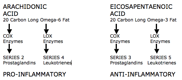
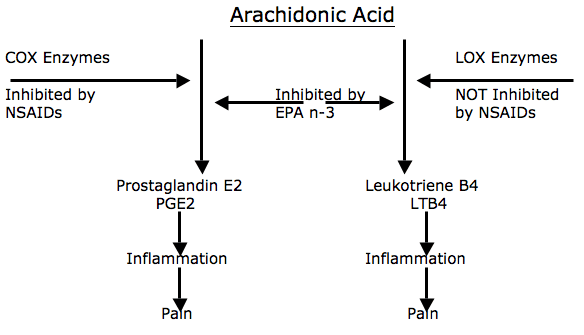
    This phase lasts up to 72 hours. It is characterized by vasodilation, immune system activation of phagocytosis to remove debris, and the release of prostaglandins which cause inflammation. Prostaglandins also play a prominent part in pain production and increased capillary permeability (swelling). During this phase the wound is hypoxic, but macrophages can perform the phagocytosis duties anaerobically.

    Phase 2: The Repair or Regeneration Phase

    This phase lasts 48 hours to 6 weeks. It is characterized by the synthesis and deposition of collagen. The collagen that is deposited during this phase is not fully oriented in the direction of tensile strength, but rather laid down in an irregular pattern, different from that of the original tissue. This phase is “largely one of increasing the quantity of the collagen” but this collagen is not laid down in the direction of stress. Additionally, collagen fibers tend to contract between 3 and 14 weeks after injury, and perhaps for as long as 6 months, decreasing tissue elasticity.

    Phase 3: The Remodeling Phase

    This phase blends with the latter part of Phase 2, the Regeneration Phase, and may last up to 12 months or more. During this phase, the collagen is remodeled to increase the functional capabilities of the tendon or ligament to withstand the stresses imposed upon it. This phase is largely “an improvement of the quality” (orientation and tensile strength) of the collagen. It appears that the tensile strength of the collagen is quite specific to the forces imposed on it during the remodeling phase: i.e. the maximum strength will be in the direction of the forces imposed on the ligament.

    This brief review by Dr. Kellett immediately suggests that immobilization of injured soft tissues would be detrimental to the healing process and that early mobilization would improve the timing and quality of healing. In fact, Dr. Kellett makes the following statements:

    “Early mobilization, guided by the pain response, promotes a more rapid return to full activity.”

    “Early mobilization, guided by the pain response, promotes a more rapid return to full functional recovery.”

    “Progressive resistance exercises (isotonic, isokinetic, and isometric) are essential to restore full muscle and joint function.”

    Dr. Kellett advocates the use of early mobilization, based upon the premise that the stress of movement on repairing collagen is largely responsible for the orientation and tensile strength of the tendons and ligaments. The stressing of repairing tissues with controlled motion induces adaptive response to functionally stronger connective tissues. According to Dr. Kellett, the benefits of early mobilization include:

    1) Improvement of bone and ligament strength, which reduces recurrence of injury.

    2) The strength of repaired ligaments is proportional to the mobility of the ligament, resulting in larger diameter collagen fiber bundles and more total collagen.

    3) Collagen fiber growth and realignment is stimulated by early tensile loading of muscle, tendon, and ligament.

    4) Collagen formation is not confined to the healing ligaments, but adheres to surrounding tissues. The formation of these adhesions between repairing tissues and adjacent structures is minimized by early movement.

    5) With motion, “joint proprioception is maintained or develops earlier after injury, and this may be of importance in preventing recurrences of injuries and in hastening full recovery to competitive fitness.”

    6) The nutrition to the cartilage is better maintained with early mobilization.

    In contrast, Dr. Kellett notes that immobilization of soft tissue injuries decreases aerobic capacity, causes muscle wasting and loss of strength that may delay full recovery for a year or more.

    At the microscopic level, Dr. Kellett notes that the healing of injured soft tissues usually contains granulation, fibrotic, or scar tissue. It is these poor quality-healing components that often account for residual reductions of strength and elasticity, impairing functional capacity and accounting for residual symptoms. Sports physicians Steven Roy and Richard Irvin note in their 1983 text titled Sports Medicine: Prevention, Evaluation, Management, and Rehabilitation (2) that early motion helps to prevent these fibrotic soft tissue changes. They state:

    “The injured tissues next undergo remodeling, which can take up to one year to complete in the case of major tissue disruption. The remodeling stage blends with the later part of the regeneration stage, which means that motion of the injured tissues will influence their structure when they are healed. This is one reason why it is necessary to consider using controlled motion during the recovery stage. If a limb is completely immobilized during the recovery process, the tissues may emerge fully healed but poorly adapted functionally, with little chance for change, particularly if the immobilization has been prolonged. Another reason for encouraging controlled motion is that any adhesions that develop will be flexible and will thus allow the tissues to move easily on each other.”

    Additional support for early mobilization in the management of soft tissue injuries is from physician James Cyriax. In his 1982 text Orthopaedic Medicine, Diagnosis of Soft Tissue Lesions, Dr. Cyriax states (3):

    “Tension within the granulation tissue lines the cells up along the direction of stress. Hence, during the healing of mobile tissues, excessive immobilization is harmful. It prevents the formation of a scar strong in the important direction by avoiding the strains leading to due orientation of fibrous tissue and also allows the scar to become unduly adherent, e.g. to bone.”

    ••••

    In 2000, physician Pekka Kannus, MD, PhD, adds to the understanding of the healing of inured soft tissues by employing immobilization or early mobilization. Dr. Kannus is chief physician and head of the Accident and Trauma Research Center and sports medicine specialist at the Tampere Research Center of Sports Medicine at the UKK Institute in Tampere, Finland. His article on the subject was published in the journal The Physician and Sports Medicine, and titled (4):

    Immobilization or Early Mobilization
    After an Acute Soft-Tissue Injury?

    In this article, Dr. Kannus states:

    “Experimental and clinical studies demonstrate that early, controlled mobilization is superior to immobilization for primary treatment of acute musculoskeletal soft-tissue injuries and postoperative management.”

    “The current literature on experimental acute soft-tissue injury speaks strongly for the use of early, controlled mobilization rather than immobilization for optimal heating.”

    “Controlled clinical trials of acute soft-tissue injuries support the results of experimental studies and have shown that early controlled mobilization is superior to immobilization, not only in primary treatment, but also in postoperative management.”

    “The superiority of early controlled mobilization has been especially clear in terms of quicker recovery and return to full activity without jeopardizing the subjective or objective long-term outcome.”

    “Evidence has been systematic and convincing for many injuries.”

    Importantly, Dr. Kannus indicates that in animal studies, immobilization for longer than one week resulted in marked atrophy of the injured muscle. This article, adds to the support for using early mobilization following soft tissue injury and avoiding immobilization.

    ••••

    In 1986, orthopedist K Mealy and colleagues specifically looked at the clinical outcomes of patients who received a cervical collar versus early mobilization for the treatment of acute soft tissue whiplash injuries. They published their study in the British Medical Journal in an article titled (5):

    Early Mobilization of Acute Whiplash Injuries

    This study is a prospective randomized trial where 61 consecutive patients with acute whiplash injuries were randomized to receive active treatment (31 patients) or standard (cervical collar) treatment (30 patients). The group assigned to receive active treatment received applications of ice in the first 24 hours and then neck mobilization and daily exercises of the cervical spine. Daily exercises were performed every hour at home, within the limits of pain; no analgesia was needed for this mobilization treatment or the exercises. The group given standard treatment received a soft cervical collar and was advised to rest for two weeks before beginning gradual mobilization. Their findings are summarized below:

    “Though pain in both groups was similar initially, pain in the group given active treatment was significantly less than that in the group given standard treatment at both four weeks and eight weeks.”

    “Movement increased significantly in the group given active treatment at four weeks and eight weeks.” “At eight weeks movement in the group given active treatment was significantly greater than that in the group given standard treatment.”

    “Many patients with whiplash injuries present late, after a period of immobility, with persistent pain and stiffness.”

    “We found that patients who are treated actively show significantly greater improvement in both cervical movement and intensity of pain compared with patients treated in the standard way.”

    “At four weeks a significant increase in cervical movement occurred in the patients given active treatment but not in those given standard treatment. At eight weeks cervical movement was significantly greater in the patients given active treatment than those given the standard treatment, indicating that the increase in cervical mobility occurred earlier and to a significantly greater degree with active treatment.”

    “At both four and eight weeks the improvement in pain was significantly greater in the group given active treatment, so that these patients had significantly less pain at four and eight weeks compared with the patients given standard treatment.”

    “In conclusion, our results confirmed expectations that initial immobility after whiplash injuries gives rise to prolonged symptoms whereas a more rapid improvement can be achieved by early active management without any consequent increase in discomfort.”

    This article is the first prospective randomized clinical trial addressing the outcomes of whiplash-injured patients who are treated with a cervical collar or with early mobilization. Clearly, early mobilization was superior to the cervical collar in this study.

    ••••

    Physical therapist Mark Rosenfeld and colleagues published another study specifically evaluating the outcomes of whiplash-injured patients with either a cervical collar or with early mobilization. Their study appeared in the journal Spine in 2000, and is titled (6):

    Early Intervention in
    Whiplash-Associated Disorders
    A Comparison of Two Treatment Protocols

    This study is a prospective randomized trial in 97 patients with a whiplash injury caused by a motor vehicle collision. The study evaluates early active mobilization versus a standard treatment protocol that included the use of a cervical collar. These patients were followed-up and reevaluated at six months.

    Rosenfeld and colleagues indicate that there are three studies showing the detrimental effect of rest and use of a cervical collar in the treatment of whiplash-injured patients. Yet, this type of treatment is still commonly recommended to patients in the early management of whiplash injuries.

    The active treatment group was instructed to perform gentle, active, small-range and amplitude rotational movements of the neck, first in one direction, then the other. The movements were repeated 10 times in each direction every waking hour.

    The standard treatment group was told that a soft collar could provide comfort and prevent the neck from excessive movement. They were also instructed to begin performing active movements, two or three times daily a few weeks after the injury.

    Improvements during the first 6 months after trauma in range of motion and pain level were compared between the groups. “Evaluation of the two treatment protocols showed that 6 months after treatment, the reduction in pain was greater for those receiving active treatment than in those receiving standard [cervical collar] treatment.” The authors state:

    “The main finding in this study was that active treatment of WAD resulted in a significantly greater pain reduction than standard treatment.”

    “It seems that active mobilization is important. The current result confirms that frequent active mobilization exercises decrease symptoms more than a gradual mobilization program. Thus, a soft collar for the first period after injury is not the best treatment for WAD.”

    “In patients with WAD caused by a motor vehicle collision, early treatment with frequently repeated active submaximal movements combined with mechanical diagnosis and therapy is more effective in reducing pain than treatment with initial rest, recommendation of a soft collar, and a gradual introduction of home exercises.”

    In this study by Rosenfeld and colleagues, once again, early mobilization was clearly superior to the cervical collar in the treatment of acute whiplash injury.

    Importantly, Mark Rosenfeld and colleagues published a three-year prospective follow-up as to the clinical status of these original 97 patients. This study was also published in the journal Spine in 2003, and titled (8):

    Active Intervention in Patients with Whiplash-Associated Disorders Improves Long-Term Prognosis:
    A Randomized Controlled Clinical Trial

    At this three-year follow-up, the authors found that pain intensity and sick leave were significantly reduced if patients received active intervention compared to those treated with a cervical collar. In addition, only patients receiving early active intervention had a total cervical range of motion similar to that of matched unexposed individuals. The authors concluded:

    “In patients with whiplash-associated disorders, active intervention is more effective in reducing pain intensity and sick leave, and in retaining/regaining total range of motion than a standard intervention [cervical collar].”

    “The reduction of pain intensity was greater and the need for sick leave was lower for patients receiving active intervention than in those receiving the standard [cervical collar] intervention.”

    “The main finding in this study was that active intervention in patients with whiplash associated disorders resulted in a significantly greater reduction in pain intensity, a greater chance to retain or regain cervical range of motion, and reduced sick leave compared with a standard intervention.”

    “The main clinical implication is that patients with acute WAD 0, 1, or 2 should be instructed in self-mobilization as soon as possible.”

    Rosenfeld and colleagues propose that the most effective active motion is rotational, which “encourages regional blood flow and facilitates the removal of exudate, thus allowing healing to occur by aiding nutrition of joint structures.” Cervical spine rotation mobilizes the nerve structures on the contralateral side, thus preventing scar tissue from forming adhesions that will later cause dysfunction. Rotation mobilization may also affect the possible inhibition of intraneural microvascular blood flow caused by compression.

    ••••

    The most comprehensive and referenced guideline pertaining to whiplash disorders was published in the journal Spine in 1995, and is titled (9):

    Scientific monograph of the Quebec Task Force on Whiplash-associated Disorders: Redefining ‘Whiplash’ and its Management

    A comprehensive summary of this Quebec Task Force on Whiplash-associated Disorders was published in the May, 1998 Spine State of the Art Reviews (11) by Task Force members Richard Leclaire, a physiatrist, and Guy Bouvier, a neurosurgeon. Both authors are from Montreal, Quebec, Canada. They review the history and details of the creation of the Quebec Task Force on Whiplash-Associated Disorders. Both the original Task Force and its summary by Leclaire and Bouvier contain treatment guidelines. Pertaining to the use of cervical collars in the management of whiplash injury, they note that in the three randomized controlled trials cited, the groups receiving soft collars had delayed recovery in terms of pain rating and range of motion compared to the groups receiving mobilization. They state:

    “Collars may promote inactivity, which can delay recovery in patients with WAD.”

    They conclude that in the treatment of whiplash injuries, cervical collars should not be used for grade I whiplash associated disorders and that for grade II or III, cervical collars should be used for no more than 72 hours. Pertaining to rest, the authors state:

    “The cumulative evidence suggests that prolonged periods of rest are detrimental to recovery from whiplash associated disorders.”

    “Rest should not be prescribed for whiplash associated disorders I. Rest longer than 4 days should not be prescribed for whiplash associated disorders II and III.”

    ••••

    In 2007, orthopedic surgeon Jerome Schofferman and colleagues published an evidence-based review of chronic whiplash disorders. Their articles appeared in the Journal of the American Academy of Orthopedic Surgeons, and was titled (11):

    Chronic whiplash and whiplash-associated disorders: An evidence-based approach

    In this evidence-based review of the literature pertaining to whiplash injuries, Dr. Schofferman and colleagues make the following points:

    1) Following a motor vehicle collision, up to 40% of patients with acute neck pain develop chronic neck pain.

    2) Following a motor vehicle collision, up to 7% of patients become permanently partially or totally disabled.

    2) The cervical facet joint is the most common source of chronic neck pain after whiplash injury, followed by disk pain. Some patients experience pain from both structures.

    3) Treatment includes advising the patient to remain active.

    4) Recovery rates from whiplash trauma are:

    A)) 44% are not recovered at 3 months.

    B)) 30% are not recovered at 6 months.

    C)) 24% are not recovered at 12 months.

    D)) 18% are not recovered at 24 months.

    Dr. Schofferman and colleagues make the following prognostic points:

    1) Compensation or litigation is not associated with an adverse prognosis.

    2) Older age, female sex, radicular symptoms, multiple areas of pain, and being unprepared for impact are associated with a worse prognosis.

    3) “There is no pre-injury personality that renders individuals more likely to develop chronic pain after whiplash.”

    4) There are no psychosocial factors that are useful predictors of whiplash injury chronicity.

    5) There is no correlation between initial psychological testing and whiplash chronicity.

    6) In most instances, psychological problems are a “consequence rather than a cause of pain.”

    7) “There was no difference in the prevalence of pre-injury psychological illness in those who did or did not return to work.”

    8) “The strongest predictor of prolonged disability [after whiplash injury] was intensity of [initial] symptoms.”

    9) Differences in speed between vehicles is not predictive of whiplash symptom chronicity.

    10) “Multiple studies have demonstrated that personal injury litigation does not adversely affect the outcome of whiplash injury.”

    11) Patients with more pain and more objective findings are more likely to file litigation claims.

    12) There is no evidence for symptom improvement after litigation is settled.

    Dr. Schofferman and colleagues indicate that treatments for acute neck pain include remaining active despite ongoing pain, performance of prescribed exercises, and possible inclusion of spinal manipulation, which can improve outcomes over exercise alone.

    In the treatment of chronic neck pain, “exercise alone is rarely curative” but exercise is helpful, and should be prescribed. The exercise should be directed to strengthen the weak muscle groups, usually the anterior, posterior, and interscapular muscle groups. Strength training is superior to stretching exercises, and intense exercise is superior to light exercise.

    Spinal manipulation is one of the most popular treatments for chronic neck pain, and it can be beneficial. Serious complications from spinal manipulation are “quite low.”

    Lastly, Dr. Schofferman and colleagues state that cervical collars are of “no value in the patient with acute whiplash injury.”

    ••••

    The modern chiropractor is extensively trained in the application of early mobilization, exercise, and manipulation techniques to properly manage both the acute and chronic whiplash-injured patient. The application of these management techniques include:

    1) Dispersion of the accumulation of inflammatory exudates that increase pain. This accelerates the timing of the healing process.

    2) Puts controlled motion into the developing granulation tissue. This helps in the alignment of the tissues in the directions of stress and strain. This improves the quality of the end product of healing, reducing the chances that future increased stress will result in an exacerbation of signs and symptoms.

    3) Reduces the formation of adhesions that often cause prolonged disability after soft tissue injury and healing.

    REFERENCES:

    1. Kellett J; Acute soft tissue injuries–a review of the literature; Medicine and Science in Sports and Exercise; October 1986;18(5):489-500.
    2. Roy, Steven, M.D., and Irvin, Richard, Sports Medicine: Prevention, Evaluation, Management, and Rehabilitation, Prentice-Hall, Inc. (1983).
    3. Cyriax, James, M.D., Orthopaedic Medicine, Diagnosis of Soft Tissue Lesions, Bailliere Tindall, Vol. 1, (1982).
    4. Pekka Kannus, MD, PhD; Immobilization or Early Mobilization After an Acute Soft-Tissue Injury? The Physician And Sports Medicine; March, 2000; Vol. 26 No 3, pp. 55-63.
    5. K Mealy, H Brennan, GCC Fenelon; Early mobilisation of acute whiplash injuries; British Medical Journal; Vol. 292, March 8, 1986, pp 656-657.
    6. Mark Rosenfeld; Ronny Gunnarsson; Peter Borenstein; Early Intervention in Whiplash-Associated Disorders: A Comparison of Two Treatment Protocols; Spine, 2000;25:1782-1787.
    7. Bunketorp L, Nordholm L, Carlsson J. A descriptive analysis of disorders in patients 17 years following motor vehicle accidents. European Spine Journal, 2002; 11: 227-34.
    8. Mark Rosenfeld, RPT; Aris Seferiadis, RPT; Jane Carlsson, RPT, PhD; Ronny Gunnarsson, MD, PhD; Active Intervention in Patients with Whiplash-Associated Disorders Improves Long-Term Prognosis: A Randomized Controlled Clinical Trial; Spine 2003; 28(22):November 15, 2003; 2491-2498.
    9. Spitzer WO, Skovron ML, Salmi LR; Scientific monograph of the Quebec Task Force on Whiplash-associated Disorders: Redefining ‘whiplash’ and its management; Spine; April 15, 1995, 20 (supplement 8): 1S-73S.
    10. Leclaire R, Bouvier G, A summary of the Quebec Task Force on Whiplash-Associated Disorders: Classification, evaluation, and treatment guidelines, Spine State of the Art Reviews, Vol. 12, No. 2, 1998, 271-286.
    11. Schofferman J, Bogduk N, Slosar P. Chronic whiplash and whiplash-associated disorders: An evidence-based approach; Journal of the American Academy of Orthopedic Surgeons; October 2007;15(10):596-606.


  • A Review of the Life and Science of Ruth Jackson, MD

    A Review of the Life and Science of Ruth Jackson, MD

    Dr. Ruth Jackson had such an accomplished life that shortly after her death in 1994 she was profiled in both the American Journal of Bone and Joint Surgery (1) in 1995 and in the Iowa Orthopedic Journal in 2001 (3).

    Dr. Jackson’s profile in the Journal of Bone and Joint Surgery was simply titled:

    RUTH JACKSON, M.D. 1902-1994

    “Ruth Jackson, a prominent orthopaedic surgeon, died on August 28, 1994, in Dallas, Texas.”

    Ruth Jackson was born in 1902, graduated from high school in 1920, and attended the University of Texas at Austin, where her major was sociology. In her junior year, while on a class assignment to see if a poor family needed groceries, she noted that the father could not work because of a knee problem. In her desire to help with such problems she switched to a premedical major, graduating in 1924 with a Bachelor’s degree in zoology and a minor in chemistry. She then entered Baylor University College of Medicine in Dallas as one of only four women in a class of 164.

    “While in medical school, Ruth protested that women were not allowed to examine male patients and suggested that a woman planning to practice medicine required the same firsthand knowledge of men as a future male physician needed of women. She graduated in 1928, ranked eighth in her class.”

    Dr. Jackson accepted a residency in orthopaedics at the University of Iowa, and began working with crippled children. She began her private practice in Dallas in 1932.

    “When The American Academy of Orthopaedic Surgeons was founded in 1933, all practicing male orthopaedic surgeons were automatically admitted as members. However, Dr. Jackson was told that she could become a member only if she passed the Board examination. Undaunted, she completed this requirement in 1937 and thus became the first woman to be admitted to The Academy. She was also the first woman to be certified by the American Board of Orthopaedic Surgery.”

    “From 1936 to 1941, Dr. Jackson was Chief of Orthopaedics at Parkland Hospital in Fairmont. In 1945, she had her own private clinic built in Dallas. The clinic comprised several examining rooms, an operating room, physical therapy facilities, and a radiology suite. She conducted her medical practice in these offices for the next thirty-eight years, performing all operations, including lumbar discectomy, with use of local anesthesia.”

    Dr. Jackson had a special interest in injuries of the cervical spine. Her interest arose after a neck injury she sustained in a motor-vehicle accident. To treat these “whiplash” injuries, she invented the Jackson Cervi-Pillow. In 1956 she published her acclaimed, authoritative book entitled The Cervical Syndrome. The fourth and final edition of her book was published in 1978 (2).

    In her career, Dr. Jackson published more than twenty-five articles, and she lectured extensively in the United States and throughout the world. Two of her articles pertaining to cervical spine trauma are reviewed below.

    Dr. Jackson’s other noted professional accomplishments include:

    • She started The Ruth Jackson Research Foundation at Baylor University.
    • She funded the Ruth Jackson Visiting Professorship at Baylor University, starting in 1961.
    • She established the Ruth Jackson Library at Baylor University.
    • The Ruth Jackson Orthopaedic Society was founded in 1983. It currently has more than 500 members. This organization salutes her pioneering spirit and her accomplishments and seeks to advance the careers of women in orthopaedics.

    Dr. Jackson retired from clinical practice in 1989 at the age of 87 years.

    ••••

    Dr. Ruth Jackson’s profile in the Iowa Orthopedic Journal was titled (3):

    RUTH JACKSON, M.D.:
    A WOMAN OF DETERMINATION AND ACCOMPLISHMENT

    Additional information from this profile indicates that her whiplash injury occurred in 1936. Subsequently:

    “She had significant cervical pain for several years and that experience stimulated her interest in cervical spine problems.”

    The article indicates that Dr. Jackson had treated over 15,000 patients with cervical spine problems.

    ••••

    In her career, Dr. Jackson published six studies in the medical literature pertaining to cervical spine trauma, as follows:

    1) The Cervical Syndrome As a Cause of Migraine. Journal of the American Medical Women’s Association. December 1947, Vol. 2, No. 12, pp. 529-534.

    2) The cervical syndrome. Dallas Medical Journal. 1949 Oct;35(10):139-46.

    3) The Positive Findings in Alleged Neck Injuries. American Journal of Orthopedics. 1964 Aug-Sep;6:178-87.

    4) Headaches associated with disorders of the cervical spine. Headache. 1967 Jan;6(4):175-9.

    5) Traumatic arthritis of the cervical spine. Comprehensive Therapy. 1981 Feb;7(2):58-61.

    6) Cervical trauma: not just another pain in the neck. Geriatrics. 1982 Apr;37(4):123-6.

    Two of these articles are reviewed below:

    The Cervical Syndrome As a Cause of Migraine

    Journal of the American Medical Women’s Association
    December 1947, Vol. 2, No. 12, pp. 529-534

    In this article, Dr. Jackson makes the following key points:

    1) At least half of patients suffering from cervical syndrome causing shoulder disability will also complain of headache as one of their principle symptoms.

    2) The cervical syndrome is caused by “cervical nerve root irritation.”

    3) There is a relationship between the cervical syndrome, cervical nerve root irritation, and the sympathetic nervous system. It is this sympathetic nervous system involvement that is responsible for headaches, including migraine.

    4) “The cervical nerve roots are more vulnerable to pressure or irritation from ruptured discs, hemorrhage, inflammatory processes of the capsules, spurs, and abnormal motion of the joints due to relaxation or tearing of the capsular and ligamentous structures.”

    5) Sympathetic nerves that originate at the T1 and T2 levels ascend into the cervical spine becoming the cervical sympathetic trunk, with its inferior, middle, and superior ganglia. From the superior cervical sympathetic ganglia, post-ganglionic sympathetic efferents pass to the anterior rami of the upper four cervical nerves. Other postganglionic sympathetic fibers travel via the internal carotid and ophthalmic arteries to join the orbit and supply the dilator muscle of the pupil and smooth muscles of the upper eyelid, the back of the orbit, and the blood vessels of the eyeball. It is the irritation of these sympathetic nerves in the cervical spine that gives rise to migraine-type headaches.

    6) “Irritation of the cervical nerve roots before they divide into anterior and posterior primary rami may occur as a result of any mechanical derangement in or about the intervertebral foramina. The most common cause of irritation is abnormal motion or subluxation of the joints due to relaxation of the capsular and ligamentous structures following trauma.”

    7) Following whiplash trauma, as time passes, “abnormal motion or subluxations of the articular processes, will cause irritation of one or more nerve roots.”

    8) “Any unguarded motion or prolonged relaxation of the neck in one position may allow a subluxation to occur,” which may cause cervical nerve root irritation.

    9) Cervical trauma can cause chronic pain syndrome, including pain decades later.

    10) Approximately 2/3 of the patients with neck symptoms are women.

    11) As a consequence of increased mobility and reduced stability of the upper cervical spine, the C1-C2-C3 nerve roots are most vulnerable to irritation. When the C1-C2-C3 nerve roots are involved, typical symptoms include:

    A)) Neck pain

    B)) Limitation of neck motion

    C)) Headache, which usually “starts at the back of the neck or base of the skull and then involves one or both sides of the head, and pain in the mastoid region or in the ears”

    D)) Blurring of the vision (found in 20% of patients)

    E)) Dizziness and nausea

    F)) Numbness of the sides of the neck

    G)) Tightness of the neck muscles

    H)) Pain in the supraclavicular region

    12) “If C4 is involved there may be shortness of breath, palpitations, anterior chest pain and pain and muscle spasm in the muscles supplied by C4.”

    13) “When the lower nerve roots are involved, the symptoms are commensurate with the segmental character of the nerve roots which are irritated.” These patients often have numbness or tingling of the fingers when they awaken.

    14) “There is always tenderness to deep pressure over the vertebrae, usually just lateral to the spinous process of the side of the nerve root irritation.” “If the irritation is above the fourth nerve root there may be tender areas over the occiput and the mastoid. If the fourth nerve root is involved, there may be tender areas in the ridge of the trapezius and/or in the sternomastoid muscles.” Irritation of the fourth nerve root may also cause referred pain to the diaphragm, the pericardium, and shoulder.

    15) “The most constant finding in all of our cases was the presence of myalgic areas in the upper or lower cervical portion of the rhomboid muscles which is indicative of fifth nerve root irritation.”

    16) “Often there seems to be localized fibrosis of the muscle. Irritation causes spasm which if allowed to persist causes ischemia with eventual formation of localized fibrosis.”

    17) Dr. Jackson stresses the importance of cervical spine x-rays in cases of cervical spine trauma. She especially emphasizes the importance of maximum flexion and maximum extension lateral views. The abnormal forward or backward slipping of a vertebral segment on flexion or extension is called a “subluxation.”

    18) 96% of patients with cervical syndrome headaches will show subluxations at more than one level, and in 77% the subluxation was of C2 on C3, irritating the C3 nerve root. “This indicates that the irritation of the third cervical nerve root must have been responsible for the greatest percentage of headache.” “Cervical nerve root irritation (C3 usually) is an etiological factor in migraine.”

    19) Dr. Jackson believes that the blurring of the vision (and ipsilateral pupil dilation, when present) seen in many cervical syndrome/headache patients is caused by irritation of the superior sympathetic ganglion caused by cervical muscle spasm.

    20) Irritation of the cervical nerve roots will cause spasm of the associated cervical spine muscles. Spasm of the muscles that surround the superior sympathetic ganglia causes irritation to both the ganglia and/or the postganglionic sympathetic fibers. This sympathetic irritation can in turn cause symptoms of migraine-like headache.

    21) Injured patients should use a cervical contour pillow for rest and healing. “This pillow has been our greatest adjunct in the treatment of cervical nerve root irritation.”

    SUMMARY AND CLINICAL SIGNIFICANCE

    In this article, Dr. Jackson stresses that the physiological basis for neck-shoulder-headache complaints is cervical nerve root irritation. She stresses that the cervical nerve roots are quite vulnerable to pressure or irritation from ruptured discs, hemorrhage, inflammatory processes of the capsules, spurs, and abnormal motion of the joints due to relaxation or tearing of the capsular and ligamentous structures.

    Irritation of the cervical nerve roots occurs as a result of any mechanical derangement in or about the intervertebral foramina. The most common cause of irritation is abnormal motion or subluxation of the joints due to relaxation of the capsular and ligamentous structures following trauma. Following whiplash trauma, as time passes there is an increased probability of abnormal motion or subluxations of the articular processes, which will cause irritation of one or more nerve roots. Additionally, any unguarded motion or prolonged postural distortion of the neck may allow a subluxation to occur, causing irritation of a cervical nerve root.

    Irritation of the cervical nerve roots will cause spasm of the associated cervical spine muscles. Spasm of the muscles that surround the superior sympathetic ganglia causes irritation to both the ganglia and/or the postganglionic sympathetic fibers. This sympathetic irritation can in turn cause symptoms of migraine-like headache.

    Additionally, this nerve root irritation and attendant muscle spasm, if not properly treated, causes muscle ischemia with eventual formation of localized fibrosis. This muscle spasm can prolong symptoms and lead to chronicity of symptoms.

    Diagnostically, Dr. Jackson insists on acquiring cervical spine stress (maximum flexion-extension) radiographs. She notes that such x-rays can display abnormal forward or backwards slippage of the vertebral segments. Such abnormal movement is once again associated with increased nerve root irritation and symptoms.

    A physical diagnostic indicator of cervical nerve root irritation is tenderness to deep pressure over the vertebrae, usually just lateral to the spinous process of the side of the nerve root irritation.

    Fortunately, post-traumatic cervical nerve root irritation and its associated abnormal segmental mobility (subluxations), muscle spasm, and even chronic muscle fibrosis are treatable. Chiropractic approaches to such problems include passive manual mobilizations, exercise, soft tissue work, myotherapy, traction, and joint manipulation (adjustments).

    ••••

    The Positive Findings In Neck Injuries

    American Journal of Orthopedics
    August-September, 1964, pp. 178-187

    In this article, Dr. Jackson makes the following key points:

    1) 90% of neck disorders are caused by trauma.

    2) Of the trauma cases, 85% are caused by motor vehicle collisions.

    3) “The forces which are imposed on the cervical spines of the passengers of colliding vehicles are tremendous, and if one attempts to calculate mathematically the amount of such forces, the results are unbelievable.”

    4) Dr. Jackson emphasizes the lack of relationship between vehicle damage and passenger injury. She states:

    “The damage to the vehicles involved in collisions is no indication of the extent of the injuries imposed on the passengers.”

    The extent of damage to the vehicles is in no way proportional to the extent of damage imposed upon the cervical spines of the passengers.”

    5) “Healing of sprained ligamentous structures takes place by the formation of scar tissue which is less elastic and less functional than normal ligamentous tissue.” Therefore, sprains result in some degree of permanent injury.

    6) “Injuries of the intervertebral disc structures are frequent and they may vary from a slight rent in the annulus fibrosis to a complete avulsion of the disc from its attachments.”

    7) “Any injury of the disc causes a disturbance in the dynamics of the motor unit of which the disc is a part. This leads to degeneration of the disc and the proximate joints.”

    8) The nerve roots, the cervical sympathetic nerves and the spinal cord may suffer injuries of varying degrees.

    9) The location of segmental motion problems can be verified by lateral radiographs of the cervical spine in maximum flexion and extension.

    10) Upper extremity deep tendon reflexes may be hyperactive immediately following neck injury, but after a few days they may become hypoactive.

    11) Superficial sensation may initially be hyperesthesia, but after a few weeks may become hypoesthetic.

    12) Grip strength should be measured by a dynamometer, and should be performed 4 times in sequence to look for muscle fatigue. Normally each of the 4 readings should be essentially the same.

    13) Unilateral dilation of the pupil “indicates irritation of the sympathetic nerve.”

    14) Whiplash-caused partial deafness may be improved with cervical traction.

    15) “Irritation of the cervical sympathetic nerve supply may give rise to vasoconstriction of the arteries which are supplied by the sympathetic fibers.” Blood pressure in the two arms often varies as much as 10 – 20 points following neck injuries. This is a completely objective finding.

    16) Pre-existing pathological conditions of the cervical spine, when injured, “result in more damage than would be anticipated in a so-called ‘normal’ cervical spine.”

    17) Dr. Jackson notes that in order to ascertain injuries, an “adequate radiographic examination is essential.” She recommends 8 x-rays be exposed following whiplash trauma, including an AP cervical with a “caudad-angled view.” This view “may show fractures of the laminae and of the inter-articular isthmuses which may not be demonstrable in any other view.” She also notes that initial x-rays may be normal, but subsequent x-rays may reveal multiple problems; therefore, repeat x-rays are essential. Dr. Jackson states:

    “All radiographs should be repeated periodically. Subsequent findings may be very revealing.”

    18) X-rays showing “any segmental instability, any segmental restriction of motion, and any segmental angulation may indicate severe injury.”

    19) The intervertebral foramina may suffer from encroachment as the result of “hemorrhage, swelling, or hyperplasia of the proximate capsular and ligamentous structures,” even in the presence of normal oblique x-rays.

    21) The alar ligaments of the upper cervical spine are very vulnerable to injury.

    22) “Repeated physical and radiographic examinations are necessary for final analysis.”

    SUMMARY AND CLINICAL SIGNIFICANCE

    Dr. Ruth Jackson was one of the most experienced clinicians in world history pertaining to whiplash trauma to the cervical spine. In this article she repeatedly states the observation that there is no relationship between vehicle damage and passenger injury. She also makes the important observation that pre-existing pathological conditions of the cervical spine, when injured, “result in more damage than would be anticipated in a so-called ‘normal’ cervical spine.” Although these observations were made nearly half a century ago, these concepts remain contentious today and a point of frustration for both patients and clinicians who are thrust into the med-legal arena when caring for whiplash-injured patients.

    Once again, Dr. Jackson emphasized the relationship between cervical spine trauma and irritation of the cervical nerve roots. Cervical spine nerve root irritation often affects the function of the sympathetic nervous system, either directly or indirectly as a consequence of muscle spasm. Sympathetic nerve irritation is in turn associated with cranial symptoms, including headaches.

    Sadly, Dr. Jackson observes that injured soft tissues often display healing problems, helping to explain why some whiplash-injured patients suffer from long-term symptoms. She states:

    “Healing of sprained ligamentous structures takes place by the formation of scar tissue which is less elastic and less functional than normal ligamentous tissue.”

    “Sprains result in some degree of permanent injury.”

    In this article, Dr. Jackson establishes the pathophysiological basis for manual therapy. She states:

    “Any injury of the disc causes a disturbance in the dynamics of the motor unit of which the disc is a part. This leads to degeneration of the disc and the proximate joints.”

    “Disturbances in the dynamics of the motor unit” is classic terminology for the chiropractic subluxation complex. The primary goal for manual manipulative therapy (adjustment) is to improve the motor unit functional disturbance.

    Additionally, Dr. Jackson notes that traumatic inflammation “leads to post-traumatic fibrosis and adhesions.” Both fibrosis and adhesions are mechanical lesions that impair motion and function. Chiropractic manual manipulative adjustive approaches have been shown to benefit these mechanical lesions.

    ••••

    The greatest achievement of Dr. Ruth Jackson’s incredible life is the publication of her book The Cervical Syndrome. The fourth and final edition of this book was published in 1978. The text is 399 pages in length and contains 158 Figures. The majority of these Figures involve sequential series of radiography showing the acceleration of degenerative joint disease as a time consequence of sustaining a cervical spine trauma.

    When this book was reviewed by the Journal of the American Medical Association, it stated:

    “This valuable book should be useful to the general practitioner, orthopedist, and neurosurgeon.”

    The back cover of this book states:

    “Treatment methods are dealt with in the final portion of the text. This section stresses more fully the importance of prevention and updates its reportage of proven therapeutic techniques. Nonsurgical treatment modalities covered span a wide area, from heat to massage, to traction, the cervical contour pillow and drugs, as well as exercises and manipulation.”

    “Residents and practitioners in orthopaedics, of course, will want to take advantage of the knowledge presented in this outstanding text. It will also be useful to neurologists, radiologists, physical medicine and rehabilitation specialists, and general practitioners. Chiropractors and osteopathic physicians will find a wealth of information applicable to their practices.”

    In this text, Dr. Jackson once again established the pathophysiological basis for manual manipulative therapy. She states:

    “Healing occurs by the formation of scar tissue, which is less elastic and less functional than normal ligamentous tissue.”

    “Immobilization of joints or functional inactivity results in stasis of circulation and is a common cause of posttraumatic joint stiffness. A small amount of movement in injured joints, rather than complete immobilization, reduces inflammation by mobilizing or dispersing the tissue breakdown products from the site of injury and prevents to some extent fibrous arthrosis.”

    In the treatment section of her book, Dr. Jackson advocates many approaches and options, including heat and/or cold, massage, traction, postural correction, a cervical contour pillow to restore cervical lordosis during healing, vitamin C to aid healing, postural advice (while sleeping, working, reading, writing, driving, sitting, shaving, drinking), exercise, and manipulation. Pertaining to manipulation, Dr. Jackson states:

    “Joint dysfunction is the result of loss of movement which is involuntary, and that movement can only be restored by being reproduced by the manipulator who restores normal function by the use of normal movement. There are many manipulators who manipulate the cervical spine and who claim excellent results.”

    “Manipulation of the cervical spine should not be undertaken by any person who is not well versed in the anatomy of this very complex structure.”

    Of course, chiropractors agree with these statements by Dr. Jackson. Chiropractors contend that their manipulative approach to joint problems is essential to restore normal function and movement when an involuntary loss of normal motion exists. Chiropractors study four years of anatomy, mechanics, orthopedics, neurology, radiology, clinical coaching as well as clinical applications to identify and safely manage these types of joint dysfunctional problems.

    Dr. Jackson concludes that with proper treatment, most patient’s symptoms will subside within a period of three to six months. She also acknowledges that no two patients will respond exactly the same, and that “some treatment may be necessary in some cases over a longer period of time.”

    References

    1) R. M. and L. R., editors, Journal of Bone and Joint Surgery (American), RUTH JACKSON, M.D.: 1902-1994; VOL. 77-A, NO. 2. February 1995, pp. 331.

    2) Jackson R, The Cervical Syndrome, Thomas, 1978.

    3) Zillmer DA; Ruth Jackson, M.D.: A Woman of Determination and Accomplishment; The Iowa Orthopaedic Journal; Volume 21, 2001.

  • Whiplash Injuries

    Whiplash Injuries

    Prognosis, Chronic Pain, and Treatment Options

    A Review of Studies by Martin Gargan, MD and Gordon Bannister, MD

    Two of the most published physicians on the topic of outcomes from whiplash trauma are Martin Gargan and Gordon Bannister from the University of Bristol in the United Kingdom. Dr. Gargan is a specialist in pediatric orthopedic surgery. Dr. Bannister is also an orthopedic surgery specialist who has published 127 peer reviewed papers and 2 books.

    A recent PubMed database search of the National Library of Medicine shows that Drs. Gargan and Bannister have published 15 studies pertaining to whiplash injuries, 11 of which they appeared together. Their first study was published in 1990 and their most recent study was published in July 2009. A review of their studies pertaining to prognosis, chronic pain and treatment options follows:

    In 1990, Drs. Gargan and Bannister published a long-term follow-up study on whiplash-injured patients in the Journal of Bone and Joint Surgery (British), titled (1):

    Long-Term Prognosis of Soft-Tissue Injuries of the Neck

    In this study, Gargan and Bannister reviewed 43 patients who had sustained soft-tissue injuries of the neck after a mean 10.8 years. Of these, only 12% had recovered completely and 88% suffered from residual symptoms. Of these residual symptoms, 28% were intrusive and 12% were severe. Pain in the neck and lower back was the commonest complaint. Older patients had a worse prognosis for recovery. After two years, symptoms did not alter with further passage of time.

    Gargan and Bannister subdivided the patients into four groups on the basis of their symptoms, as follows:

    Group A (12%) were free of any discomfort and considered that they had made a complete recovery from their accident.

    Group B (48%) were left with mild symptoms which did not interfere with their work or leisure activities.

    Group C (28%) complained of intrusive symptoms which handicapped work and leisure and caused them to seek relief by frequent intermittent use of analgesia, orthoses or physiotherapy.

    Group D (12%) suffered from severe problems, had lost their jobs, relied continually on orthoses or analgesics, and had undergone repeated medical consultations.

    This indicates that in this study, 40% of whiplash-injured patients continued to suffer from significant residual symptoms more than a decade after being injured.

    The specific residual symptoms found at follow-up (10.8 years) were:

    Neck pain 74%
    Paraesthesia 45%
    Lower back pain 42%
    Headache 33%
    Dizziness 19%
    Auditory symptoms 14%
    Dysphagia 2%
    Visual symptoms 2%

    Other interesting finding in this study by Gargan and Bannister were:

    1) 35% of those injured in motor vehicle collisions experienced a delay in symptoms, meaning they did not suffer from immediate symptomatology following the collision.

    2) Seat-belts did not offer protection against hyperextension whiplash injuries.

    3) Injured patients tended to reach their final level of improvement by two years after injury.

    4) Patients with objective neurological signs and restriction of neck movement were more likely to experience continuing symptoms.

    5) The fact that symptoms do not resolve even after a mean 10 years supports the conclusion that litigation does not prolong symptoms; all litigation had resolved by the follow-up analysis.

    ••••

    In 1991, Drs. Gargan and Bannister and colleague (Watkinson) published their second long-term follow-up study on whiplash-injured patients, but this time included a detailed radiological analysis of their patients. This article was published in the journal Injury and was titled (2):

    Prognostic factors in soft tissue injuries of the cervical spine

    In this study, the authors clinically and radiographically reviewed 35 patients presenting with soft tissue injuries of the cervical spine after an average of 10.8 years. Symptoms persisted in 86% and were intrusive or worse in 23%. On presentation, paraesthesia, thoracolumbar back pain and multiple symptoms were predictive of continuing symptoms and their severity. Degenerative changes of the cervical spine were present in 68% of the patients, of whom 87% were symptomatic. Of those with normal radiographs, 80% were symptom-free. Degenerative changes occurred significantly more frequently in patients who had sustained soft tissue injuries than in the control population.

    Once again, the severity of symptoms was graded using the same classification used in their 1990 study (1):

    A asymptomatic

    B nuisance

    C intrusive

    D disabling

    These authors found that continued complaints usually occurred in those who had radiographically proved degenerative disease of the cervical spine. In contrast, most of those who had recovered did not show degenerative changes in their cervical spines. The authors concluded:

    “The increased symptomatic degenerative changes after 10 years suggests that the complaints of patients with whiplash injuries are organic. The condition does not recover with time and its prevention and treatment merit further consideration.”

    ••••

    In 1993, Drs. Gargan and Bannister and colleagues (Hamer and Nelson) published a study pertaining to whiplash injury and surgical cervical disc pathology. The study was published in the journal Injury and titled (3):

    Whiplash injury and surgically treated cervical disc disease

    In this study, the authors reviewed the incidence of a previous whiplash injury in 215 patients who underwent an anterior cervical discectomy and fusion. The rate of this disc surgery was found to be twice that of a control population of 800 general orthopaedic outpatients. The mean age at which the whiplash injury occurred in the surgical group was 37 years and in the control group 36 years. The mean age at operation of those patients with a previous whiplash injury (45 +/- 12 years) was significantly less than those patients without a previous whiplash injury (55 +/- 14 years). These authors concluded:

    “This study provides further evidence that whiplash injury causes structural changes predisposing to premature degenerative disc disease.”

    “These data suggests that the symptoms and signs of whiplash injury cannot be attributed solely to psychological factors and the organic pathology is a more constant explanation.”

    ••••

    In 1994, Drs. Gargan and Bannister published a study pertaining to whiplash injury recovery rates. The study was published in the European Spine Journal and titled (4):

    The rate of recovery following whiplash injury

    In this study, Gargan and Bannister prospectively studied 50 consecutive patients with soft-tissue neck injuries following rear end collisions to assess their rate of recovery. Patients were seen within 5 days of the accident, after 3 months, 1 year and 2 years, and their symptoms were classified into one of four groups (A, asymptomatic; B, nuisance; C, intrusive; D, disabling) as was done in their earlier studies. Their findings were:

    • 93% of patients who were asymptomatic after 3 months remained symptom-free after 2 years.
    • 86% of patients with symptoms after 3 months remained symptomatic after 2 years.
    • After 1 year, 52% stated that they had recovered completely, but after 2 years this had fallen to 38%.
    • 60% of the patients who had improved between 3 months and 1 year deteriorated to their previous status, or worse, between 1 and 2 years.

    They concluded:

    “In asymptomatic cases, a prognosis that is 93% accurate after 2 years can be given after 3 months, and 86% of patients who are symptomatic after 3 months will remain so after 2 years. However, the severity of their symptoms will change during this period and will be at the same degree of severity in less than 50%.”

    ••••

    In 1996, Drs. Gargan and Bannister and colleague (Squires) published a 15-year study pertaining to whiplash injury recovery. The study was published in the Journal of Bone and Joint Surgery (British) and titled (5):

    Soft-tissue Injuries of the Cervical Spine
    15-year Follow-up

    In this study, 40 patients with a whiplash injury who had been reviewed previously 2 and 10 years after injury were assessed again after a mean of 15.5 years by physical examination, pain and psychometric testing.

    Twenty-eight (70%) continued to complain of symptoms referable to the original accident. Neck pain was the commonest, but low-back pain was present in half. Women and older patients had a worse outcome. Radiating pain was more common in those with severe symptoms. Evidence of psychological disturbance was seen in 52% of patients with symptoms. Between 10 and 15 years after the accident 18% of the patients had improved whereas 28% had deteriorated.

    Once again, the severity of symptoms was graded using the same classification used in their prior studies:

    A asymptomatic

    B nuisance

    C intrusive

    D disabling

    At follow-up (15 years after injury):

    30% of the patients were asymptomatic (group A)

    28% had mild symptoms (group B)

    33% complained of intrusive symptoms (group C)

    10% were unable to work and relied heavily on analgesics or alternative therapy (group D)

    At the 15-year follow-up, neck pain was present in 65% and low-back pain was present in 48%. 80% of women and 50% of men continued to have symptoms at 15 years. Patients with continued symptoms had stiffer necks. Paraesthesia occurred in 35% of patients. Radiating pain was eight times more common in patients with intrusive or severe symptoms.

    Once again these authors found that degenerative changes of the cervical spine were associated with a worse prognosis for recovery, stating:

    “80% of the patients who had deteriorated in the last five years had degenerative changes, compared with 67% of those whose symptoms had stayed the same and 50% of those who had improved.”

    Key points from this article include:

    • At a mean of 15.5 years post whiplash trauma, 70% of patients continued to complain of symptoms referable to the original accident.
    • Long-term symptoms from whiplash injury include neck pain, arm paraesthesia, back pain, headache, dizziness, and tinnitus.
    • At the 15-year follow-up, neck pain was present in 65% and low-back pain was present in 48%.
    • 80% of women and 50% of men continued to have symptoms at 15 years after whiplash injury.
    • Degenerative changes are associated with a worse prognosis for recovery from whiplash injury.
    • 60% of symptomatic patients had not seen a doctor in the previous five years because the doctors were unable to help them.
    • 18% of these patients had taken early retirement due to health problems which they related to the whiplash injury.
    • Whiplash symptoms do not improve after settlement of litigation.
    • In this study, 100% of patients with severe ongoing problems had cervical spinal degeneration.

    ••••

    In 1997, Drs. Gargan and Bannister published a long-term follow-up comparison study of whiplash-injured patients to a group of matched controls. This study appeared in the Journal of Orthopedic Medicine, titled (6):

    The Comparative Effects of Whiplash Injuries

    This study is a cross-sectional age and sex-matched comparison of the clinical signs and radiographic features of 41 patients 10 years after whiplash injury, with 80 clinical and 100 radiographic controls. Specifically, the symptoms and signs of 41 patients who had sustained a whiplash injury 10 years previously were compared with 80 age-matched controls and their radiographs with 100 age-matched controls. Their findings in the whiplash-injured group include:

    Neck pain was 8 times more prevalent than in the control group.

    Paraesthesia was 16 times more prevalent than in the control group.

    Headaches were 11 times more prevalent than in the control group.

    The combination of both back pain and neck pain was 32 times more prevalent than in the control group.

    Importantly, the x-rays showed that radiographic degenerative changes in the cervical spine appeared 10 years earlier in the whiplash group as in the control group, leading Gargan and Bannister to conclude:

    “The prevalence of degenerative changes in the younger cervical spine [of the whiplash group] suggests that the condition has an organic basis.”

    “After soft tissue neck trauma, degenerative change presented over 4 times as frequently in those aged between 31 and 40 and twice as often between 41 and 50.”

    “Only one of the 24 patients with degenerative changes after soft tissue injury was symptom free.”

    “Patients with degenerative changes had more restricted neck movements than controls.”

    “Degenerative change and its association with neck stiffness support an organic basis for the symptoms that follow soft tissue injuries of the neck.”

    ••••

    In 2000, Drs. Gargan and Bannister published a long-term follow-up comparison study of whiplash-injured patients to a group of matched controls. This study appeared in the Journal of Orthopedic Medicine, titled (6):

    Prognosis following a second whiplash injury

    In this study, Gargan and Banister note that 43% of whiplash-injured patients suffer long-term symptoms. They undertook a retrospective study of 79 patients who had suffered two whiplash injuries. The severity of each patient’s symptoms was assessed after the first and second injuries using the Gargan and Bannister classification (A, B, C, and D, as noted above).

    Overall, 84% of patients reported increased symptoms following the second injury. Ninety-seven percent of patients who had been symptom free before the second injury reported persisting discomfort. This indicates that nearly all patients who are involved in a second whiplash injury will develop chronic symptoms.

    Gargan and Bannister explain their findings in this study with the following comments:

    “The increased severity of residual symptoms following a second whiplash injury may result from an increased vulnerability to trauma caused by the first injury.”

    “This suggests that whiplash injury appears to cause structural damage to the neck, rendering it more vulnerable to subsequent trauma, which results in more severe symptoms and a poorer prognosis.”

    The authors end by noting that the differences in neck pain shown in this study, as compared to studies reporting neck pain in the general population “are greater by many multiples suggesting that our observations are not confounded by age-related neck pain.”

    ••••

    In 2007, Drs. Gargan and Bannister and a colleague (Tomlinson) published a long-term prospective study of recovery rates in whiplash-injured patients. This study appeared in the journal Injury, titled (8):

    The fluctuation in recovery following whiplash injury:
    7.5-year prospective review

    In this study, 42 patients with a whiplash injury were assessed at the time of injury, after 3 months, after 2 years and after a mean of 7.5 years. Patients were assessed using both physical and psychometric tests.

    Once again, the severity of symptoms was graded using the same classification used in their earlier studies:

    A asymptomatic

    B nuisance

    C intrusive

    D disabling

    In this prospective study, 7.5 years after sustaining whiplash injury:

    • 29% of the patients had no symptoms.
    • 48% had mild symptoms that did not interfere with work or leisure.
    • 21% had intrusive symptoms that interfered with work and leisure and required continued treatment and drugs.
    • 2% had severe problems that required ongoing medical investigations and drugs.

    This means that 71% of patients had symptoms 7.5 years after being injured, and these symptoms were clinically significant in 23%.

    Their data shows that it takes 2 years for whiplash symptoms to stabilize, stating:

    “Between 3 months and 2 years symptoms fluctuate significantly and during this time any estimation of patients’ prognosis will be unreliable.”

    The cause of this fluctuation is “important in medico-legal reporting since patients’ outcome can only be predicted at 3 months and not confirmed until 2 years.”

    ••••

    In July 2009, Drs. Gargan and Bannister and colleagues (Amirfeyz, Kelley) published a study pertaining to whiplash injuries. This study appeared in the Journal of Bone and Joint Surgery, (British), titled (9):

    Whiplash Injury

    This review article has 100 references. These authors make the following key points:

    • Patients who sustain low-velocity whiplash injuries often will have more pain than those who sustain a fracture.
    • Patients who sustain low-velocity whiplash injuries often will have more psychological distress than those who sustain a fracture.
    • 90% of all road-traffic collisions occur at speeds less than 14 mph and “it is in these that whiplash occurs.”
    • “Since the mid 1950s it has been recognized that the disability from whiplash is associated less with tire skid marks or the degree of vehicle damage than the effect of differential velocity on the head and upper torso.”
    • Being rear-ended by a larger/heavier vehicle increases inertial injuries.
    • Because women have a thinner less rigid neck they have twice the whiplash-injury rate as men.
    • All human volunteer rear-end crash tests at collision speeds of 5 mph have produced neck pain in a proportion of their subjects.
    • The most common whiplash-injury symptoms are:

    Neck pain

    Neck stiffness

    Occipital headache

    Thoracolumbar back pain

    Upper limb paraesthesia

    • Symptoms are more prognostic than signs. [Important]
    • Signs that have prognostic value are, in order of severity:

    Neck tenderness < neck stiffness < neurological deficit.

    • In reviewing 15 studies on whiplash-injury outcomes:

    Fewer than 50% of all patients made a full recovery; 4.5% were permanently disabled.

    • Whiplash-injured patients are 5 times more likely to suffer from chronic neck pain than control populations.
    • The view that a whiplash-injured patient’s symptoms will improve once litigation has finished “is unsupported by the literature.”
    • “Patients whose necks are spondylotic at the time of their accident have an incidence of pain of 53% after two years.”
    • “Patients who sustain a whiplash injury in their third decade and undergo radiography ten years later show a level of cervical spondylosis which is typical of necks 15 years older.”

    ••••

    Drs. Gargan and Bannister have authored two studies pertaining to the management of chronic whiplash injury pain. Both articles evaluated the effectiveness of chiropractic spinal manipulation on these chronic patients.

    Their first chiropractic study was published in 1996 in the journal Injury, and titled (10):

    Chiropractic treatment of chronic ‘whiplash’ injuries

    In this study, the authors note that 43% of patients will suffer long-term symptoms following ‘whiplash’ injury, for which no conventional treatment has proven to be effective. Therefore, they completed a retrospective study to determine the effects of chiropractic in a group of 28 patients who had been referred with chronic ‘whiplash’ syndrome. The severity of patients’ symptoms was assessed before and after treatment using the Gargan and Bannister classification as noted above.

    These author also note that if whiplash patients are still symptomatic after 3 months then there is almost a 90% chance that they will remain so, and that no conventional treatment has proven to be effective in these established chronic cases. However, they did not consider chiropractic to be “conventional treatment.”

    The 28 chronic whiplash patients in this study were treated by chiropractor J. Cook, using “specific spinal manipulation, proprioceptive neuromuscular facilitation, and cryotherapy.” The treatment was evaluated by an independent orthopedic surgeon, M. Woodward, who was blinded as to the treatment. “Spinal manipulation is a high-velocity low-amplitude thrust to a specific vertebral segment aimed at increasing the range of movement in the individual facet joint, breaking down adhesions and stimulating production of synovial fluid.” The 28 patients in this study had initially been treated with anti-inflammatories, soft collars and physiotherapy. These patients had all become chronic, and were referred for chiropractic at an average of 15.5 months after their initial injury. Following chiropractic 93% of the patients had improved. These authors concluded:

    “The results of this retrospective study would suggest that benefits can occur in over 90% of patients undergoing chiropractic treatment for chronic whiplash injury.”

    ••••

    Their second chiropractic study was published in 1999 in the Journal of Orthopaedic Medicine, and titled (11):

    A symptomatic classification of whiplash injury and the implications for treatment

    In this study, the authors retrospectively reviewed 93 consecutive patients seen in a chiropractic clinic for whiplash injures. They documented that 74% of the patients being treated by chiropractic improved. They concluded:

    “The results from this study provide further evidence that chiropractic is an effective treatment for chronic whiplash symptoms.”

    “Chiropractic is the only proven effective treatment in chronic [whiplash] cases.”

    ••••

    In summary, the studies by Gargan and Bannister indicate that a substantial number of individuals injured in whiplash trauma will develop chronic pain, lasting more than a decade; this chronic pain is organic, often involving joint degeneration, and is not related to litigation. Chiropractic management of these chronic whiplash cases, including specific joint manipulation to reestablish normal joint motion, is often effective in helping these patients, especially when other treatment approaches have failed.

    References:

    1) Gargan MF, Bannister GC. Long-Term Prognosis of Soft-Tissue Injuries of the Neck. Journal of Bone and Joint Surgery (British); Vol. 72-B, No. 5, September 1990, pp. 901-3.

    2) Watkinson A, Gargan MF, Bannister GC. Prognostic factors in soft tissue injuries of the cervical spine. Injury. 1991 Jul;22(4):307-9.

    3) Hamer AJ, Gargan MF, Bannister GC, Nelson RJ. Whiplash injury and surgically treated cervical disc disease. Injury. 1993 Sep;24(8):549-50.

    4) Gargan MF, Bannister GC. The rate of recovery following whiplash injury. Eur Spine J. 1994;3(3):162-4.

    5) Squires B, Gargan MF, Bannister CG. Soft-tissue Injuries of the Cervical Spine: 15-year Follow-up. Journal of Bone and Joint Surgery (British). November 1996, Vol. 78-B, No. 6, pp. 955-7.

    6) Gargan MF, Bannister GC. The Comparative Effects of Whiplash Injuries. The Journal of Orthopaedic Medicine, 19(1), 1997, pp. 15-17.

    7) Khan S, Bannister G, Gargan M, Asopa V, Edwards A. Prognosis following a second whiplash injury. Injury 2000 May 1;31(4):249-251.

    8) Tomlinson PJ, Gargan MF, Bannister GC. The fluctuation in recovery following whiplash injury: 7.5-year prospective review. Injury. Volume 36, Issue 6, June 2005, Pages 758-761.

    9) Bannister GC, Amirfeyz R, Kelley S, Gargan MF. Whiplash Injury. Journal of Bone and Joint Surgery (British). July 2009, Vol. 91B, no. 7, pp. 845-850.

    10) Woodward MN, Cook JCH, Gargan MF, Bannister GC. Chiropractic treatment of chronic ‘whiplash’ injuries. Injury. Volume 27, Issue 9, November 1996, Pages 643-645.

    11) Khan S, Cook JCH, Gargan MF, Bannister GC. A symptomatic classification of whiplash injury and the implications for treatment. The Journal of Orthopaedic Medicine 21(l) 1999, 22-25.

  • Meniere’s Disease and the Cervical Spine

    Meniere’s Disease and the Cervical Spine

    Meniere’s Disease was first described by Prosper Meniere in 1861. Dr. Meniere was a French physician who focused on diseases of the ear after securing the position of physician-in-chief at the Institute for Deaf-Mutes in Paris. Dr. Meniere died in 1862 at the age of 63.

    Meniere’s disease is a disease of the inner ear, which includes the entire labyrinth (cochlea and semicircular canals). The cochlea is the organ of hearing. The semicircular canal/vestibule is the organ of balance. It was probably Dr. Meniere who first recognized that “vertigo and hearing loss commonly occur together with inner ear disease (1).”

    The Eustachian tube is also an important part of the inner ear, and this discussion will elaborate on the possible role of the Eustachian tube in Meniere’s disease.

    The various canals of the inner ear, or labyrinth, contain a fluid known as endolymph. The endolymph pressure normally remains constant. For unknown reasons, increases in the endolymphic pressure may cause dilation and distention of the labyrinth, a condition known as “hydrops,” or “endolymphatic hydrops.” Although there are clearly those with hydrops that do not have Meniere’s disease, most patients with Meniere’s disease do have hydrops.

    Meniere’s disease is characterized by attacks of dizziness, nausea, vomiting, ringing in the ears (tinnitus), a feeling of fullness or pressure in the ear, and fluctuating hearing loss with occasional deafness. A typical attack of Meniere’s disease is preceded by fullness in one ear. Hearing fluctuation or changes in tinnitus may also precede an attack. A Meniere’s episode generally involves severe vertigo (spinning), imbalance, nausea and vomiting. The average attack lasts two to four hours. Following a severe attack, most people find that they are exhausted and must sleep for several hours. There is a large amount of variability in the duration of symptoms.

    In Meniere’s disease, the attacks of vertigo can be severe, incapacitating, and unpredictable. Some patients experience “drop attacks” where a sudden, severe attack of vertigo or dizziness causes the patient to fall.

    Meniere’s disease is a disease of middle age. The age of onset is usually around age 40. The disease is very rare in children. Meniere’s disease is initially unilateral, but becomes bilateral over a period of 5–30 years in about half of patients.

    Meniere’s attacks often occur in clusters. Several attacks may occur within a short period of time. However, years may pass between episodes. Between the acute attacks, most people are free of symptoms or note mild imbalance and tinnitus.

    In most patients with Meniere’s disease, the underlying cause is unknown. It is most often attributed to viral infections of the inner ear, head injury, allergy, or autoimmune inner ear disease. Importantly, especially for this discussion, is understanding that stress has been identified as a factor that can bring on an attack of Meniere’s disease. This suggests that increased sympathetic tone is physiologically an aspect of Meniere’s disease.

    Meniere’s disease has a severe impact on people’s lives. In acute episodes, Meniere’s disease is one of the most debilitating diseases experienced by people who survive any illness. Meniere’s disease may persist for decades, and it is generally a chronic disease after its first middle age episode.

    Harry Lee Parker, MD (d. 1959), professor of neurology at the Mayo Clinic in Rochester, Minnesota, describes a 50-year old patient with Meniere’s disease as follows (2):

    I have a man here who complains of dizziness. This strikes him where and when he least expects it. The world revolves around him; he is nauseated and vomits, and he falls down prostrate. His life is spent in dread of such an occurrence, and he can not depend on his coming or his going.

    For ten years he has been annoyed by a sound in his right ear. It is high-pitched and is compared to that of a water mill. Hearing in the same ear has been failing, so that he now uses a telephone receiver altogether on his left side. Two years before he had his first attack of vertigo. This was terrific! At his place of business he was struck as if by lightning, and fell on his face amongst his papers and vomited all over them. He was carried home and put to bed, where he lay for twenty-four hours with his eyes tightly shut. He did not dare to move trunk or limb. Each time he opened his eyes the room swam around him, and he vomited anew and retched thereafter incessantly.

    He recovered from this attack by being quiet and motionless, for a week, and was free from any trouble for three months. He regarded this as a single episode never to be repeated. At that time the iron had not entered his soul.
    Remorselessly, however, the attacks of vertigo, nausea and vomiting recurred and kept occurring. These attacks of ringing in his ears, nausea and dizziness come more frequently, but as a recompense are less severe and of shorter duration. The noise in his ear is greater before an attack of vertigo, and the deafness is more marked.

    The psychologic side is paramount here. Displacement in space is a grievous affliction. Deafness can be adjusted to, tinnitus can be ignored, but to be suddenly distorted, avulsed and misplaced in the old, well-known, solid, happy earthly medium is something that produces a nervous shock, invites a neurosis. An attack of vertigo is more demoralizing than a headache. Pain is in one’s being, but vertigo is a translation to another sphere.

    As noted above, the Eustachian tube (also known as the pharyngotympanic tube and/or the auditory tube) is an important part of the inner ear. The Eustachian tube links the nasopharynx to the middle ear. It extends from the middle ear to the lateral wall of the nasopharynx.

    Normally the human Eustachian tube is closed, but it can open to let a small amount of air through to equalize the pressure between the middle ear and the environment. Muscles control this opening event.

    Another important function for the Eustachian tube is to drain mucus from the middle ear. Chronic muscular contractions, upper airway infections or allergies can cause the Eustachian tube to become swollen, impairing normal function.

    Two of the muscles associated with the function of the Eustachian tube are innervated by the motor (mandibular) division of the fifth cranial nerve (trigeminal). They are the tensor tympani and the tensor veli palatini. Abnormal tone in these muscles may cause dysfunction of the Eustachian tube, disrupting normal atmospheric pressure regulation and impairing inner ear mucus drainage. These functions can be related to Meniere’s disease.

    There is evidence that a contributing component to the pathophysiology of Meniere’s disease is altered biomechanical function of the cervical spine, especially the upper cervical spine. Supporting studies for a cervical-Meniere’s relationship can be found in the literature for nearly half a century:

    • In 1961, physician Karel Lewit, MD, published a study titled (3):

    Meniere’s Disease and the Cervical Spine

    In this article, Dr. Lewit notes that many cases of Meniere’s disease are related to biomechanical problems of the upper cervical spine.

    • In 1962 orthopedic surgeon Murray Braaf, MD, and neurosurgeon Samuel Rosner, MD, published a study titled (4):

    Meniere-like Syndrome Following Whiplash Injury of the Neck

    In this study, the authors reviewed 200 cases of cervical trauma in which the patients appeared to have developed symptoms consistent with Meniere’s disease. In their article, Drs. Braaf and Rosner state:

    Meniere’s syndrome may be part of the cervical syndrome in much as the symptoms of equilibratory disturbances are very much the same in both instances and are due to reflex stimulation of the sympathetic nerve supply to the inner ear and to the eye.

    Importantly, these authors both discuss cervical spine biomechanics as a component of Meniere’s disease and attribute the neurophysiologic lesion to the sympathetic nervous system. As we will see, other contemporary authors will publish similar pathophysiologic observations.

    • In 1971, the authoritative text written by Georg Schmorl (d. 1932) and Herbert Junghanns (d. 1986) The Human Spine in Health and Disease (5), reference Lewit (3) in noting that Meniere’s disease is a “disturbance originating in the cervical spine.” Georg Schmorl was a German physician and pathologist. Herbert Junghanns was the Chief of the Occupational Accident Hospital, Surgical Clinic, and Head of the Institute for Spinal Column Research, in Frankfurt, Germany.
    • In 1998, Howard Vernon, DC, published his book, Upper Cervical Syndrome (6). Again referencing Lewit (3), Dr. Vernon states:

    Although the classic consideration is that this disease [Meniere’s] is a form of labyrinthitis, a large majority of cases are related to functional disturbances of the upper cervical spine.

    • In 1996, Swedish dentists Assar Bjorne and Goran Agerberg published an article in the Journal of Orofacial Pain titled (7):

    Craniomandibular disorders in patients
    with Menière’s disease: a controlled study

    In their study, Drs. Bjorne and Agerberg compared the frequency of signs and symptoms of craniomandibular disorders and dental conditions in thirty-one patients diagnosed with Meniere’s disease and compared them to a control population of thirty-one subjects. Both groups were subjected to a screening of their symptoms with a self-administered questionnaire and to a routine stomatognathic examination. The function of the masticatory system was further calculated for both anamnestic dysfunction and clinical dysfunction state. Clinical symptoms of craniomandibular disorders such as pain in the face or jaw; pain on movement of the mandible; fatigue of the jaws; and pain located in the vertex area, the neck/shoulder area, and the temples all occurred significantly more often in the Meniere’s patient group. Findings at the clinical examination included a statistically higher frequency of tenderness to palpation of the masticatory muscles, the temporomandibular joint, and the upper part of the trapezius muscle in the patient group compared to that of the control group. The authors concluded that there is a much higher prevalence of signs and symptoms of craniomandibular disorders in patients diagnosed with Meniere’s disease than in the general population.

    It is important to recall that the upper cervical spine and the craniomandibular joint function interdependently (8). In fact, in this study, Drs. Bjorne and Agerberg state:

    Palpation of the upper part of the trapezius muscle in the area of the atlas, the axis, and the third cervical vertebra showed statistically significant differences between the two groups.

    Without doubt, the great soreness at palpation in the patient group express signs of neck disorders and correlates well with their symptoms.

    • In 1998, Drs. Bjorne and Agerberg and physiotherapist Agenta Berven published a study in the Journal of Craniomandibular Practice (Cranio) titled (9):

    Cervical signs and symptoms in patients
    with Meniere’s disease: a controlled study

    This study compared the frequency of signs and symptoms from the cervical spine in 24 patients diagnosed with Meniere’s disease and 24 control subjects. Symptoms of cervical spine disorders, such as head and neck/shoulder pain, were all significantly more frequent in the patient group than in the control group. Most of the patients (75%) reported a strong association between head neck movements in the atlanto-occipital and atlanto-axial joints and that they triggered attacks of vertigo. Also, 29% of the patients could influence their tinnitus by mandibular movements. Signs of cervical spine disorders, such as limitations in side-bending and rotation movements, were significantly more frequent in the patient group than in the control group. Tenderness to palpation of the transverse processes of the atlas and the axis, the upper and middle trapezius, and the levator scapulae muscle were also significantly more frequent in the patient group. The study shows a much higher prevalence of signs and symptoms of cervical spine disorders in patients diagnosed with Meniere’s disease compared with control subjects from the general population. These authors concluded:

    The results of this study show that patients diagnosed with Meniere’s disease have signs and symptoms of cervical spine disorder that are much more severe than does the general population.

    Patients diagnosed with Meniere’s disease report that a shifted posture and tension could trigger their vertigo. Most of the patients also reported an association between head/neck movements in the atlanto-occipital and the atlanto-axial joints that triggered vertigo attacks. Some also reported that they could influence their tinnitus by mandibular movements.

    Patients with Meniere’s disease have more limitations in neck mobility, both in rotation and side-bending movements.

    • In 1999, Burkhard Franz and colleagues from the Department of Anatomy and Cell Biology of the University of Melbourne and the Tinnitus Research and Balance Clinic, Victoria, Australia, published a study in the International Tinnitus Journal titled (10):

    The cervicogenic otoocular syndrome:
    a suspected forerunner of Meniere’s disease

    In this study, the authors, over a period of 4 years, observed 420 patients with fullness in the ear, episodic vertigo, fluctuating hearing, and tinnitus. They persistently found a relationship between Meniere’s disease signs and symptoms, eustachian tube dysfunction, and a functional disorder of the upper cervical spine. They referred to this combination of signs and symptoms as “cervicogenic otoocular syndrome.” These patients often responded favorably to mechanically based conservative management. They concluded:

    The cervicogenic otoocular syndrome is suspected to be a forerunner of Meniere’s disease.

    In January 2003, Drs. Bjorne and Agerberg published another study in the Journal of Craniomandibular Practice (Cranio) titled (11):

    Symptom relief after treatment of temporomandibular
    and cervical spine disorders in patients
    with Meniere’s disease: a three-year follow-up

    This study describes the coordinated treatment of temporomandibular disorders (n = 31) and cervical spine disorders (n = 24) in patients diagnosed with Meniere’s disease. The study patients were followed for three years with examinations every six months. At each follow-up, their symptoms were evaluated using self-administered questionnaires and visual analog scales.

    The results of the coordinated treatment showed simultaneous decreases in the intensities of vertigo, nonwhirling dizziness, tinnitus, feeling of fullness in the ear, pain in the face and jaws, pain in the neck and shoulders, and headache that were both longitudinal and highly significant. Significant longitudinal reductions in the frequencies of vertigo, nonwhirling dizziness, and headache were also reported by the patients as well as a complete disappearance of pain located in the vertex area. A significant relief of temporomandibular disorder symptoms and a decrease in nervousness was also achieved. The results showed that a coordinated treatment of temporomandibular disorder and cervical spine dysfunction in patients with Meniere’s disease is an effective therapy for symptoms of this disease. The results also suggested that Meniere’s disease has a clear association with temporomandibular disorder and cervical spine dysfunction and that these three ailments appeared to be caused by the same stress, nervousness, and muscular tension.

    Once again, these authors state: “the movements of the mandible and the upper cervical joint (C0/C1) constitute an integrated motor system.” This is an important therapeutic biomechanical consideration.

    The comments by these authors pertaining to “same stress, nervousness, and muscular tension” additionally suggests an involvement of the sympathetic nervous system. Hubbard and Berkoff (12) from the Department of Neurosciences, University of California, San Diego (my alma mater), were able to show that increased sympathetic tone will cause increased muscle tension. This will also have biomechanical therapeutic implications, below.

    • In April 2003, Drs. Bjorne and Agerberg published another study in the Journal of Craniomandibular Practice (Cranio) titled (13):

    Reduction in sick leave and costs to society of patients
    with Meniere’s disease after treatment of temporomandibular and
    cervical spine disorders: a controlled six-year cost-benefit study

    In this study the authors conclude:

    The costs to society for sick leave and disability pension due to Meniere’s disease are substantial compared to the same costs for control subjects from the population.

    The present study also shows that the coordinated treatment of the Meniere patients’ temporomandibular disorder and cervical spine disorders appears to substantially reduce the costs to society of sick leave due to the decrease in frequency and intensity of their symptoms of Meniere’s disease.

    The benefit in reduced costs to society of days of sick leave are more than five times the treatment costs for the patients.

    • In 2006, German physician A Reisshauer and colleagues published a study in the journal HNO titled (14):

    Functional disturbances of the cervical spine in tinnitus

    In this study, 189 patients with tinnitus, Meniere’s disease, and sudden hearing loss underwent manual therapeutic examination at the Department of Physical Medicine and Rehabilitation in the setting of an interdisciplinary program for the management of patients of the tinnitus daycare center and inpatients of the ENT department of the Charité Medical School.

    In all patients, global and segmental joint mobility of the cervical spine, cervicothoracic junction, first rib, and craniomandibular system was assessed using standardized documentation. Muscle extensibility and trigger points were determined for the sternocleidomastoid muscle, the descending part of the trapezius muscle, the levator muscle of the scapula, and the masseter muscle.

    Results showed that patients with tinnitus have characteristic and specific patterns of abnormalities in the joints and paravertebral muscles. The dominant finding is an overall impairment of cervical spine mobility, to which various factors contribute. These include disturbed function of segmental joints of the head and the cervicothoracic junction as well as muscular imbalances of the shoulder and neck muscles.

    This study adds support for the evidence that cervical spine biomechanical function may be involved in the pathophysiology of Meniere’s disease.

    • In 2007, Dr. Assar Bjorne published an article in the journal Progressive Brain Research, titled (15):

    Assessment of temporomandibular
    and cervical spine disorders in tinnitus patients

    In this study, Dr. Bjorne reviews his prior work on Meniere’s disease, stating:

    Many of such patients had also symptoms of cervical spine disorders, head, neck and shoulder pain, and limitations in side bending and rotation were also frequent complaints. One-third of these patients could influence tinnitus by jaw movements and 75% could trigger vertigo by head or neck movements. Treatment of jaw and neck disorders in 24 patients with Meniere’s disease had a beneficial effect on not only their episodic vertigo but also on their tinnitus and aural fullness. At the 3-year follow-up, intensity of all symptoms were significantly reduced.

    • Also in 2007, Burkhard Franz and Colin Anderson from the Department of Anatomy and Cell Biology of the University of Melbourne and the Tinnitus Research and Balance Clinic, Victoria, Australia, published an article in the International Tinnitus Journal, titled (16):

    The potential Role of Joint Injury and Eustachian Tube Dysfunction
    in the Genesis of Secondary Meniere’s Disease

    In the article abstract, these authors note:

    Meniere’s disease not only includes the symptom complex consisting of attacks of vertigo, low-frequency hearing loss, and tinnitus but also comprises symptoms related to the eustachian tube, the upper cervical spine, the temporomandibular joints, and the autonomic nervous system.

    Clinical practice also shows that treating disorders of the upper cervical spine and temporomandibular joints can lessen Meniere’s disease symptoms, suggesting a relationship.

    Similarly, stellate ganglion blocks can be beneficial in controlling Meniere’s disease symptoms, highlighting the influence of the autonomic nervous system.

    Thus, contrasting symptoms associated with the eustachian tube, the upper cervical spine, the temporomandibular joints, and the autonomic nervous system relate to Meniere’s disease.

    We made an attempt in this study to describe a hypothetical reflex pathway that links joint injury and the autonomic nervous system, where eustachian tube function is under their influence and is the critical link.

    In this hypothetical reflex pathway, irritation of facet joints can first lead to an activated anterior cervical sympathetic system via an independent pathway in the mediolateral cell column; it can simultaneously lead to an axon reflex involving nociceptive neurons, resulting in neurogenic inflammation and the prospect of a eustachian tube dysfunction.

    The eustachian tube dysfunction is responsible for a disturbed middle ear-inner ear pressure relationship, circumstances that have the potential to develop into secondary Meniere’s disease.

    These authors also note that it has been observed for about five decades that disorders and/or injury of the cervical spine is involved in Meniere’s disease. They propose the following:

    A disorder of the upper cervical spine or of the temporomandibular joint will initiate a reflex to the mandibular branch cranial nerve V motor which may result in ineffective Eustachian tube opening secondary to hyperactivity of the tensor veli palatini. The eustachian tube has “quite a remarkable representation of sensory neurons” that can be activated through the mandibular branch of the trigeminal nerve that innervates the temporomandibular joint and upper cervical facet joints.

    Both the sensory divisions of the Trigeminal nerve (cranial V) and the sensory branches of C1-C2-C3 converge in the trigeminal-cervical nucleus of the medulla and upper cervical cord. Both can subsequently activate the motor division of the Trigeminal nerve (cranial V), which innervates the tensor tympani muscle, affecting eustachian tube function.

    Both the sensory divisions of the Trigeminal nerve (cranial V) and the sensory branches of C1-C2-C3 converge in the trigeminal-cervical nucleus of the medulla and upper cervical cord.

    Both can subsequently activate the motor division of the Trigeminal nerve (cranial V), which innervates the tensor tympani muscle, affecting eustachian tube function.

    The inner ear receives neurological input from the trigeminal and sympathetic nerves through the tympanic plexus. Sympathetic hyperactivity leads to dysfunction in the eustachian tube which could influence middle ear pressure relationships.

    These authors state:

    An upper cervical facet joint disorder (or temporomandibular joint disorder) could simultaneously release inflammatory mediators in the eustachian tube via an axon reflex and activate the anterior cervical sympathetic system, the latter enhancing neurogenic inflammation in the eustachian tube resulting in reduced middle-ear ventilation. This imbalance of a middle ear—inner ear pressure relationship has the potential to develop into secondary Meniere’s disease.

    Unquestionably, the upper cervical spine, the temporomandibular joints, the eustachian tube, and the autonomic nervous system can contribute to the global symptom complex of Meniere’s disease.

    • In 2008, Michael Burcon, DC, published a case study of ten patients with Meniere’s disease who were analyzed and treated chiropractically. The study was published in the Journal of Vertebral Subluxation Research and titled (17):

    Upper Cervical Protocol to Reduce Vertebral Subluxation
    in Ten Subjects with Meniere’s: A Case Series

    In this study, all patients had a history of cervical trauma, particularly that of a prior whiplash trauma. All patients showed biomechanical dysfunctions of the upper cervical spine. All but one patient showed significant improvement of clinical symptoms following specific spinal adjusting (manipulation) of the upper cervical spine. Dr. Burcon concluded:

    It is possible that the true cause of Meniere’s disease is not only endolymphatic hydrops as theorized, but that vertebral subluxation [neurobiomechanical lesions of the upper cervical spine] plays a role.

    In conclusion, Meniere’s disease is debilitating and chronic. The discussion presented here shows that there exists a relationship between Meniere’s disease and the neurobiomechanical function of the upper cervical spine and the temporomandibular joints. It is documented that both upper cervical and temporomandibular afferents reflex into the trigeminal-cervical nucleus, altering the motor control of the tensor veli palitini muscle, which in turn adversely influences the proper function of the Eustachian tube. Both upper cervical and temporomandibular afferents also reflex into the sympathetic neuronal pools in the upper thoracic spinal cord which can influence the tone of the tensor veli palitine muscles an well as alter the visceral function of the Eustachian tube. Both mechanisms may be related to Meniere’s disease. It is suggested that all patients suffering from Meniere’s disease have assessment and treatment of the cervical spine and temporomandibular joints as a component of their overall management. The studies presented here show that such an approach is often effective in improvement of signs and symptoms, and improvement in quality of life.

    REFERENCES

    1. Baloh RW, Honrubia V, “Endolymphatic Hydrops (Meniere’s Syndrome),” Chapter 10, in Clinical Neurophysiology of the Vestibular System, Third Edition, Oxford University Press, 2001.
    2. Parker,HL. Clinical Studies in Neurology. Charles C Thomas Publisher, second edition, 1956.
    3. Lewit K. Meniere’s Disease and the Cervical Spine. Rev Czech Med. 1961;7:129-39.
    4. Braaf MM, Rosner S. Meniere-like Syndrome Following Whiplash Injury of the Neck. Journal of Trauma. September 1962, pp. 494-501.
    5. Junghanns H; Schmorl’s and Junghanns’ The Human Spine in Health and Disease; Grune & Stratton; 1971.
    6. Vernon H. Upper Cervical Syndrome. Williams & Wilkins, 1998.
    7. Bjorne A, Agerberg G. Craniomandibular disorders in patients with Menière’s disease: a controlled study. J Orofac Pain. 1996 Winter;10(1):28-37.
    8. Kaplan A, Assael L, Temporomandibular Disorders, Diagnosis and Treatment, WB Saunders Company, 1991.
    9. Bjorne A, Berven A, Agerberg G. Cervical signs and symptoms in patients with Meniere’s disease: a controlled study. Cranio. 1998 Jul;16(3):194-202.
    10. Franz B, Altidis P, Altidis B, Collis-Brown G. The cervicogenic otoocular syndrome: a suspected forerunner of Ménière’s disease. International Tinnitus Journal. 1999;5(2):125-30.
    11. Bjorne A, Agerberg G. Symptom relief after treatment of temporomandibular and cervical spine disorders in patients with Meniere’s disease: a three-year follow-up. Cranio. 2003 Jan;21(1):50-60.
    12. Hubbard DR, Berkoff GM. Myofascial trigger points show spontaneous needle EMG activity. Spine, October 1, 1993;18(13):1803-7.
    13. Bjorne A, Agerberg G. Reduction in sick leave and costs to society of patients with Meniere’s disease after treatment of temporomandibular and cervical spine disorders: a controlled six-year cost-benefit study. Cranio. 2003 Apr;21(2):136-43.
    14. Reisshauer A, Mathiske-Schmidt K, Küchler I, Umland G, Klapp BF, Mazurek B. Functional disturbances of the cervical spine in tinnitus. HNO. 2006 Feb;54(2):125-31.
    15. Bjorne A. Assessment of temporomandibular and cervical spine disorders in tinnitus patients. Prog Brain Res. 2007;166:215-9.
    16. Franz B, Anderson C. The potential Role of Joint Injury and Eustachian Tube Dysfunction in the Genesis of Secondary Meniere’s Disease. International Tinnitus Journal, 2007, Vol. 13, No. 2, pp. 132-137.
    17. Burcon M. Upper Cervical Protocol to Reduce Vertebral Subluxation in Ten Subjects with Menieres: A Case Series. Journal of Vertebral Subluxation Research, June 2, 2008, pp 1-8.

    “Authored by Dan Murphy, D.C.. Published by ChiroTrust® – This publication is not meant to offer treatment advice or protocols. Cited material is not necessarily the opinion of the author or publisher.”

  • Temporomandibular Joint Dysfunction

    Temporomandibular Joint Dysfunction

    Perhaps the best reference book pertaining to the jaw is the 1991 text by Andrew Kaplan, DMD and Leon Assael, DMD, titled Temporomandibular Disorders, Diagnosis and Treatment (1).

    This book has 31 distinguished contributing authors, 35 chapters, and 754 pages.

    Dr. Kaplan’s credentials include:

    Director, Temporomandibular Disorder/Facial Pain Clinic, The Mount Sinai HospitalAssistant Clinical Professor, The Mount Sinai Hospital

    Coordinator, Department of Dentistry, The Mount Sinai Hospital Clinical Instructor, Department of Medicine, Hospital Division, New     York University

    Dr. Assael’s credentials include:

    Associate Professor and Residency Program Director, Department of Oral and Maxillofacial Surgery, School of Dental Medicine Associate Professor of Surgery, School of Medicine, University of Connecticut

    Associate Chief of Staff, John Dempsey Hospital

    Attending Oral and Maxillofacial Surgeon, Hartford Hospital, Hartford, Connecticut

    Dr. Kaplan and Assael’s text discusses scenarios whereby temporomandibular joint disorders may occur as a consequence of direct mechanisms or as a consequence of altered neurobiomechanical function of the cervical spine. Their list of direct mechanisms of temporomandibular disorders includes:

    • A blow to the chin
    • Yawning
    • Unexpectedly biting down on a hard substance while chewing soft food
    • Whiplash trauma
    • A fall from a bicycle
    • A non-impact jolting of the mandible, like a “banana-peel” fall on to the back
    • Iatrogenic dental procedures
    • Epileptic seizures
    • Teeth clenching during childbirth–labor and delivery
    • Frequent gum chewing
    • Frequent nail biting
    • Frequent resting of the jaw on one’s hand
    • Frequent lip biting
    • Frequent bruxism
    • Frequent clenching
    • Postural microtrauma during scuba diving, playing the violin, etc.
    • Prolonged forward head posture

    Drs. Kaplan and Assael note a delay of symptomatology, or latency, is often observed in patients with temporomandibular disorders. They state:

    “Temporomandibular joint pain may develop weeks or months after initial trauma.”

    They note in the case of temporomandibular pain as a consequence of forward head posture that symptoms may be delayed for as long as a “few years.”

    Supporting Drs. Kaplan and Assael’s comment on temporomandibular joint delay of symptoms is a recent (2007) prospective study published in the Journal of the American Dental Association, titled (2):

    Delayed temporomandibular joint pain and dysfunction induced by whiplash trauma: a controlled prospective study

    In this study, the authors studied 60 consecutive patients who had neck symptoms after whiplash trauma and were seen at a hospital emergency department. They followed up 59 subjects one full year later. At the initial examination and at follow-up, each subject completed a self-administered questionnaire, followed by a comprehensive interview. Fifty-three frequency-matched control subjects followed the same protocol concurrently. The authors found that the incidence of new symptoms of temporomandibular joint pain, dysfunction or both between the initial examination and follow-up was five times higher in subjects (34 percent) than in control subjects (7 percent).

    At the follow-up evaluation, 20 percent of all subjects reported that temporomandibular joint symptoms were their main complaint. These authors concluded that one in three people who are exposed to whiplash trauma are at risk of developing delayed temporomandibular joint symptoms that may require clinical management.

    A summary of the initial and final (one year later) evaluation is as follows:

    Initial Evaluation Injured Patients Initial Evaluation Control Subjects Final Evaluation Injured Patients Final Evaluation Control Subjects
    Disturbed Sleep 44% 2% 41% 0%
    Symptoms Negatively Impacting Daily Life 53% 2% 41% 4%
    TMJ Symptoms as Main Complaint 5% 2% 19% 3%


    Additional findings from this study include:

    1)      Temporomandibular joint symptoms were consistently located “at the site immediately in front of the ear canal and tragus.”

    2)      Typical temporomandibular joint symptoms presented were clicking, crepitations, transient locking, locking with restricted mandibular movements, mandibular deflection and pain.

    3)      This study shows that “delayed temporomandibular joint symptoms frequently appear after whiplash trauma.”

    4)      “One in five subjects reported that temporomandibular joint symptoms were their main complaint one full year after the accident. This was quadruple the number of subjects reporting temporomandibular joint symptoms as their main complaint directly after the accident, and the increase was found in female subjects.”

    5)      At follow-up, “one in three subjects reported having temporomandibular joint pain, which was five times more frequent than in control subjects.”

    6)      “The majority of subjects with temporomandibular joint symptoms as their main complaint at follow-up reported the onset of new symptoms that were consistent with painful non-reducing temporomandibular joint disk displacement.”

    7)      “One in three people who are exposed to whiplash trauma, which induces neck symptoms, is at risk of developing delayed TMJ pain and dysfunction with onset during the year after the accident.”

    Consistent with this article (2), of the direct mechanisms of temporomandibular disorders, significant information is published concerning whiplash trauma as an etiology. Most reference books on whiplash trauma have sections or entire chapters pertaining to temporomandibular disorders (3, 4, 5, 6, 7, 8). In fact, there is a 1992 text titled (9):

    Whiplash And Temporomandibular Disorders

    The basic theme of these reference books is that there exists the potential for temporomandibular joint injury during whiplash because the mandible has a different inertia that the rest of the skull. Therefore, the temporomandibular joint injury from whiplash trauma is an inertial injury, there often being no direct blow to the jaw.

    A recent article has quantified the risk of developing painful jaw movements following whiplash trauma and comparing such risk to a control population (10). It was published in 2007 in the Journal of the American Dental Association, and titled:

    Reduced or painful jaw movement after collision-related injuries
    A large population-based study

    In this study, all adults filing collision-related personal injury claims during an 18-month period in Saskatchewan, Canada, were evaluated via questionnaire to determine demographic characteristics, pre-collision health (including jaw pain), collision parameters and collision-related symptoms, including reduced and/or painful jaw movement and injury-related neck pain. The authors excluded patients who were hospitalized for more than two days and those who sustained injuries as a pedestrian, bicyclist or motorcyclist. The authors also excluded those who had had jaw pain before the collision.

    The incidence of reduced and/or painful jaw movement was 15.8% in subjects with whiplash injuries. The incidence of reduced and/or painful jaw movement was 4.7% in subjects without whiplash injuries. Therefore, those with whiplash injuries were 336% more likely to have reduced and/or painful jaw movement than those without whiplash injuries.

    These authors concluded that reduced or painful jaw movement is more common in whiplash-injured persons than in those with other collision-related injuries. Reduced or painful jaw movement is an important aspect of whiplash injuries. These authors make the following comments:

    “Temporomandibular Disorder consists of varying combinations of jaw pain or dysfunction, headache, dizziness   and auditory disturbance, and is considered to be clinically relevant after collision-related injuries, especially in people with whiplash injuries.”

    “After a motor vehicle collision resulting in whiplash injuries,       17.4% of people reported experiencing jaw pain in the collision; among those with no history of jaw pain, 15.8% reported experiencing jaw problems after the collision.” This “suggests that whiplash injuries in a motor vehicle collision are associated with temporomandibular disorder.”

     “We found that reduced and/or painful jaw movement was more common after a collision-related whiplash injury than after other collision-related injuries.”

    Very recently (2008), The Journal of Contemporary Dental Practice published an article qualifying the nature of the temporomandibular joint disorders seen in a clinical setting. The article is titled (11):

    Temporomandibular Joint Internal Derangement:
    Association with Headache, Joint Effusion, Bruxism, and Joint Pain

    In this study, the authors assessed the correlation of temporomandibular joint internal derangement in patients with the presence of headache, bruxism, and joint pain using magnetic resonance imaging (MRI). They evaluated 42 symptomatic patients and compared them to 16 asymptomatic controls.

    These authors found that common temporomandibular disorder complaints include:

    Headache
    Jaw ache
    Earache
    Facial pain

    Importantly, they found that the most common type of temporomandibular joint dysfunction involves alterations of the condyle-disk relationship. The most common condyle-disk derangement of the temporomandibular joint is anterior disc displacement. Temporomandibular joint disc displacement is associated with joint effusion. They state:

    “Temporomandibular joint effusion represents an inflammatory response to a dysfunctional disk-condyle relationship,” and this is associated with pain and headache.

    “Patients with unexplained headaches should be considered for evaluation of the presence of internal derangement and inflammation of the temporomandibular joint.”

    This article suggests the following pathophysiological model:

    Abnormal mechanical stresses within the temporomandibular joint result in accumulation of irritating agents in the tissue fluid and inflammatory changes in the synovial membrane leading to subsequent joint effusion, temporomandibular joint pain, and headache.

    This would suggest that a reasonable approach to management would start with effective treatment of the abnormal mechanical stresses of the temporomandibular joint. As noted above, the primary abnormal mechanical stress is an anterior displacement of the temporomandibular joint disc. The 1991 book by Andrew Kaplan and Leon Assael above (1), shows and discusses how the temporomandibular joint anterior disk displacement can be manipulated with an inferior/anterior line of drive in an effort at repositioning the disk. Please refer to the following illustrations:

    The Most common Temporomandibular Dysfunction:
    Posterior Condyle Displacement / Anterior Disc Displacement

    Normal temporomandibular disc alignment

    Anterior temporomandibular disc displacement

    Reductive adjustment of the temporomandibular joint. The mandible is manually distracted anterior and inferior while the skull is stabilized “in an effort to manually reduce the displaced disc.” The adjustment involves controlled distraction only, there is no thrust.(After Kaplan and Assael)


    Cervical Spine and Temporomandibular Relationships

    It is increasingly being accepted that disorders of the cervical spine can cause temporomandibular disorders, even in the absence of direct injury or stress to the temporomandibular joint. In such cases, although the symptoms may be attributed to the jaw, the treatment is to the cervical spine.

    In 1998, the journal Clinical Oral Investigations, published a study titled (12):

    Correlation Between Cervical Spine and Temporomandibular Disorders

    These authors evaluated 31 consecutive patients with symptoms of temporomandibular disorders and 30 controls with a standardised clinical examination of the masticatory system, evaluating range of motion of the mandible, temporomandibular joint function and pain of the temporomandibular joints and masticatory muscles. Afterwards subjects were referred for clinical examination of the cervical spine, evaluating segmental limitations, tender points upon palpation of the muscles, hyperalgesia and hypermobility.

    The results indicated that segmental limitations (especially at the Occiput-C3 levels) and tender points (especially in the m. sternocleidomastoideus and m. trapezius) are significantly more present in patients than in controls.

    These authors concluded that there are neuroanatomical interconnections and neurophysiological relationships between the cervical spine and the jaw. Sensory information from the cervical spine converges with trigeminal afferents in the trigeminal spinal nucleus; these trigeminal fibers descend at least to C2-C3 and perhaps as low as C6. Therefore, the “temporomandibular system and the cervical spine function as a single entity.”

    Additional support for a cervical spine and temporomandibular functional relationship was published in 2004 in the European Journal of Oral Sciences and titled (13):

    Deranged jaw–neck motor control in whiplash-associated disorders

    These authors note that jaw movements are the result of activation of jaw as well as neck muscles, leading to simultaneous movements in the temporomandibular, atlanto-occipital and cervical spine joints. Therefore, injury to the neck would disturb natural jaw function. Anatomical, biomechanical, neuroanatomical, neurophysiological and clinical studies indicate that the mandibular and the craniocervical regions are functionally linked. Natural jaw activities require a healthy state of both the mandibular and the head–neck motor systems. Natural jaw actions require a healthy state not only of the temporomandibular joint but also of the atlanto-occipital and the cervical spine joints. Head-neck trauma can be an etiological factor behind temporomandibular disorders. Neck injury from whiplash is associated with deranged control of mandibular movements which compromise jaw function.

    These authors are indicating that the pain in the jaw following whiplash injury is not primarily from direct trauma to the jaw. Rather, they are presenting evidence that supports that whiplash injuries to the cervical spine alter the normal function of the jaw. Injury to the upper cervical spine causes a reflex to the muscle spindles of the jaw muscles, which are extensive. The jaw muscles contract, resulting in more dysfunction and pain. These authors are using a neurological model rather than an orthopedic model for temporomandibular dysfunction following whiplash trauma.

    Additional support for a cervical spine and temporomandibular functional relationship was published very recently (2008) in the Swedish Dental Journal and titled (14):

    Impaired jaw function and eating difficulties
    in whiplash-associated disorders

    These authors evaluated 50 whiplash-injured patients with pain and dysfunction in the jaw-face region and 50 healthy age- and sex-matched controls without any history of neck injury. For the whiplash-injured group, there were significant differences in jaw pain and dysfunction and eating behavior after the accident. Both the healthy and the whiplash-injured group reported no or few jaw-face symptoms before the accident. The whiplash-injured patients after the accident reported pain and dysfunction during mouth opening, biting, chewing, swallowing and yawning and felt fatigue, stiffness and numbness in the jaw-face region. In addition, a majority also reported avoiding tough food and big pieces of food, and taking breaks during meals.

    These authors note that jaw opening-closing movements are the result of coordinated activation of both jaw and neck muscles, with simultaneous movements in the temporomandibular, atlanto-occipital and cervical spine joints. Consequently, pain or dysfunction in any of the three joint systems involved could impair jaw activities.

    These authors concluded that examination of jaw function should be recommended as part of the assessment and rehabilitation of whiplash-injured patients.

    These cervical spine / temporomandibular relationships are summarized in the following graph:

    Both the sensory divisions of the Trigeminal nerve (cranial V) and the sensory branches of C1-C2-C3 converge in the trigeminal-cervical nucleus of the medulla and upper cervical cord.

    Both can subsequently activate the motor division of the Trigeminal nerve (cranial V), which innervates the temporalis, masseter, medial and lateral pterygoid muscles.

    Since all four of these muscles cross the temporomandibular joint, muscular imbalance, temporomandibular joint imbalance, abnormal joint biomechanical stress, inflammation and pain can result.

    In summary, in patients with upper neck, jaw, face or head pain, both the cervical spine and temporomandibular joints should be evaluated. The commonly found anterior disc displacement of the temporomandibular joint can be carefully manipulated back into proper position with a distraction maneuver by those who are trained in manual therapy. Primary dysfunction of the joints of the upper cervical spine can cause secondary dysfunction of the temporomandibular joints; in such cases, treatment is to the dysfunctional joints of the cervical spine.

    Dan Murphy, DC

    REFERENCES

    1)      Andrew Kaplan, DMD and Leon Assael, DMD, Temporomandibular

    Disorders, Diagnosis and Treatment, WB Saunders Company, 1991.

    2)      Salé H, Isberg A; Delayed temporomandibular joint pain and dysfunction induced by whiplash trauma: a controlled prospective study; Journal of the American Dental Association; August 2007;138(8): pp. 1084-91.

    3)      Steven Foreman, Arthur Croft, Whiplash Injuries, The Acceleration / Deceleration Syndrome, Williams & Wilkins, 1988.

    4)      C. David Tollison, John Sattertwaite, Painful Cervical Trauma, Diagnosis and Rehabilitative Treatment of Neuromusculoskeletal Injuries, Williams & Wilkins, 1992.

    5)      Gerald Malanga, Cervical Flexion-Extension / Whiplash Injures, Spine State of the Art Reviews, 1998.

    6)      Bernard Swerdlow, Whiplash and Related Headaches, CRC Press, 1999.

    7)      Gerald Malanga, Scott Nadler, Whiplash, Hanley & Belfus, 2002.

    8)      Lawrence Nordhoff, Motor Vehicle Collision Injuries, Biomechanics, Diagnosis, and Management, Second Edition, Jones and Bartlett publishers, 2005.

    9)      Dennis Steigerwald, Whiplash And Temporomandibular Disorders, Keiser Publishing, 1991.

    10)    Linda J. Carroll, Robert Ferrari, J. David Cassidy; Reduced or painful jaw movement after collision-related injuries: A large population-based study; Journal of the American Dental Association January 2007, Vol. 138, No. 1, pp. 86-93.

    11)    Costa ALF, D’Abreu A, Cendes F; Temporomandibular Joint Internal Derangement: Association with Headache, Joint Effusion, Bruxism, and Joint Pain; The Journal of Contemporary Dental Practice; Volume 9, No. 6, September 1, 2008, pp. 9-16.

    12)    A. De Laat, H. Meuleman, A. Stevens, G. Verbeke; Correlation Between Cervical Spine and Temporomandibular Disorders; Clinical Oral Investigations; 1998, 2: pp. 54-57.

    13)    Per-Olof Eriksson, Hamayun Zafar, Birgitta Haggman-Henrikson; Deranged jaw–neck motor control in whiplash-associated disorders; European Journal of Oral Sciences, February, 2004; 112: 25–32.

    14)    Grönqvist J, Häggman-Henrikson B, Eriksson PO; Impaired jaw function and eating difficulties in whiplash-associated disorders; Swed Dent J. 2008;32(4):171-7.

  • Nonsteroidal Anti-inflammatory Drugs, Side Effects, and Two Alternatives

    Nonsteroidal Anti-inflammatory Drugs, Side Effects, and Two Alternatives

    In 1935, Swedish physiologist Ulf von Euler isolated a biologically active compound from seminal fluid. He named the compound “prostaglandin” because he believed it was derived from the prostate gland. It is now known that prostaglandins are made in tissues throughout the body. [As an interesting side-note, von Euler (and colleagues) was awarded the Nobel Prize in Medicine or Physiology in 1970 for work on neurotransmitters.]

    In 1971, biochemists Sune K. Bergström (Sweden; d.2004), Bengt I. Samuelsson (Sweden) and John R. Vane (United Kingdom; d. 2004) determined that aspirin-like drugs could inhibit the synthesis of prostaglandins. They subsequently jointly received the 1982 Nobel Prize in Physiology or Medicine for their research on prostaglandins. Their award was given:

    “for their discoveries concerning prostaglandins and related biologically active substances”

    The official the Nobel Prize press release made the following comments:

    “Prostaglandins and related substances constitute part of a new biological system. They are formed from unsaturated fatty acids, primarily arachidonic acid. Arachidonic acid is present in the cellular membrane, which also has the enzymatic capacity to form prostaglandins. A release of these compounds takes place when the function of the tissue is perturbed by trauma, disease or stress.”

    The prostaglandins and their related substances, which include thromboxane and prostacyclin, can therefore best be characterized as local tissue hormones.

    The latest members of the prostaglandin family [compounds made from arachidonic acid], the leukotrienes, are formed only in a few tissues and cells, chiefly in the lung and white blood cells. Release of leukotrienes in allergic and inflammatory conditions is probably responsible for the symptoms which characterize these diseases.”

    Compounds inhibiting the formation of prostaglandins effectively relieve pains provoked by a number of conditions.

    “Antiinflammatory compounds such as aspirin act by blocking the formation of prostaglandins and thromboxanes.”

    “Prostaglandins are formed by conversion of unsaturated fatty acids, primarily arachidonic acid. The presence of these fatty acids in most cells in the body provided the basis for a new biological system of fundamental importance for several    processes in the healthy and diseased body.”

    “Aspirin and allied anti-inflammatory drugs block the synthesis of the prostaglandins.”

    “Thanks to this important discovery the mode of action of aspirin, the most frequently used drug all over the world, was clarified.”

    “Prostaglandins are continuously formed in the stomach, where they prevent the tissue from being damaged by the hydrochloric acid. If the formation of prostaglandins is blocked a peptic ulcer can rapidly be formed.”

    If the release of prostaglandin E in the kidney is blocked, renal function rapidly deteriorates.

    “The leukotrienes are formed in only a few tissues and cells, i.e. in the lungs and white blood cells, where they evidently play an important role for the development of allergic and inflammatory manifestations. It has recently been shown that leukotrienes are formed and released in lung tissue obtained from asthmatic patients and exposed to allergy provoking agents. They induce contraction of the bronchi and cause an accumulation of liquid in the lung, thus eliciting changes which are characteristic of the asthmatic attack. No doubt the discovery of the leukotrienes constitutes a breakthrough in the research on asthma. Compounds which block the formation of leukotrienes are also supposed to become effective anti-asthmatic drugs.”

    “The cause of the often very severe pain during the menstruation periods has so far been unknown. However, it now seems as if this pain is due to an over production of prostaglandins in the uterus. Various compounds blocking the synthesis of prostaglandins have therefore been used and found to effectively reduce this pain. Similarly, these compounds are also superior in reducing pains induced by stones in the gallbladder and kidney. Thus, a completely new type of treatment against severe pain has been established.”

    As a consequence of the 1982 Nobel Prize in Medicine or Physiology, scientists and healthcare providers have a much better understanding of the mechanisms of how aspirin and other nonsteroidal antiinflammatory drugs reduce pain. However, the same Nobel Prize winning physiology notes that these same products that reduce pain may increase the risk for gastrointestinal bleeding and kidney damage. More recently, these products have been linked to increased cardiovascular risks, and even to an increased risk in Alzheimer’s dementia. A few selected reviews follow:

    Kidney Disease

    Risk of Kidney Failure Associated with the Use of Acetaminophen, Aspirin, and Nonsteroidal Antiinflammatory Drugs

    New England Journal of Medicine
    December 22, 1994
    Number 25, Volume 331:1675-1679

    Thomas V. Perneger, Paul K. Whelton, and Michael J. Klag

    These authors note “people who take analgesic drugs frequently may be at increased risk of end-stage renal disease (ESRD), but the extent of this risk remains unclear.” Therefore, they studied 716 patients treated for ESRD and 361 control subjects of similar age. The results showed that a cumulative dose of 5,000 or more pills containing NSAIDs was associated with an increased risk of ESRD by 780% compared to the control population. Because the risk was associated only with those taking large doses of NSAIDs (5,000 or more cumulative dose), the risk is associated with chronic conditions. This finding “arouses concern about the safety of persons taking large quantities of NSAIDs.”

    Gastrointestinal Bleeding

    Gastrointestinal Toxicity of Nonsteroidal Antiinflammatory Drugs

    The New England Journal of Medicine
    June 17, 1999

    Michael Wolfe, M.D., David R. Lichtenstein, M.D., Gurkirpal Singh, M.D.

    These authors are from the Section of Gastroenterology, Boston University School of Medicine and Boston Medical Center Division of Immunology and Rheumatology, and Stanford University School of Medicine. The article has 113 references. The authors make the following points:

    Aspirin was the first nonsteroidal anti-inflammatory drug (NSAID) to be synthesized, one hundred years ago, by the Bayer Industries. Forty years later there was endoscopic proof that aspirin could cause gastric mucosal damage, which has been substantiated by numerous other reports. The introduction of more potent NSAID agents increases the propensity for toxic effects. NSAID agents constitute one of the world’s most widely used classes of drugs, with more than 70 million prescriptions and more than 30 billion over-the-counter tablets sold annually in the United States.

    “It has been estimated conservatively that 16,500 NSAID-related deaths occur among patients with rheumatoid arthritis or osteoarthritis every year in the United States.” “If deaths from gastrointestinal toxic effects of NSAIDs were tabulated separately in the National Vital Statistics reports, these effects would constitute the 15th most common cause of death in the United States. Yet these toxic effects remain largely a ‘silent epidemic,’ with many physicians and most patients unaware of the magnitude of the problem. Furthermore, the mortality statistics do not include deaths ascribed to the use of over-the-counter NSAIDs.”

    Doses of aspirin as low as 30 mg are sufficient to suppress prostaglandin synthesis in the gastric mucosa initiating gastric-duodenal mucosal injury, resulting in the release of oxygen-derived free radicals. “In the majority of patients, NSAID-induced gastroduodenal mucosal injury is superficial and self-limited. However, peptic ulcers develop in some patients, and they may lead to gastroduodenal hemorrhage, perforation, and death.” 

    Epidemiology of NSAID induced gastrointestinal complications

    Journal of Rheumatology
    April 1999
    26 Suppl 56:pp. 18-24

    Gurkirpal Singh, George Triadafilopoulos

    These authors are from the Department of Medicine, Division of Immunology, Stanford University School of Medicine. These authors note:

    “Nonsteroidal anti-inflammatory drugs (NSAID) are one of the most commonly used classes of medications worldwide. It is estimated that more than 30 million people take NSAID’s daily. Gastrointestinal (GI) complications related to NSAID therapy are the most prevalent category of adverse drug reactions. Patients with arthritis are among the most frequent users of NSAID’s and are therefore particularly at risk for these side effects.”

    In this prospective observational study, which involved more than 36,000 patients with rheumatic diseases from 17 centers in the United States and Canada who have been followed for more than 300,000 patient-years, they found that “serious GI complications can occur without any prior side effects or prior evidence of mucosal damage.” Conservatively, the number of hospitalizations for serious GI complications per year is estimated to be 103,000, with a conservative estimated cost of $15,000 to $20,000 per hospitalization, the annual costs exceed $2 billion. “The overall estimates for NSAID-related deaths among patients with RA and OA are even more startling. It is conservatively estimated that 16,500 NSAID-related deaths occur in these patients every year in the United States.”

    Cardiovascular Events

    Most of us recall the voluntary withdrawal of the Cox-2 inhibitor Vioxx on September 30, 2004 as a consequence of evidence of the unacceptable risk of cardiovascular events. Consequently, other COX inhibition drugs were evaluated for cardiovascular safety. One such study is presented here:

    NSAID use and the risk of hospitalization for first myocardial infarction in the general population: a nationwide case-control study from Finland

    European Heart Journal
    July 2006;27(14):1657-63

    Helin-Salmivaara A, Virtanen A, Vesalainen R, Grönroos JM, Klaukka T, Idänpään-Heikkilä JE, Huupponen R.

    These authors are from the Centre for Pharmacotherapy Development and Postgraduate School of Clinical Drug Research, University of Turku, Helsinki, Finland. The abstract from their article includes the following:

    AIMS: To evaluate the risk of first myocardial infarction (MI) associated with the use of various non-steroidal anti-inflammatory drugs (NSAIDs) in the general population.

    METHODS AND RESULTS: We conducted a population-based matched case-control study over the years 2000-3 in outpatient residents of Finland and compared them to 138,949 controls.

    For combined NSAIDs, the adjusted odds ratio for the risk of first MI with current use was 1.40 (40% increased risk).

    Age of current user did not consistently modify the risk.

    No NSAID was associated with an MI-protective effect.

    All durations from 1 to 180 days of conventional NSAIDs and from 31 to 90 days duration of COX-2 selective NSAIDs were associated with an elevated risk of MI.

    CONCLUSION: Current use of all NSAIDs is associated with a modest risk of first time MI.

    Dementia, including Alzheimer’s disease

    Risk of dementia and AD with prior exposure to NSAIDs in an elderly community-based cohort

    Neurology
    [April 22, 2009, early publication]

    J.C.S. Breitner, MD, MPH, S.J.P.A. Haneuse, PhD, R. Walker, MS, S. Dublin, MD, PhD, P.K. Crane, MD, MPH, S.L. Gray, PharmD, M, E.B. Larson, MD, MPH. These authors are associated with the University of Washington School of Medicine.

    These authors studied 2,736 dementia-free elderly enrollees for up to 12 years to identify dementia and Alzheimer’s disease (AD). “Contrary to the hypothesis that NSAIDs protect against AD, pharmacy-defined heavy NSAID users showed increased incidence of dementia and AD, by 66%. These numbers represent only the use of prescription NSAIDs; over-the-counter use was more difficult to track and was assessed only indirectly. Sixteen different prescription NSAIDs were consumed, and 46% was one drug, Ibuprofen; a standard daily dose of prescription ibuprofen was defined as 1,200 mg per day. This is obtained by consuming two 600 mg prescription capsules per day.

    2,736 subjects use (1,200 mg per day) of prescription NSAIDs (rounded)

    Beginning of Study 12 Years Later

    Light Use
    <30 standard doses / year

    50% 41%

    Moderate Use
    30-250 standard doses / year

    37% 42%

    Heavy Use
    >250 standard doses / year

    13% 17%

    This indicates that as time progressed, subjects increased their use of NSAIDs.

    “Alzheimer disease (AD) is a large and growing public health problem and disease prevention represents the best long-term strategy for reducing its human and economic toll.” “Presently no curative therapies exist, but even a small delay in disease onset would have significant public health implications.” This study is important because it included “nearly 17,000 person-years of follow-up.” This study showed that over 50% of senior citizens are consuming moderate to heavy doses of NSAIDs, and that there are very few elderly Americans who do not consume NSAIDs.

    In this very well done study, elderly persons who were heavy users of NSAIDs were 66% more likely to have dementia and 57% more likely to have Alzheimer’s disease compared to nonusers of the drugs. “In a large cohort study of an elderly population-based sample, we observed no reduction in risk of dementia or AD among users of NSAIDs. Instead, we found that prior sustained NSAID exposure was associated with increased incidence of dementia and AD. This result was robust to comprehensive sensitivity analyses investigating features of both the design and analytic approach.”

    Despite these health risks associated with long-term use of NSAIDs, a recent analysis of NSAID use in the United States was published in the May 3, 2008 issue of the journal The Lancet. The article is authored by AB Krueger and AA Stone from the Economics Department of Princeton University, and is titled:

    Assessment of Pain: A community-based diary in the United States

    This analysis indicates that at any given point in time, as many as 28% of Americans are suffering from pain, and their primary management of that pain is by the consumption of NSAIDs. The article estimates that NSAID consumption by these individuals approaches $15 billion per year in prescription NSAIDs and an additional $2 billion per year in over-the-counter NSAIDs. One wonders if there are any management options that are at least as effective as NSAIDs with fewer adverse health risks.

    Simplified Review Of Biochemistry

    Both omega-6 and omega-3 fatty acids are essential for human health, and our bodies do not have the ability to create them from other fatty acids. Consequently, both omega-6 and omega-3 fatty acids must be consumed in the diet. Dietary deficiencies in either omega-6 or omega-3 fatty acids are deleterious to our health.

    Additionally, the ratio of omega-6 to omega-3 fatty acids is critically important. “Historical estimates place the ratio of omega-6 to omega-3 oils at nearly 1:1 for prehistoric humans.” The ratio of omega-6 to omega-3 fatty acids has changed dramatically due to the widespread use of vegetable oils (mostly n-6 fats) in cooking and foods. By 1900, the ratio of omega-6 to omega-3 fatty acids had increased to about 4:1. The current American ratio is about 25:1. This “sharp rise is due to increased vegetable oil consumption: from 2 lb. per year in 1909 to 25 lb. per year in 1985!” (Mark Boswell and B. Eliot Cole, editors; American Academy of Pain Management Weiner’s Pain Management, A Practical Guide for Clinicians; Seventh Edition, 2006, pp.584-585.)

    An important aspect of essential fatty acid biology is that the 20-carbon long omega-6 and omega-3 fatty acids are the precursors to a group of powerful but short-lived hormone-like compounds called “eicosanoids.” One category of eicosanoids is referred to as “prostaglandins.” Another group is referred to as “leukotrienes.” Clinical applications of this biochemistry is summarized in the following pictures:

    Historically, the conversion of arachidonic acid into pro-inflammatory prostaglandin E2 has been inhibited by utilization of nonsteroidal anti-inflammatory drugs (NSAIDs) that block the cyclo-oxygenase (COX) enzymes. However, as noted in the article reviews above, long-term use of these drugs can cause problems in some patients. Additionally, the article reviews below indicate that nonsteroidal anti-inflammatory drugs (NSAIDs) do not inhibit the lipo-oxygenase (LOX) pathway and therefore do not inhibit the formation of pro-inflammatory series 4 leukotrienes. In contrast the omega-3 fatty acid Eicosapentaenoic acid (EPA) inhibits both cyclo-oxygenase (COX) and lipo-oxygenase (LOX) enzymes, reducing the production of both pro-inflammatory prostaglandin E2 and series 4 leukotrienes.

    Alternative Approach #1
    Fish Oil

    Fish contains omega-3 polyunsaturated fatty acids, including the 20-carbon long oil eicosapentaenoic acid, or EPA. As noted above, EPA has a therapeutic advantage over NSAIDs in that it inhibits both the COX and LOX enzymes, reducing the production of both PGE2 and LTB4 proinflammatory eicosanoids. Additionally, EPA is also the precursor to the antiinflammatory eicosanoid prostaglandin E3, or PGE3.

    In 2006, Pittsburgh neurosurgeon and specialist in spinal degenerative disease, Joseph Maroon, MD, published an appropriate study to this discussion, as follows:

    Omega-3 Fatty acids (fish oil) as an anti-inflammatory:
    an alternative to nonsteroidal anti-inflammatory drugs for discogenic pain

     Surgical Neurology
    65 (April 2006) 326– 331

    This paper won first prize in the poster competition at the American Association of Neurological Surgeons Annual Meeting, New Orleans, LA, April 2005. The authors, Joseph Maroon, MD, Jeffrey Bost, PAC, are from the Department of Neurological Surgery, University of Pittsburgh Medical Center. Dr. Maroon also wrote a book on the same topic in 2006, titled:

    Fish Oil, The Natural Anti-Inflammatory
    Joseph Maroon, MD, Basic Health, 2006

    Additionally, Dr. Maroon is the neurosurgeon for the 2009 Super Bowl Champion Pittsburgh Steelers.

    In this article, Dr. Maroon reviews how the use of NSAIDs is associated with occasional extreme complications, including gastric ulcers, bleeding, myocardial infarction, stroke, and even death. He states:

    “NSAIDs are the most common cause of drug-related morbidity and mortality reported to the FDA and other regulatory agencies around the world.”

    “More than 70 million NSAID prescriptions are written each year, and 30 billion over-the-counter NSAID tablets are sold annually.”

    “The agent best documented by hundreds of references in the literature for its anti-inflammatory effects is omega-3 EFAs found in fish and in pharmaceutical-grade fish oil supplements.” 

    “To encourage the production of anti-inflammatory PGs and to discourage the production of inflammatory PGs, saturated fats, trans-fatty acids, and arachidonic acid should be reduced in the diet; blood glucose should be controlled; and appropriate amounts of omega-3 fatty acids found in fish oils should be consumed.”

    “Omega-3 EFA fish oil supplements appear to be a safer alternative to NSAIDs for treatment of nonsurgical neck or back pain.”

    In this study, Dr. Maroon showed that after 75 days on high dose fish oil, 59% of patients with chronic spinal pain and who had degenerative spine disease were able to discontinue their prescription NSAIDs, and 88% stated they were satisfied with their improvement and that they would continue to take the fish oil.

    In 2006, Leslie G Cleland, Michael J James and Susanna M Proudman wrote:

    Fish oil: what the prescriber needs to know

    Arthritis Research & Therapy
    Volume 8, Issue 1, 2006, pp. 402

    In 2007, Robert J. Goldberg and Joel Katz wrote:

    A meta-analysis of the analgesic effects of omega-3 polyunsaturated fatty acid supplementation for inflammatory joint pain

    Pain
    May 2007, 129(1-2), pp. 210-223

    These two articles agree that in order to achieve an antiinflammatory effect from fish oil consumption, one must consume a minimum of 2.7 g per day of EPA + DHA, and daily consumption of this amount must be continued for a period of a minimum of 2 – 4 months. Both articles claim that consumption of less than 2.7 g per day of EPA + DHA and/or consumption for a period of less than 2 months would not be beneficial when striving for an antiinflammatory benefit.

    Biochemist Barry Sears, PhD, has written three books on the omega-3 topic:

    The Omega Zone, by Barry Sears, Regan Books, 2002

    The Anti-Inflammation Zone, by Barry Sears, PhD, Regan Books, 2005.

    Toxic Fat, Barry Sears, PhD, Regan Books, 2008.

    Dr. Sears claims that if one is dealing with chronic pain, better clinical outcomes will occur if the patient consumes 5 g per day of EPA + DHA. He also claims that omega-3 dosing can be fine tuned by assessing the blood ratio of arachidonic acid/eicosapentaenoic acid (AA/EPA). In review, arachidonic acid is a 20-carbon long omega-6 fatty acid and eicosapentaenoic acid is its competing 20-carbon long omega-3 fatty acid. Dr. Sears gives the following guideline:

    Dangerous Poor Good Ideal
    AA/EPA >15 10 3 1.5

    Dr. Sears refers to arachidonic acid as “toxic fat”. Because of the contemporary abundance of arachidonic acid or its omega-6 plant based precursors in our diets, Dr. Sears, Harvard psychiatrist Andrew Stoll, MD, PhD (Omega-3 Connection, Simon & Schuster, 2001), and geneticist Artemis P. Simopoulos, MD (Omega-3 Fatty Acids and Athletics; Current Sports Medicine Reports; July 2007, 6:230–236) all recommend that the best results will be achieved with fish oil consumption if there is higher levels of EPA. Specifically, they each recommend that the EPA/DHA ratio be at least 2/1.

    Alternative Approach #2
    Spinal Manipulation

    In 1985, Dr. Kirkaldy-Willis, a Professor Emeritus of Orthopedics and director of the Low-Back Pain Clinic at the University Hospital, Saskatoon, Canada, co-authored:

    Spinal Manipulation in the Treatment of Low back Pain

    Canadian Family Physician
    March 1985, Vol. 31, pp. 535-540

    In this article, Dr. Kirkaldy-Willis suggests that the clinical benefit of spinal manipulation is linked to an improved range of motion. This improved range of motion subsequently “closes” the pain gate, as originally described by Ronald Melzack and Patrick Wall in 1965.

    In 2003, Giles and Muller published:

    Chronic Spinal Pain:
    A Randomized Clinical Trial Comparing Medication, Acupuncture, and Spinal Manipulation

    Spine July 15, 2003; 28(14):1490-1502

    In this study the medications used were prescription NSAIDs Celebrex or Vioxx. The spinal manipulation used was delivered by chiropractors. All patients were assessed using standard measurement outcomes. The 9-week follow-up analysis is summarized below:

    Treatment Drugs (Celebrex or Vioxx) Acupuncture Chiropractic Adjustments
    Years Of Chronic Spinal Pain 4.5 or 6.4 4.5 or 6.4 8.3
    % Asymptomatic within 9 weeks 5% 9.4% 27.3%
    % That suffered an adverse event 6.1% 0% 0%

    The authors concluded:

    The highest proportion of early (asymptomatic status) recovery was found for manipulation (27.3%), followed by acupuncture (9.4%) and medication (5%).

    “The results of this efficacy study suggest that spinal manipulation, if not contraindicated, may be superior to needle acupuncture or medication for the successful treatment of patients with chronic spinal pain syndrome.”

    “In summary, the significance of the study is that for chronic spinal pain syndromes, it appears that spinal manipulation provided the best overall short-term results, despite the fact that the spinal manipulation group had experienced the longest pretreatment duration of pain.”

    In summary, it has been know for decades that long-term consumption of NSAIDs is associated with undesirable and potentially fatal side effects. It has also been shown that both fish oil supplementation and spinal manipulation can be beneficial to individuals suffering from chronic spinal pain syndromes. These alternative approaches to the management of chronic spinal pain syndromes may be appropriate and beneficial for some patients, resulting in good outcomes with very low risks.

  • Discussions on Contemporary Trends in the Diagnosis and Treatment of Low Back Pain

    In February 2008, Brook Martin, MPH, and colleagues published an article in the Journal of the American Medical Association titled

    Expenditures and Health Status Among Adults With Back and Neck Problems

    This article evaluated the inpatient, outpatient, emergency department, and pharmacy expenditures related to back and neck problems in the United States from 1997 through 2005 and to examine associated trends in health status. In 1997, the mean age- and sex-adjusted medical costs for respondents with spine problems was $4,695 compared with $2,731 among those without spine problems (inflation-adjusted to 2005 dollars). This represents an increase of $1,964 per person per year. In 2005, the mean age- and sex- adjusted medical expenditure among respondents with spine problems was $6,096, compared with $3,516 among those without spine problems. This represents an increase of $2,580 per person per year. This indicates that the total expenditures among respondents with spine problems increased 65% (adjusted for inflation) from 1997 to 2005.

    These authors state:

    “Rates of imaging, injections, opiate use, and surgery for spine problems have increased substantially over the past decade.”

    There was a “423% increase in the expenditure for spine-related narcotic analgesics from 1997 to 2004.”

    “Despite rapidly increasing medical expenditures from 1997 to 2005, there was no improvement over this period in self-assessed health status, functional disability, work limitations, or social functioning among respondents with spine problems.”

    “Inflation-adjusted health care expenditures related to spine problems increased 65% between 1997 and 2005.”

    “The greatest relative increase among expenditure categories was observed for medications.”

    “Across all years, the average expenditure for respondents reporting spine problems was 73% greater than that of those without spine problems.”

    The total estimated cost for treating spine problems in 2005 was $85.9 billion.

    Importantly, this study data did not include the costs from over-the-counter drugs used in the treatment of spinal problems.

    This study shows that the cost of treating back problems is rising faster than other medical costs, and yet patients are not fairing any better. This study clearly indicates that increasing the amounts of drugs to treat back problems is not the answer, because it is clearly not working.

    •••••

    In January 2009, physician Richard Deyo, from the Department of Medicine, Oregon Health and Science University in Portland, Oregon, and colleagues, published a study in The Journal of the American Board of Family Medicine titled:

    Overtreating Chronic Back Pain: Time to Back Off?

    In this article, Dr. Deyo and colleagues state:

    “Pain complaints are a leading reason for medical visits. The most common pain complaints are musculoskeletal, and back pain is the most common of these.”

    “The prevalence and impact of back pain have led to an expanding array of tests and treatments, including injections, surgical procedures, implantable devices, and medications. Each is valuable for some patients, but use may be expanding beyond scientifically validated indications, driven by professional concern, patient advocacy, marketing, and the media.”

    “Chronic back pain is among the most common patient complaints. Its prevalence and impact have spawned a rapidly expanding range of tests and treatments. Some of these have become widely used for indications that are not well validated, leading to uncertainty about efficacy and safety, increasing complication rates, and marketing abuses.”

    These authors list recent increases in Medicare expenditures associated with low back pain as follows:

    1. A 629% increase for epidural steroid injections.
    2. A 423% increase in expenditures for opioids for back pain.
    3. A 307% increase in the number of lumbar magnetic resonance images.
    4. A 231% increase in facet joint injections.
    5. A 220% increase in spinal fusion surgery rates.

    Sadly, these increases have not been accompanied by population-level improvements in patient outcomes or disability rates. Expanded testing and treatment for back pain have not improved outcomes, but have increased complications, including deaths.

    These authors estimate that 33 – 66% of spinal computed tomography (CT) imaging and MRI are inappropriate. Because positive imaging findings, such as herniated disks, are common in asymptomatic people, inappropriate imaging may result in inappropriate treatment. Positive imaging findings result in more surgery and higher costs than those receiving plain x-rays, but the clinical outcomes are no better, including subsequent pain, function, quality of life, or overall improvement.

    Additionally, these authors state:

    “Despite concerns surrounding the use of opioids for long-term management of chronic [low back pain], there remain few high-quality trials assessing their efficacy… Based on our results, the benefit of opioids in clinical practice for the long-term management of chronic [low back pain] remains questionable.”

    “Many patients receiving opioids for noncancer pain have persistent high levels of pain and poor quality of life.”

    Ironically, “opioid use may paradoxically increase sensitivity to pain.”

    •••

    “The efficacy of spinal injections is limited. Epidural corticosteroid injections may offer temporary relief of sciatica, but both European and American guidelines, based on systematic reviews, conclude they do not reduce the rate of subsequent surgery.”

    “Facet joint injections with corticosteroids seem no more effective than saline injections.”

    “For patients with axial back pain without sciatica there is no evidence of benefit from spinal injections; however, many injections given to patients in the Medicare population seemed to be for axial back pain alone.”

    •••

    Spine fusion surgery is limited when treating degenerative discs with back pain with no sciatica, yet they have increased 220% from 1990 to 2001 in the United States.

    “Higher spine surgery rates are sometimes associated with worse outcomes.”

    •••••

    “Prescribing yet more imaging, opioids, injections, and operations is not likely to improve outcomes for patients with chronic back pain.”

    “There are no ‘magic bullets’ for chronic back pain, and expecting a cure from a drug, injection, or operation is generally wishful thinking.”

    •••••

    There are a number of studies that show that chiropractic spinal adjusting is highly effective, safe, cost effective, and results in long-termed stable outcomes in the treatment of chronic low back pain.

    In 1985, Professor Emeritus of Orthopedics and director of the Low-Back Pain Clinic at the University Hospital, Saskatoon, Canada, Dr. W. H. Kirkaldy-Willis and his colleague Dr. J. D. Cassidy, presented the results of chiropractic spinal manipulation in 283 patients with chronic, disabling, treatment resistant low back pain. Their study was published in the journal Canadian Family Physician, and titled:

    Spinal Manipulation in the Treatment of Low back Pain

    These authors state:

    “Spinal manipulation, one of the oldest forms of therapy for back pain, has mostly been practiced outside of the medical profession.”

    “Over the past decade, there has been an escalation of clinical and basic science research on manipulative therapy, which has shown that there is a scientific basis for the treatment of back pain by manipulation.”

    “Most family practitioners have neither the time nor inclination to master the art of manipulation and will wish to refer their patients to a skilled practitioner of this therapy.”

    “The physician who makes use of this resource will provide relief for many patients.”

    Drs. Kirkaldy-Willis and Cassidy note that less than 10% of low back pain is due to herniation of the intervertebral disc or entrapment of spinal nerves by degenerative disc disease. They also explain the physiology of spinal manipulation by claiming that it improves the physiology of pain transmission using Melzack and Wall’s 1965 Gate Theory of Pain. Segmental spinal adjusting (manipulation) improves segmental motion, resulting in a neurophysiological sequence of events that closes the pain gait, inhibiting muscle spasm and pain. They state:

    “The central transmission of pain can be blocked by increased proprioceptive input.” Pain is facilitated by “lack of proprioceptive input.” This is why it is important for “early mobilization to control pain after musculoskeletal injury.”

    These authors present the results of a prospective observational study of spinal manipulation in 283 patients with chronic low back and leg pain. All 283 patients in this study had failed prior conservative and/or operative treatment, and they were all totally disabled (“Constant severe pain; disability unaffected by treatment.”) These patients were given a “two or three week regimen of daily spinal manipulations by an experienced chiropractor.”

    These authors considered a good result from manipulation to be:

    1. “Symptom-free with no restrictions for work or other activities.”
    2. “Mild intermittent pain with no restrictions for work or other activities.”

    81% of the patients with referred pain syndromes subsequent to joint dysfunctions achieved the “good” result.

    48% of the patients with nerve compression syndromes, primarily subsequent to disc lesions and/or central canal spinal stenosis, achieved the “good” result.

    •••••

    In 1990, Dr. T W Meade and colleagues published a study in the British Medical Journal titled:

    Low back pain of mechanical origin:
    Randomized comparison of chiropractic and hospital outpatient treatment

    This study involved 741 patients aged 18-65. The patients were treated by chiropractors, who used chiropractic manipulation in most patients, or by the hospital staff who most commonly used Maitland mobilization or manipulation, or both. The outcomes were assessed by noting changes in the score on the Oswestry pain disability questionnaire and in the results of tests of straight leg raising and lumbar flexion. These authors state:

    “Chiropractic treatment was more effective than hospital outpatient management, mainly for patients with chronic or severe back pain.”

    “A benefit of about 7 percentage points on the Oswestry scale was seen at two years.”

    “The benefit of chiropractic treatment became more evident throughout the follow up period.”

    “Secondary outcome measures also showed that chiropractic was more beneficial.”

    “For patients with low back pain in whom manipulation is not contraindicated, chiropractic almost certainly confers worthwhile, long-term benefit in comparison with hospital outpatient management.”

    “The benefit is seen mainly in those with chronic or severe pain.”

    If all back pain patients without manipulation contraindications were referred for chiropractic instead of hospital treatment, there would be significant annual treatment cost reductions, a significant reduction in sickness days during two years, and a significant savings in social security payments.

    “There is, therefore, economic support for use of chiropractic in low back pain, though the obvious clinical improvement in pain and disability attributable to chiropractic treatment is in itself an adequate reason for considering the use of chiropractic.”

    “The results leave little doubt that chiropractic is more effective than conventional hospital outpatient treatment.”

    “The effects of chiropractic seem to be long term, as there was no consistent evidence of a return to pretreatment Oswestry scores during the two years of follow up, whereas those treated in hospital may have begun to deteriorate after six months or a year.”

    “Chiropractic was particularly effective in those with fairly intractable pain-that is, those with a history of severe pain.”

    “Whatever the explanation for the difference between the two approaches, however, this pragmatic comparison of two types of treatment used in day to day practice shows that patients treated by chiropractors were not only no worse off than those treated in hospital but almost certainly fared considerably better and that they maintained their improvement for at least two years.”

    The editors of journal The Lancet reviewed the June 2nd 1990 British Medical Journal article by Meade [immediately above], and noted:

    The article “showed a strong and clear advantage for patients with chiropractic.”

    The advantage for chiropractic over conventional hospital treatment was “not a trivial amount” and “reflects the difference between having mild pain, the ability to lift heavy weights without extra pain, and the ability to sit for more than one hour, compared with moderate pain, the ability to lift heavy weights only if they are conveniently positioned, and being unable to sit for more than 30 minutes.”

    “This highly significant difference occurred not only at 6 weeks, but also for 1, 2, and even (in 113 patients followed so far) 3 years after treatment.”

    “Surprisingly, the difference was seen most strongly in patients with chronic symptoms.”

    “The trial was not simply a trial of manipulation but of management” as 84% of the hospital-managed patients had [physiotherapy] manipulations.

    “Chiropractic treatment should be taken seriously by conventional medicine, which means both doctors and physiotherapists.”

    “Physiotherapists need to shake off years of prejudice and take on board the skills that the chiropractors have developed so successfully.”

    •••••

    The journal Spine is the top ranked orthopedic journal and the official journal for publication by the world’s top 14 orthopedic societies. In 2003, Lynton Giles and Reinhold Muller published a study titled:

    Chronic Spinal Pain:
    A Randomized Clinical Trial Comparing Medication, Acupuncture, and Spinal Manipulation

    This was a 9-week clinical trial involving 115 patients with chronic neck and/or back pain. The drugs used in this study were either Celebrex or Vioxx, cox-2 inhibitors. The spinal manipulation was specific adjustments delivered by a chiropractor. All patients were evaluated using standard measurement outcomes, including the Oswestry Back Pain Disability Index (Oswestry), the Neck Disability Index (NDI), the Short-Form-36 Health Survey questionnaire (SF-36), visual analog scales (VAS) of pain intensity and ranges of movement. These authors state:

    “The highest proportion of early (asymptomatic status) recovery was found for manipulation (27.3%), followed by acupuncture (9.4%) and medication (5%).”

    “Manipulation achieved the best overall results, with improvements of 50% on the Oswestry scale, 38% on the NDI, 47% on the SF-36, 50% on the VAS for back pain, 38% for lumbar standing flexion, 20% for lumbar sitting flexion, 25% for cervical sitting flexion, and 18% for cervical sitting extension.”

    There is “insufficient evidence for the use of NSAIDs to manage chronic low back pain, although they may be somewhat effective for short-term symptomatic relief.”

    “The results of this efficacy study suggest that spinal manipulation, if not contraindicated, may be superior to needle acupuncture or medication for the successful treatment of patients with chronic spinal pain syndrome.”

    “In summary, the significance of the study is that for chronic spinal pain syndromes, it appears that spinal manipulation provided the best overall short-term results, despite the fact that the spinal manipulation group had experienced the longest pretreatment duration of pain.”

    In January 2005, these same authors published the data on the 12-month follow-up status of these patients. These results were published in the Journal of Manipulative and Physiological Therapeutics, and titled:

    Long-Term Follow-up of a Randomized Clinical Trial Assessing
    the Efficacy of Medication, Acupuncture, and Spinal Manipulation
    for Chronic Mechanical Spinal Pain Syndromes

    This follow-up study on these patients involved reapplying the same instruments (ie, Oswestry Back Pain Index, Neck Disability Index, Short-Form-36, and Visual Analogue Scales). These authors state:

    “Comparisons of initial and extended follow-up questionnaires to assess absolute efficacy showed that only the application of spinal manipulation revealed broad-based long-term benefit: 5 of the 7 main outcome measures showed significant improvements compared with only 1 item in each of the acupuncture and the medication groups.”

    “In patients with chronic spinal pain syndromes, spinal manipulation, may be the only treatment modality of the assessed regimens that provides broad and significant long-term benefit.”

    Both the 9-week findings and the extended follow-up results are consistent with others who conclude that “those treated by chiropractic derived more short-term and long-term benefit and satisfaction than those treated by hospital therapists.”

    •••••

    The most recent, comprehensive, and authoritative

    Clinical Guidelines for the Diagnosis and Treatment of Low Back Pain

    were published in the October 2007 issue of the journal Annals of Internal Medicine. An extensive panel of qualified experts constructed these clinical practice guidelines. These experts performed a review of the literature on the topic and then graded the validity of each study. The literature search for this guideline included studies from MEDLINE (1966 through November 2006), the Cochrane Database of Systematic Reviews, the Cochrane Central Register of Controlled Trials, and EMBASE. This project was commissioned as a joint effort of the American College of Physicians and the American Pain Society. This article has 131 references, and lists 7 recommendations, as follows:

    Recommendation 1:

    Clinicians should conduct a focused history and physical examination to help place patients with low back pain into 1 of 3 broad categories:

    1. Nonspecific low back pain
    2. Back pain potentially associated with radiculopathy or spinal stenosis
    3. Back pain potentially associated with another specific spinal cause

    The history should include assessment of psychosocial risk factors, which predict risk for chronic disabling back pain.

    Recommendation 2:

    Clinicians should not routinely obtain imaging or other diagnostic tests in patients with nonspecific low back pain.

    Recommendation 3:

    Clinicians should perform diagnostic imaging and testing for patients with low back pain when severe or progressive neurologic deficits are present or when serious underlying conditions are suspected on the basis of history and physical examination.

    Recommendation 4:

    Clinicians should evaluate patients with persistent low back pain and signs or symptoms of radiculopathy or spinal stenosis with magnetic resonance imaging (preferred) or computed tomography only if they are potential candidates for surgery or epidural steroid injection (for suspected radiculopathy).

    Recommendation 5:

    Clinicians should provide patients with evidence-based information on low back pain with regard to their expected course, advise patients to remain active, and provide information about effective self-care options.

    Recommendation 6:

    For patients with low back pain, clinicians should consider the use of medications with proven benefits in conjunction with back care information and self-care.

    Clinicians should assess severity of baseline pain and functional deficits, potential benefits, risks, and relative lack of long-term efficacy and safety data before initiating therapy.

    For most patients, first-line medication options are acetaminophen or nonsteroidal anti-inflammatory drugs.

    Recommendation 7:

    For patients who do not improve with selfcare options, clinicians should consider the addition of nonpharmacologic therapy with proven benefits—for acute low back pain, spinal manipulation.

    For chronic or sub-acute low back pain, intensive interdisciplinary rehabilitation, exercise therapy, acupuncture, massage therapy, spinal manipulation, yoga, cognitive-behavioral therapy, or progressive relaxation.

    It is noted that when medication and self-care is inadequate for back pain that clinicians should “consider the addition of nonpharmacologic therapy with proven benefits.” For acute, sub-acute and chronic low back pain, spinal manipulation is listed.

    Following these Clinical Guidelines for the Diagnosis and Treatment of Low Back Pain, an article reviewing the evidence for nonpharmacologic therapies for the treatment of back pain was presented, and titled:

    Nonpharmacologic Therapies for Acute and Chronic Low Back Pain:
    A Review of the Evidence for an American Pain Society
    And
    American College of Physicians Clinical Practice Guideline

    This article, also published in the Annals of Internal Medicine, October 2007, is probably the most comprehensive review of the literature concerning non-drug therapies used in the treatment of low back pain, citing 188 references.

    These authors note that there are many nonpharmacologic therapies available for treatment of low back pain. They therefore assessed the benefits and harms of acupuncture, back schools, psychological therapies, exercise therapy, functional restoration, interdisciplinary therapy, massage, physical therapies (interferential therapy, low-level laser therapy, lumbar supports, shortwave diathermy, superficial heat, traction, transcutaneous electrical nerve stimulation, and ultrasonography), spinal manipulation, and yoga for acute or chronic low back pain (with or without leg pain).

    In this review, the only non-drug treatment that has proven evidence to benefit acute, subacute, and chronic back pain is spinal manipulation. Manipulation was defined as “Manual therapy in which loads are applied to the spine using short- or long-lever methods. High-velocity thrusts are applied to a spinal joint beyond its restricted range of movement.”

    Below is a summary chart of the treatments assessed and their efficacy. The bottom line is that there exists substantial evidence that chiropractic spinal manipulation is safe, effective, cost effective and offers long-term clinical benefits to those suffering from acute an chronic low back pain syndromes.

    The Following Chart Summarizes The
    Treatment Benefit For Low Back Pain

    Acute Subacute Chronic
    Manipulation yes yes yes
    Massage insufficient insufficient yes
    Acupuncture no no yes
    Exercise Therapy no no yes
    Yoga no no yes
    Back Schools no no no
    Psychological Therapies no no no
    Interdisciplinary Rehabilitation no no yes
    Interferential Therapy no no no
    Low-Level Laser Therapy no no yes
    Lumbar Supports no no no
    Shortwave Diathermy no no no
    Superficial Heat yes no no
    Traction no no no
    TENS no no no
    Ultrasound no no no


    References

    1. Brook I. Martin, MPH, Richard A. Deyo, MD, MPH, Sohail K. Mirza, MD, MPH, Judith A. Turner, PhD, Bryan A. Comstock, MS, William Hollingworth, PhD Sean D. Sullivan, PhD; Expenditures and Health Status Among Adults With Back and Neck Problems; Journal of the American Medical Association; February 13, 2008, Vol. 299, No. 6, pp. 656-664.
    2. Richard A. Deyo, MD, MPH, Sohail K. Mirza, MD, MPH, Judith A. Turner, PhD and Brook I. Martin, MPH; Overtreating Chronic Back Pain: Time to Back Off?; The Journal of the American Board of Family Medicine; Volume 22 Number 1, January 2009, pp. 62-68.
    3. W. H. Kirkaldy-Willis and J. D. Cassidy; Spinal Manipulation in the Treatment of Low back Pain; Canadian Family Physician; March 1985, Vol. 31, pp. 535-540.
    4. T W Meade, Sandra Dyer, Wendy Browne, Joy Townsend, A 0 Frank; Low back pain of mechanical origin: Randomized comparison of chiropractic and hospital outpatient treatment; British Medical Journal; Volume 300, June 2, 1990, pp. 1431-7.
    5. Chiropractors and Low Back Pain; The Lancet; July 28, 1990, p. 220.
    6. Lynton G. F. Giles, DC, PhD; Reinhold Muller, PhD; Chronic Spinal Pain: A Randomized Clinical Trial Comparing Medication, Acupuncture, and Spinal Manipulation; Spine; July 15, 2003; 28(14):1490-1502.
    7. Reinhold Muller, PhD, Lynton G.F. Giles, DC, PhD; Long-Term Follow-up of a Randomized Clinical Trial Assessing the Efficacy of Medication, Acupuncture, and Spinal Manipulation for Chronic Mechanical Spinal Pain Syndromes; Journal of Manipulative and Physiological Therapeutics; January 2005, Volume 28, Number 1.
    8. Roger Chou, MD; Amir Qaseem, MD, PhD, MHA; Vincenza Snow, MD; Donald Casey, MD, MPH, MBA; J. Thomas Cross Jr., MD, MPH; Paul Shekelle, MD, PhD; and Douglas K. Owens, MD, MS; Diagnosis and Treatment of Low Back Pain: A Joint Clinical Practice Guideline from the American College of Physicians and the American Pain Society; Annals of Internal Medicine; Volume 147, Number 7, October 2007, pp. 478-491.
    9. Roger Chou, MD, and Laurie Hoyt Huffman, MS; Nonpharmacologic Therapies for Acute and Chronic Low Back Pain: A Review of the Evidence for an American Pain Society And American College of Physicians Clinical Practice Guideline; Annals of Internal Medicine; October 2007, Volume 147, Number 7, pp. 492-504.

    “Authored by Dan Murphy, D.C.. Published by ChiroTrust® – This publication is not meant to offer treatment advice or protocols. Cited material is not necessarily the opinion of the author or publisher.”

  • The Cervical Spine’s Role in Back Pain

    The Cervical Spine’s Role in Back Pain

    One of the best reference books pertaining to the spine is appropriately titled:

    The Spine

    The editors of The Spine are Richard Rothman, MD, PhD, and Frederick Simeone, MD. When the second edition of their book was published in 1982, Dr. Rothman was a Professor of Orthopaedic Surgery, The University of Pennsylvania School of Medicine, and Chief of Orthopedic Surgery at the Pennsylvania Hospital in Philadelphia. A recent internet search reveals that Dr. Rothman remains in clinical practice, has published 13 textbooks and over 200 original research papers, and serves as the Editor-in-Chief of the Journal of Arthroplasty.

    In 1982, Dr. Frederick Simeone was Professor of Neurosurgery, The University of Pennsylvania School of Medicine; Chief of Neurosurgery at the Pennsylvania Hospital; and Director of Neurosurgery at the Elliott Neurological Center of Pennsylvania Hospital. Dr. Simeone also remains in clinical practice.

    The Spine, edited by Rothman and Simeone, also includes 30 distinguished contributing authors. Chapter 2 of the book is titled:

    “Applied Anatomy of the Spine”

    This chapter is written by Wesley Parke, PhD. In 1982, Dr. Parke was Professor and Chairman, Department of Anatomy, University of South Dakota School of Medicine. In this chapter, Dr. Parke writes:

    “Although the 23 or 24 individual motor segments must be considered in relation to spinal column as a whole, no congenital or acquired disorder of a single major component of a unit can exist without affecting first the functions of the other components of the same unit and then the functions of other levels of the spine.”

    I believe that the point of Dr. Parke’s comments is that although spinal biomechanical function and pathology is often discussed in terms of the segmental motor unit and all of its components, that in fact the entire spinal column is an integrated functioning unit. Specifically, this would indicate that a cervical spine disorder could influence the function and symptomatology of the lower back, and visa versa.

    The concept of the entire spine acting as a single integrated functioning entity is further supported by the reference text written by rheumatologist John Bland, MD, in his 1987 text:

    Disorders of the Cervical Spine

    Dr. Bland is a Professor of Medicine at the University of Vermont College of Medicine. Dr. Bland writes:

    “We tend to divide the examination of the spine into regions: cervical, thoracic, and lumbar spine clinical studies.

    This is a mistake.

    The three units are closely interrelated structurally and functionally – a whole person with a whole spine.

    The cervical spine may be symptomatic because of a thoracic or lumbar spine abnormality, and vice versa!

    Sometimes treating a lumbar spine will relieve a cervical spine syndrome, or proper management of cervical spine will relieve low backache.”

    Over the decades, a number of publications indicate that cervical spine problems can influence the biomechanics, the perception of pain and the neurology of the low back. All of the afferent neurology from the low back and lower extremities must pass through the cervical spinal cord on its way to the brain. All of the motor controls from the brain must travel through the cervical spinal cord on its way to the low back and lower extremities. Mechanical problems of the cervical spine may interfere with the normal transmission of this afferent and efferent neurological transmission.

    The bombing of Pearl Harbor on December 7, 1942, resulted in unbelievable injuries to American soldiers and civilians. The only neurosurgeon in the Hawaiian Islands was a young Army Lieutenant named Ralph B Cloward (1908-2000). Dr. Cloward operated on the wounded for 4 straight days without sleep, saving countless lives. For the next 5 weeks, Dr. Cloward traveled from hospital to hospital, performing life-saving surgeries from early morning to late at night. Throughout his career, Dr. Cloward pioneered numerous diagnostic and spinal surgical techniques.

    In December 1959, Dr. Cloward published in the journal Annals of Surgery an article titled:

    Cervical Diskography

    In this article, Dr. Cloward clearly notes that spinal cord compression by a midline cervical disc protrusion can cause pain extending down to the feet (see picture). He writes that a “small midline protrusion [of a cervical disc] often causes no pain or mild discomfort in the back of the neck.” Yet irritation of the long sensory pathways of the spinal cord, as originally described by L’Hermitte, can illicit pain in the coccyx or down the spine, into the lower extremities, and to the toes.

    In 1964, physician FW Gorham published an article in the journal California Medicine titled:

    Cervical Disc Injury
    Symptoms and Conservative Treatment

    Dr. Gorham writes:

    “Primary traumatic cervical disc disease and chronic disc disease associated with spondylitis aggravated by injury causes referred pain to the head, face, neck, arms, shoulders and chest, and even in the lower back.

    Such pain may be reproduced by the injection of contrast medium for cervical discography.”

    “Backache and coccygeal pain occur and may be related to dural irritation.”

    Although Dr. Gorham’s paper is well referenced, he infuses significant observations from his clinical practice in Santa Ana, California.

    In May of 1967, physicians Thomas Langfitt and Frank Elliott published an article in the Journal of The American Medical Association, titled:

    Pain in the Back and Legs Caused by Cervical Spinal Cord Compression

    Drs. Langfitt and Elliott were from the Department of Neurosurgery at the Pennsylvania Hospital and from the University of Pennsylvania School of Medicine. Their article is both a literature review and the presentation of three demonstrative case studies. These authors write:

    “Cervical spinal cord compression by tumor or degenerated disk material can cause low back and leg pains which simulate the lumbar disk syndrome.”

    “The pain caused by [cervical] cord compression tends to be diffuse, involves both legs, and is burning or aching in quality.”

    “The results of neurologic examination may be normal at a time when [cervical] cord compression is sufficient to produce severe pain.”

    “The mechanical signs of lumbar disk herniation, limitation in back mobility, and a positive reaction to the straight leg raising test are absent with [cervical] cord compression.”

    “The most common causes of spinal cord compression are degeneration of intervertebral disks with herniation of the nucleus pulposus into the spinal canal or the formation of osteofibrous ridges at the junction of the disk and the vertebral body. This later conditions is termed spondylosis.”

    “The earliest symptoms [of cervical spinal cord compression secondary to spondylosis] may be sensory, in the form of numbness or paresthesia of the legs, or weakness of the legs due to involvement of the corticospinal tracts.”

    Importantly, these author note that the lower extremity symptoms can be unilateral or bilateral, that the patients often have no neurological signs of spinal cord involvement, and there may be no cervical or upper extremity symptoms.

    In October 1973, physicians Herbert Lourie and colleagues publish an article in the Journal of the American Medical Association, titled:

    The Syndrome of Central Cervical Soft Disk Herniation

    Dr. Lourie and colleagues are from the Department of Neurosurgery, State University of New York, Upstate Medical Center in Syracuse, New York. In this article, these physicians present six case studies. These authors note that in cervical myelopathy:

    “The common initial complaints are those of an unsteady gait and of a subjective feeling of numbness in the trunk and lower extremities.”

    “There may be some variability of the clinical picture, but spasticity with or without weakness in the lower extremities is a uniform feature.”

    In support of the clinical observations noted above is the 1980 book by Herman Kabat, MD, PhD, titled:

    Low Back and Leg Pain From Herniated Cervical Disc

    Dr. Kabat is a specialist in physical medicine. In 1946, with the industrialist Henry J. Kaiser (1882-1967), he established the Kabat-Kaiser Institute in Vallejo, California. One of the purposes of the institute was to conduct medical research in neuromuscular disorders. Although Dr. Kabat was born in 1913, a current internet search suggests he is still living. Dr. Kabat writes:

    “Herniated cervical disc has been shown to be caused by trauma.

    This disorder is much more common than was previously recognized and can occur at almost any age, even in childhood.

    This investigation has demonstrated conclusively that compression of the cervical spinal cord by the herniated nucleus pulposus of the cervical disc is the most common cause of low back and leg pain.

    Conservative treatment exclusively of the herniated cervical disc in a large series of cases has routinely produced complete and lasting relief of pain in the low back and leg.

    The low back and leg pain and associated complaints from compression of the cervical spinal cord by the herniated nucleus pulposus of the cervical disc are usually indistinguishable from the characteristic symptoms of a herniated lumbar disc.

    In many cases, pain in the low back and leg is the only complaint from herniation of the cervical disc, without pain in the neck or arm that would call attention to a disorder of the cervical spine.

    In a large series of cases previously attributed to a variety of disorders in the lumbosacral region, the low back and leg pain was shown to be entirely associated to the herniated cervical disc.

    In patients for whom immediate lumbar laminectomy was recommended elsewhere for herniated lumbar disc confirmed by recent lumbar myelography, the pain in the low back and leg proved to be the result solely of herniated cervical disc.

    Furthermore, patients who had failed to obtain relief from lumbar disc surgery were found to have a herniated cervical disc which was exclusively responsible for the low back and leg pain.

    Pain in the low back and leg without neurological findings has been reported from compression of the cervical spinal cord by tumors, cervical spondylosis and a herniated nucleus pulposus of a cervical disc.

    Although it is well established that compression of the cervical spinal cord can cause low back and leg pain, this has not been generally known and it has been largely ignored in management of patients with this complaint.”

    Dr. Kabat attributes the low back and leg symptoms caused by herniation of a cervical disc to impingement upon the long tracts of the cervical spinal cord. He indicates that the low back and leg symptoms include pain, paresthesia, hypoesthesia, paresis and scoliosis. Dr. Kabat stresses that the primary complaint is pain to the lower back and leg, while noting that any combination can occur: low back and leg pain; low back pain alone, or leg pain alone. The leg pain may be unilateral or bilateral, and on occasion it will alternate. He notes that the pain may be constant or intermittent. Most importantly, Dr. Kabat writes:

    Because the compression of the cervical spinal cord is by a protruded soft disc, these complaints, even if intense or of long duration, are completely reversible by conservative treatment exclusively of the herniated [cervical] disc, except for the rare manifestation of myelopathy.”

    Perhaps, the best explanation for a cervical disc herniation causing low back and leg symptoms without producing symptoms in the upper extremities can be found in reference text Gray’s Anatomy. Gray’s Anatomy shows an axial view picture of the spinal cord showing its somatotopic organization (below, Gray’s Anatomy, 39th edition, 2005, p. 318). In oversimplification, the spinal cord appears like a bull’s eye target. The outer rings of the target (spinal cord) are the motor and sensory innervation to the perineum, legs and low back; while inner rings of the target (spinal cord) are the motor and sensory innervation to the upper extremities.

    Central canal spinal stenosis of the cervical spine (subsequent to disc herniation, spondylosis, etc.) is essentially an irritation to the spinal cord from the outside towards the center. Consequently, central canal spinal stenosis primarily adversely affects the outer rings of the target (spinal cord), thereby affecting the nerve fibers that innervate the perineum, legs and low back. This would adequately explain the presence of more low back and leg symptoms that upper extremity symptoms.

    Consistent with these studies and anatomical descriptions, in 1996, physician Shinichi Kikuchi and colleagues published an article in the journal Spine, titled:

    Spinal Intermittent Claudication Due to Cervical and Thoracic Degenerative Spine Disease

    Dr. Kikuchi and colleagues are from the Department of Orthopedic Surgery, Fukushima Medical Center in Fukushima, Japan, and the Department of Orthopedic Surgery, Japan Red Cross Medical Center in Tokyo,Japan. In their study pertaining to cervical and/or thoracic central canal stenosis subsequent to spondylosis and resulting in spinal cord compression, the primary documented impairment was “walking intolerance.”

    Most recently, and once again consistent with these studies and anatomical descriptions, in January 2009, physician Yasutsugu Yukawa and colleagues published in the journal Spine titled:

    “Ten Second Step Test” as a New Quantifiable Parameter of Cervical Myelopathy

    Dr. Yukawa and colleagues are from the Department of Orthopedic Surgery, Chubu Rosai Hospital in Nagoya, Japan. In their article they designed a clinical and cohort study to test the clinical effectiveness of a quantifiable measure of severity in cervical compressive myelopathy, which they termed the “Ten-Second Step Test.” Their study included 163 preoperative patients with cervical compressive myelopathy and 1,200 healthy volunteers. The study population included 99 men and 64 women with a mean age of 63.3 years (range, 33-92). Importantly, these authors are assessing the severity, prognosis and clinical outcomes of patients suffering from cervical compressive myelopathy by counting the number of lower extremity steps they could perform in a ten-second period of time. These authors write:

    “Cervical compressive myelopathy is one of the most common neurologic disorders increasing in the geriatric population. It is caused by cervical spondylosis, disc herniation, and ossification of the longitudinal ligament. Symptoms include sensory disturbances of the extremities, clumsiness of hands, gait disturbance, and urinary dysfunction.”

    “Patients with myelopathy experience difficulty in taking a step while walking, due to disorders of position sense and locomotor disability in the lower extremities, which reflect long tract pathology.”

    “Hence, it was assumed that the step test could be used as a scale to quantify the severity of cervical compressive myelopathy.”

    In this study, the patients and controls “were instructed to take a step by lifting their thighs parallel to the floor (hip and knee joints in 90° flexion) in the same place without holding onto any object for balance. The number of steps in 10 seconds was counted. Each patient and control was requested to perform the test at maximum speed.” These authors conclude:

    “A new locomotor scale 10 second step test was investigated to determine if it could be used as a quantifiable parameter for cervical compressive myelopathy.

    The present study demonstrates that the step test can reflect and quantify the severity of cervical compressive myelopathy.

    This test can easily be performed anywhere and at any time without the requirement of a special instrument and repeated if necessary, as it is sensitive to neurologic impairment, particularly locomotor function of the lower extremities.”

    “This test is reproducible and comprehensively performed worldwide and is not affected by the difference in language and life style.”

    “The number of steps was significantly lower in patients than in control and decreased with age in both groups.”

    “A ten-second step test is an easily performed, quantitative task, and useful in assessing the severity of cervical spine myelopathy.”

    Moreover, it can be used in determining the effects of treatment.

    These authors also note that cervical myelopathy secondary to spondylosis has an insidious onset, developing over a prolonged period, and that an additional advantage to this test is to reveal the silent patient, as a screening test, who does not recognize they may be suffering from sub-clinical myelopathy. Worsening of performance on the step test indicates increasing damage to the long tracts of the spinal cord. They state:

    “A test result of less than 12.8 indicates the possibility of cervical compressive myelopathy, if no other locomotor disorders are present. This value might be used as a screening test for cervical compressive myelopathy.”

    Once again, it is shown that central canal stenosis of the cervical spine adversely affects the lower extremities, and such patients often have a walking intolerance. Testing lower extremity function is an assessment of the motor and sensory integrity of the outer fibers of the cervical spinal cord. The key points from this study include:

    1) Myelopathy secondary to spondylosis has an insidious onset, developing over a prolonged period.

    2) “Cervical compressive myelopathy is one of the most common neurologic disorders increasing in the geriatric population. It is caused by cervical spondylosis, disc herniation, and ossification of the longitudinal ligament. Symptoms include sensory disturbances of the extremities, clumsiness of hands, gait disturbance, and urinary dysfunction.”

    3) “Patients with myelopathy experience difficulty in taking a step while walking, due to disorders of position sense and locomotor disability in the lower extremities, which reflect long tract pathology.”

    4) The patients and controls “were instructed to take a step by lifting their thighs parallel to the floor (hip and knee joints in 90° flexion) in the same place without holding onto any object for balance. The number of steps in 10 seconds was counted. Each patient and control was requested to perform the test at maximum speed.”

    5) Normal controls can perform about 20 steps in 10 seconds.

    6) Proven pre-surgical patients with cervical compressive myelopathy can perform about 10 steps in 10 seconds.

    7) Post-surgical patients can perform about 14 steps in 10 seconds, which is significant postoperative improvement.

    8) “A test result of less than [13 steps in 10 seconds] indicates the possibility of cervical compressive myelopathy, if no other locomotor disorders are present. This value might be used as a screening test for cervical compressive myelopathy.”

    9) A 10-second step test is an easily performed, quantitative task, and useful in assessing the severity of cervical spine myelopathy. Moreover, it can be used in determining the effects of treatment.

    10) “The present study demonstrates that the step test can reflect and quantify the severity of cervical compressive myelopathy. This test can easily be performed anywhere and at any time without the requirement of a special instrument and repeated if necessary, as it is sensitive to neurologic impairment, particularly locomotor function of the lower extremities.”

    11) Worsening of performance on the step test indicates increasing damage to the long tracts of the spinal cord.

    12) “This test is reproducible and comprehensively performed worldwide and is not affected by the difference in language and life style.”

    The application of these studies is to understand that patients with low back and/or leg pain may have a primary involvement of the cervical spine. All such patients should not only have an adequate evaluation of the lumbar spine, but also an evaluation of the cervical spine as well. The cervical spine evaluation should include at the minimum, imaging for cervical canal stenosis (discussed in a future Newsletter), and performance of the “ten-second step test.” If cervical spine involvement is documented, then treatment, of course, is to the cervical spine.

    An often-reported protocol for the conservative management of cervical intervertebral disc herniation was written by physician Joel Saal and colleagues, and reported in the journal Spine in August of 1996. The article is titled:

    Nonoperative Management ofHerniated Cervical Intervertebral Disc With Radiculopathy

    Spine
    Volume 21(16) August 15, 1996, pp 1877-1883

    The authors, Joel Saal, MD, Jeffrey Saal, MD, and Elizabeth Yurth, MD, are from SOAR, The Physiatry Group in Menlo Park, California, and Mapleton Hill Orthopaedics in Boulder, Colorado. Their paper was presented at the 9th Annual Meeting of the North American Spine Society, Minneapolis, Minnesota, October 19-22, 1994. The primary premise for this study was:

    “For nonvalidated reasons, cervical disc extrusions have been frequently considered a definite indication for surgery.”

    These authors note that at their clinic, “patients with cervical herniated nucleus pulposus and radiculopathy are treated with an aggressive physical rehabilitation program.” In this study, 26 consecutive patients with cervical herniated nucleus pulposus and radiculopathy were evaluated, treated, and followed-up after more than 1 year. The data analyzed included symptom level, activity and function level, medication and ongoing medical care, job status, and satisfaction. The physical rehabilitation program consisted of traction and specific physical therapeutic exercise, as follows:

    “All patients were treated with ice, relative rest, a hard cervical collar worn for up to 2 weeks in a position to maximize arm pain reduction (all patients), NSAIDs for 6-12 weeks, manual and mechanical traction in physical therapy, followed by home cervical traction (all patients), and progressive strengthening exercises of the shoulder girdle and chest with training in postural control and body mechanics training.”

    “A physical rehabilitation program was used for all patients, including instruction in body mechanics and adaptation of the basic and advanced activities of daily living and occupation to proper cervical spine mechanics.”

    “A specific upper and lower body strength and endurance and posture program was used as well as cervicothoracic spine stabilization training.”

    “The duration of this portion of the program was 3 months, at which time the patient was discharged to an independent exercise program.”

    The “nonoperative treatment in the patients in the present study averaged 9 months.”

    “Forceful joint manipulation was not used.”

    Although the majority of patients presented with neurologic loss, the outcome results were impressive:

    Twenty-four patients were successfully treated without surgery.
    [24/26 = 92%]

    Twenty patients achieved a good or excellent outcome, of these 19 had disc extrusions.

    Two patients underwent cervical spine surgery.

    Twenty-one patients returned to the same job.

    These authors concluded:

    “Many cervical disc herniations can be successfully managed with aggressive nonsurgical treatment (24 of 26 in the present study).

    Progressive neurologic loss did not occur in any patient, and most patients were able to continue with their preinjury activities with little limitation.

    High patient satisfaction with nonoperative care was achieved on outcome analysis.”

    “A small percentage of patients with cervical herniated nucleus pulposus do require surgery for radiculopathy. However, the majority can be treated successfully with a carefully applied and progressive nonoperative program.”

    Key points from this article include:

    1) The most important point made by this article is that herniated cervical discs, even extrusion of the cervical disc, with radiculopathy (motor and sensory signs), can be successfully conservatively managed by a regime that consists primarily of exercise, traction, and mobilization.

    2) The second most important point made by this article is that herniated cervical discs, even extrusion of the cervical disc, with radiculopathy (motor and sensory signs), rarely require surgery, even if they have significant extremity weakness or severe pain longer than 8 weeks.

    3) The third most important point made by this article is that herniated cervical discs, even extrusion of the cervical disc, with radiculopathy (motor and sensory signs), should always be conservatively managed before surgery is warranted.

    4) In this study, the success rate for conservative management of patients with herniated cervical discs, including extrusion, with radiculopathy was 24/26 = 92%.

    5) Patients are unlikely to achieve good results with conservative management if the central canal stenosis is < 12 mm, or if they have clinical findings of myelopathy.

    6) In this study, all patients did office and home mechanical traction.

    7) The intense, in office portion of the physical rehabilitation program was 3 months.

    8) The “nonoperative treatment in the patients in the present study averaged 9 months.”

    9) Multilevel degenerative changes with disc herniation worsens the clinical outcome with conservative management, but these patients still did not require surgery.

    10) Extruded disc material reabsorbs better than a contained disc, giving a more favorable nonoperative prognosis.

    11) “The presence of radicular neurologic loss or nuclear extrusion should not be used solely as the criterion for surgical intervention.”

    12) The patients did not receive “forceful joint manipulation,” but they did receive joint mobilization along with manual traction.

    13) In this study, “no patients achieved an outcome in the poor category” with conservative nonsurgical management.

    References

    Rothman RH and Simeone FA, The Spine, second edition, WB Saunders Company, 1982.

    Parke WW, “Applied Anatomy of the Spine” chapter 2 in Rothman and Simeone, The Spine, second edition, WB Saunders Company, 1982.

    Bland J, Disorders of the Cervical Spine, WB Saunders Company, 1987.

    Cloward RB, Cervical Diskography, Annals of Surgery, December, 1959, Vol. 150, No. 6, pp. 1052-1064.

    Gorham FW, Cervical Disc Injury: Symptoms and Conservative Treatment, California Medicine, 1964, pp. 363-367.

    Langfitt TW, Elliott FA. Pain in the back and legs caused by cervical spinal cord compression, Journal of the American Medical Association, May 1967, 200(5):382-5.

    Lourie H, Shende MC, Stewart DH Jr, The syndrome of central cervical soft disk herniation, Journal of the American Medical Association, October 15, 1973 ;226(3):302-5.

    Kabat H, Low Back and Leg Pain From Herniated Cervical Disc, Warren H Green, 1980.

    Gray’s Anatomy, 39th edition, 2005, p. 318.

    Kikuchi S, Watanabe E, Hasue M, Spinal intermittent claudication due to cervical and thoracic degenerative spine disease, Spine, February 1, 1996, 21(3):313-8.

    Yukawa, Yasutsugu; Kato, Fumihiko; Ito, Keigo; Horie, Yumiko; Nakashima, Hiroaki; Masaaki, Machino; Ito, Zen-ya; Wakao, Norimitsu, “Ten Second Step Test” as a New Quantifiable Parameter of Cervical Myelopathy, Spine

    January 1, 2009; Volume 34; Issue 1; pp 82-86.

    Saal, Joel S. MD; Saal, Jeffrey A. MD; Yurth, Elizabeth F. MD; Nonoperative Management of Herniated Cervical Intervertebral Disc With Radiculopathy, Spine, Volume 21(16) August 15, 1996, pp 1877-1883.

  • Spinal Segmental Spondylosis

    Spinal Segmental Spondylosis

    In 1921, physician Henry Winsor, MD, from the University of Pennsylvania, performed meticulous necropsies on 50 cadavers, and published the results in the journal Medical Times. A unique interest of Dr. Winsor was the stages of spinal dysfunction and spinal degenerative disease.

    Dr. Winsor discovered that 49 of the 50 cadavers displayed minor curvatures of the spine, and that these regions of spinal curvature shared another characteristic: they were quite fixed, rigid. He noted that at the area of the involved regions, “there was intense rigidity of the segments.” His exact description was:

    “All curves and deformities of the spine were rigid, apparently of long duration; irreducible by ordinary manual force: extension, counter-extension, rotation, even strong lateral movements failed to remove them or even cause them to change their relative positions.”

    Based upon his observation of cadavers at varying stages of life, Dr. Winsor noted that spondylosis is a process, with the last stage being fixation of segments. He described the spondylosis process, using the example of a short leg resulting in pelvic obliquity, sacroiliac joint adaptation, and spinal compensation. His exact description was:

    A “sacro-iliac subluxation, an apparent shortening of the leg, comparative elevation of the posterior superior iliac spine of the ilium, combined with lateral curve in the lumbar region, lumbar curve and sacro-iliac subluxation (rotation of the innominate) appear to be interdependent.”

    Subsequent to this series of malpositions, Dr. Winsor describes the sequence of the spondylotic process in three stages of histological change, as follows:

    1)         Minor spinal curves develop, such as the so-called sacroiliac subluxations.

    2)         The regions of spinal curvature become stiffer. The muscles are converted into ligaments, and the ligaments are converted to bone.

    3)         Ultimately true spinal segmental bony ankylosis occurs.

    Dr. Winsor concludes that this progressive stiffening of the spinal segments is not desirable. He terms this spinal stiffening as a “disease,” and implies that avoidance of this stiffening is preferred. His statements include:

    “The disease appears to precede old age and to cause it. The spine becomes stiff first and old age follows. Therefore, we may say a man is as old as his spine.”

    The Human Spine in Health & Disease

    In their authoritative book, The Human Spine in Health and Disease, George Schmorl and Herbert Junghanns describe the pathology of the “Inefficient Motor Segment” or “Intervertebral Insufficiency.”

    George Schmorl (1861-1932) was a German physician and pathologist. Herbert Junghanns (1902-1986) was the Chief of the Occupational Accident Hospital, Surgical Clinic, and Head of the Institute for Spinal Column Research, in Frankfurt, Germany. The first edition of their book was published in 1932, shortly before the death of Dr. Schmorl. The fifth German edition of their book was translated into English in 1971. Their pioneering reference book contains 500 figures of radiographs, histological sections, photographs, and drawings. The book has more than 500 pages and approximately 5,000 references in the bibliography.

    In their discussion pertaining to the “Inefficient Motor Segment” or “Intervertebral Insufficiency,” Schmorl and Junghanns describe a series of events beginning with loosening, resulting in irritation to muscles, joints, and nerves; this is followed, eventually by stiffening and perhaps, occasional joint locking. At first, the loosening is asymptomatic because the associated muscles adequately compensate it for. However, a trivial “additional stimulus” may cause collapse of the compensating mechanism, and the quiescent mechanical problem becomes symptomatic.

    Schmorl and Junghanns note:

    “For the great majority of patients with complaints of vertebral insufficiency (loosening, fusion, stiffening, immobility, locking), the choice of treatment is conservative.”

    “The possibilities of conservative treatment are manifold. Every physician involved with the treatment of spondylogenic symptoms and syndromes should acquire extensive knowledge of the various methods of treatment.”

    “Conservative treatment methods have been known since antiquity. They were applied in the presence of symptoms which today are considered part of the concept of intervertebral insufficiency.”

    “Previously and into our own time, methods of ‘manual spinal treatment’ have been in the hands of lay therapists. Yet, these methods of treatment have gained access into the medical thinking and treatment and are now known under different names: chiropractice, chirotherapy, osteopathy, forcible manual corrections, manual vertebral therapy, etc.” 

    “The first goal of treatment can be achieved by strengthening the muscles which are of importance for the spine. This requires loosening chronically tense muscles, strengthening weak muscles and restoring improperly exposed muscles (due to deformities of spinal curvatures, inclined positions, etc.) to the best possible normal function.” 

    Manual therapy methods “include chiropractice, osteopathy and similar methods which can be termed as manual therapy of the spine. Their beginnings can be traced back into medical history. The proper application and success of these methods within the framework of spinal therapy shows that they are successful in selective cases of spondylogenic disease.” “They often bring sudden relief of nervous pressure or interrupt the improper reflex chain and thereby produce an immediate relief of symptoms.”

    Nearly six decades after the publication of the first edition of The Human Spine in Health and Disease, in 1990 Dr. Herbert Junghanns published a book titled Clinical Implications of Normal Biomechanical Stresses on Spinal Function. In the chapter titled “The Intervertebral Motor Unit or Segment,” Dr. Junghanns describes how the intervertebral disc requires the influence of proper segmental motion in order to stay healthy. Specifically, because the intervertebral disc is avascular, nutrients arrive from a motion-dependent pumping mechanism through the cartilaginous end plates of the vertebral bodies above and below an individual disc. Consequently, impaired segmental motion begins the process of disc disease and the first phase of segmental insufficiency. Dr. Junghanns states:

    “One external influence is certainly lack of motion, which leads to a slower pump mechanism or even to a complete halt of the pump mechanism. Because of the insufficient interplay in the diffusional flow, nutrients and substances that need to be removed accumulate in the intervertebral disc tissues that are insufficiently supplied. Therefore a lack of or insufficient motion may damage the segmental element with the largest mass, ie, the intervertebral disc.”

    The Pathophysiology of Vertebral Segmental Insufficiency

    Consistent with his descriptions of the pathophysiology of vertebral segmental insufficiency, Dr. Junghanns suggests the applications of therapy that restores the initial lack of proper motion. Because his described vertebral segmental insufficiency is neuro-mechanically based, his recommendations are as follows:

    “The distinct as well as the hidden syndromes of intervertebral insufficiency require a treatment adapted to the state of the illness. Often, manual therapeutic manipulations, physical therapy, and massages will be emphasized.”

    These descriptions of vertebral segmental insufficiency, pathophysiological process, and treatment are remarkably similar to those presented by Dr. Vert Mooney when he delivered his Presidential Address of the International Society for the Study of the Lumbar Spine. This speech was delivered at the 13th Annual Meeting of the International Society for the Study of the Lumbar Spine, May 29-June 2, 1986, Dallas, Texas. It was published in the journal Spine in 1987.

    Dr. Mooney’s speech pertained to the lumbar spine, and was titled:

    Where Is the Pain Coming From?

    In his presentation, Dr. Mooney describes the avascular nature of the intervertebral disc, the disc dependence on a pumping motion to ensure proper nutrition and health, and the value of mechanical therapy. Specifically, Dr. Mooney states:

    “Anatomically the motion segment of the back is made up of two synovial joints and a unique relatively avascular tissue found nowhere else in the body – the intervertebral disc. Is it possible for the disc to obey different rules of damage than the rest of the connective tissue of the musculoskeletal system?”

    “Mechanical events can be translated into chemical events related to pain.”

    “Mechanical activity has a great deal to do with the exchange of water and oxygen concentration” in the disc.

     An important aspect of disc nutrition and health is the mechanical aspects of the disc related to the fluid mechanics.

     The pumping action maintains the nutrition and biomechanical function of the intervertebral disc. Thus, “research substantiates the view that unchanging posture, as a result of constant pressure such as standing, sitting or lying, leads to an interruption of pressure-dependent transfer of liquid. Actually the human intervertebral disc lives because of movement.”

    “The fluid content of the disc can be changed by mechanical activity.”

    “In summary, what is the answer to the question of where is the pain coming from in the chronic low-back pain patient? I believe its source, ultimately, is in the disc. Basic studies and clinical experience suggest that mechanical therapy is the most rational approach to relief of this painful condition.”

    “Prolonged rest and passive physical therapy modalities no longer have a place in the treatment of the chronic problem.”

    “Managing Low Back Pain…”

    Perhaps the best discussion of the pathophysiological process leading to spinal segmental spondylosis is presented by the late (d. 2006) William H Kirkaldy-Willis, MD. Dr. Kirkaldy-Willis was Emeritus Professor, Department of Orthopaedic Surgery, University Hospital, University of Saskatchewan College of Medicine. In his 1988 (second edition) book Managing Low Back Pain, there are 19 international multidisciplinary distinguished contributing authors. Dr. Kirkaldy-Willis authored a chapter in the book titled:

    The Three Phases of the Spectrum of Degenerative Disease

    Dr. Kirkaldy-Willis describes how spinal segments are comprised of a three-joint complex: the two posterior facet joints and the intervertebral disc. He notes that the three joints always work together, stating:

    “Changes affecting the posterior joints also affect the disc, and visa versa.”

    Consequently, injury or stress to any single component of the three-joint complex will mechanically affect the other two components. His breakdown of the three phases of spinal degenerative disease is as follows:

    First Phase of Spondylosis
    “Dysfunction”

    In the first phase, the normal function of the three-joint complex is interrupted as a consequence of injury or chronic stress. This causes the posterior musculature of the involved segment to go into a state of hypertonic contraction. This restricts normal movement. The hypertonic contraction of the muscles also causes muscle ischemia, causing pain. The muscle hypertonicity also causes a slight misalignment of the posterior facet joints, which is known as a “subluxation.” Eventually tissue fibrosis begins to appear.

    Second Phase of Spondylosis

    “Instability”

    If the first phase is allowed to persist, the second phase will eventually ensue. In the second phase, there is abnormal increased movement. Laxity of the posterior joint capsule and of the annulus fibrosus is seen in anatomical sections. Local fibrosis is problematic because “the collagen of scar tissue is not as strong as normal collagen.” Therefore, there is increased propensity for additional injury, inflammation, pain, and muscle hypertonicity.

    Third Phase of Spondylosis
    “Stabilization”

    If the second phase is allowed to persist, the third phase will eventually ensue. In the third phase, degenerative changes begin to appear. As the degenerative changes advance, the unstable segment regains its stability because of fibrosis and osteophytes form around the posterior joints and within and around the disc. In this stabilization phase, the facet joints will progress through the following sequence:

    Events Of The Stabilization Phase

    Synovitis

    Degeneration of Articular Hyaline Cartilage

    Development of Intra-articular Adhesions

    Increasing Capsular Laxity

    Subluxation of the Joint Surfaces

    Formation of Subperiosteal Osteophytes

    Enlargement of Both Inferior and Superior Facets

    Ultimately Greatly Reduced Movement

    Simultaneously with this sequence of changes in the facet joints, there are parallel changes in the intervertebral disc. These changes follow this sequence:

    The Development of Small Circumferential Tears in the Annulus Fibrosis

    The Circumferential Tears in the Annulus Become Larger and Coalesce to form Radial Tears that Pass From the Annulus to the Nucleus Pulposus

    Eventual Internal Disruption of the Disc

    Disc Degeneration with Disc Resorption

    Peripheral Osteophytes Around the Circumference of the Disc

    Ultimately Greatly Reduced Movement

    Importantly, Dr. Kirkaldy-Willis indicates that during the First Phase or Dysfunction Phase is when most patients experience their first episode of back pain. Even more important, Dr. Kirkaldy-Willis indicates that the pathological changes during the First Phase are minor and potentially reversible. He states:

    “During this phase [Dysfunction Phase] the pathological changes are relatively minor and perhaps reversible.”

    Only the First Phase, the Phase of Dysfunction, has altered biomechanical properties that are wholly reversible. An important question is?

    What treatment is appropriate for these altered biomechanical properties?

    Dr. Kirkaldy-Willis and his colleague Dr. J. David Cassidy answer this question with the following paragraph:

    “Manipulation is an art that requires much practice to acquire the necessary skill and competence. Few medical practitioners have the time or inclination to master it. This modality has much to offer to the patient with low back pain, especially in the earlier stages during the phase of dysfunction. The majority of patients are first seen while in this phase. Most practitioners of medicine, whether family physicians, or surgeons, will wish to refer their patients to a practitioner of manipulative therapy with whom they can cooperate, whose work they know, and whom they can trust. The physician who makes use of this resource will have many contented patients and save himself many headaches.”

    In September of 2008, researchers (Alyas, Connell, Saifuddin) from the Department of Radiology, The Royal National Orthopaedic Hospital NHS Trust, Stanmore, Middlesex, UK, compared recumbent spinal MRI to upright dynamic (motion) spinal MRI. They published their findings in the prestigious journal Clinical Radiology. These authors claim that “dynamic changes in soft-tissue structures” which are related to a patient’s clinical presentation are more appropriately ascertained by upright dynamic MRI than with the more conventional recumbent MRI. They note:

    “Upright MRI in the flexed, extended, rotated, standing, and bending positions, allows patients to reproduce the positions that bring about their symptoms and may uncover MRI findings that were not visible with routine supine imaging. Assessment of the degree of spinal stability in the degenerate and postoperative lumbar spine is also possible.”

    “Clinical symptoms can develop with sitting, standing, or dynamic maneuvers (including flexion and extension) and may not be adequately assessed by supine MRI.”  

    “Development of these symptoms reflects the morphological changes in normal or degenerate disco-ligamentous structures due to the effects of gravity, changes in size of the intervertebral foramen, and relative motion between adjacent vertebrae on assumption of the upright posture and with dynamic maneuvers.”

    “Therefore, upright and dynamic imaging is important.”

    “Imaging in the physiologically representative upright position and with kinetic maneuvers, allows accurate assessment and measurement of changes in the relationship between the components of the functional spinal unit and the potential to correlate radiological signs with positional symptoms.”

    “Imaging in the supine position and with non-dynamic methods can only identify indirect radiological signs of instability (i.e., degenerative changes of the disc, ligaments, and facet joints) and some direct signs (malalignment of the vertebral bodies). Upright and positional MRI can demonstrate changes in intersegmental motion that may correlate with clinical symptoms of low back pain and neurogenic claudication.”

    In their evaluation of dynamic upright spinal MRI, these authors describe the phases of spinal spondylosis in a similar fashion to Drs. Winsor, Schmorl, and Junghanns. The three phases they describe are:

    1)         Phase One:

    “Initially, there is abnormal motion of the spinal segment (disc, adjacent vertebrae, ligaments, facet joints) and pathological signs of degeneration are minimal; this stage being termed ‘spinal dysfunction’”. 

    “The signs of relative spinal motion (e.g., translation and sagittal rotation of the vertebral bodies with respect to each other) can be uncovered with upright/positional MRI.”

    2)         Phase Two or “instability phase.”

    “Signs of degeneration are more prominent and there is increased and abnormal intersegmental movement. Instability can be demonstrated as relative hypermobility at the spinal motion segment compared with adjacent motion segments on positional MRI.”

    “The disc below a degenerate spinal level can be susceptible to degeneration and can be identified by increased degree of motion.”

    3)         Phase Three:

    “As degeneration progresses, fibrosis and osteophytosis result in re-stabilization and consequential reduction in movement.”

    The analysis of spinal segmental spondylosis by Dr. Winsor was based upon his detailed assessment of human cadavers. The analysis of spinal segmental spondylosis by Dr. Kirkaldy-Willis was based upon clinical evaluation, varying forms of imaging, and an assortment of histological collections. The analysis of spinal segmental spondylosis by Dr. Alyas and colleagues was based on dynamic motion MRI performed in the upright weight-bearing position. All three authors found a similar sequence of events that lead to end-stage spinal degenerative disease. Dr. Vert Mooney emphasized the importance of vertebral segmental motion in the health and pain management of the intervertebral disc. Drs. Schmorl, Junghanns, and Kirkalady-Willis discuss the value of manual therapy, mobilizations, exercise, massage and manipulation in the management of especially the earliest phases of spinal segmental dysfunction.

    In his 1964 book Joint Pain, Diagnosis and Treatment Using Manipulative Techniques, John McM. Mennell, MD well describes the pathophysiology of pre-arthritic joint pain. Dr. McM. Mennell was Associate Professor, Department of Physical Medicine and Rehabilitation, University of Pennsylvania School of Medicine and Attending Physician, Veterans Administration Hospital, Philadelphia. In the preamble of his text, Dr. McM. Mennell makes the following points:

    “There can be few physicians who, at one time or another, have not been baffled by the clinical fact that, after a joint has been immobilized for any reason, there is pain and stiffness in it. It is striking that the pain will disappear only after the stiffness has disappeared.”

    “Joint dysfunction is a pain producing pathological condition that causes loss of movement, and restoration of normal joint play by manipulation is the logical and the only reasonable treatment to relieve pain from it and to restore normal voluntary movement.” 

    “The recognition of joint dysfunction can only be achieved by clinical means.”

    “The completion of any movement of joint play can be demonstrated by stress x-rays. Stress x-rays, however, cannot be made routinely.”

    …Reduction Of Segmental Vertebral Motion Is Not Correctable By Performing Voluntary Exercises.

    The summary of the references cited above indicates that spinal segmental spondylosis is a process that begins with an irritation or stress that results in hypertonic segmental muscles and a consequent reduction in motion. This reduction of segmental vertebral motion is not correctable by performing voluntary exercises.

    Following this initial reduction of segmental vertebral motion, there is a progression of pathophysiology that eventually leads to significant degenerative disease and a marked limitation of spinal motion. Historically, the various stages of spinal degeneration could be proven with stress x-rays, as noted by Dr. McM. Mennell in his 1964 text. Most recently, Dr. Alyas and colleagues have shown that the various stages of spinal degeneration could be proven with dynamic upright weight-bearing MRI. However, it is not reasonable to routinely obtain such diagnostic imaging on most patients.

    In contrast, subtle alterations in normal segmental vertebral motions can be ascertained by a chiropractor trained in the ability to assess joint end-play. The authors above also have a consensus that most back pain begins in the earliest phases of segmental joint dysfunction, and that appropriate management at that time can reverse the pathophysiological process. Appropriate management mentioned by these authors includes manual therapy, mobilizations, exercise, massage and manipulation.

    This approach to understand the actual pathophysiological process of spinal segmental spondylosis, the training to be able to ascertain subtle segmental motion dysfunctions, and skills necessary to manually abort the process are the educational emphasis of today’s modern chiropractor.

    References

    Winsor H; Sympathetic Segmental Disturbances: The Evidences of the Association, in Dissected Cadavers, of Visceral Disease with Vertebral Deformities of the Same Sympathetic Segments; Medical Times, November 1921, pp. 1-7.

    Junghanns H; Schmorl’s and Junghanns’ The Human Spine in Health and Disease; Grune & Stratton; 1971.

    Junghanns H; Clinical Implications of Normal Biomechanical Stresses on Spinal Function; Aspen, 1990.

    Mooney V; Where Is the Pain Coming From? Spine, 12(8), 1987, 754-759.

    Kirkaldy-Willis H; Managing Low Back Pain, Second Edition, Chruchill Livingstone, 1988.

    Alyas F, Connell D, Saifuddin A; Upright Positional MRI of the Lumbar Spine; Clinical Radiology; Volume 63, Issue 9, September 2008, Pages 1035-1048.

    McM. Mennell J; Joint Pain, Diagnosis and Treatment Using Manipulative Techniques, Little, Brown, 1964.

  • Musculoskeletal Function & Children

    Musculoskeletal Function & Children

    In 2003, Donald E Ingber, MD, PhD, published an article in the journal Annals of Medicine (1) titled:

    Mechanobiology and Diseases of Mechanotransduction

    Dr. Ingber is from the Vascular Biology Program, Departments of Surgery and Pathology, Children’s Hospital and Harvard Medical School. In this article, Dr. Ingber emphasizes the importance of mechanics and physical forces in influencing cellular physiology, health and disease. Dr. Ingber defines mechanotransduction as “the molecular mechanism by which cells sense and respond to mechanical stress.” Dr. Ingber notes:

    “The current focus of medicine on molecular genetics ignores the physical basis of disease even though many of the problems that lead to pain and morbidity, and bring patients to the doctor’s office, result from changes in tissue structure or mechanics.”

    “A wide range of diseases included within virtually all fields of medicine and surgery share a common feature: their etiology or clinical presentation results from abnormal mechanotransduction.”

    “Mechanical forces are critical regulators of cellular biochemistry and gene expression as well as tissue development.”

    “There is a huge disconnect between ‘genome-age’ technologies and the reality of how diseases manifest themselves. From the time the first human looked, listened and felt for what is wrong with a sick friend, caregivers have recognized the undeniable physical basis of disease.”

    There is a “strong mechanical basis for many generalized medical disabilities, such as lower back pain and irritable bowel syndrome, which are responsible for a major share of healthcare costs world-wide.”

    “Altered cell or tissue mechanics may contribute to disease development.”

    Because our bodies are complex hierarchical structures, “mechanical deformation of whole tissues results in coordinated structural rearrangements on many different size scales.”

    “All cells also contain ‘stress-sensitive’ (mechanically-gated) ion channels that either increase or decrease ion influx when their membranes are mechanically stressed.”

    “Many ostensibly unrelated diseases may share a common dependence on abnormal mechanotransduction for their development or clinical presentation.”

    “The therapeutic value of physical therapy, massage, and muscle stimulation is also well known.”

    “The current focus in medicine is on the genetic basis of disease. However, it is not necessary to correct the underlying genetic defect in order to treat clinically relevant symptoms or relieve the pain and morbidity of disease. Moreover, most of the clinical problems that bring a patient to the doctor’s office result from changes in tissue structure and mechanics. Although these physical alterations have been commonly viewed as the end-result of the disease process, recent advances in mechanobiology suggest that abnormal cell and tissue responses to mechanical stress may actively contribute to the development of many diseases and ailments. Thus, it might be wise to search for a physical cause when chemical or molecular forms of investigation do not suffice.”

    A summary of Dr. Ingber’s work indicates that the entire body is mechanically integrated through an extracellular matrix which attaches to cell membranes; cell membranes are attached to cell organelles through a filamentous cytoskeleton, including attachments to the nuclear membrane; the nuclear membrane is attached to the chromosomes through a nucleoskeleton. This is known as tensegrity or the tensegrenous matrix.

    Altered alignment in gravity or altered movement patterns adversely affect this tensegrenous matrix, altering the function of cell membranes, cellular organelles, and genetic expression. Fortunately, applied mechanical forces improve altered alignment and abnormal movement patterns, and also improves the influence of the tensengrous matrix on a variety of cellular processes that in turn influence health and disease.

    •••••••••

    The two largest professional organizations for chiropractors are the American Chiropractic Association (2) and the International Chiropractors Association (3). The two organizations are politically distinct, but they share a single post-graduate education certification program known as PEDIATRICS. The program is a three-year educational and hands-on learning certification program pertaining to the diagnosis, analysis and management of primary mechanical-based musculoskeletal problems in infants and children.

    History indicates that manual therapy in children is not new. In the introduction of his 2004 text titled Manual Therapy in Children (4), German physician Heiner Biedermann states:

    “In 1727 Nicolas Andry de Boisregard, who coined the word ‘orthopedics’, had mentioned the treatment of torticollis as one important field of this new discipline. In going back to the roots we understand that good posture in children was at the forefront of orthopedics diagnostics and treatment: Ortho-Pedics – ‘righting the young’ was so important for Andry that he used this concept as the definition of the medical procedures he published in his book.”

    “Audry’s seminal book on orthopedics (published in 1741) contains entire chapters about the treatment of newborn babies with postural asymmetries.” (5)

    •••••••••

    Dr. Biedermann is extensively published on the topic of pediatrics, but the majority of his work is only written in his native German. Dr. Biedermann is from the Surgical Department of the University of Witten-Herdecke, Germany. Fortunately, in 1992, Dr. Biedermann published important research in the Journal of Manual Medicine (6), which at the time was printed in both its original German language and it was also translated into English. The title of the article is:

    Kinematic Imbalances Due To Suboccipital Strain In Newborns

    In this article, Dr. Biedermann discusses the immense pathogenic potential for mechanical problems of the craniovertebral junction in newborn and young children. He refers to the symptom complex “kinematic imbalances due to suboccipital strain” (KISS syndrome), and notes that it is responsible for a “wide range of clinical signs and can in many cases be dealt with effectively with manual therapy.”

    Dr. Biedermann’s paper is based upon the evaluation of more than 600 children, all less than 2 years of age. He notes that the main signs of the KISS syndrome are:

    • Torticollis
    • Unilateral face asymmetry
    • C-shaped spinal scoliosis
    • Motor asymmetries, often accompanied by unilateral retarded maturation of the hip joints
    • Slowed motor development

    The main risk factors for the development of the KISS syndrome include:

    • Intrauterine misalignment
    • Application of extraction aids
    • Prolonged labor
    • Multiple fetuses

    Most of the babies in his study had asymmetric posture, and the postural deformity did not correct during sleep. The babies also displayed the following:

    1) Tilt posture of the head / torticollis.

    2) The head is held in extension to the point where the baby was unable to lie on the back.

    3) “Uniform sleeping posture, the child cries if the mother tries to change its position.”

    4) Asymmetries of movement patterns.

    5) Asymmetric posture of trunk or extremities.

    6) “Sleeping disorders, the baby wakes up crying every hour.”

    7) “Extreme sensitivity of the neck.” [Roy Sweat scanning test]

    8) Asymmetry or swelling of the face / head.

    9) Asymmetries of the gluteal muscles.

    10) “Asymmetric development and range of movement of the hips.” “Retarded development of the hip joints.”

    11) Fever of unknown origin.

    12) Loss of appetite.

    13) Feet deformities.

    14) Pathological reflexes.

    15) Reduced mobility of the cervical spine or other spinal regions by more than 30%.

    16) The parent reporting that the baby does not eat or drink well.

    Dr. Biedermann indicates that studies show that postural correction therapies developed for children with mild cerebral damage “greatly improved the changes of rehabilitation in these little patients.” “In many cases the duration of the treatment can be shortened by combining and/or replacing it with manual therapy of the suboccipital segments of the upper cervical spine.”

    The suboccipital joints are most likely to be involved when the baby has a combination of asymmetry of motion, facial asymmetry, and sleeping disorders. The history of the affected babies reveals a high incidence of birth stress/trauma, including multiple fetuses, prolonged labor, and use of extraction aids.

    Dr. Biedermann routinely exposed spinal radiographs on the babies that were referred to his clinic. The authors determine the direction of the manipulation with an “exact evaluation of the lateral displacement of atlas and/or axis against the occiput.” Additionally, Dr. Biedermann states:

    • “An A-P radiograph of the upper cervical spine is imperative.”
    • “The radiological evaluation helps to find malformations and aids in determining the direction of the manipulation.”
    • “In most cases the direction of the manipulation is determined by radiological findings (85%).”
    • “Selection of the direction of the treatment without x-ray seems the most plausible cause of the less encouraging results of some colleagues.”

    Dr. Biedermann describes the treatment of these babies as “basically an impulse manipulation.” He further states:

    “The manipulation itself consists of a short thrust of the proximal phalanx of the medial edge of the second finger.”

    “The [manipulation] technique itself needs subtlety and long years of experience in the manual treatment of the upper cervical spine.”

    “In the hands of the experienced the risk is minimal; we have not yet encountered any serious complications. Most children cry for a moment, but stop as soon as they are in their mother’s arms. In two cases (of ± 600) these children vomited after treatment; this had no negative effect on the outcome in either case.”

    Pertaining to the KISS syndrome, Dr. Biedermann makes the following comments:

    “The ‘typical’ KISS baby is first seen at the age of 6 to 12 months. Such babies are suffering from birth problems in the widest sense.”

    “The pathogenic importance of asymmetric posture and motion in small children is often played down if recognized at all.”

    “In most children, the upper cervical spine remains a weak spot, which is why we re-examine them routinely before they start school at the age of 6.”

    Upper cervical “delicate structures undergo considerable stress during delivery.” “The birth canal is one of the most dangerous obstacles we ever have to traverse.”

    “Suboccipital strain is not confined to local complaints or even mechanical symptoms and is not taken into account when these children show signs of restlessness and concentration difficulties, etc.”

    “The immense pathogenic potential of the proprioceptive afferents of the suboccipital region has until now been widely underestimated.”

    During delivery, “A majority of newborns suffer from microtrauma of brain stem tissues in the periventricular areas.” Forgotten trauma of early childhood has a significant impact on “perceptuomotor development.”

    “Head stabilization is a complex process involving the interaction of reflexes elicited by vestibular, visual and proprioceptive signals. Most of the afferent proprioceptive signals originate from the craniocervical junction. Any obstacle impeding these afferents will have much more extensive consequences for a nervous system in formation, which depends on appropriate stimuli to organize itself. Most of the cerebral development [occurs after birth]; this development begins at the head.”

    “Traumatization of the suboccipital structures inhibits functioning of the proprioceptive feedback loops.” Consequently, the motor development cannot develop normally. “The price for this is a reduced capacity to absorb additional stress later on. These children may show only minor symptoms in the first months of their life” like fixation of the head in one position. “Later on, at the age of 5 or 6, they suffer from headaches, postural problems or diffuse symptoms like sleep disorders, being unable to concentrate, etc.”

    Dr. Biedermann indicates that suboccipital strain does not always lead to clinical symptoms. This opens the prospect of having asymptomatic infants and children assessed for mechanical lesions of the upper cervical spine so that they can be appropriately treated. This would be done in an effort to avoid clinical problems later in life.

    Dr. Biedermann cautions that babies with a contracted sternocleidomastoid muscle should not be subjected to operative measures to lengthen the muscle because they will nearly always respond perfectly to manipulation of the upper cervical spine. He also notes that in treating pediatric C-scoliosis and movement asymmetries, manipulation of the suboccipital region is superior to physical therapy because “suboccipital strain is the leading factor.” Also, manipulation of the occipital-cervical region leads to disappearance of problems that the parents had not reported because they did not see a connection with the spine. These parents would regularly note that their child would eat or sleep much better since the treatment.

    The author presents 3 case histories successfully treated by specific upper cervical manipulation. In each instance, the problems were resolved shortly following the manipulation. The presented case studies include:

    1) 4 month old with difficulty controlling head position, using her left arm, uniform sleeping position, and asymmetry of face and skull.

    2) 5 month old with C-scoliosis, reduced use of left arm, poor muscle tone on left side of body, poor head control, and asymmetry of face and skull.

    3) 6 month old, unable to turn head to left (since birth), pronounced facial asymmetry, cried when picked up, severely retarded movement development, recurrent fever of unknown origin.

    In summary, babies tend to injure their upper cervical spine during the birth process. This is because the upper cervical spine is the “weak spot.” The potential for injury to the upper cervical spine is greater when there is intrauterine misalignment, multiple fetuses, prolonged labor, and the use of extraction aids. The upper cervical spine contains most of the proprioceptive afferent signals to the central nervous system. The brain requires appropriate afferent proprioceptive input from the upper cervical spine to organize itself during early development. Birth injury to the upper cervical spine robs the brain of the required proprioceptive afferent input it requires to organize itself, including visuomotor function. Typical early signs displayed by babies with upper cervical injury include asymmetric posture, tilted head, torticollis, using only one posture for sleeping, asymmetries of movement patterns, asymmetries or swelling of the face / head, asymmetries of the gluteal muscles, asymmetric development and range of movement of the hips, fever of unknown origin, and deformities of the feet. Typical early symptoms displayed by babies with upper cervical injury include fussiness when picked up, the baby wakes up crying every hour, extreme sensitivity of the neck to pressure, loss of appetite, the baby does not eat or drink well. Later symptoms displayed by these children at age 5 or 6 include restlessness, concentration difficulties, a reduced capacity to absorb stress, headaches, postural problems, and diffuse symptoms like sleep disorders. X-rays are important, “imperative.” They identify malformations and identify the direction of the manipulation, improving clinical outcomes. All children should be checked for restricted movement of the head and for increased pain sensitivity of the upper cervical spine. If positive findings are identified, the child should have a specific line-of-drive manipulation of the upper cervical spine.

    •••••••••

    Mechanical Irritation & Heart Rate Changes
    In Infant Patient Populations

    General practitioner physician LE Koch from Germany is the primary author of a study published in the journal Forensic Science International, August 28, 2002, titled (7):

    Heart rate changes in response to mild mechanical irritation of the high cervical spinal cord region in infants

    In this study, Dr. Koch and colleagues are specifically assessing the hypothesis that there exists a relationship between mechanical dysfunction of the upper cervical spine and the incidence of sudden infant death syndrome. Their investigation is based on a survey of 695 infants between the ages of 1 and 12 months. These infants were strictly analyzed and manually treated using the KISS protocols of Dr. Biedermann, above.

    Dr. Koch and colleagues indicate that in first world countries, sudden infant death (SID) is the most common cause of death during the first 12 months of postnatal life. A major risk for sudden infant death is the prone position of the sleeping baby, which enhances the death rate. Dr. Koch and colleagues indicate, referencing the work of Biedermann above, that manual therapy of the upper cervical spine causes alterations of visceral function, including heart and respiration rates. Consequently they hypothesize that there may be a relationship between upper cervical spine mechanical irritations and the risks of sudden infant death syndrome. These authors state:

    “A pronounced sensitivity of the atlanto-occipital region was first noticed during routine chiropractic treatment of 6,000 infants that were diagnosed with ‘kinematic imbalance due to suboccipital strain’ (KISS).”

    “This therapy included an impulse [adjustment] applied to the atlanto-occipital region which was often associated with vegetative responses.”

    All of the infants involved in this study were diagnosed with orthopedic abnormalities, in particular asymmetries in the horizontal and sagittal plane of body posture and motion. The deficits included asymmetry such as wryneck and c-scoliosis of the spinal column. The asymmetry in the atlanto-occipital-C2 region was determined by X-ray analysis. The infants examined showed pathological neuromuscular development, yet none of the infants examined in this study had neurological disorders, idiopathic cerebral palsy, floppy babies, vitium cordis, basilar impression, assimilation of atlas, or other anomalies of the spinal column and spinal cord. All of the children in this study “were all taken for chiropractic treatment because of some sort of abnormality in their motor pattern (695/695).”

    The manual therapy delivered was describes as follows:

    “The therapeutic impulse used to treat KISS consisted of a short, gentle thrust administered onto the suboccipital region with the inner side of the interphalangeal portion of the second digit.”

    “Although the force of the thrust was not measured in every case, representative impulses were measured and were ranged from 30 to 70 N, being in the order of 50 N and never exceeding these values.”

    “For the chiropractic therapy the infants were positioned on their back while the chiropractor was sitting perpendicular to the child’s head. Great care was taken that the infant was comfortable before the impulse was administered. The child’s body was relaxed and any rotation of the spine was avoided. The impulse was applied to the side of the asymmetry.”

    After observing the vegetative responses of these children to the manual therapy treatment of the upper cervical spine, these authors made the following conclusions:

    “A mild irritation of the cervical region will more likely lead to a severe bradycardia in the first 3 months.”

    “Thus, our findings are consistent with the possibility that a minor mechanical irritation of the cervical region may trigger the first step in the events that lead to sudden infant death.”

    “A delayed maturity in motor activity should, therefore, be considered as a possible cofactor in the events that lead to SID.”

    “Children with a disturbed symmetry of the atlanto-occipital region could be of higher risk for SID.”

    As a reminder, the authors of this study are medical physicians, yet they are using chiropractic analysis and adjustments (manual therapy) to the joints of the upper cervical spine in infants between 1 – 12 months of age when they showed an asymmetry in spinal posture or motion. Consequently these authors included comments pertaining to the safety of chiropractic spinal adjusting on such a young patient population. Their commends are as follows:

    “How safe is chiropractic treatment for young infants?”

    “The chiropractic therapy has proven to be a successful technique which can be used to treat disorders, especially cerebral disturbances of motor patterns of various etiology (wryneck, c-scoliosis, irritation of the plexus brachialis), sensomotoric disturbances of integration ability (retardation of sensation and coordination), as well as pain related entities such as cry-babies with ‘3-month colic’ or hyperactivity with sleeplessness.”

    “In older children disturbances of this kind are known as retardation of development in motor patterns as well as in sensory abilities.”

    “The epidemiological prevalence of such disturbances has been estimated to be as high as 16.8-17.8%.”

    “In many cases, chiropractic treatment seems to be the most successful therapy which helps to treat such disorders.”

    “Therefore, chiropractic treatment and manual therapy have become increasingly popular over the past decade.”

    “We can report more that 20,000 children treated without serious complications.”

    In summary, these experienced medical physicians provided an analysis

    of the vegetative responses in appropriately selected infants to upper cervical spine adjustments (manual therapy). They document that chiropractic spinal adjusting of the upper cervical spine causes visceral responses. They concluded that minor mechanical irritations of the upper cervical spine may be related to the risk of sudden infant syndrome. They concluded that chiropractic spinal adjusting of infant upper cervical spines is successful in the management of “cerebral disturbances of motor patterns of various etiology (wryneck, c-scoliosis, irritation of the plexus brachialis), sensomotoric disturbances of integration ability (retardation of sensation and coordination), as well as pain related entities such as cry-babies with ‘3-month colic’ or hyperactivity with sleeplessness.” They concluded that chiropractic spinal adjusting of infant upper cervical spines is both safe and appropriate when a mechanical lesion is present.

    •••••••••

    Manual Therapy in Children, edited by Heiner Biedermann, MD, is the most comprehensive book written on the subject. This 2004 text boasts 22 international experts from Germany, Russia, Japan, the United Kingdom, The Netherlands, Switzerland, and the United States. The book has 27 chapters that are broken into five sections that include anatomy/physiology, clinical insights, radiology, and practical insights of pediatric manual therapy.

    The primary concept expressed in the book is that the developing infant has an amazing variety of potential developmental paths that can be followed. The primary influence on the development of the neuromotor system is the use or nonuse of a variety of orthopedics symmetries. Consequently, asymmetries of posture and/or movement will adversely influence the development of the neuromotor system. Proper function of the upper cervical spine influences the proprioceptive development of the central nervous system. Disturbances of proprioceptive input complicate the computation of spatial information, so that abnormal patterns acquired in early childhood can influence behaviour years or decades later. “This makes the understanding of neuromotor development at the beginning of our life so important.” The authors state:

    “The basic trigger which makes pediatricians send the babies to a specialist in manual therapy is the hypersensitivity of the neck region in combination with a restricted range of movement of the head.”

    Altered proprioception not only alters posture, it also alters locomotion. Spontaneous motor development in the first year of life includes tactile, vestibular, and proprioceptive information, all of which are directly connected with movement. Uncorrected alterations in proprioceptive input to the central neural axis can result in long-term changes in walking behaviors, which in turn further alters proprioceptive input. “The afferent impulses of the cervical receptor region are integrated into the motor system for control of body support.”

    Mechanical problems in newborn babies, children, and adults can have their roots in pregnancy and birth. The birth process can be particularly mechanically difficult for the newborn infant, especially if the process involves rotational forces.

    “Mechanical obstructions of the functioning of the vertebral joints termed ‘blockages’, occur in all age groups, with infants and the newborn being no exception.”

    “Craniocervical blockages in infants and the newborn have special potency.”

    “In newborns it is safe to declare the occipitocervical junction by far the most important part of the vertebral spine with a potential for functional disorder vastly greater than its size.”

    Biedermann and colleagues also note that spinal mechanical lesions not only impair appropriate proprioception, neuromuscular integration and coordination, they also impair the ability of an infant to adequately use their developing auditory and visual systems. They note:

    “The quality of the cervical system comes into play, as a proprioceptive organ and as an effector of head movements directing eyes and ears towards points of interest.”

    Biedermann and colleagues cite several references that may account for the mechanical obstructions observed in infants and children, including forceps deliveries, vacuum extractions, multiple pregnancies, breech presentations, prolonged expulsion period, and transverse lie.

    Biedermann and colleagues indicate that they apply manual therapy to infants and children only if they have orthopedic indications, particularly asymmetries of posture and/or motion. Therefore, the main symptoms of KISS are:

    • Wry Neck
    • Fixed and Bent Trunk (asymmetry of posture)
    • Asymmetry of the Face
    • Flattened Back of the Head
    • Asymmetrical use of Arms or Legs

    Other observations reported by parents include:

    • Fixed posture of the head to one side or to the back
    • Insufficient control of the head
    • Fixed retroflexion of the head with the arms pulled back
    • Fixed posture while sleeping with the head bent back
    • Difficulties getting the child to sleep
    • Frequent waking up at night crying
    • Uneven maturation of the hip joints
    • A bent and curved foot (pes adductus)
    • Highly irritable neck; the baby does not want neck to be touched
    • Head banging, especially against the sides of the bed
    • Asymmetric position of the ears
    • Colic
    • Incessant crying

    Biedermann and colleagues also suggests to parents that there may be certain non-orthopedic observations that may indicate a need for a mechanical evaluation to see if manual therapy may be appropriate for their children. Their list includes:

    • Restlessness
    • Impulsiveness
    • Disturbs other children
    • Short attention span, does not finish the work
    • Constant fidgeting
    • Inattentive, easily divertible
    • Cannot wait, easily disappointed
    • Cries quickly
    • Mood changes quickly and drastically
    • Prone to fits of rage
    • Starts a lot and does not finish the work

    Biedermann and colleagues suggest that if early life mechanical problems are not corrected, that the child will experience other difficulties as they age, including:

    Problems learning to bicycle or walking on stilts. Lack of confidence in their own perception often leads to fear of heights and being afraid of unknown situations.

    Hearing impairment, which makes it difficult for them to concentrate.

    They become impatient, aggressive, and they ‘never listen’.

    They become too slow, too timid, and too clumsy.

    They have trouble dealing with stress.

    They suffer from headaches.

    These authors conclude:

    “Manual therapy seems to be able to alter the course of an individual’s development profoundly at some well-defined moments of ontogenetical development.”

    “Anything improving the symmetry of sensory input early on can only exert a positive influence on the further development of the child.”

    In regards to the safety of manual therapy in children, in the July-August 2008 issue of the Journal of Manipulative and Physiological Therapeutics, Dr. Joyce Miller presented a three-year retrospective study of pediatric case files from the Anglo-European College of Chiropractic teaching clinic in Bournemouth, England. All files (781) of children younger than 3 years of age were reviewed. Most of these patients (73.5%) were infants 12 weeks of age or younger. 697 children received a total of 5,242 chiropractic treatments, with 85% of parents reporting improvement. Seven parents reported an adverse effect, or 1 reaction reported for every 749 treatments. Dr. Miller concluded:

    “There were no serious complications resulting from chiropractic treatment (reactions lasting greater than 24 hours or severe enough to require hospital care).”

    “This study shows that for the population studies, chiropractic manipulation produced very few adverse effects and was a safe form of therapy in the treatment of patients in this age group.”

    •••••••••

    In conclusion, it appears that newborn infants may suffer from mechanical lesions, primarily to the upper cervical spine, as a consequence of various events surrounding the birth process. These mechanical lesions may impair both the proprioceptive/locomotion development and neurological integration for the child. These mechanical lesions may also impair aspects of auditory and visual development. These mechanical lesions can be easily and safely corrected by a well-trained chiropractor.

    References:

    1) Ingber DE; Mechanobiology and Diseases of Mechanotransduction; Annals of Medicine; 2003;35(8), pp.564-77.

    2) American Chiropractic Association at www.amerchiro.org.

    3) International Chiropractors Association at www.chiropractic.org

    4) Biedermann H; Manual Therapy in Children; Churchill Livingstone; 2004.

    5) Andry de Boisregard N; 1741; L’orthopedie ou l’art de prevenir et de corriger dans les enfants les difformites de corps; Vv Alix, Paris.

    6) Biedermann H; Kinematic Imbalances Due To Suboccipital Strain In Newborns; Journal of Manual Medicine; June (No. 6) 1992, pp. 151-156.

    7) Koch LE, Koch H, Graumann-Brunt S, Stolled, Ramireze JM, Saternus KS; Heart rate changes in response to mild mechanical irritation of the high cervical spinal cord region in infants; Forensic Science International Volume 128, Issue 3, August 28, 2002, Pages 168-176.

    8) Miller J, Adverse effects of spinal manipulative therapy in children younger than 3 years: a retrospective study in a chiropractic teaching clinic; Journal of Manipulative and Physiological Therapeutics; July-August, 2008; 31(6): pp. 419-423.