Author: tmpl_admin

  • Chiropractic And Safety

    Chiropractic And Safety

    How Safe Are Chiropractic Adjustments?

    In recent years the safety and efficacy of Chiropractic adjustments / manipulation have come into question. And simultaneously the accusations against spinal manipulation / Chiropractic adjustments have come under equal fire. It is the hope of this months issue to alleviate the questions surrounding this clinical approach and it’s utilization

    In 1985, the late physician W. H. Kirkaldy-Willis (d. 2006 at age 92) was the lead author in an article published in the journal Canadian Family Physician (1), titled:

    “Spinal Manipulation in the Treatment of Low back Pain”

    Dr. Kirkaldy-Willis was a Professor Emeritus of Orthopedics and director of the Low-Back Pain Clinic at the University Hospital, Saskatoon, Canada. In this article, Dr. Kirkaldy-Willis notes:

    “Spinal manipulation, one of the oldest forms of therapy for back pain, has mostly been practiced outside of the medical profession.”

    “Over the past decade, there has been an escalation of clinical and basic science research on manipulative therapy, which has shown that there is a scientific basis for the treatment of back pain by manipulation.”

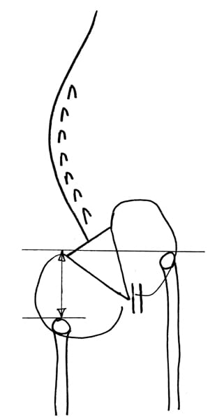
    In his explanation of the biomechanics of spinal manipulation, Dr. Kirkaldy-Willis  uses the following graphic:


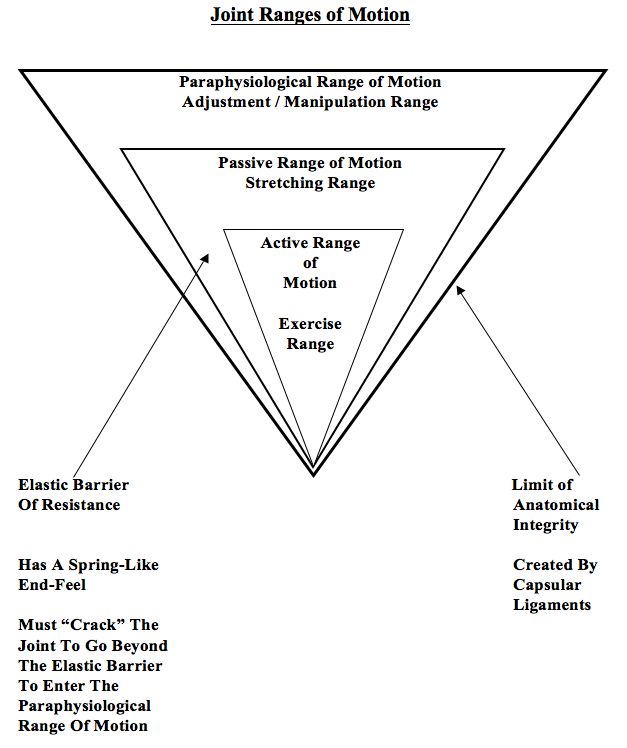
    The graphic shows that joint motion is divided into three distinct categories:

    1) Active Range of Motion

    This is the range of motion the patient can achieve through their own active efforts.

    2) Passive Range of Motion

    This range of motion is primarily achieved by the physician / therapist who pushes the involved joint beyond the active range of motion. The passive range of motion is greater than the active range of motion. The passive range of motion will influence a larger range of tissue than does the active range of motion.

    Dr. Kirkaldy-Willis states (1):

    “Beyond the end of the active range of motion of any synovial joint, there is a small buffer zone of passive mobility.” A joint can only move into this zone with passive assistance, and going into this passive range of motion “constitutes mobilization.”

    [This is mobilization, not manipulation]

    3) Paraphysiological Range of Motion

    This range of motion is only achieved by moving beyond the Passive Range of Motion.

    Dr. Kirkaldy-Willis states (1):

    “At the end of the passive range of motion, an elastic barrier of resistance is encountered. This barrier has a spring-like end-feel.”

    “If the separation of the articular surfaces is forced beyond this elastic barrier, the joint surfaces suddenly move apart with a cracking noise.”

    “This additional separation can only be achieved after cracking the joint and has been labeled the paraphysiological range of motion.”

    “This constitutes manipulation.” [Important]

    “The cracking sound on entering the paraphysiological range of motion is the result of sudden liberation of synovial gases—a phenomenon known to physicists as cavitation.”

    Following cavitation, a synovial bubble can be observed on x-rays, which is reabsorbed over the following 30 minutes. During this “refractory period” there is no resistance between the passive and paraphysiological zones.

    Concerning spinal manipulation, Dr. Kirkaldy-Willis also makes the following

    Points (1):

    “At the end of the paraphysiological range of motion, the limit of anatomical integrity is encountered. Movement beyond this limit results in damage to the capsular ligaments.”

    Joint manipulation [adjusting] “requires precise positioning of the joint at the end of the passive range of motion and the proper degree of force to overcome joint coaptation” [to overcome the resistance of the joint surfaces in contact].

    “With experience, the manipulator can be very specific in selecting the spinal level to be manipulated.”

    “Most family practitioners have neither the time nor inclination to master the art of manipulation and will wish to refer their patients to a skilled practitioner of this therapy.”

    “The physician who makes use of this resource [manipulation] will provide relief for many back pain patients.”

    Dr. Kirkaldy-Willis suggests… (1) that during spinal manipulation, there is a stretching of facet joint capsules which fires the capsular mechanoreceptors. This will reflexly “inhibit facilitated motoneuron pools” which are responsible for the muscle spasms that commonly accompany low back pain. The result of this inhibition is an improvement of joint movement. The improved movement initiates the Gate Theory of Pain, originally published in 1965 by Ronald Melzack and Patrick Wall in the journal Science (2).

    This theory has “withstood rigorous scientific scrutiny.” “The central transmission of pain can be blocked by increased proprioceptive input.” Pain is facilitated by “lack of proprioceptive input.” This is why it is important for “early mobilization to control pain after musculoskeletal injury.”

    The facet capsules are densely populated with mechanoreceptors. “Increased proprioceptive input in the form of spinal mobility tends to decrease the central transmission of pain from adjacent spinal structures by closing the gate. Any therapy which induces motion into articular structures will help inhibit pain transmission by this means.”

    Furthermore, in chronic cases, there is a shortening of periarticular connective tissues and intra-articular adhesions may form; manipulations [chiropractic adjustments] can stretch or break these adhesions. Consequently, “In most cases of chronic low back pain, there is an initial increase in symptoms after the first few manipulations [probably as a result of breaking adhesions]. In almost all cases, however, this increase in pain is temporary and can be easily controlled by local application of ice.”

    The main point stressed by Dr. Kirkaldy-Willis is that chronic spinal pain is

    often associated with reduced motion, which opens the pain gate. Spinal manipulation is an excellent method to restore the range of motion, close the pain gate, and reduce pain. However, appropriate spinal manipulation is a skill that requires special training and initial supervised applications. When the practitioner is appropriately trained, spinal manipulation is both safe and effective.

    Manipulation & The Source Of It’s Checkered Past

    In an important and revealing article published in 1995 (3), Alan Terrett reviewed the published literature pertaining to the incidence of reported adverse events associated with chiropractic spinal adjusting (manipulation). Astonishingly, his results revealed that in many of the published adverse events ascribed to chiropractic manipulation were, in fact, not associated with chiropractic in any manner. Apparently, the authors of the articles assumed “chiropractic” and “manipulation” were synonyms. When untrained laypersons or physicians performed a manipulation resulting in a reported adverse event, authors would claim that the manipulation was performed by a chiropractor. The list of discovered manipulators included:

    A Blind Masseur
    An Indian Barber
    A Wife
    A Kung-Fu Practitioner
    Self Manipulation

    Often the manipulation was performed by a medical doctor, an osteopath, a naturopath, or a physical therapist.

    Dr. Terrett concluded:

    “This study reveals that the words chiropractic and chiropractor commonly appear in the literature to describe spinal manipulative therapy, or practitioner of spinal manipulative therapy, in association with iatrogenic complications, regardless of the presence or absence of professional training of the practitioner involved.”

    “The words chiropractic and chiropractor have been incorrectly used in numerous publications dealing with spinal manipulative therapy injury by medical authors, respected medical journals and medical organizations.”

    “In many cases, this is not accidental; the authors had access to original reports that identified the practitioner involved as a non-chiropractor. The true incidence of such reporting cannot be determined.”

    “Such reporting adversely affects the reader’s opinion of chiropractic and chiropractors.”

    “It has been clearly demonstrated that the literature of medical organizations, medical authors and respected, peer-reviewed, indexed journals have, on numerous occasions, misrepresented the facts regarding the identity of a practitioner of manual therapy associated with patient injury.”

    “Such biased reporting must influence the perception of chiropractic held by the reader, especially when cases of death, tetraplegia and neurological deficit are incorrectly reported as having been caused by chiropractic.”

    “Because of the unwarranted negative opinion generated in medical readers and the lay public alike, erroneous reporting is likely to result in hesitancy to refer to and underutilization of a mode of health care delivery.”

    In 1996, Woodward and colleagues (4) from the University Department of Orthopaedic Surgery, Bristol, UK, published a retrospective study to determine the effects of chiropractic in a group of 28 patients who had been referred with chronic ‘whiplash’ syndrome. The 28 patients in this study had initially been treated with anti-inflammatories, soft collars and physiotherapy. These patients had all become chronic, and were referred for chiropractic care at an average of 15.5 months (range was 3 – 44 months) after their initial injury. Following chiropractic care, 93% of the patients had improved. The title of the article was Chiropractic treatment of chronic ‘whiplash’ injuries, and it was published it the prestigious journal Injury.

    Woodward and colleagues defined spinal manipulation as:

    “Spinal manipulation is a high-velocity low-amplitude thrust to a specific vertebral segment aimed at increasing the range of movement in the individual facet joint, breaking down adhesions and stimulating production of synovial fluid.”

    Woodward and colleagues also note that complications from cervical manipulations are rare, and when they are reported in the literature, they often…

    “arose as a result of spinal manipulation performed by non-chiropractors, who had been misrepresented in the literature as being trained chiropractors.” [Important]

    With respects to risk associated with cervical manipulation, there is particular concern pertaining to vertebral artery dissection. All chiropractors are well aware of the issue. Vertebral artery dissection is extensively discussed in both chiropractic undergraduate and post graduate continuing educational programs. Entire books are written on the subject and are a part of core curriculum at chiropractic colleges (5). Chiropractors are well schooled on the pertinent anatomy, signs/symptoms, clinical presentations, examination findings, and procedures that may possibly be associated with increased risk.

    In 2002, the Journal of Neurology (6) published a study titled:

    “Hyperhomocysteinemia: A potential risk factor for cervical artery dissection following chiropractic manipulation of the cervical spine”

    In this study, the authors measured total fasting plasma homocysteine concentration in 4 subjects with manipulation-related cervical artery dissection and compared them to a series of patients with spontaneous dissection of the neck arteries. Both groups were then compared to 36 control subjects. Median plasma homocysteine levels were significantly higher in patients with manipulation-related cervical artery dissection (18.2 µmol/l, range 14.3 to 30.0) compared with controls (8.9 µmol/l, range 5 to 17.3). These authors concluded that hyperhomocysteinemia may represent a potential risk factor for manipulation-related cervical artery dissection, leading to structural abnormalities of the arterial wall and increasing the susceptibility to mechanical stress.

    Importantly for chiropractors, the authors note that the risk factors used by chiropractors to screen patients at risk for cervical artery dissection from manipulation are usually unrevealing. Therefore, “the population at risk cannot be identified a priori” using standard risk factor screening.

    In 2002, Dr. Scott Haldeman from the Department of Neurology, University of California, Irvine, and colleagues, published a study titled (7):

    “Unpredictability of cerebrovascular ischemia associated with
    cervical spine manipulation therapy: a review of
    sixty-four cases after cervical spine manipulation”

    The study, published in Spine, was a retrospective review of 64 medicolegal records describing cerebrovascular ischemia after cervical spine manipulation.

    The authors note, that in 2002, only about “117 cases of post-manipulation cerebrovascular ischemia have been reported in the English language literature.”

    The authors further indicate that proposed risk factors for cerebrovascular ischemia secondary to spinal manipulation include age, gender, migraine headaches, hypertension, diabetes, birth control pills, cervical spondylosis, and smoking, and that it is often assumed that these complications may be avoided by clinically screening patients and by pre-manipulation positioning of the head and neck to evaluate the patency of the vertebral arteries.

    After an extensive review, these authors conclude:

    “This study was unable to identify factors from the clinical history and physical examination of the patient that would assist a physician attempting to isolate the patient at risk of cerebral ischemia after cervical manipulation.”

    “Cerebrovascular accidents after manipulation appear to be unpredictable and should be considered an inherent, idiosyncratic, and rare complication of this treatment approach.”

    Earlier this year (2008), Dr. David Cassidy and colleagues published the most comprehensive study to date pertaining to the risk of vertebral artery dissection as related to chiropractic cervical spine manipulation (8). The article was published in Spine, and titled:

    “Risk of Vertebrobasilar Stroke and Chiropractic Care:
    Results of a Population-Based Case-Control and Case-Crossover Study”

    Key points from this article include:

    1. “Vertebrobasilar artery stroke is a rare event in the population.”
    2. “We found no evidence of excess risk of vertebral artery stroke associated chiropractic care.”
    3. “Neck pain and headache are common symptoms of vertebral artery dissection, which commonly precedes vertebral artery stroke.”
    4. “The increased risks of vertebral artery stroke associated with chiropractic and primary care physicians visits is likely due to patients with headache and neck pain from vertebral artery dissection seeking care before their stroke.”
    5. Most cases of vertebral arterial dissection occur spontaneously.
    6. “Because patients with vertebrobasilar artery dissection commonly present with headache and neck pain, it is possible that patients seek chiropractic care for these symptoms and that the subsequent vertebral artery stroke occurs spontaneously, implying that the association between chiropractic care and vertebral artery stroke is not causal.”
    7. “Since it is unlikely that primary care physicians cause stroke while caring for these patients, we can assume that the observed association between recent primary care physician care and vertebral artery stroke represents the background risk associated with patients seeking care for dissection-related symptoms leading to vertebral artery stroke. Because the association between chiropractic visits and vertebral artery stroke is not greater than the association between primary care physicians visits and vertebral artery stroke, there is no excess risk of vertebral artery stroke from chiropractic care.”
    8. Neck manipulation “is unlikely to be a major cause” of these rare vertebral artery stroke events.
    9. “Our results suggest that the association between chiropractic care and vertebral artery stroke found in previous studies is likely explained by presenting symptoms attributable to vertebral artery dissection.”
    10. “There is no acceptable screening procedure to identify patients with neck pain at risk of vertebral artery stroke.”

    In 2004, the American Academy of Orthopedic Surgeons published a monograph titled Neck Pain (9). The second to last chapter in the monograph, chapter 7, is titled:

    “Manual Therapy Including Manipulation For
    Acute and Chronic Neck Pain”

    The editor of the monograph is Jeffery Fischgrund, MD, from the Department of Orthopaedic Surgery at William Beaumont Hospital in Royal Oaks, Michigan. This monograph has twelve respected contributors, including the authors of chapter 7, Scott Haldeman, Clinical Professor of Neurology at the University of California, Irvine, and Eric Hurwitz, Associate Professor of Epidemiology at the University of California, Los Angeles. With respect to the safety of spinal manipulation, the authors make the following comments:

    “Major complications from manual therapies are extremely rare but, nonetheless, have been a source of much discussion.”

    “Estimates of vertebral artery dissections or stroke rates associated with cervical manipulation have ranged form 1 per 400,000 to 1 per 10 million manipulations.”

    “An estimate of 1 per 5.85 million manipulations, based on 1988 to 1997 medical record and chiropractic malpractice data from Canada, reflects the experience of practitioners of manipulation.”

    “No serious complications from spinal manipulation or other chiropractic forms of manual treatment have been reported from any of the published clinical trials involving manipulation or mobilization for neck pain.”

    “It should be noted that complications rates from medications, surgery, and most other neck pain treatments for which data are available are estimated to be higher than those from manual and manipulative therapies.”

    The largest prospective study to date assessing the safety of chiropractic spinal manipulation to the cervical spine was published in Spine, October 2007, and titled:

    “Safety of Chiropractic Manipulation of the Cervical Spine
    A Prospective National Survey”

    This study assessed the risk of serious and relatively minor adverse events following chiropractic manipulation of the cervical spine by a sample of U.K. chiropractors. The authors studied treatment outcomes from 19,722 patients from 377 chiropractors that involved 50,276 cervical spine manipulations. Manipulation was defined as the application of a high-velocity/low-amplitude or mechanically assisted thrust to the cervical spine. A serious adverse event was defined as referral to hospital and/or severe onset/worsening of symptoms immediately after treatment and/or resulted in persistent or significant disability/incapacity. Minor adverse events were reported by patients as a worsening of presenting symptoms or onset of new symptoms, were recorded immediately, and up to 7 days, after treatment.

    The authors concluded:

    “Safety of treatment interventions is best established with prospective surveys, and this study is unique in that it is the only prospective survey on such a large scale specifically estimating serious adverse events following cervical spine manipulation.”

    “There were no reports of serious adverse events.”

    “Although minor side effects following cervical spine manipulation were relatively common, the risk of a serious adverse event, immediately or up to 7 days after treatment, was low to very low.”

    “No significant adverse event was reported by the chiropractors using the definition criteria.”

    “The risk rates described in this study compare favorably to those linked to drugs routinely prescribed for musculoskeletal conditions in general practice.”

    “The risks reported here are also lower than those reported for acupuncture, which were described as a very safe intervention in the hands of a competent practitioner.”

    “Although minor side effects were found to be relatively common, the risk of a serious adverse event, immediately and up to 7 days after treatment, was estimated to be low to very low in these consultations.”

    “On this basis, this survey provides evidence that cervical spine manipulation is a relatively safe procedure when administered by registered U.K. chiropractors.”

    No therapeutic intervention is without risk. Spinal manipulation has the potential to exceed the limit of anatomic integrity and thus cause injury. The study by Terrett (3) shows that when spinal manipulation is attempted by untrained individuals, there is an increased incidence of adverse events. Chiropractors are well schooled in the potential for injury with certain manipulative maneuvers, and extremely well trained in the effective delivery of safe spinal manipulations. The best-published studies indicate that chiropractic spinal manipulation is both safe and effective.

    References

    • Kirkaldy-Willis WH and Cassidy JD; Spinal Manipulation in the Treatment of Low back Pain; Canadian Family Physician; March 1985, Vol. 31, pp. 535-540.
    • Melzack R and Wall PD; Pain Mechanisms: a new theory; Science; 1965, 150, pp. 971-9.
    • Terrett AG; Misuse of the literature by medical authors in discussing spinal manipulative therapy injury; Journal of Manipulative and Physiological Therapeutics; 1995 May;18(4):203-10.
    • N. Woodward, J. C. H. Cook, M. F. Gargan and G. C. Bannister; Chiropractic treatment of chronic ‘whiplash’ injuries; Injury; Volume 27, Issue 9, November 1996, pages 643-645.
    • Terrett AGJ; Current Concepts in Vertebrobasilar Complications Following Spinal Manipulation; Second Edition, NCMIC Group, 2001.
    • Pezzini A, Del Zotto E, Padovani A; Hyperhomocysteinemia: A potential risk factor for cervical artery dissection following chiropractic manipulation of the cervical spine; Journal of Neurology, October 2002, Vol. 249, Issue 10, pp. 1401-1403.
    • Haldeman S, Kohlbeck FJ, McGregor M; Unpredictability of cerebrovascular ischemia associated with cervical spine manipulation therapy: a review of sixty-four cases after cervical spine manipulation; Spine; 2002 Jan 1;27(1):49-55.
    • Cassidy, J David DC, PhD; Boyle, Eleanor PhD; Côté, Pierre DC, PhD; He, Yaohua MD, PhD; Hogg-Johnson, Sheilah PhD; Silver, Frank L. MD; Bondy, Susan J. PhD; Risk of Vertebrobasilar Stroke and Chiropractic Care: Results of a Population-Based Case-Control and Case-Crossover Study; Spine; Volume 33(4S), February 15, 2008 pp S176-S183.
    • Fischgrund, JS; Neck Pain, 2004.
    • Thiel, Haymo W. DC, PhD; Bolton, Jennifer E. PhD; Docherty, Sharon PhD; Portlock, Jane C. PhD; Safety of Chiropractic Manipulation of the Cervical Spine, A Prospective National Survey; Spine; Volume 32(21), October 2007, pp 2375-2378.


  • Headache Symptoms & The Commonly Overlooked Connection To The Cervical Spine

    Headache Symptoms & The Commonly Overlooked Connection To The Cervical Spine

    Understanding that cervical spine problems can cause headache is not a new understanding, yet is still all too often overlooked. In 1956, Emil Seletz, MD… a noted Beverly Hills, CA, neurosurgeon. He was member of the esteemed faculty at the University of California, Los Angeles, medical school, and had treated more than 20,000 injury patients at the Los Angeles General Hospital. In January of 1957, Dr. Seletz published in the Journal of the International College of Surgeons an article titled (1):

    Craniocerebral Injuries and the Postconcussion Syndrome

    In this article, Dr. Seletz discusses post-traumatic headaches arising from whiplash injuries. He states:

    “Analysis of the symptoms of several thousands of such patients will reveal that headaches persisting for months or years after a cerebral concussion are real and that they are extracranial in origin.”

    He states that there are three primary extracranial sources for these post-traumatic headaches:

    1) The upper cervical spinal nerve roots.

    2) Elements of the trigeminal nerve.

    3) A combination of the two.

    In every whiplash injury, the head is involved in sudden acceleration or deceleration, causing some unnatural movement, rotation or strain of the upper cervical spinal roots and muscles.

    The 2nd cervical nerve root becomes the greater occipital nerve and supplies the major portion of the scalp, the upper portion of the neck and portions of the face. [PICTURE #1]

    The 2nd cervical nerve root is more vulnerable to trauma than other nerve roots because it is not protected by pedicles and facets. The 2nd cervical nerve root leaves the interspace between C1-C2, courses down and winds under the inferior oblique muscle, then proceeds upward as the greater occipital nerve, and pierces the tendon of the trapezius muscle. [PICTURE #2]

    The spinal accessory nerve takes its origin from the entire length of the cervical cord, always as low as the fifth and often as low as the seventh cervical level (see drawing below). Any acute flexion, extension or torsion of the neck will exert traction on the delicate filaments of the spinal accessory nerve, resulting in spasm of the trapezius and sternocleidomastoid muscles. When the trapezius and suboccipital muscles (especially the inferior oblique muscle) are in spasm, traction is produced upon the greater occipital nerve as it pierces the fascial attachment of these muscles.

    Sensory changes extending over the trigeminal innervation area following whiplash injury to the neck are explained by the communication between the 2nd and 3rd cervical nerve roots and the greater occipital nerve with the trigeminal nerve in the spinal fifth tract in the medulla (trigeminal cervical nucleus). [PICTURE #3] This has been called the greater occipital/trigeminus syndrome, and is a cause of post-whiplash headache. As the ophthalmic fibers descend the deepest into the cervical spine, the headaches are often in the distribution of the ophthalmic branch of cranial nerve V (around and behind the eye).

    Following his 1957 publicaton on the subject of craniocerebral injury he was back in 1958 with yet another contribution, this time discussing the cervical spine and its involvement in injuries, Dr. Seletz publishes an article in the Journal of the American Medical Association, titled (2):

    Whiplash Injuries
    Neurophysiological Basis for Pain and
    Methods Used for Rehabilitation

    Once again, Dr. Seletz states that whiplash trauma results in “direct trauma to the spinal accessory nerve and to the roots of the cervical nerves.” He states:

    “A major portion of the headaches associated with this syndrome are derived from a traction injury to the second cervical nerve root.”

    The second cervical nerve root is particularly vulnerable to injury because it is not protected by pedicles and facets, as are the other cervical nerve roots. Additionally, the second cervical nerve root exits between the atlas and axis, “the point of greatest rotation of the head on the neck.” The second nerve root becomes the greater occipital nerve, which innervates the majority of the scalp, the side of the upper neck, and portions of the face.

    “The physiological communication between the second cervical and the trigeminal nerves in the spinal fifth tract of the medulla [trigeminal-cervical nucleus] involves the first division of the trigeminal nerve [opthalamic] and thereby gives attacks of hemicrania with pain radiating behind the corresponding eye. This is the mechanism whereby a great many chronic and persistent headaches have their true origin in injury to the second cervical nerve,” noting:

    “‘Many headaches are not headaches at all, but really a pain in the neck.’”

    Since the greater occipital nerve pierces the tendinous attachment of the trapezius muscle at the base of the skull, trapezius spasm aggravates greater occipital nerve (C2) sensory disturbance. The trapezius is innervated by the spinal accessory nerve, cranial nerve XI. The spinal accessory nerve originates from filaments of the entire length of the cervical spinal cord, ascends through the foramen magnum and leaves the skull through the jugular foramen for a long descent to the trapezius muscle. Persistent spasm of the trapezius muscle is not due to primary injury to the muscles but to traction injury to the delicate filaments of origin of the spinal accessory nerve.

    Although Dr. Seletz expertly described the neuroanatomical basis for headaches arising from the cervical spine in the late 1950’s, it took nearly a quarter of a century for officials to include the category “Cervicogenic Headache” in their classification schemes (3). Additions to Dr. Seletz’s concepts on cervicogenic headache continue through today. A recent (August 2008) PubMed search of the National Library of Medicine database using the key words “cervicogenic headache” listed 588 articles, with publication dates ranging from 1949 to 2008.

    Perhaps the most detailed anatomical description for the physiological basis for cervicogenic headache was written by Australian physician and researcher Nikoli Bogduk, MD, PhD, in 1995. Dr Bogduk published an article in the journal Biomedicine and Pharmacotherapy titled (4):

    Anatomy and Physiology of Headache

    In this article, Dr. Bogduk notes that all headaches have a common anatomy and physiology in that they are all mediated by the trigeminocervical nucleus, and are initiated by noxious stimulation of the endings of the nerves that synapse on this nucleus or by irritation of the nerves themselves. The trigeminocervical nucleus is a region of grey matter in the brainstem.

    The trigeminocervical nucleus is “defined by its afferent fibers,” and it receives afferents from the following sources:

    1) Trigeminal Nerve (Cranial Nerve V)

    2) Upper three cervical nerves

    3) Cranial Nerve VII (Facial Nerve)

    4) Cranial Nerve IX (Glossopharyngeal Nerve)

    5) Cranial Nerve X (Vagus Nerve)

    All of these sources of afferents terminate on common second-order neurons in the trigeminocervical nucleus, which can then relay the sensations of pain to the cortical brain. The trigeminocervical nucleus is the sole nociceptive nucleus of the head, throat and upper neck. “All nociceptive afferents from the trigeminal, facial, glossopharyngeal and vagus nerves and C1-C3 spinal nerves ramify in this single column of grey matter.” Stimulation of cervical afferents to a second-order neuron that also receives trigeminal afferents may result in the source of stimulation being interpreted as arising in the trigeminal receptive field, resulting in the perception of headache. Pain in the forehead can arise as a result of stimulation by cervical afferents of second-order neurons in the trigeminocervical nucleus that happen also to receive forehead trigeminal nerve afferents.

    Dr. Bogduk indicates that because the ophthalmic branch of the trigeminal nerve extends the farthest into the trigeminocervical nucleus, cervical afferent stimulation is most likely to refer pain to the frontal-orbital region of the head. Yet, importantly, any structure innervated by any of the three branches (ophthalmic, maxillary, mandibular) of cranial nerve V (trigeminal) can be painful as a consequence of irritation of any of the structures innervated by the C1, C2, and/or C3 nerve roots. The following lists are helpful:

    Structures innervated by ophthalmic branch (V1) cranial V (trigeminal):

    Skin of forehead
    Orbit
    Eye
    Frontal sinus
    Dura mater of the anterior cranial fossa
    Anterior and posterior ends of the falx cerebri
    Superior sagittal sinus
    Proximal ends of the anterior and middle cerebral arteries
    Superior surface of the tentorium cerebelli
    Cavernous sinus
    Venous sinuses
    Temporal artery

    Structures innervated by maxillary branch (V2) cranial V (trigeminal):

    Nose
    Paranasal sinuses
    Upper teeth
    Upper jaw
    Dura mater of the middle cranial fossa

    Structures innervated by mandibular branch (V3) cranial V (trigeminal):

    Dura mater of the middle cranial fossa
    Lower teeth
    Lower jaw
    Temporomandibular joint
    External auditory meatus (ear)
    Anterior aspect of the tympanic membrane

    Structures innervated by C1-C3:

    Dura mater of the posterior cranial fossa
    Inferior surface of the tentorium cerebelli
    Anterior and posterior upper cervical and cervical-occiput muscles
    OCCIPUT-C1, C1-C2, and C2-C3 joints
    C2-C3 intervertebral disc
    Skin of the occiput
    Vertebral arteries
    Carotid arteries
    Alar ligaments
    Transverse ligaments
    Trapezius muscle
    Sternocleidomastoid muscle

    Dr. Bogduk agrees with the writings from Dr. Seletz decades before in that the C1 and C2 nerve roots are particularly likely to be involved in the genesis of cervicogenic headache because the C1 and C2 spinal nerve roots “do not emerge through intervertebral foramina.” This make these nerve roots more vulnerable to stretch or compressive stresses. Additionally, Dr. Bogduk also agrees with Dr. Seletz in noting that the sensory component of the C2 posterior primary rami becomes the greater occipital nerve. C2 winds under the inferior oblique muscle, ascends and pierces the shared aponeurosis of the trapezius and sternocleidomastoid muscle to supply the posterior scalp.

    Two other clinically relevant points
    Dr. Bogduk makes in this article include:

    1) Arthritic changes of the upper cervical synovial joints (including C2-C3) can cause neck pain and headache.

    2) C2 neuralgia can cause headache as a consequence of “scar tissue following trauma to the lateral atlanto-axial joint.”

    In 2005, adding to the more recent data on this topic, Dr. David Biondi, an instructor in Neurology Harvard Medical School, published an article titled (5):

    Cervicogenic Headache:
    A Review of Diagnostic and Treatment Strategies

    In this article, Dr. Biondi makes the following key points:

    1. “Cervicogenic headache is a relatively common cause of chronic headache that is often misdiagnosed or unrecognized.”
    2. Cervicogenic headache is chronic hemicranial pain that is referred to the head from tissues of the neck.
    3. Head pain that is referred from the tissues of the neck is called cervicogenic headache.
    4. Cervicogenic headache was not officially recognized until 1983.
    5. The key neurological structure in cervicogenic headache is the trigeminocervical The trigeminocervical nucleus is a region in the upper cervical spinal cord where sensory nerve fibers from the trigeminal nerve (cranial V) interact with sensory fibers from the upper cervical nerve roots.
    6. The convergence of upper cervical and trigeminal sensory fibers is the basis for upper cervical problems causing pain in the face and head.
    7. Cervicogenic headache is often a sequelae of head or neck injury but may occur in the absence of trauma.
    8. The prevalence of cervicogenic headache is as high as 20% of patients with chronic
    9. Cervicogenic headache is four times more prevalent in women.
    10. “Patients with cervicogenic headache will often have altered neck posture or restricted cervical range of motion.”
    11. Cervicogenic headache pain can be “triggered or reproduced by active neck movement, passive neck positioning especially in extension or extension with rotation toward the side of pain, or on applying digital pressure to the involved facet regions or over the ipsilateral greater occipital nerve.”
    12. Zygapophyseal joint, cervical nerve, or medial branch blockade is used to confirm the diagnosis of cervicogenic headache.
    13. Trauma to or pathologic changes to the C1-C2-C3 joints can cause head pain.
    14. The third occipital nerve (dorsal ramus C3) innervates the C2–C3 facet joint; the C2- C3 facet joint and the third occipital nerve are the most vulnerable to trauma from acceleration-deceleration whiplash injuries of the neck. Therefore, injury to the C2-C3 facet joint is the most common cause of post-traumatic cervicogenic
    15. It can take a year or longer for post-whiplash cervicogenic headache to resolve.
    16. Disc problems as low as C5–C6 can cause chronic cervicogenic headache.
    17. The following structures can cause cervicogenic headache:
      1. The greater occipital nerve (dorsal ramus C2)
      2. The lesser occipital nerve
      3. The atlanto-occipital joint
      4. The atlantoaxial joint
      5. The C2 or C3 spinal nerve
      6. The third occipital nerve (dorsal ramus C3)
      7. The zygapophyseal joint(s) as low as C5
      8. The intervertebral discs as low as C5-C6

    In addition to these points, Dr. Biondi notes that in the management of cervicogenic headache, drugs alone are often ineffective. He states that “Many patients with cervicogenic headache overuse or become dependent on analgesics.” He also notes that COX-2 inhibitors cause both gastrointestinal and renal toxicity after long-term use, and they cause an increased risk of cardiovascular and cerebrovascular events.

    Dr. Biondi is an osteopathic physician, and therefore has an understanding of manual and manipulative techniques. He states:

    “All patients with cervicogenic headache could benefit from manual modes of therapy and physical conditioning.”

    Dr. Biondi notes that the treatment of cervicogenic headache usually requires manipulation of the upper cervical facet joints, and that manipulative techniques are particularly well suited for the management of cervicogenic headache, including high velocity, low amplitude manipulation. These techniques are commonly used by chiropractors in the management of cervicogenic headaches.

    In October of 1997, a randomized, blinded study on the treatment of cervicogenic headache that included chiropractic adjustments to the cervical spine was published (6). The study participants had cervicogenic headaches in accordance with the standards of the International Headache Society. The group receiving chiropractic cervical spine adjustments obtained statistically significant improvements in all three measurement criteria. Specifically, subjects receiving chiropractic adjustments for their cervicogenic headache:

    • Decreased their analgesic use by 36%.
    • Decreased their headache hours by 69%.
    • Decreased their headache intensity by 36%.

    Using gold standard research protocols, this study reveals that chiropractic spinal adjusting can benefit a large number of patients suffering from cervicogenic headache.

    Diagnostic Criteria for Cervicogenic Headache
    (Developed by the Cervicogenic Headache International Study Group)

    THE PATIENT MUST HAVE AT LEAST ONE OF THE FOLLOWING:

    1. The head pain must be preceded by:
      Neck Movement

      or

      Sustained Awkward Head Positioning

      or

      External Pressure Over the Upper Cervical (C1-2-3-4) or
      Occipital Region on the Symptomatic Side

    2. Restricted Cervical Spine Range of Motion (Active and Passive)
    3. Ipsilateral Neck, Shoulder, or Arm Pain of a Vague Nonradicular Nature

      or

      Occasional Arm Pain of a Radicular nature

    If All Three Criteria Are Present, One Is Essentially Assured of Cervicogenic Headache

    CHARACTERISTICS OF CERVICOGENIC HEADACHE

    Frequently a History of Indirect Neck Trauma [Whiplash injury]

    Unilateral Headache That Does Not Change Sides

    Occasionally the Pain May Be Bilateral

    The Pain is Located Occipitally, Frontally, Temporally, or Orbitally

    The Pain Can Last Hours to Days

    The Headache Usually Begins in the Neck

    The Headache is Moderate to Severe

    The Headache is Non-Throbbing

    The Headache is Non-Lancinating

    THE FOLLOWING FEATURES
    MAY ALSO BE OCCASIONALLY NOTED

    Nausea

    Phonophobia

    Photophobia

    Dizziness

    Difficult Swallowing

    Ipsilateral Blurred Vision

    Vomiting

    Ipsilateral Lacrimation

    Ipsilateral Edema, Especially in the Periocular Region

    REFERENCES

    • Seletz E; Craniocerebral Injuries and the Postconcussion Syndrome; Journal of the International College of Surgeons; January, 1957; 27(1):46-53.
    • Seletz E; Whiplash Injuries, Neurophysiological Basis for Pain and Methods Used for Rehabilitation; Journal of the American Medical Association; November 29, 1958, pp. 1750 – 1755.
    • Sjaastad O; “Cervicogenic Headache” An Hypothesis; Cephalagia; December 1983; 3(4):249-256.
    • Bogduk N; Anatomy and Physiology of Headache; Biomedicine and Pharmacotherapy; 1995, Vol. 49, No. 10, 435-445.
    • Biondi DM; Cervicogenic Headache: A Review of Diagnostic and Treatment Strategies; Journal of the American Osteopathic Association; April 2005, Vol. 105, No. 4 supplement, pp. 16-22.
    • Nilsson N; The effect of spinal manipulation in the treatment of cervicogenic headache; Journal of Manipulative and Physiological Therapeutics; June 1997; 20(5):326-330.
  • The Meniscoid Block, Acute Neck / Back Pain & Two German Physicians…

    The Meniscoid Block, Acute Neck / Back Pain & Two German Physicians…

    Perhaps the most authoritative book written pertaining to the spine is Schmorl’s and Junghanns’ The Human Spine in Health and Disease. Georg Schmorl (1861-1932) was a German physician and pathologist. Herbert Junghanns (1902-1986) was the Chief of the Occupational Accident Hospital, Surgical Clinic, and Head of the Institute for Spinal Column Research, in Frankfurt, Germany. The first edition of their book was published in 1932, shortly before the death of Dr. Schmorl. The fifth German edition of their book was translated into English in 1971 (1). This incredible book contains 500 figures of radiographs, histological sections, photographs, and drawings. The book has more than 500 pages and approximately 5,000 references in the bibliography.

    In their book, Drs. Schmorl and Junghanns note:

    “Like other body articulations, the apophyseal joints are endowed with articular capsules, reinforcing ligaments and menisci-like internal articular discs.” p. 251

    “Like any other joint, the motor segment may become locked. This is usually associated with pain.” Chiropractors refer to such events as subluxations. These motor unit disturbances can cause torticollis and lumbago. “Various processes may cause such ‘vertebral locking.’ It may happen during normal movement. The incarceration of an articular villus or of a meniscus in an apophyseal joint may produce locking.” pp. 221-222

    If a joint is suddenly incarcerated within the range of its physiologic mobility, as occurs with the meniscus incarceration of the knee joint, it is an “articular locking or a fixed articular block.”

    “Such articular locking is also possible in the spinal articulations (apophyseal joints, intervertebral discs, skull articulations, lumbosacral articulations). They may be mobilized again by specific therapeutic methods (stretchings, repositioning, exercises, etc.). Despite many opinions to the contrary, this type of locking is today increasingly recognized by physicians. Many physicians are employing manipulations which during the past decades were the tools of lay therapists only (chiropractors, osteopaths). However, these methods have at times been recommended by physicians. They have also been known in folk medicine and in medical schools of antiquity.” p. 376

    Accompanying these quotes and other pertinent discussions are two photographs of anatomical sections through the facet joints showing these “menisci-like internal articular discs,” or meniscus. They also included three radiographs and one drawing showing abnormal gapping of an articulation as a consequence of meniscus entrapment in a facetal articulation. They note that such a meniscoid incarceration can cause acute torticollis, and they show a “follow-up roentgenogram after manual repositioning” resulting in “immediate relief of complaints and complete mobility.” p. 222

    In September 1975, the Department of Physical Medicine and Rehabilitation of the California College of Medicine organized an international conference concerning Approaches to the Validation of Manipulation Therapy. The conference was held at the University of California, Irvine. Twenty-one global top experts on spinal manipulation participated in the conference. The proceedings from the conference were published in 1977, edited by Alfred Buerger, PhD, and Jerome Tobis, MD. Dr. Buerger was an assistant professor from the Department of Physical Medicine & Rehabilitation and Physiology at the medical school at the University of California Irvine, and Dr. Tobis was a Professor and Chairman of the Department of Physical Medicine and Rehabilitation of the medical school at the University of California Irvine (2). Chapter 14 of the conference proceedings was authored by physician James Fisk from New Zealand (3). The title of Dr. Fisk’s chapter 14 is:

    An Evaluation of Manipulation in the Treatment of the Acute Low Back Pain Syndrome in General Practice

    In this chapter, Dr. Fisk, in the section regarding possible lesions that cause low back pain, lists:

    “Entrapment of a Meniscoid”

    “These meniscoids are richly innervated by pain producing nerve endings. There should be a rapid response to manipulation.”

    In 1977, the National Institute of Neurological and Communicative Disorders and Stroke of the National Institutes of Health (USA), funded a grant for a Research Workshop on Neurobiologic Mechanisms in Manipulative Therapy. The workshop was held at Michigan State University, and had 38 worldwide participant experts. The Proceedings for the workshop were published the following year (4), 1978, edited by Irvin Korr, Professor from the Department of Biomechanics, College of Osteopathic Medicine, Michigan State University. The first chapter of the workshop proceedings is authored by Dr. Karel Lewit from Czechoslovakia, and is titled (5):

    The Contribution of Clinical Observation to
    Neurobiological Mechanisms in Manipulative Therapy

    In this chapter, Dr. Lewit notes:

    “The meniscoid has a soft base and a hard edge.”

    “If the meniscoid is caught between the gliding surfaces of the joint facets the hard edge produces a cavity in the cartilage in which it is trapped.”

    “The implications for the theory of manipulation are obvious: if we separate the joint facets the meniscoid can slip out.”

    Dr. Lewit includes a similar discussion in his 1985 text titled Manipulative Therapy in Rehabilitation of the Locomotor System (6).

    Also in 1985, 30 distinguished international multidisciplinary experts collaborated on a text titled Aspects of Manipulative Therapy (7). The comments in this text pertaining to the interarticular meniscus include:

    “Histologically, meniscoids are synovial tissue.”

    “Their innervation is derived from that of the capsule.”

    The current hypothetical model of the mechanism involved in acute joint locking is based on a phenomenon in which the “meniscoid embeds itself, thereby impeding mobility.”

    “It is highly probable that the meniscoids do play an important role in acute joint locking, and this is confirmed by the observation that all the joints afflicted by this condition are equipped with such structures.” pp. 90-91

    In 1986, physical therapist Gregory Grieve authored a text titled Modern Manual Therapy of the Vertebral Column (8). This text boasts 61 international multidisciplinary contributors, contains 85 topic chapters, and is 898 pages in length. In the chapter titled “Acute Locking of the Cervical Spine” the text notes that a cause of acute cervical joint locking includes:

    “Postulated mechanical derangements of the apophyseal joint include nipped or trapped synovial fringes, villi or meniscoids.” p. 351

    In 1988, Rene Cailliet, MD, published the fourth edition of his book Low Back Pain Syndrome (9). At the time, Dr. Cailliet was the retired Chairman of the Department of Physical Medicine and Rehabilitation of the medical school at the University of California Los Angeles. Dr. Cailliet states:

    “A meniscus exists within facet joints that becomes entrapped.”

    as a cause of low back pain. He also includes an explanation of the benefits of spinal manipulation to treat the meniscal entrapment.

    In the second edition of his blockbuster reference text, Common Vertebral Joint Problems, physical therapist Gregory Grieve includes a section on the meniscoid block. Mr. Grieve’s 1988 text is 787 pages in length and includes 2,472 references. Mr. Grieve (d. 2001) had been an Honorary Fellow of the Chartered Society of Physiotherapy and Clinical Tutor at the Department of Rheumatology and Rehabilitation at the Royal National Orthopaedic Hospital, London. While discussing Sudden Backache or Acute Lumbago, Greive’s text states (10):

    “The small ‘meniscoid’ structures in the facet-joints are apparently susceptible to temporary impaction at times, with the chronic sequelae of the joint tissue damage probably adding to the natural process of senescence.” p. 201

    “Impacted Synovial Meniscoid Villus”

    “The patient with sudden backache due to presumed locking of a facet-joint is usually a young female but may be a young man, and there is often a degree of hypermobility. They often excel at athletics or ballet dancing, and during some activity which may be reaching up to open a window or adjust curtains, a lumbar synovial joint locks. No outside force is applied, the condition being consequent upon a body movement involving reaching or stretching. It is reasonable to suppose that the opposed joint faces of the hypermobile segment come apart more easily than usual, and the normally slight negative pressure within the joint cavity is further lowered by the greater distraction. A villus of synovial tissue is presumably ‘sucked in and nipped,’ and thus the meniscoid structure is impacted between joint surfaces. On resumption of normal posture the pain of impaction induces reactive muscle spasm, fixing the articulation rigidly to produce a locked joint.” p. 407

    In her 1994 text Physical Therapy of the Cervical and Thoracic Spine, professor of physiotherapy from the University of South Australia, Ruth Grant writes (11):

    “Acute locking can occur at any intervertebral level, but is most frequent at C2-C3. Classically, locking follows an unguarded movement of the neck, with instant pain over the articular pillar and an antalgic posture of lateral flexion to the opposite side and slight flexion, which the patient is unable to correct. Locking is more frequent in children and young adults. In many, the joint pain settles within 24 hours without requiring treatment (because the joint was merely sprained or because it unlocked spontaneously), but other patients will require a localized manipulation to unlock the joint.” p. 298

    In his 2004 text titled The Illustrated Guide to Functional Anatomy of the Musculoskeletal System (12), renowned physician and author Rene Cailliet, MD also comments on the anatomy of the interarticular meniscus. Dr. Cailliet is Professor emeritus at the University of Southern California School of Medicine and a clinical professor at the department of Physical Medicine Rehabilitation at the University of California Los Angeles (UCLA) School of Medicine. Dr. Cailliet writes:

    “The uneven surfaces between the zygapophyseal processes are filled by an infolding of the joint capsule, which is filled with connective tissue and fat called meniscoids. These meniscoids are highly vascular and well innervated.” p. 95

    In this comment, Dr. Cailliet, along with comments from Dr. James Fisk (3) and physical therapist Gregory Grieve (7) above, adds a critical feature to the understanding of the meniscoid block syndrome: the meniscus is “well innervated.” Presumably it is this innervation that produces the pain associated with meniscoid entrapments.

    Physician, anatomist, and researcher Nikolai Bogduk from Australia has had a most distinguished career. A recent (September 2008) search of the National Library of Medicine database using the PubMed search engine found that Dr. Bogduk has written or contributed to 183 peer reviewed articles in the database. Additionally, Dr. Bogduk has written and contributed to dozens of reference texts pertaining to a variety of anatomic, orthopaedic, neurological, and clinical syndromes. In the fourth edition of his textbook Clinical Anatomy of the Lumbar Spine and Sacrum (13), Dr. Bogduk writes:

    “The largest of the meniscoid structures are the fibro-adipose meniscoids. These project from the inner surface of the superior and inferior capsules. They consist of a leaf-like fold of synovium which encloses fat, collagen and some blood vessels.”

    “Fibro-adipose meniscoids are long and project up to 5 mm into the joint cavity.” p. 35

    “A relatively common clinical syndrome is ‘acute locked back.’ In this condition, the patient, having bent forward, is unable to straighten because of severe focal pain on attempted extension.”

    “Normal lumbar zygapophysial joints are endowed with fibroadipose meniscoids, and following trauma, segments of articular cartilage still attached to joint capsules may be avulsed from the articular surface to form an acquired cartilaginous meniscoid. These meniscoid structures could feasibly act as loose bodies within the joint or be trapped in the subscapsular pockets of the joints”

    “Maintaining flexion is comfortable for the patient because that movement disengages the meniscoid. Treatment by manipulation becomes logical.”

    “This condition of meniscoid entrapment is only theoretical, for it is difficult, if not impossible, to visualize meniscoids radiologically. However, it reigns as one of the plausible explanations for some cases of acute locked back, particularly those amenable to manipulative therapy.” p. 195

    The January 15, 2007 publication of the top ranked orthopaedic journal Spine contains an article titled (14):

    High-Field Magnetic Resonance Imaging of Meniscoids in the Zygapophyseal Joints of the Human Cervical Spine

    Key Points From this article include:

    1) Pain originating from the cervical spine is a frequent condition.

    2) Neck pain can be caused by pathologic conditions of meniscoids within the zygapophysial joints.

    3) “Cervical zygapophysial joints are well documented as a possible source of neck pain, and it has been hypothesized that pathologic conditions related to so called meniscoids within the zygapophysial joints may lead to pain.”

    4) The meniscoids of the cervical facet joints contain nociceptors and may be a source of cervical facet joint pain.

    5) Proton density weighted MRI image sequence is best for the evaluation of the meniscoid anatomy and pathology.

    6) Meniscoids are best visualized with high-field MRI of 3.0 T strength.

    7) Meniscoids are best depicted in a sagittal slice orientation.

    8) The meniscoids in C1-C2 differ from those in the rest of the cervical spine.

    9) Meniscoids may become entrapped between the articular cartilages of the facet joints. This causes pain, spasm, reduced movement, and “an acute locked neck syndrome.” “Spinal adjusting can solve the problem by separating the apposed articular cartilages and releasing the trapped apex.”

    Although Dr. Bogduk (13) claims that these meniscoid blocks are “theoretical” because they cannot be visualized radiographically, recall that they have been demonstrated in anatomical sections, as shown in the text by Dr. Junghanns in 1971 (1). More recently, these articular meniscoids have been histologically documented and published in the 1988 text, Managing Low Back Pain by orthopaedic surgeon William H. Kirkaldy-Willis (d. 2006) (15). Dr. Kirkaldy-Willis was an emeritus professor from the Department of Orthopaedic Surgery, University Hospital, University of Saskatchewan College of Medicine. The 2007 article by physicians Freidrich (14) and colleagues in Spine further support that meniscoid entrapments are not “theoretical” because they can be imaged with proton density weighted MRI technology.

    Clinical Applications

    Decades of evidence support the perspective that the inner aspect of the facet capsules have a process that extends into and between the facet articular surfaces. This evidence includes anatomical sections, histological sections, MR imaging, and clinical evaluations. This meniscoid can become entrapped between the facet articulating surfaces, producing pain, spasm, and antalgia. Published terminology for the anatomy includes synovial fold, synovial villus, meniscoid, meniscoid block, and joint locking.

    Using the cervical spine as a representative model, a classic clinical presentation would be that of an acute torticollis. If the meniscoid is entrapped on the left side of the cervical spine, the patient would present with an antalgia of right lateral flexion; in other words, the patient bends away from the side of entrapment. The patient’s primary pain symptoms will be on the side of entrapment, in this example, the left side. Active range of motion examination will show that the patient is capable of additional lateral flexion to the right, but will not laterally flex to the left because of increased pain; once again this is because the meniscus is entrapped on the left side and left lateral flexion increases meniscus compression, pain, and spasm. This is also why the patient is antalgic to the right; such positioning reduces left sided meniscus compression, pain, and spasm.

    Additional clinical evaluation will reveal no sings of radiculopathy; no alterations of superficial sensation in a dermatomal pattern, and no signs of motor weakness or altered deep tendon reflexes. An important clinical feature is in that although the patient will not laterally flex the cervical spine to the left because of increased pain and spasm, left cervical lateral flexion against resistance without motion (the doctor holds the patient’s head so that there is no motion even though the left-sided cervical muscles are contracting) will not increase the patient’s pain. This is because the involvement is not muscular. Muscle contraction against resistance will not increase pain as long as the joint does not move in the meniscoid block syndrome.

    A typical treatment protocol to manage the meniscoid block syndrome is that the patient is manipulated in an effort to free the entrapped meniscus. Post-graduate teachings in chiropractic orthopedics (Richard Stonebrink, DC, DABCO) and clinical experience indicate that the most successful manipulation would induce additional right lateral flexion; in other words, the manipulation would cause further right side antalgia. Such a maneuver would cause both a gapping of the facets on the left side as well as a tensioning of the left side facet capsules, together pulling free the entrapped meniscus. When the precise level of meniscoid entrapment is ascertained and that precise level is manipulated in the appropriate direction to cause the intended neurobiomechanical changes, it is referred to by chiropractors as a “spinal adjustment.” The depth and speed of such an adjustment must be sufficient to overcome local muscle spasm that reflexly exist as a consequence of the pain the patient is experiencing. Following this first manipulation/adjustment, the patient may benefit from 10-15 minutes of axial traction to the cervical spine. Experience suggests that most patients will benefit from the application of a soft cervical collar, worn continuously until the following day. The patient is evaluated and manipulated/adjusted again the second day, followed once again by optional axial cervical traction, but there is no need for the soft cervical collar on the second day. The patient is given the third day off, returning the fourth day for a final evaluation and adjustment/manipulation. It is typical for complete symptomatic resolution in a period of 3 – 5 days following onset and treatment.

    An important caution in adjusting/manipulating the meniscoid block lesion is to not do so in such a manner that it straightens the right antalgic lean. Recall that the patient is antalgic to the right because the meniscoid is entrapped on the left side. To attempt to straighten the right antalgic lean out will increase the meniscoid compression, pain and spasm, making the patient truly unhappy. In contrast, the adjustment/manipulation should be made in such a manner that the right antalgic lean is enhanced, gapping the left sided articulations, freeing the entrapped meniscus, reducing pain and spasm.

    As described in the eight edition of his book (1982) Textbook of Orthopaedic Medicine (16), orthopaedic surgeon Sir James Cyriax describes how the fibers of the multifidus muscles blend with the facet joint capsular fibers. Chiropractic orthopedic training indicates that at the beginning of any joint movement, appropriate local articular proprioception will quickly initiate a contraction of the multifidus muscle, tightening the capsular ligaments, and pulling the meniscus of that joint into such a position that it cannot become entrapped. This suggests that the etiology of the meniscoid block syndrome is a failure of appropriate proprioceptive driven reflexes, indicative of a long-standing biomechanical problem. It is reasonable and appropriate to treat the long-standing biomechanical problem with a more prolonged series of spinal adjustments/manipulations and indicated rehabilitation. Failure to do so often results in frequent reoccurrences of the meniscoid block syndrome following trivial mechanical environmental stresses.

    References

    1) Junghanns H; Schmorl’s and Junghanns’ The Human Spine in Health and Disease; Grune & Stratton; 1971.

    2) Buerger AA and Tobis JS; Approaches to the Validation of Manipulation Therapy; Thomas, 1977.

    3) Fisk JW; “An Evaluation of Manipulation in the Treatment of the Acute Low Back Pain Syndrome in General Practice” in Approaches to the Validation of Manipulation Therapy; Thomas, 1977.

    4) Korr IM; Neurobiologic Mechanisms in Manipulative Therapy; Plenum; 1978.

    5) Lewit K; “The Contribution of Clinical Observation to Neurobiological Mechanisms in Manipulative Therapy” in Korr IM; Neurobiologic Mechanisms in Manipulative Therapy; Plenum; 1978.

    6) Lewit K; Manipulative Therapy in Rehabilitation of the Locomotor System, Butterworths, 1985.

    7) Idczak GD; Aspects of Manipulative Therapy; Churchill Livingstone; 1985.

    8) Grieve G; Modern Manual Therapy of the Vertebral Column; Churchill Livingstone; 1986.

    9) Cailliet R; Low Back Pain Syndrome; FA Davis, 1988.

    10) Grieve G; Common Vertebral Joint Problems, second edition; Churchill Livingstone; 1988.

    (11) Grant R; Physical Therapy of the Cervical and Thoracic Spine, second edition; Churchill Livingstone, 1994.

    (12) Cailliet R; The Illustrated Guide to Functional Anatomy of the Musculoskeletal System, American Medical Association, 2004.

    (13) Bogduk N; Clinical Anatomy of the Lumbar Spine and Sacrum, fourth edition; Elsevier, 2005.

    (14) Friedrich KM. MD, Trattnig S, Millington SA, Friedrich M, Groschmidt K, Pretterklieber ML; High-Field Magnetic Resonance Imaging of Meniscoids in the Zygapophyseal Joints of the Human Cervical Spine; Spine; January 15, 2007, Volume 32(2), January 15, 2007, pp. 244-248.

    (15) Kirkaldy-Willis WH; Managing Low Back Pain, second edition; Churchill Livingstone, 1988.

    (16) Cyriax J; Textbook of Orthopaedic Medicine, Diagnosis of Soft Tissue Lesions, eighth edition; Bailliere Tindall, 1982.

  • The Influence of Pre-Accident Degenerative Joint Disease

    The Influence of Pre-Accident Degenerative Joint Disease

    This month it’s time for us to discuss the remarkably common nature of cervical spine degenerative joint disease in the general population. In a book titled Painful Cervical Trauma, John Hopkins University School of Medicine neurosurgeon John Aryanpur, MD, states:

    “Degenerative spondylosis, or osteoarthritis, of the cervical spine is common in all individuals over 50 years of age.”

    Cervical spine degenerative joint disease is also frequently found in individuals younger than age 50 years. As a rule, cervical spine degenerative joint disease is asymptomatic, with the exception of the possibility of stiffness and reduced range of cervical motion. Cervical spine degenerative joint disease is usually considered to be a part of the normal aging process, and this process can be accelerated by single macrotraumatic event or from repeated microtraumatic events. For example, in 1997, physicians Martin Gargan and Gordon Bannister published a study in which 41 patients who had sustained a whiplash injury 10 years previously were radiographically compared with 100 age-matched control subjects. They found that:

    “Radiographic degenerative changes in the cervical spine appeared 10 years earlier in the whiplash group.”

    This indicates that a single macrotraumatic event, a whiplash injury in this study, accelerates degenerative changes of the cervical spine.

    In another example, in 2004, Alparslan Kartal and colleagues assessed the development of radiological and MRI changes and degeneration of the cervical spine in soccer players as compared to matched control subjects. The authors specifically looked at the association between repeated “heading” of the soccer ball and cervical spine degenerative changes. These authors noted:

    In soccer, “scoring, defending and passing the ball with the head is an integral part of this game; so chronic degenerative changes should be common in the cervical spine.” 

    “The cervical spine absorbs a significant amount of the force generated due to heading the ball. This type of repetitive force during competition or training may increase the risk of degeneration at the intervertebral joints, intervertebral discs or the spinal cord.”

    “Continuous micro- and macro-trauma to the cervical spine due to heading the ball in soccer may cause early degenerative changes.”

    “The onset of such [degenerative] changes was 10–20 years earlier [in the soccer players] than that of the normal population.”

    “Magnetic resonance findings of degeneration were more prominent in soccer players when compared to their age-matched controls.”

    “Low-impact recurrent trauma mainly due to heading the ball may initiate degenerative changes at the cervical spine.”

    “In conclusion, biomechanical, radiological, and MR findings present a tendency towards early degenerative changes of the cervical spine most probably due to heading the ball in soccer.”

    Repeated heading of the soccer ball is an example of a repeated microtraumatic event. This study confirms that such repeated microtraumatic events accelerate the development of cervical spine degenerative changes.

    When it comes to whiplash trauma, if the radiographs exposed shortly after the injury show cervical spine degenerative joint disease, these arthritic changes are certainly not attributable to the accident. Rather, these degenerative changes were pre-existing. Often, the patient’s post-injury symptomatology is attributed to the pre-injury degenerative joint disease, even in cases where the patient was completely asymptomatic prior to injury. This is clearly not logical and not fair to the injured patient.

    In dealing with the whiplash-injured patient, an important question is:

    What is the Influence of Pre-Accident Degenerative Joint Disease?

    Over the past four and a half decades, a number of books and articles have addressed this question. The comments and research found in these publications are remarkably consistent, and summarized below.

    •••

    In 1964, whiplash injury expert and pioneer, Ruth Jackson, MD, published an article titled “The Positive Findings In Neck Injuries” in the American Journal of Orthopedics. Dr. Jackson’s conclusions in this article were based on her evaluation of 5,000 injured patients. She notes:

    “An adequate radiographic examination of the cervical spine is essential for diagnosis.”

     Pre-existing pathological conditions of the cervical spine, when injured, “result in more damage than would be anticipated in a so-called ‘normal’ cervical spine.”

     “Any injury of the disc causes a disturbance in the dynamics of the motor unit of which the disc is a part. This leads to degeneration of the disc and the proximate joints.”

     “All radiographs should be repeated periodically. Subsequent findings may be very revealing.”

    •••

    In 1977, Samuel Turek, MD, clinical professor from the Department of Orthopedics and Rehabilitation at the University of Miami School of Medicine, and author of the reference text, Orthopaedic Principles and their Applications, states:

    “The injury may be compounded by the presence of degenerative disease of the spine.”

    “With advancing age, especially in the presence of degenerative disease, the tissues become inelastic and are easily torn.”

    •••

    In 1981, Rene Cailliet, MD, professor and rehabilitation specialist from the University of Southern California, and author of the book Neck and Arm Pain, states:

    “The pre-existence of degeneration may have been quiescent in that no symptoms were noted, but now minor trauma may ‘decompensate’ the safety margin and symptoms occur.”

    •••

    In 1983, Norris and Watt followed 61 whiplash-injured patients for a minimum of six months in order to establish factors that were prognostic for recovery. They published their findings in the prestigious British Journal Of Bone And Joint Surgery, titled “The Prognosis Of Neck Injuries Resulting From Rear-End Vehicle Collisions.” Their conclusions include:

    “Factors which adversely affect prognosis include the presence of objective neurological signs, stiffness of the neck, [loss of cervical lordosis], and pre-existing degenerative spondylosis.”

    Entirely normal radiographs were found in 30% of patients with no objective findings and in 13% of patients with reduced cervical range of motion; all radiographs in patients with neurological loss were abnormal [showing degenerative changes].

    Degenerative spondylosis was detected in 26% of patients with no objective findings, 33% of patients with reduced cervical range of motion, and 40% of patients with neurological loss, indicating that cervical spine degenerative changes are associated with greater injury and worse prognosis for recovery.

    This “study suggests that prognosis is predictable on the basis of the initial presentation of the patient.” “Two features on plain radiographs seem relevant.”

     1)     “Pre-existing degenerative changes in the cervical spine, no matter how slight, do appear to affect the prognosis adversely.”

    2)     Abnormal curves in the cervical spine “are more common in patients with a poor outcome.”

    “The prognosis may be modified by the presence or absence of  degenerative changes, by an abnormality [degeneration] of the cervical spine on the initial radiograph, or by both.”

    •••

    In 1985, Webb in his article titled “Mechanisms and Patterns of Tissue Injury” notes:

    “Degenerative joint disease is recognized as a major influence on subsequent tissue damage both in severity and pattern.”

     “In any individual where changes consistent with degenerative joint disease are present, one can expect the injury to be more severe or a very minor injury to produce severe symptoms requiring prolonged treatment.”

    •••

    In 1986, Arthur Ameis, MD, who practices physical medicine and rehabilitation, and is on the Faculty of Medicine at the University of Toronto, notes:

    “For the elderly, neck injury can be very serious. The degenerative spine is biomechanically ‘stiffer’, behaving more like a single long bone than like a set of articulating structures. Deforming forces are less evenly dissipated, and more damage is done.”

    •••

    In 1987, physicians Edward Dunn and Steven Blazar authored “Soft-Tissue Injuries of the Lower Cervical Spine” for the American Academy of Orthopedic Surgeons. In this publication they note:

    “If present, degenerative changes should be duly noted as they may affect the prognosis.”

    “…pre-existing degenerative changes adversely affected the outcome.”

    •••

    In 1988, Mairmaris and colleagues published a study titled “Whiplash Injuries of the Neck.” They reviewed 102 whiplash-injured patients 2 years after injury. They concluded:

    “The analysis of the radiological results showed that pre-existing degenerative changes in the cervical spine are strongly indicative of a poor prognosis.”

    •••

    In October of 1988, physician Hirsch and colleagues published a paper titled “Whiplash Syndrome, Fact or Fiction?” in Orthopedic Clinics of North America.

    These authors note:

    Pre-existing structural changes and degenerative changes are “frequently associated with a more difficult, more prolonged, and less complete recovery.”

    “The films should be inspected especially for evidence of pre-existing structural changes or for alteration, which are frequently associated with a more difficult, more prolonged, and less complete recovery.”

    “These changes may include the presence of osteophytes, foraminal encroachment on the oblique projections, and the presence of intervertebral disc space narrowing.”

    “When hyperextension injury occurs in the presence of pre-existing osteophyte formation, there is further narrowing of the spinal canal, which increases the potential for injury to the nerve roots or cord.”

    •••

    In their 1988 reference text on whiplash injuries titled Whiplash Injuries, The Acceleration/Deceleration Syndrome, Steve Foreman and Arthur Croft note:

    “…the presence of preexisting degenerative changes, no matter how slight, appears to alter the prognosis adversely.”

    •••

    In 1989, physician Porter published an article in the British Medical Journal titled “Neck Sprains After Car Accidents.” He noted:

    “Pre-existing degenerative changes may worsen the prognosis.”

    •••

    In 1991, Watkinson, along with Gargan and Bannister, radiographically reviewed 35 whiplash-injured patients 10.8 years after injury. In this study, 87% of patients with spondylosis on initial radiographs reported continued symptoms, compared with only 20% of patients with normal initial radiographs. They concluded:

    “Patients with degenerative change initially have more symptoms after 2 years than those with normal radiographs at the time of injury.”

    “Degenerative changes occurred significantly more frequently in patients who had sustained soft tissue injuries than in a control population.”

    Also in 1991, Lawrence Friedmann, MD (Chairman, Physiatrist-In-Chief at the New Youk Nassau County Medical Center, Edgar Marin, MD (Associate Chairman of the Department of Physical Medicine and Rehabilitation at the State University of New York), and Patricia Padula, DPM (Professor of Orthopedic Sciences at the New Youk College of Podiatric Medicine), wrote in the reference text Painful Cervical Trauma, Diagnosis and Rehabilitative Treatment of Neuromusculoskeletal Injuries:

    “The elasticity of tissues decreases with an increase in age. The range of motion in the cervical spine also decreases. In both cases, the potential for injury is increased because the neck is less resilient.”

    •••

    In 1995, physician Jerome Schofferman and colleague Dr. S. Wasserman published in Spine an article titled: “Successful treatment of low back pain and neck pain after a motor vehicle accident despite litigation.” In this study, the authors evaluated 39 consecutive patients with low back pain or neck pain that resulted from a motor vehicle accident who had litigation pending. The patients were treated until they became pain free, or until they reached maximum improvement. Maximum improvement was claimed after “mild-to-moderate pain remained stable for approximately 8 weeks.” These authors also noted:

    “Pre-existing degenerative changes on initial x-rays, no matter how slight, had a worse prognosis.”

    •••

    In 1996, Squires, along once again with Drs. Martin Gargan and Gordon Bannister, published a 15.5-year follow-up evaluation of 40 patients who had been injured in a motor vehicle collision. They published their results once again in the prestigious British Journal of Bone and Joint Surgery, titled “Soft-tissue Injuries of the Cervical Spine. 15-year Follow-up.” In this article, these authors note: 

    “Symptoms had remained static in 54%, improved in 18% and deteriorated in 28%.”

    “The patients who had deteriorated were on average five years older than the rest of the group.”

    “80% of the patients who had deteriorated in the last five years had degenerative changes, compared with 67% of those whose symptoms had stayed the same and 50% of those who had improved.”

    “Older patients were more likely to continue to experience symptoms, and only 5% of those who were aged over 40 years at the time of the accident were free from symptoms at follow-up.”

    “100% of patients with severe ongoing problems had cervical degeneration at 11 years after injury.”

    This study clearly shows that the older the patient at the time of injury, the greater the cervical spine degenerative disease, and the less likely that they would have recovered from their injuries more than 15 years later.

    •••

    In 1999, the reference text Whiplash and Related Headaches, by neurologist Bernard Swerdlow, MD, makes the following point:

    Risk factors that may lead to chronicity include “pre-existing degenerative osteoarthritic changes.”

    “Other conditions that may pre-exist the accident that may contribute to a chronic state following the accident are osteoarthritis, degeneration of vertebral body joints, disc degeneration and inflammatory processes.”

    “Studies indicate that pre-existing osteoarthritic changes contributed to alter the prognosis adversely.”

    “As we get older there is a degeneration of the intervertebral disc. This degeneration affects the height of the disc. When there is loss of disc height, then this may cause a decrease in motion of the posterior facets and lead to restriction of motion at that level. Therefore the biomechanical function of these vertebrae are affected.”

    “If there is restricted motion and a cervical acceleration/deceleration accident takes place, an insult to the facet joint and disc is more probable and can lead to the chronicity of the pain.”

    •••

    In 2002, in their reference text titled Whiplash, Gerard Malanga, MD and Scott Nadler, DO, state:

    Radiographic spondylosis is not rare in any age groups typically affected by whiplash associated disorders.”

    “Several researchers have associated poor clinical outcomes with spondylosis, reporting a higher prevalence of spondylosis in patients with continued symptoms.”

    “It is certainly theoretically possible that symptoms from a previously asymptomatic cervical spondylosis are precipitated by trauma and are responsible for the continuing pain.” 

    “It is generally accepted, for example, that a previously asymptomatic hip or knee with long-standing radiographic degenerative changes can become painful after an apparently minor injury.”

    “It seems reasonable to presume that a similar outcome can occur with so-called soft tissue strains to the cervical spine.”

    •••

    In 2005, physician Schenardi published a study titled “Whiplash injury, TOS and double crush syndrome, Forensic medical aspects.” In this article he addresses the issue of pre-injury cervical spine degeneration by stating:

    A substantial percentage of people will have whiplash symptoms for more than a few months, “especially the elderly or those with pre-existing neck problems who may develop chronic long-term problems which may never resolve.”

    •••

    In his 2005 reference text titled Motor Vehicle Collision Injuries, Lawrence Nordhoff notes:

    “Patients who have clinically significant pre-existing medical conditions may have more severe injuries, slower recoveries and poorer prognoses.”

    Dr. Nordhoff clearly lists “spinal degeneration” as one such pre-existing medical factor.

    •••

    In conclusion, for more than 40 years, published studies, primary

    research, and reference texts pertaining to whiplash trauma have evaluated the significance of pre-injury cervical spine degenerative joint disease. The consensus from these publications is that pre-existing degenerative joint disease renders such joints less capable of adequately handling and dispersing the forces of a new injury; therefore, injury to these articulations and the surrounding soft tissues is greater; the amount of treatment required for maximum improvement is greater; there are more long-term subjective, objective, and functional residuals. It appears that the traumatic event not only adversely affects the pre-injury degenerative joints, but places greater stresses on adjacent normal joints, altering their

    neuro-biomechanics as well; this probably becomes an additional factor in post-whiplash chronic pain syndrome, requiring prolonged treatment to achieve maximum improvement. In 1989, Mason Hohl, MD, wrote “Soft-Tissue Neck Injuries,” in The Cervical Spine, The Cervical Spine Research Society, stating:

    “In a follow-up study of patients with similar [whiplash] injuries but with preexisting degenerative changes in the neck, it was observed that after an average of 7 years 39% had residual symptoms, and roentgenographic evidence of new degenerative change at another level occurred in 55%.”

    All injured patients, including the frail with pre-accident degenerative joint disease, are entitled to proper and adequate treatment. The take home message for clinicians treating these patients includes:

    1)      Degenerative joint disease of the cervical spine is common in the population, and nearly universal in those older than age 50.

    2)      When individuals with degenerative joint disease of the cervical spine are injured in a motor vehicle collision, their degenerative joints are less capable of adequately dealing with the traumatic forces.

    3)      Consequently, whiplash trauma to individuals with pre-accident degenerative joint disease increases the injury to these joints and to adjacent joints.

    4)      Whiplash trauma to individuals with pre-accident degenerative joint disease accelerates the degeneration of these joints and also accelerates the degeneration of adjacent joints that did not initially show signs of degeneration.

    5)      The greater injury and accelerated spinal degenerative disease in such patients is probably a contributing factor to chronic whiplash injury pain syndrome.

    6)      The greater injury and accelerated spinal degenerative disease in such patients creates a rational for the reason these patients often require longer treatment, more frequent treatment, and have a worse prognosis for complete recovery.

    7)      To ascribe whiplash injury symptomatology to pre-existing cervical spine degenerative changes is wrong, especially if those changes were quiescent prior to the accident.

    Bibliography

    Aryanpur, J; “Associated Conditions and Differential Diagnosis” in Painful Cervical Trauma, Diagnosis and Rehabilitative Treatment of Neuromusculoskeletal Injuries, Edited by C. David Tollison and John R. Satterthwaite, Williams and Wilkins, 1991, p. 102.

    Gargan MF, Bannister GC; The comparative effects of whiplash injuries; Journal of Orthopaedic Medicine; 1997 Vol. 19, pp. 15-17.

    Kartal A, Yildiran B, Senköylü A and Korkusuz F; Soccer causes degenerative changes in the cervical spine; European Spine Journal, February 2004, 13(1):76-82.

    Ruth Jackson, MD; The Positive Findings In Neck Injuries; American Journal of Orthopedics; August-September, 1964, pp. 178-187.

    Turek S; Orthopaedics Principles and their Applications, Lippincott, 1977, p. 740.

    Cailliet R; Neck And Arm Pain, F. A. Davis Company, 1981, p. 103.

    Norris SH, Watt I; The Prognosis Of Neck Injuries Resulting From Rear-end Vehicle Collisions; The Journal Of Bone And Joint Surgery (British); November 1983, Vol. 65-B.

    Webb; Whiplash: Mechanisms and Patterns of Tissue Injury, Journal of the Australian Chiropractors’ Association, June, 1985.

    Ameis A; Cervical Whiplash: Considerations in the Rehabilitation of Cervical Myofascial Injury, Canadian Family Physician, September, 1986.

    Dunn EJ, Blazar S; Soft-tissue injuries of the lower cervical spine; Instructional course lectures; 1987;36:499-512.

    Maimaris C, Barnes MR, Allen MJ; ‘Whiplash injuries’ of the neck: a retrospective study. Injury. 1988 Nov;19(6):393-6.

    Hirsch SA, Hirsch PJ, Hiramoto H, Weiss A; Whiplash syndrome. Fact or fiction? Orthop Clin North Am. 1988 Oct;19(4):791-5.

    Foreman S and Croft A; Whiplash Injuries, The Acceleration/Deceleration Syndrome, Williams & Wilkins, 1988, p. 389 and p. 395.

    Porter KM; Neck sprains after car accidents; British Medical Journal; 1989 Apr 15;298(6679):973-4.

    Hohl M; “Soft-Tissue Neck Injuries,” in The Cervical Spine, The Cervical Spine Research Society, Sherk editor, Lippincott, 1989, p. 440.

    Watkinson A, Gargan M, Bannister G; Prognostic factors in soft tissue injuries of the cervical spine, Injury: the British Journal of Accident Surgery, July 1991, pp. 307-309.

    Friedmann L, Marin E, Padula P; “Biomechanics of Cervical Trauma” in Painful Cervical Trauma, Diagnosis and Rehabilitative Treatment of Neuromusculoskeletal Injuries, Edited by C. David Tollison and John R. Satterthwaite, Williams and Wilkins, 1991, p. 17.

    Schofferman J, Wasserman S; Successful treatment of low back pain and neck pain after a motor vehicle accident despite litigation; Spine, May 1, 1994;19(9):1007-1010.

    Squires B, Gargan M, Bannister G: Soft-tissue Injuries of the Cervical Spine,

    15-year Follow-up; Journal of Bone and Joint Surgery (British); November 1996, Vol. 78-B, No. 6, pp. 955-7

    Swerdlow B; Whiplash and Related Headaches, CRC press, 1999, p. 1040.

    Malanga G and Nadler S; Whiplash, Hanley & Belfus, 2002, p. 91.

    Schenardi C; Whiplash injury, TOS and double crush syndrome, Forensic medical aspects; Acta Neurochirurgica, supplement, Vol. 92, 2005, pp. 25-27.

    Nordhoff L; Motor Vehicle Collision Injuries, Biomechanics, Diagnosis, and management, Second Edition, Jones and Bartlett, 2005, pp. 537-538.

  • Thoracic Outlet Syndrome

    Thoracic Outlet Syndrome

    A Common Sequela Of Neck Injuries

    In 1991, Denver Thoracic Outlet surgeon Richard Sanders, MD, wrote a book with the above title (1). In this months issue we are going to discuss many of Dr. Sanders’ remarkable observations… beginning with…

    His Definition of Thoracic Outlet Syndrome (TOS)

    “TOS is neurovascular symptoms in the upper extremity due to pressure on the nerves and vessels in the thoracic outlet area. The specific structures compressed are usually the nerves of the brachial plexus and occasionally the subclavian artery or subclavian vein.”

    Synonyms for TOS include brachiocephalic syndrome, cervicobrachial syndrome, and cervicothoracic syndrome. 

    Mechanisms

    Scalene muscle injury, secondary to cervical spine trauma, is the commonest etiology of thoracic outlet syndrome. In some studies, trauma was the cause of TOS in 86% of patients, and automobile accidents were the commonest type of injury. Anatomically, the scalene muscles normally touch the nerves of the brachial plexus.

    A typical breakdown of etiology, from Dr. Sanders (1), is as follows:

    Trauma 86%
    Rear-end MVC 32%
    Front or Side MVC 24%
    Work Injury 20%
    Work Stress Injury 2%
    Other Trauma 8%
    Cervical Rib 2%
    Axillary Vein Occlusion 1%
    Arterial Insufficiency 1%
    Soft Tissue Other Than Trauma 10%

    In studies in which every patient is asked if an accident or injury to the neck preceded the onset of TOS symptoms, the incidence of trauma increased to 91%.

    The cervical spine trauma that causes TOS “need not be of great severity.”

    Dr. Sanders (1) further notes:

    “In the large majority of patients, the onset of TOS symptoms follows neck injury and is accompanied by head and neck complaints as well as extremity symptoms. It is easy to explain the symptoms of TOS as the result of scalene muscle injury followed be scarring, tightening or spasm of the scalenes, and subsequent compression of the plexus.”

    “Over 80% of patients with TOS have a history of some type of trauma preceding the onset of their illness.” “The usual cause is an auto accident, most often a rear-end collision, but impacts from the front or side are fairly frequent causes. The common denominator is acute hyperextension of the neck (whiplash injury).”

    In 2001, Ide and colleagues (2) from the Department of Orthopaedic Surgery, Kumamoto University School of Medicine, Japan, investigated the incidence of brachial plexus irritation in 119 patients with whiplash injuries. They concluded that the primary injury to the thoracic outlet/brachial plexus nerves was a stretching injury. They noted that these thoracic outlet/brachial plexus injuries occur in a significant proportion of patients after a whiplash injury, and are associated with a poor outcome.

    Cervical ribs are a predisposing factor rather than the primary cause of thoracic outlet syndrome. No more than 10% of people with cervical ribs ever develop thoracic outlet syndrome symptoms. Those who do become symptomatic usually relate the onset of symptoms to some type of trauma.

    Congenital bands and ligaments are also found attached to the first rib in some patients with thoracic outlet syndrome. While the ligaments have been present since birth, it usually requires neck injury to elicit symptoms. It is the injury, rather than the congenital band, that is the immediate cause of the thoracic outlet syndrome.

    Risk factors for the development of post-traumatic thoracic syndrome include a longer neck, cervical kyphotic malalignment, and sloping shoulders (3). An assessment of the biomechanics of cervical risk factors can be done radiographically. Increased risk factors for post-traumatic thoracic outlet syndrome included long cervical spine, narrowed central neural canal, and reduced cervical lordosis. The measurements were made as follows (3):

    The radiographic measurements of neutral lateral view included:

    (1)       The lowest level of vertebral body.

    (2)       LCS (length of cervical spine) measured from the tip of the superior margin at C1 to the lowest level.

    (3)       Anterior–posterior canal diameter of C5–C6 (*).

    (4)       Cervical lordotic angle formed by straight line at posterior margin of C2 and C7.

    Types Of Thoracic Outlet Syndrome

    There are two primary types of thoracic outlet syndrome, Neurogenic and Vascular. Neurogenic thoracic outlet syndrome compromises more than 95% of cases. Vascular thoracic outlet syndrome compromises less than 5% of cases.

    The vascular symptoms of thoracic outlet syndrome are more often arterial than venous. Arterial thoracic outlet syndrome is compression of the subclavian artery in the region of the scalene triangle and costoclavicular space. The artery can also be afflicted by aneurysm, thrombosis, or emboli. Arterial thoracic outlet syndrome is uncommon, occurring in less than 5% of all cases of thoracic outlet syndrome, and usually in less than 1-2% of cases. Although small in absolute numbers, arterial thoracic outlet syndrome accounts for a large percentage of the serious disabilities resulting from thoracic outlet syndrome. Most cases of arterial thoracic outlet syndrome are associated with a bony abnormality, usually a complete cervical rib. Also, incomplete cervical ribs are usually attached to the first rib by a dense fibrous band that can exert pressure against an arterial wall similar to that imposed by a cervical rib.

    The incidence of venous obstruction in all thoracic outlet syndrome patients is small, from 1.5 to 5%. Venous thoracic outlet syndrome symptoms include swelling, cyanosis, and aching.

    VASCULAR v. NEUROLOGIC SYMPTOMS

    Arterial symptoms include coldness, pallor, Raynauld’s phenomenon (color changes), claudication, and gangrene of fingertips (from emboli). The etiology of such vascular symptoms is usually irritation of the sympathetic nerves accompanying the somatic nerve plexus; therefore they are indicative of neurologic compression. Dr. Sanders (1) states:

    “Irritation of the sympathetic nerve fibers that accompany C8 and T1 produce peripheral vasoconstriction and symptoms of coldness, color changes, excessive sweating, and on occasion, even ischemic lesions in the finger tips.”

    These symptoms mimic those of arterial emboli, but are usually due not to emboli, but to irritation of the sympathetic nerves. Therefore they are neurologic rather than vascular symptoms.

    In 2007, Drs. Richard J. Sanders, Sharon L. Hammond and Neal M. Rao from the Department Surgery, Rose Medical Center, Denver, CO, and the University of Colorado Health Science Center, presented a paper based upon their more than 2,500 thoracic outlet surgeries over a period of 40 years (6). They state:

    Arterial TOS                    Less than 1% of cases

    Venous TOS                     About 3% of cases

    Neurogenic TOS               Over 95% of cases

    Dr. Sanders and colleagues (6, 2007) make the following points:

    “Most patients with neurogenic TOS have a history of neck trauma preceding their symptoms, auto accidents being the most common and repetitive stress at work being next most common.”

    Venous TOS may be preceded by excessive activity with the arms.

    “Symptoms of arterial TOS usually develop spontaneously, unrelated to trauma or work. Arterial TOS is almost always associated with a cervical rib or an anomalous first rib and, thus, a normal neck x-ray is a good screening test to rule out ATOS.”

    Cervical ribs occur in less than 1% of the population and most cervical ribs are asymptomatic. “However, the cervical rib is a predisposition to develop Neurogenic TOS following neck trauma, most often whiplash injuries.”

    “The only patients we have seen develop arterial TOS had either a complete cervical rib or an anomalous first rib. Since arterial TOS is usually asymptomatic until arterial emboli occur, asymptomatic patients found to have one of these rib anomalies are followed with duplex scans every few years to detect silent arterial abnormalities. If arterial abnormalities develop, surgical repair of the artery and excision of the rib should be performed before arterial TOS develops.” 

    Symptoms

    Typical symptoms of thoracic outlet syndrome are:

    Paresthesia in the fingers, hand, or arm. This paresthesia is most common in all five fingers, but it is often worse in the fourth and fifth fingers.

    Pain in the upper extremity, shoulder, neck, and sometimes above or along the medial edge of the scapula.

    Arm weakness.

    Occipital headaches.

    Aggravation of the symptoms with the arm elevated.

    Chest pain, facial pain, hand swelling, and color changes are seen in a few patients, but are less frequent.

    Other documented symptoms from thoracic outlet syndrome include pain in the neck, face, mandible, ear, occipital headaches, dizziness, vertigo, and blurred vision.

    While the textbook description of thoracic outlet syndrome describes numbness and tingling in the fourth and fifth digits, more patients have involvement of all five fingers, with the fourth and fifth fingers often being worse.

    Dr. Sanders (1) notes:

    “Within 24 hours of the injury, headaches, neck pain, and neck stiffness develop. Dorsal spine pain and aching in the shoulders are other early symptoms. Numbness and tingling in the arms and fingers usually do not occur immediately but appear several days, weeks, or sometimes even a few months after the accident. Elevating the arms typically elicits or aggravates the symptoms.”

    RECENT SYMPTOM NUMBERS, 223 CASES (1)

    History of Trauma                                          91%

    Arm Paresthesias                                            90%

    Neck Pain                                                        85%

    Headache                                                         83%

    Arm Pain                                                         74%

    Shoulder Pain                                                  68%

    Arm Weakness                                                29%

    Chest Pain                                                       9%

    Facial Pain                                                       4%

    Raynaud’s Phenomenon                                 2%

    Arm Swelling                                                  1%

    Dr. Sanders and colleagues in 2007 (6) make the following symptomatic divisions:

    Arterial TOS Symptoms

    The symptoms of ATOS include digital ischemia, claudication, pallor, coldness, paresthesia, and pain in the hand but seldom in the shoulder or neck. These symptoms are the result of arterial emboli or from thrombus forming just distal to subclavian artery stenosis. The pallor and coldness are due to arterial ischemia.

    Venous TOS Symptoms

    “Swelling of the arm, plus cyanosis, is strong evidence of subclavian vein obstruction, either thrombotic or nonthrombotic. Pain or aching is often present, but may also be absent. The arm swelling seen in VTOS is not a feature of either ATOS or NTOS. Paresthesia in the fingers and hands is common in venous TOS and may be secondary to swelling in the hand rather than to nerve compression in the thoracic outlet area.”

    Neurogenic TOS Symptoms

    “Pain, paresthesia, and weakness in the hand, arm, and shoulder, plus neck pain and occipital headaches, are the classical symptoms of NTOS. Raynaud’s phenomenon, hand coldness and color changes, is also frequently seen.”

    Pathology 

    Any entity that causes swelling and/or fibrosis of the scalene muscles can elicit symptoms of thoracic outlet syndrome. Since 1990, histochemical studies have demonstrated fiber changes as well as scar tissue in both anterior and middle scalene muscles of thoracic outlet syndrome patients. Histological studies show increased connective tissue in the scalenes of most thoracic outlet syndrome patients. Trauma caused inflammation, fibrosis and contracture of the anterior scalene muscle, compresses the brachial plexus and subclavian artery to produce thoracic outlet syndrome symptoms.

    Dr. Sanders (1) notes:

    “Injury to the scalenes results in muscle fibrosis and mild plexus compression by tightened or spastic anterior and middle scalene muscles. The scalene muscles might compress the nerves of the brachial plexus when they contract, given that fibrosed muscle would be less flexible than normal.”

    In 2005, Schenardi (4) notes:

    “Injury can cause a fibrosis of the plexus because of the change of microenvironment of the nerves.”

    Post-traumatic edema stimulates the “production of connective tissue which eventually might lead to endoneurial scar tissue.”

    When the neck in hyper-extended, “the scalene muscles, which hold the neck in place, are torn, causing blood and other fluids to leak into the brachial plexus injury. This causes scar tissue in the brachial plexus.”

    Also in 2005, Alexandre (5) and colleagues confirm that the pathology of thoracic outlet syndrome is “posttraumatic brachial plexus entrapment in fibrotic scarring.” Their operative finding on patients with thoracic outlet syndrome was “moderate to dense scar tissue surrounding completely the offended nerve trunks at the point of their exit from the interscalenic space.” Post-traumatic thoracic outlet fibrotic scar tissue is probably the physical basis for prolonged complaints of patients who suffered from low-speed rear-end accidents. Importantly, the greatest degree of brachial plexus scarring was adhered nerves to the anterior scalene muscle. Therefore, the anterior scalene muscle is an important treatment target.

    Post Traumatic Muscle Injury and Fibrosis; Treatment with Transverse Friction, based on Cyriax (7):

    Brachial Plexus Fibrosis From Jones (8):

    In 2007, Dr. Sanders and colleagues note (6):

    In Neurogenic TOS, the coldness and color changes are due to an “overactive sympathetic nervous system whose fibers run on the circumference of the nerve roots of C8, T1, and the lower trunk of the brachial plexus.”

    “When the nerves are irritated or compressed, the sympathetic fibers are activated, producing Raynaud’s phenomenon. This explains how the coldness and color changes are frequently seen with NTOS.”

    Diagnosis

    Schenardi (4) notes that x-rays, MRI, and CT usually fail to show the pathology in thoracic outlet syndrome, and that electrodiagnostic studies may be normal.

    Tight scalene muscles explain the non-neurologic symptoms seen in thoracic outlet syndrome patients, such as neck pain, neck stiffness, and occipital headaches.

    Although osseous anomalies are rarely the cause of thoracic outlet syndrome symptoms, their presence can be very important. Therefore, Dr. Saunders recommends that “Cervical Spine x-rays should be obtained in all patients.”

    Adson’s test for thoracic outlet syndrome diagnosis has been around since 1927. The patient takes a long deep breath, elevates the chin, and turns the chin to the affected side. A decrease or obliteration of the radial pulse is a sign of scalenes anticus syndrome. However, positive tests occur in over 50% of normal people. Therefore Adson’s test cannot be relied upon in diagnosing thoracic outlet syndrome.

    Pulling the shoulders backward and downward to diagnose thoracic outlet syndrome [Eden’s test] has been around since 1943, and is usually positive in costoclavicular syndrome. However, positive tests occur in as high as 68% of normal people. Therefore, this test also cannot be relied upon to diagnose thoracic outlet syndrome.

    Dr. Sanders (1) notes that the history is more important than the physical examination in making a diagnosis of thoracic outlet syndrome.

    The only two physical findings that are present in over 90% of thoracic outlet syndrome patients are tenderness over the scalene muscles and reproduction of symptoms with the arms abducted to 90 degrees in external rotation (90-degree AER position). Dr. Sanders (1) states:

    “The most important aspects of physical examination in establishing a diagnosis of thoracic outlet syndrome are palpation for supraclavicular tenderness [directly over the anterior scalene muscle, 3 cm lateral to the trachea and 2-3 cm above the clavicle] and checking for symptoms with the arms in the 90-degree AER position.”

    “The second point to palpate is in the supraclavicular space directly over the brachial plexus, located 1 cm posterior to the anterior scalene muscle. The thumb presses over the plexus, holding pressure for 20-30 seconds. A positive response is the onset of paresthesia or pain radiating to the arm and hand, similar to the patient’s symptoms.”

    The 90-degree AER position will reproduce thoracic outlet syndrome symptoms in over 90% of thoracic outlet syndrome patients. Positive responses in normal people occur only 5 – 10% of the time. The neck should be extended while performing this maneuver. The pioneering Thoracic Outlet Surgeon Dr. David Roos describes exercising the fingers in this position. However, studies and experience suggests that a positive response will occur as easily without exercise.

    About half of thoracic outlet syndrome patients have weakness of the affected hand. Yet, the majority of thoracic outlet syndrome patients do not have muscle atrophy. Their symptoms are sensory, not motor, and EMG results are usually normal.

    Nerve Conduction Velocity testing measures both sensory and motor nerves. Importantly, normal nerve conduction studies do not rule out the diagnosis of thoracic outlet syndrome.

    Arterial thoracic outlet syndrome often has a cervical rib on x-ray, or a pulsatile supraclavicular mass with a subclavian bruit with the arm at rest. Unfortunately, the presence of subclavian bruit with positional change is found in many normal people, a fact that renders this sign unreliable for diagnosing thoracic outlet syndrome in the absence of symptoms. However, a history of sudden onset of unilateral extremity symptoms in the absence of trauma should raise suspicion of arterial emboli. 

    Treatment

    Fewer than 25% of the patients seen for thoracic outlet syndrome are operated upon, as most patients are managed conservatively. Conservative management includes control of post-traumatic inflammation, early persistent motion to minimize the adverseness of scar formation and adhesions, and shoulder/neck strengthening and stretching exercises.

    There is evidence for a Double Crush component to thoracic outlet syndrome (2, 3, 4). Consequently, appropriate management of cervical spine mechanical problems is essential in the overall management of thoracic outlet syndrome. Studies (3) indicate that the primary Double Crush component of thoracic outlet syndrome is discogenic. Conservative treatment duration for patients with traumatic neurogenic thoracic outlet syndrome can exceed 4 months, and even exceed 1 year if the cervical discs are involved.

    Surgery for thoracic outlet syndrome should not be performed until the patient has failed to improve while on conservative management for at least a few months. Dr. Sanders (1) states:

    “Surgery is a last resort; conservative treatment should be tried first.”

    “If symptoms do not improve with several months of conservative management, the patient regards the symptoms as disabling, and all other treatable conditions have been excluded, the patient must either live with the symptoms or undergo surgery. With surgery not an ideal solution, if there is persistent nerve compression, surgery is the only alternative.”

    “Recurrent symptoms develop in 15-20% of patients who have received operations for thoracic outlet syndrome.”

    “The almost constant finding at reoperations for thoracic outlet syndrome is the presence of scar tissue around the nerves of the plexus.”

    “Scar tissue lies not only around the entire neurovascular bundle, but also around the individual nerves comprising the plexus. Presumably it is the maturation and contraction of this scar tissue that produces brachial plexus compression.”

    In 2001, Yukihiro Kai and colleagues (3) indicated that post-traumatic cervical

    spine kyphosis is a perpetuating factor in thoracic outlet symptoms. They note that cervical kyphosis will lead to total spinal malalignment, increasing whole body stress and symptoms. They indicate that medical care will not improve the spinal alignment and therefore will not be effective for long-term relief. Consequently, management of cervical kyphosis and related whole body misalignment is important in the treatment of thoracic outlet syndrome.

    The late James Cyriax, MD, from the Department of Orthopedic Medicine, St. Thomas’s Hospital in London, and Professor of Orthopaedic Medicine, University of Rochester Medical School, notes in his 1983 text Illustrated Manual of Orthopaedic Medicine, the value of transverse friction in the treatment of post-traumatic muscle fibrosis (7).

    Thoracic Outlet Syndrome Questionnaire,
    To Be Completed By The Patient

    INSTRUCTIONS:    Circle YES or NO for each question
    Circle RIGHT or LEFT or both RIGHT and LEFT if on both sides

    •Do you have pain in any of these areas?
    Head (Headache) YES NO If yes, BACK or FRONT of head?
    Neck YES NO RIGHT         LEFT
    Between Shoulders YES NO
    Shoulders YES NO RIGHT         LEFT
    Elbow YES NO RIGHT         LEFT
    Forearm (below elbow) YES NO RIGHT         LEFT
    Hand YES NO RIGHT         LEFT
    •Do you have numbness or tingling in your?
    Fingers YES NO
    If YES, circle which fingers:
    RIGHT Hand: Thumb           Index         Middle         Ring         Baby
    LEFT Hand: Thumb         Index        Middle         Ring         Baby
    Forearm (below elbow) YES NO RIGHT         LEFT
    Arm (above elbow) YES NO RIGHT         LEFT
    •Do you have weakness in your hand or arm?
    YES NO RIGHT         LEFT
    •Does elevating your hand over you head make your symptoms worse?
    YES NO RIGHT         LEFT
    •Were you in an accident?
    YES NO DATE:                                   
    •Where any of your arm symptoms present prior to your accident?
    YES NO DATE:                                   
    •Where any of your arm symptoms present prior to your accident?
    YES NO  
    •Have you had any other accidents involving your head or neck prior or after this one?
    YES NO DATE:                                   


    Thoracic Outlet Syndrome
    The Scalene Triangle


    Thoracic Outlet Syndrome; The Scalene Triangle


    References

    1)         Sanders, Richard; Thoracic Outlet Syndrome: A Common Sequela Of Neck Injuries, Sanders, 1991.

    2)         Ide M, Ide J, Yamaga M, Takagi K; Symptoms and Signs of Irritation of the Brachial Plexus in Whiplash Injuries; Journal of Bone and Joint Surgery (Br); 2001 Mar;83(2):226-9.

    3)         Yukihiro Kai; Masanobu Oyama; Shinnosuke Kurose; Tatsuro Inadome; Yutaka Oketani; Yoshitake Masuda. Neurogenic Thoracic Outlet Syndrome in Whiplash Injury; Journal of Spinal Disorders, 2001 December;14(6)487-493.

    4) Schenardi C; Whiplash injury, TOS and double crush syndrome: Forensic medical aspects; Acta Neurochirurgica, supplement, Vol. 92, 2005, pp. 25-27.

    5)         Alexandre A, Coro L, Azuelos A, Pellone M. Thoracic outlet syndrome due to hyperextension-hyperflexion cervical injury; Acta Neurochir Supplement, November 29, 2005;92:21-4.

    6)         Richard J. Sanders MD, Sharon L. Hammond MD and Neal M. Rao BA; Diagnosis of thoracic outlet syndrome; Journal of Vascular Surgery; Volume 46, Issue 3, September 2007, Pages 601-604.

    7)         Cyriax J and Cyriax P, Illustrated Manual of Orthopaedic Medicine, Butterworths, 1983.

    8)         Jones HR, Netter’s Neurology, Icon Learning, 2005.

  • The 3 Critical Components Of A Whiplash Injury

    The 3 Critical Components Of A Whiplash Injury

    When considering whiplash injuries, three questions are important:

    1. What are the primary tissues that are injured during whiplash trauma?
    2. Which injured tissues are responsible for chronic whiplash injury pain?
    3. Which conservative treatments are best at preventing and treating chronic whiplash injury pain?

    Discussions to help answer these questions are to come in the following pages. However, I believe first a brief review of fundamental yet often misunderstood whiplash biomechanics is warranted.

    Historically, it had been thought and extensively taught that whiplash injury was the consequence of hyperextension of the cervical spine. The usual explanation was based upon Sir Isaac Newton’s Law of Inertia, first published in 1687 (1).

    However, in 1995, Whitman E. McConnell and colleagues from Biodynamic Research Corporation performed a series of 28 low speed automobile collisions (velocity changes up to 6.8 mph) on 7 male volunteers (2).

    All 7 of the volunteer test subjects initially sustained typical whiplash symptoms such as neck discomfort and headaches. Prior to being exposed to the collision, each subject had his sitting range of cervical spine extension measured.

    The video assessment of cervical extension during the collision clearly showed that none of the volunteer subject’s cervical spines actually hyperextended. In fact, in some cases the extension of the cervical spine during the collision was up to 40° less extension that the subject could perform while seated prior to the collision. The authors concluded that…

    Hyperextension Of The Cervical Spine
    Was Not The Cause Of The Subject’s Symptoms.

    These findings by McConnell and colleagues initiated a series of experiments to determine the mechanism by which whiplash trauma could cause injury and symptoms without causing cervical spine hyperextension.

    The first widely published experiment appeared in the journal Spine two years later, in November 1997 (3).

    In this study, Grauer and associates from the Department of Orthopaedics and Rehabilitation at Yale University School of Medicine performed a series of rear-end collisions on human cadavers. Because the subjects in this series were cadavers, exposure to ionizing radiation was moot, allowing the team to view cervical spine dynamics during the collision using cineradiography.

    This unique method of assessment brought forth the following conclusions and opinions:

    1. In the earliest phase of the cervical spine dynamics following a rear-end collision, the cervical spine forms an “S” shaped configuration, with flexion of the upper cervical spine and simultaneous significant hyperextension of the lower cervical spine.
    2. The tissue distortion noted during this “S” configuration of the cervical spine was of a magnitude that is injurious.
    3. This injurious “S” configuration of the cervical spine occurs very quickly, between 50 – 75 milliseconds following impact.
    4. The quickness of this “S” configuration of the cervical spine is shorter than the time required by the stretched muscles to react and to afford meaningful protection of the cervical spine joints. Therefore, the injury is primarily imparted to the joints of the cervical spine.
    5. In most cases, this quick injurious “S” configuration of the cervical spine occurs before the head contacts the head restraint, meaning the head restraint often does not offer adequate protection.

    Several other cadaver studies confirmed this “S” configuration of the cervical spine in the initial phase of whiplash injury.

    In 1999, similar cineradiography studies were performed on live human volunteers by Kaneoka and colleagues (4), and the results were the same as those of the cadaver studies.

    This 1999 live human volunteer study generated this following official Point of View, published in Spine:

    POINT OF VIEW
    Nikolai Bogduk, MD, PhD, DSc, FAFRM
    Department of Anatomy and Musculoskeletal Medicine
    University of Newcastle
    Newcastle Bone and Joint Institute
    Royal Newcastle Hospital
    Newcastle, New South Wales, Australia

    “The study of Kaneoka et al now fills a critical gap in the story of cervical facet pain. It provides the missing biomechanical link. Their’s is the most significant advance in the biomechanics of whiplash since the pioneering studies of Severy et al in 1955.”

    “As a result of this study, we no longer rely on inference or speculation; we have a direct demonstration of the mechanism of injury in whiplash.”

    Essentially all articles published regarding whiplash biomechanics since 1997 – 1999 cite these studies that agree the pathology of whiplash primarily occurs during this “S” configuration very early on (50–75 ms) following the collision. As an example, last fall (October 2007), a review article by Schofferman and colleagues (5) titled:

    Chronic whiplash and whiplash-associated disorders: An evidence-based approach
    Journal of the American Academy of Orthopedic Surgeons
    October 2007;15(10):596-606

    makes the following comments:

    “In a typical rear-end motor vehicle collision, the injury is caused by the abnormal biomechanics of neck motion resulting from the forward and upward motion of the torso while the head lags behind as the result of inertia.” 

    “Whiplash injury is any structural damage sustained because of the whiplash forces.”

    “The forward acceleration of the torso deforms the cervical spine into a nonphysiologic S-shaped curve, with extension developing between the lower segments and flexion developing between the uppermost segments. Most of the whiplash injury occurs during this deformation phase.”

    With this basic and fundamental review complete now we’ve laid the groundwork for this month’s discussion of “The 3 Critical Components Of A Whiplash Injury…

    Critical Whiplash Component #1
    What are the primary tissues that are injured during whiplash trauma?

    Importantly, the 1999 live human cineradiography cervical spine biomechanical study by Kaneoka and colleagues (4) showed that the primary injury from whiplash trauma was to the facet joints and to the intervertebral disc. Their article makes the following points:

    “The zygapophysial joint is the suspected origin of neck pain after rear-end car collision.”

    “The facet joint collision that occurs during the first phase of whiplash trauma creates a bending moment. “If this bending moment is large enough, this motion is likely to cause the disruption of the disc from the vertebral rim (rim lesion) or to cause a zygapophysial joint injury.”

    “Most whiplash injuries occur during low-speed rear-end collisions and rarely produce morphologic changes such as fracture of the joint. The zygapophysial joint is a synovial joint and has a synovial fold (meniscus), between the articular facets that is innervated with nociceptive receptors. Thus, we hypothesize that facet collisions are likely to impinge on and inflame the synovial folds in the zygapophysial joints, causing neck pain (facet synovial fold impingement syndrome).”

    Once again, in the POINT OF VIEW by Dr. Nikolai Bogduk noted previously following the Kaneoka study, the following comments are found:

    “The critical observation is that in whiplash the lower cervical segments undergo sagittal rotation about an abnormally high instantaneous axis of rotation. As a result, there is no translation; there is only rotation. As the vertebra spins, its anterior elements separate from, while the posterior elements crunch into, the vertebra below. This mechanism predicts that the resultant lesions should be tears of the anterior annulus and fractures of the zygapophysial joints or contusions of their meniscoids. These are the very lesions seen at postmortem.”

    In 2002, additional evidence for whiplash trauma causing injury to the facet joints and intervertebral disc of the lower cervical spine was presented by Lars Uhrenholt and colleagues from the Institute of Sports Science and Clinical Biomechanics, University of Southern Denmark, Odense, Denmark (6). Their study, published in Spine, was titled:

    Cervical spine lesions after road traffic accidents: a systematic review

    The authors concluded that occult pathoanatomical lesions in the cervical intervertebral disc and zygapophysial joints were possible in survivors of motor vehicle collisions. This article was also well summarized in the Point Of View from Dr. Nikoli Bogduk, as follows:

    Point of View
    Nikolai Bogduk, MD

    This study has “harvested the best available evidence concerning the possible pathology of whiplash.”

    The injuries documented include:

    (1) Articular fractures
    (2) Intra-articular contusions
    (3) Tears of the anterior annulus

    “The credibility of these injuries is enhanced because different lines of investigation, using totally independent methods, point to the same conclusion. “This constitutes convergent validity.”  

    “In the case of whiplash, postmortem studies, biomechanics studies, and clinical studies converge.”

    “Postmortem studies point to lesions in the zygapophysial joints.”

    “Biomechanical studies show how these joints can be injured to produce the lesions seen at mortem.”

     “Clinical studies have shown that zygapophysial joint pain is common in patients with chronic neck pain after whiplash.”

    “All three lines of investigation point to the same culprit,” the facet joint.”

    Two years later, in 2004, Pearson and colleagues from the Biomechanics Research Laboratory, Department of Orthopaedics and Rehabilitation, Yale University School of Medicine, published in the journal Spine the most detailed evidence to date showing the biomechanics of facet joint injury during whiplash mechanism (7). Their article is titled:

    Facet joint kinematics and injury mechanisms during simulated whiplash

    The same year, 2004, the same group from Yale, lead by Panjabi, published in the journal Spine the most detailed experimental evidence on the biomechanics of intervertebral disc injury during the whiplash mechanism (8). Their article is titled:

    Injury mechanisms of the cervical intervertebral disc during simulated whiplash

    Critical whiplash component #1 answers the question… “what are the primary tissues that are injured during whiplash trauma?”

    The answer appears to be the facet and the intervertebral disc. These studies conclusively show that the primary soft-tissue injury caused by whiplash collisions is to the facets and intervertebral discs of the lower cervical spine.

    Critical Whiplash Component #2
    Which injured tissues are responsible for chronic whiplash injury pain?

    The tissue sources for chronic post-traumatic neck pain were thoroughly evaluated by Drs. Nikoli Bogduk and Charles Aprill in 1993 and published in the journal Pain (9). In this study, the authors evaluated the sources of chronic neck pain by using both provocation discography and cervical zygapophysial joint blocks. Comments found in their study include:

    “Both a symptomatic disc and a symptomatic zygapophysial joint were identified in the same segment in 41% of the patients.”  

    “Discs alone were symptomatic in only 20% of the sample.”

    “Zygapophysial joints were symptomatic but discs were asymptomatic in 23%.”

    “Only 17% of the patients had neither a symptomatic disc nor a symptomatic zygapophysial joint at the segments studied.”

    Neck muscle injury “does not provide a satisfying model for persistent or chronic neck pain” because extremity muscle injuries heal rapidly, “in a matter of days or weeks.”

    “Persistent neck pain suggests injury to tissues that heal poorly or slowly, such as the intervertebral disc and the facet joints. “However, painful disorders of these structures are not demonstrable by plain radiography, computed tomography or magnetic resonance images.”

    No findings on plain radiography, computed tomography or magnetic resonance images are correlated with pain.

    The most frequent finding was “both a symptomatic disc and a symptomatic zygapophysial joint at the same segment,” seen in 41%.

    The second most frequent finding was a symptomatic zygapophysial joint, alone, with no disc involvement, which was found in 23%.

    “This indicated that 64% of the sample had a symptomatic zygapophysial joint.” [41% + 23% = 64%]

    The third most frequent finding was a symptomatic disc alone, with no zygapophysial joint involvement, found at 20%.

    “This indicated that 61% of the sample had a symptomatic disc.”[41% + 20% = 61%]

    “If cervical segments are fully investigated, it emerges that cervical discs are not the most common, primary source of neck pain.”

    “A large proportion, if not the majority, of patients with post-traumatic neck pain have symptomatic zygapophysial joints.”

    In 1995, members of the Cervical Spine Research Unit from the Faculty of Medicine, University of Newcastle, Callaghan, Australia, published a study in Spine (10) to determine the prevalence of cervical zygapophysial joint pain in patients with chronic neck pain after whiplash. In this study, these authors note and conclude:

    “In a significant proportion of patients with whiplash, chronic, refractory neck pain develops.”

    “Painful joints were identified in 54% of the patients.” 

    “In this population, cervical zygapophysial joint pain was the most common source of chronic neck pain after whiplash.”

    The following year, in 1996, the same Australian group repeated their study, this time including a placebo control. Once again they published their findings in Spine (11). Their findings and conclusions include:

    “Overall, the prevalence of cervical zygapophysial joint pain (C2-C3 or below) was 60%.”

    “Cervical zygapophysial joint pain is common among patients with chronic neck pain after whiplash.”

    The evidenced based review of chronic whiplash and whiplash-associated disorders, published in the Journal of the American Academy of Orthopedic Surgeons, October 2007 (5), makes the following statements:

    “The cervical facet joint is the most common source of chronic neck pain after whiplash injury, followed by disk pain. Some patients experience pain from both structures.”

    “The facet joints are the most common source [more than half of the cases] of chronic neck pain after whiplash injury.”

    “Some patients have pain that arises from a disk, and some have a combination of facet joint pain and discogenic pain.”

    Critical whiplash component #2 answers the question, “Which injured tissues are responsible for chronic whiplash injury pain?”

    The answer appears to be the facets and the intervertebral disc. Note, the same tissues responsible for chronic whiplash injury pain (Critical Whiplash Component #2) are the same tissues primarily injured during whiplash trauma (Critical Whiplash Component #1): the facet joints and the intervertebral disc.

    Critical Whiplash Component #2
    Which conservative treatments are best at preventing and treating chronic whiplash injury pain?

    Less is known about the successful conservative treatment of whiplash injuries than is known about the biomechanics of whiplash injuries. However, with respects to the healing of injured soft tissues, studies indicate that early persistent mobilization is significantly superior to immobilization. Two such studies include the 1986 article by physician John Kellett (12), published in the journal Medicine and Science in Sports and Exercise and titled:

    Acute Soft Tissue Injuries
    A Review of the Literature

    The second article is by Pekka Kannus, MD, PhD (13). Dr. Kannus is chief physician and head of the Accident and Trauma Research Center and a sports medicine specialist at the Tampere Research Center of Sports Medicine at the UKK Institute in Tampere, Finland. His article was published in the journal The Physician And Sports Medicine in 2000, and titled:

    Immobilization or Early Mobilization After an Acute Soft-Tissue Injury?

    Specifically pertaining to whiplash injury, Mark Rosenfeld and colleagues (14) compared the six-month outcome of whiplash-injured patients who were treated either with a cervical collar or no collar and in contrast with early mobilization. Their article was published in Spine in 2000, and titled:

    Early Intervention in Whiplash-Associated Disorders
    A Comparison of Two Treatment Protocols

    Clearly, early mobilization was superior in clinical improvement as compared to the use of a cervical collar. Sadly, the patients who were immobilized earliest following their injury reported a 90% incidence of chronic pain at the six-month follow-up evaluation.

    In 2002, physical therapist Jan Hoving and colleagues published a randomized clinical trial in the treatment of acute neck pain involving physician care v. exercise v. manual manipulative therapy (15). The article was published in the Annals of Internal Medicine and titled:

    Manual Therapy, Physical Therapy, or Continued Care by a General Practitioner for Patients with Neck Pain A Randomized, Controlled Trial

     In this study, “Manual Therapy” was defined as:

    “Orthopedic manipulative (manual) therapy is a specialization within physical therapy and provides comprehensive conservative management for pain and other symptoms of neuro-musculo-articular dysfunction in the spine and extremities.”

    These authors also made the following points and conclusions:

    “At 7 weeks, the success rates were 68.3% for manual therapy, 50.8% for physical therapy, and 35.9% for continued [physician] care.”

    “Manual therapy scored consistently better than the other two interventions on most outcome measures.”

    “In daily practice, manual therapy is a favorable treatment option for patients with neck pain compared with physical therapy or continued care by a general practitioner.”

    “Primary care physicians should consider manual therapy when treating patients with neck pain.”

    “The success rates for manual therapy were statistically significantly higher than those for physical therapy.”

    “Patients receiving manual therapy had fewer absences from work than patients receiving physical therapy or continued [physician] care.”

    “In our study, mobilization, the passive component of the manual therapy strategy, formed the main contrast with physical therapy or continued care and was considered to be the most effective component.”

    There are two studies evaluating the chiropractic management of chronic whiplash injuries. The first was published in the journal Injury in 1996 (16), and titled:

    Chiropractic treatment of chronic ‘whiplash’ injuries

    The authors of this study are from the University Department of Orthopaedic Surgery, Bristol, UK. The authors retrospectively evaluated the effects of chiropractic in a group of 28 patients who had been referred with chronic ‘whiplash’ syndrome.

    The 28 chronic whiplash patients in this study were treated by a chiropractor using “specific spinal manipulation, proprioceptive neuromuscular facilitation, and cryotherapy.”

    The treatment was evaluated by an independent orthopedic surgeon, M. Woodward, who was blinded as to the treatment. The results showed that following chiropractic treatment, 93% of the patients had improved. The authors stated:

    “The results of this retrospective study would suggest that benefits can occur in over 90% of patients undergoing chiropractic treatment for chronic whiplash injury.”

    The second article pertaining to the chiropractic management of chronic whiplash appeared in the Journal of Orthopedic Medicine in 1999 (17), and is titled:

    A symptomatic classification of whiplash injury and the implications for treatment

    In this study, the authors retrospectively evaluated 93 consecutive patients seen in chiropractic clinics for chronic whiplash symptoms. All patients underwent spinal manipulation, a high velocity, low amplitude thrust to a specific vertebral segment by a licensed chiropractor. These authors made the following points and conclusions:

    “Conventional treatment of patients with whiplash symptoms is disappointing.”

    “In chronic cases, no conventional treatment has proved successful.”

    “Chiropractic is the only proven effective treatment in chronic [whiplash] cases.”

    “Our results confirm the efficacy of chiropractic, with 69 of our 93 patients (74%) improving following treatment.”

    “The results from this study provide further evidence that chiropractic is an effective treatment for chronic whiplash symptoms.”

    Lastly, in the evidenced-based review article on whiplash from the October 2007 Journal of the American Academy of Orthopedic Surgeons noted above (5), Dr. Schofferman and colleagues note:

    Treatments for acute neck pain include remaining active despite ongoing pain, performance of prescribed exercises, and possible inclusion of spinal manipulation, which can improve outcomes over exercise alone.

    Critical whiplash component #3 answers the question, “Which conservative treatments are best at preventing and treating chronic whiplash injury pain?” And it clearly appears to be early mobilization, including manual therapy and manipulation, along with exercise.

    Summary and Conclusion

    In summary and conclusion, this article supports the following:

    1. Whiplash biomechanically injures the facets and intervertebral discs of the lower cervical spine.
    2. The facets and intervertebral discs are proven to be the most probable source of chronic post-traumatic whiplash pain.
    3. Early persistent mobilization is the most effect treatment for post-traumatic whiplash pain. Superior clinical outcomes are achieved if the early mobilization includes a combination of passive motions, manipulation, and exercise. This protocol has the best chance to avoid post-traumatic chronic pain. If post-traumatic chronic pain does occur in a whiplash-injured patient, specific joint manipulation is appropriate treatment with a high percentage of clinical improvement.

    References:

    1)         Newton I. Principia Mathematica; July 5, 1687.

    2)         McConnell, Whitman E; Howard, Richard P; Van Poppel, Jon; Krause, Robin; Guzman, Herbert M; Bomar, John B; Raddin, James H; Benedict, James V; Human Head and Neck Kinematics After Low Velocity Rear-End Impacts–Understanding “Whiplash”; Society of Automobile Engineers Document Number: 952724, November 1995.

    3)         Grauer JN, Panjabi MM, Cholewicki J, Nibu K, Dvorak J. Whiplash produces an S-shaped curvature of the neck with hyperextension at lower levels. Spine. 1997 Nov 1;22(21):2489-94.

    4)         Kaneoka K, Ono K, Inami S, Hayashi K. Motion analysis of cervical vertebrae during whiplash loading. Spine. 1999 Apr 15;24(8):763-9.

    5)         Schofferman J, Bogduk N, Slosar P. Chronic whiplash and whiplash-associated disorders: An evidence-based approach; Journal of the American Academy of Orthopedic Surgeons; October 2007;15(10):596-606.

    6)         Uhrenholt L, Grunnet-Nilsson N, Hartvigsen J. Spine. Cervical spine lesions after road traffic accidents: a systematic review; 2002 Sep 1;27(17):1934-41.

    7)         Pearson AM, Ivancic PC, Ito S, Panjabi MM. Facet joint kinematics and injury mechanisms during simulated whiplash; Spine; 2004 Feb. 15; 29(4):390-7.

    8)         Panjabi MM, Ito S, Pearson AM, Ivancic PC. Injury mechanisms of the cervical intervertebral disc during simulated whiplash; Spine; 2004 Jun 1; 29(11):1217-25.

    9)         Bogduk N, Aprill C. On the nature of neck pain, discography and cervical zygapophysial joint blocks; Pain; August 1993;54(2):213-7.

    10)       Barnsley L, Lord SM, Wallis BJ, Bogduk N. The prevalence of chronic cervical zygapophysial joint pain after whiplash. Spine. 1995 Jan 1;20(1):20-5.

    11)       Lord SM, Barnsley L, Wallis BJ, Bogduk N. Chronic cervical zygapophysial joint pain after whiplash. A placebo-controlled prevalence study. Spine. 1996 Aug 1;21(15):1737-44.

    12)       Kellett J. Acute soft tissue injuries–a review of the literature; Medicine and Science in Sports and Exercise. Oct. 1986;18(5):489-500.

    13)       Kannus P. Immobilization or Early Mobilization After an Acute Soft-Tissue Injury?; The Physician And Sports Medicine; March, 2000; Vol. 26 No. 3, pp. 55-63.

    14)       Rosenfeld M, Gunnarsson R, Borenstein P. Early Intervention in Whiplash-Associated Disorders: A Comparison of Two Treatment Protocols; Spine July 15, 2000;25:1782-1787.

    15)       Hoving JL, Koes BW, de Vet HCW, van der Windt DAWM, Assendelft WJJ, van Mameren H, Devillé WLJM, Pool JJM, Scholten RJPM, Bouter LM. PhD. Manual Therapy, Physical Therapy, or Continued Care by a General Practitioner for Patients with Neck Pain A Randomized, Controlled Trial; Annals of Internal Medicine, May 21, 2002, Vol. 136 No. 10, pp. 713-722.

    16)       M. N. Woodward MN, Cook JCH, Gargan MF, and Bannister GC. Chiropractic treatment of chronic ‘whiplash’ injuries; Injury; Volume 27, Issue 9, November 1996, pp 643-645.

    17)       Khan S, Cook J, Gargan M, Bannister G. A symptomatic classification of whiplash injury and the implications for treatment; The Journal of Orthopaedic Medicine 21(1) 1999, 22-25.


  • The Potentially Devastating Effects Of ‘Low Impact’ Motor Vehicle Collisions On Spine And Joint Pain And Degeneration

    The Potentially Devastating Effects Of ‘Low Impact’ Motor Vehicle Collisions On Spine And Joint Pain And Degeneration

    Can Injury Occur In Low Impact Motor Vehicle Collisions?

    Today in 2008 its almost a foregone conclusion and certainly professional experience confirms that automobile accident insurance company claim adjusters, defense attorneys, and medical experts for the defense continue to proclaim that an individual within a vehicle involved in a collision cannot be injured if their vehicle sustains only minimum structural damage.

    Yet there can be no doubt that individuals involved in minimum structural damage collisions not uncommonly develop symptomatology consistent with whiplash type neck distortion soft tissue injuries.

    Health care providers both medical and chiropractic who examine these patients regularly document findings that are consistent with occult soft tissue trauma (alterations of segmental motion, alterations of joint end play, altered regional posture, alterations of normal tissue textures, abnormal sensitivity to local pressure, etc.).

    With adamant and conformational claims by patients that their symptoms are genuine and substantiated by doctors that their findings are in fact real, ultimately has seemingly created the cynical perspective that the patient’s prime objective is secondary/monetary gain and the objective of the doctor is greed.

    While the mathematical principles of ‘collision physics’ are complex and unique for each accident. They can in fact be simplified, as many of the forces involved are so small that for practical purposes they are negligible.

    However, these principles often support the position of the patient and his/her doctor. The example used here is that of an automobile…

    Rear Impact Collision In Which The Struck Vehicle Was Stationary At The Moment Of Collision…

    The type of injuries chiropractors and medical physicians deal with frequently result from rear impact motor vehicle collisions that are classified as “inertial acceleration injuries.”

    Popular terminology within both professions is “cervical acceleration / deceleration syndrome,” or CAD (Foreman and Croft).

    The acceleration that results in passenger inertial injury is the result of energy. The acceleration achieved by the struck vehicle in a rear impact is dependent upon the weight and speed of the striking vehicle (Macnab).

    Understanding energy is the key to understanding the physics of automobile rear impact collision vehicle damage and ultimately and most importantly for this articles purpose… passenger injury.

    Published data on these principles of collision physics (Smith) indicate that the suppliers of energy are called sources, or Esources

    In a rear impact collision the supplier of energy, or Esources is the kinetic energy of the striking vehicle.

    The kinetic energy of the striking vehicle is dependent upon the striking vehicle’s weight and speed. This kinetic energy is mathematically represented as:

    KEstriking vehicle = 1/2 m v2

    Because of the sizeable mass that makes up a motor vehicle, even small speed collisions generate significant kinetic energy.

    This kinetic energy is transferred into the struck vehicle. This kinetic energy is dissipated through several mechanisms, including the generation of sound, heat, vehicle crushing, and vehicle acceleration.

    The energy dissipated through sound and heat are so small that they can be ignored as they are minor.

    The energy that is dissipated through acceleration of the struck vehicle is critically important, as this is the energy that results in potential inertial acceleration injuries of the patients involved.

    The energy that is dissipated through crushing of vehicles is important because this crushing is related to vehicle damage.

    To make this complicated process completely clear:

    The source of energy in a rear impact collision is the kinetic energy of the striking vehicle, or:
    Esource = KEstriking vehicle

    Published principles on collision physics (Smith) indicate that the receivers of energy are called sinks, or Esinks

    In a rear impact collision there are two primary receivers of energy, or Esinks. They are the kinetic energies possessed by the two vehicles after the collision, and the energy that went into crushing or damaging the vehicles. The kinetic energies possessed by the two vehicles after the collision is represented by:

    KEafter impact

    The energy that went into crushing or damaging the vehicles after the collision is represented by:

    VD, short for vehicle damage.

    Restating the above: the receivers of energy in a rear impact collision is the kinetic energy of the vehicles after impact, plus the vehicle damage, or:

    Esinks = KEafter impact + VD

    The principle of conservation of energy requires that in an automobile collision the suppliers of energy must equal the receivers of energy. This energy balance equation is represented as:

    Esource = Esinks

    Since Esource = KEstriking vehicle and Esinks = KEafter impact + VD, we can write:

    KEstriking vehicle = KEafter impact + VD

    The most common injury from rear impacts are inertial acceleration injuries.

    These inertial acceleration injuries are related to the kinetic energy after impact, or KEafter impact. By looking at this equation, it is obvious that the magnitude of passenger injury cannot be correlated to the amount of their vehicle damage.

    In fact, the smaller the vehicle damage, the greater the kinetic energy available to cause injury. This effect is especially relevant in low speed rear impacts (Navin and Romilly, Smith, Nordhoff and Emori, Robbins).

    The development of safety or “no-damage” bumpers has been the standard for several decades. They are designed specifically to minimize vehicle damage in low speed rear impact collisions, and there is clear evidence that insurance property losses have decreased dramatically as a result (Smith).

    Studies clearly indicate that such vehicles can withstand a reasonably high-speed impact with little or no accompanying vehicle damage (Navin). Unfortunately, when vehicle damage energy is reduced, the energy is transferred into the kinetic energy that causes patient injury.

    Current bumper standards have the effect of reducing property damage while subjecting the occupants to a more violent ride and increasing the probability of occupant injury (Navin, Smith).

    Published experts in motor vehicle collisions have completed experiments (Navin, Emori) or made observations which conclude that the degree of patient/passenger injury from automobile collisions is not related to the size, speed, or magnitude of damage of the involved vehicles. Navin and Romilly state (1989):

    “…experimental results indicate that some vehicles can withstand a reasonable high speed impact without significant structural damage. The resulting occupant motions are marked by a lag interval, followed by a potentially dangerous acceleration up to speeds greater that of the vehicle.

    A review of accident reports indicates that a significant percentage occur with little or no accompanying vehicle damage.

    As the vehicle becomes stiffer, the vehicle damage costs are reduced as less permanent deformation takes place. However, the occupant experiences a more violent ride which increases the potential for injury.

    …the average acceleration experienced by the occupant in the elastic [no damage] vehicle would be approximately twice that of the plastic [structurally damaged] vehicle. This theory implies that vehicles which do not sustain damage in low speed impacts can produce correspondingly higher dynamic loadings on their occupants than those which plastically deform under the same of more severe impact conditions.”

    Emori and Horiguchi state (1990):

    “…neck extension became almost 60° which is the potential danger limit of whiplash, at collision speed as low as 2.5 km/h.”

    Robbins notes (1997) that it is false reasoning and a misconception to claim that vehicle crash damage offers a correlation to the degree of occupant injury. He states:

    “This false reasoning is often applied by insurance adjusters, attorneys and physicians and frequently results in costly unjustified litigation. Due to this litigation process, the injured parties often are not compensated, resulting in unjustified hardship to the party who has already been injured.”

    Historically, a number of authors have made the observation that vehicle damage is not an indicator of occupant injury. In 1964, physician and whiplash expert/author Ruth Jackson, MD, wrote:

    “The forces which are imposed on the cervical spines of the passengers of colliding vehicles are tremendous, and if one attempts to calculate mathematically the amount of such forces, the results are unbelievable.” “The damage to the vehicles involved in collisions is no indication of the extent of the injuries imposed on the passengers.”

    “The extent of damage to the vehicles is in no way proportional to the extent of damage imposed upon the cervical spines of the passengers.”

    Macnab states (1982):

    “The amount of damage sustained by the car bears little relationship to the force applied. To take an extreme example: If the car was struck in concrete, the damage sustained might be very great but the occupants would not be injured because the car could not move forward, whereas, on ice, the damage to the car could be slight but the injuries sustained might be severe because of the rapid acceleration permitted.”

    Carroll et. al. state (1986):

    The amount of damage to the automobile bears little relationship to the force applied to the cervical spine of the occupants. The acceleration of the occupant’s head depends upon the force imparted, the moment of inertia of the struck vehicle, and the amount of collapse of force dissemination by the crumpling of the vehicle.

    Ameis states (1986):

    “Each accident must be analyzed in its own right. Auto speed and damage are not reliable parameters.”

    Hirsch et. al. state (1988):

    “The amount of damage to the automobile may bear little relationship to the forces applied to the cervical spine and to the injury sustained by the cervical spine.”

    Smith states (1993):

    “The absence or presence of vehicle damage is not a reliable indicator of injury potential in rear impacts. Based upon the principle of conservation of energy, any energy which does not go into damaging the vehicle must be converted into kinetic energy, the source of injuries.”

    Nordhoff and Emori state (1996):

    “Historically, insurance company claims adjusters have assumed that collision injuries correlate to the vehicle external structural damage and cost repair. … The assumption that injuries relate to the amount of external vehicle damage in all types of crashes has no scientific basis.”

    “There is little correlation between neck injury and vehicle damage in the low-speed rear-end collision.”

    Importantly, published studies have reviewed both the presenting and long-term clinical status of consecutive patients injured in motor vehicle collisions. Their conclusions support the mathematical principles of collision physics, the experimental studies of staged collisions, and the observations of published experts. Specifically, Parmar and Raymakers (1993) reviewed 100 patients who had injured their necks in rear impact road traffic accidents. They state:

    “There was no relationship between the prognosis and the type of car or the severity of damage it sustained.

    Some factors bore no relationship to the prognosis and they included…the amount of damage sustained by the vehicle.”

    Sturzenegger et. al. (1994) reviewed 137 consecutive patients after whiplash injury. Their study specifically excluded patients with fractures, dislocations, head trauma, and preexisting neurological disorders. The article states:

    “The amount of damage to the automobile and the speed of the cars involved in the collision bear little relationship to the injury sustained by the cervical spine.

    …the velocities of the involved vehicles and the extent of car damage are not directly related to the forces acting on the cervical spine.”

    Ryan et. al. (1994) reviewed 29 individuals who sustained a neck strain as a result of a car crash, and followed them for a period of six months. They conclude:

    “No statistically significant associations between crash severity and 6-month injury status were found.

    …there were no statistically significant relationships between injury status at 6 months and either measure of crash severity.

    …there were no statistically significant associations between crash severity variables and injury status at 6 months…”

    Sturzenegger et. al. in another published study (1995) followed 117 consecutive whiplash patients for more than 12 months. Again the authors state:

    “Attempts to correlate outcome with extent of damage to the involved cars and their speed has previously been shown to be of little prognostic value.”

    In 2002, accident reconstructionists Batterman and Batterman published research that concludes that…

    no damage and low damage collisions do indeed produce forces that are injurious.

    They note that literature that proclaims one cannot sustain whiplash injury in low speed accidents is scientifically and methodologically flawed and invalid.

    In 2004, Duffy and colleagues presented a case of disability following a bumper car collision. The patient suffered debilitating, chronic neck pain after a low-velocity bumper car collision, with negative MRI, CT scan, and electromyography. They state:

    “The myriad of dynamic variables between occupant and vehicle precludes a definition of change-in-velocity thresholds for neck injury from car collisions.”

    In 2005, Gun and colleagues prospectively followed 135 whiplash-injured patients for 1 year. They concluded:

    “Disability appears unrelated to the severity of the collision.”

    “The degree of damage to the vehicle was not a predictor of outcome.”

    Also in 2005, Pobereskin followed 503 whiplash-injured patients prospectively for 1 year. Some of his comments include:

    Striking vehicle speeds are not related to initial neck VAS scores.

    Striking vehicle speeds are not related to the number of days the victim will have neck pain.

    Striking vehicle speeds are not related to neck pain severity initially or at one year or neck VAS scores at one year.

    “There is little evidence that the severity of the impact predicts the early onset of neck pain or pain at 1 year.”

    “It is surprising that it has not been possible to relate estimated striking speeds to early whiplash or to any measure of neck pain severity either early on or at 1 year.”

    In this study, driving a large car and being struck increased the risk of neck pain. This “seems counterintuitive.” “Large cars are less likely to deform and therefore more of the energy of the collision was transmitted to the occupants.”

    The question arises then, why do occupants involved in seemingly small collisions have such significant symptoms and poor prognosis?

    Part of the answer is because the kinetic energy that creates occupant injury is increased, as explained above.

    A second part of the answer is that these low speed rear impacts are capable of producing high accelerations to the vehicle occupants. McConnell et. al. (1995) analyzed the head and neck kinematics of eighteen human volunteers subjected to rear impacts between 3.6 – 6.8 mph. All volunteers were male of apparently good health, and of course were “aware” of the fact that they were to be in a rear impact collision.

    All test subjects reported some test related awareness or discomfort symptoms. The tangential acceleration was found to typically reach values exceeding 10 G’s during the period up to 150 msec after the impact.

    The third part of the answer concerns itself with the specific moment of impact biomechanics of the vehicle occupant.

    Historically, authors have published an empirical association between whiplash type neck injuries and patient awareness prior to impact, and position of patient’s head prior to impact.

    Importantly, research by Sturzenegger et. al. (1994), Ryan et. al. (1994), and Sturzenegger et. al. (1995) substantiates the empirical historical perspective that occupant awareness and head position are significant factors in injury and prognoses.

    Awareness Factor

    With respect to awareness, Emori and Horiguchi state (1990):

    “If the passenger is aware of and anticipates a collision, and makes his neck muscle tense, he can tolerate more severe impact.”

    Teasell and McCain state (1992):

    “Injury results because the neck is unable to adequately compensate for the rapidity of head and torso movement resulting from the acceleration forces generated at the time of impact. This is particularly true when the impact is unexpected and the victim is unable to brace for it.”

    Smith states (1993):

    “Research has shown that an occupant aware of an impending impact may possess sufficient muscle control to prevent hyperflexion and hyperextension during low velocity impacts.”

    Lord states (1993):

    “In a whiplash injury, the acceleration-deceleration movements of the neck are typically completed within 250 msec. The brevity of this period precludes any voluntary or reflex muscle response that might arrest, limit, or control the movements of a cervical motion segment. Without muscle control the normal arcuate movement of a cervical motion segment must be disturbed, and the forces to which individual segments are subjected can be resisted only by passive ligamentous elements or bony contact. This sets the scene for a variety of possible injuries.”

    Teasell (1993) states that injury is greater

    “..when the impact is unexpected and the victim is unable to brace.”

    Research by Sturzenegger et. al. (1994) state:

    “Patients struck when they were unprepared for the impact had a significantly higher frequency of multiple symptoms, higher headache intensity, and shorter latency of headache onset.

    The state of preparedness proved to be the first significant factor with respect to initial injury findings.”

    Research by Ryan et. al. (1994) state:

    “…awareness appears to have a strong protective influence and may prove to be a useful prognostic indicator in clinical settings.

    …subjects who were unaware of the impending collision had a greatly increased likelihood of experiencing persisting symptoms and/or signs of neck strain, compared to those who were aware.

    Subjects who were unaware of the impending collision were 15 times more likely to have a persisting condition than those who were aware.”

    Research by Sturzenegger et. al. (1995) states the following set of variables predicted persistence of symptoms at 1 year:

    “…unpreparedness at the time of impact…”

    Primary research by Brault and Wheeler (1998) indicates that if the patient is caught by surprise during a rear-end collision, the threshold for injury begins at a change in velocity of only 2.5 mph.

    Head Position Factor

    With respect to head position at the moment of impact, Turek states (1977):

    “When the direction of force is from the side, or when a frontal or rear force occurs while the head is turned to one side, the spine is less flexible and the force is expended upon the articulations where the small bone elements may be fractured.”

    Cailliet (1981) indicates that if the head is turned at the moment of impact, there is increased injury on the side to which the head is turned, as:

    “not only will the already narrowed foramen be compressed more, but the torque effect on the facets, capsules, and ligaments will be far more damaging.”

    Webb states (1985):

    “When the hyperflexion-hyperextension or hyperextension-hyperflexion occurs with head rotation present, the pattern of tissue injury is different, and the extent of damage produced is always more severe. Rotation increases stress in certain soft tissue structures, which then reach their limit of motion at an earlier point, thus resulting in more severe injury with less application of force.”

    “It has also been shown that extension with pre-existing rotation is more likely to rupture the anterior longitudinal ligament than simple extension.”

    Barnsley states (1993):

    “If the head is in slight rotation, a rear-end impact will force the head into further rotation before extension occurs. This has important consequences because cervical rotation prestresses various cervical structures, including the capsules of the zygapophseal joints, intervertebral discs, and the alar ligament complex, making them more susceptible to injury.”

    Havsy states (1994):

    “Injuries are greater when nonsymmetrical loads are applied to the spine. This occurs when the spine sustains a rotatory injury. The injuries are increased because the facet joints lock-out spinal motion, making the neck rigid, less resilient, and more susceptible to injury.

    When the head is rotated 45° to one side, the amount of extension that side of the spine is capable of is decreased by 50%. This results in increased compressive loads on the facet joints, articular pillars on the ipsilateral side, and increased tensor loads at the facet joints on the contralateral side. The intervertebral foramen are smaller on the side of rotation and lateral flexion, and the neurovascular bundles are more vulnerable to compressive injuries.”

    Research by Sturzenegger et. al. (1994) state:

    “Rotated and inclined head position both led to a significantly higher frequency of multiple symptoms and increased neck pain and headache intensity, and showed a trend to shorter latency of headache onset. In addition, inclined head position caused more frequent cranial nerve or brainstem dysfunction and more frequent visual disturbances. Both rotated and inclined head positions showed a significant relationship with signs of radicular deficit.”

    Research by Sturzenegger et. al. (1995) state the following set of variables predicted persistence of symptoms at 1 year:

    “…rotated or inclined head position…”

    “Rotated as well as inclined head position showed a significantly higher incidence in the symptomatic group.”

    Conclusion

    Motor vehicle collision patient/passenger injury and clinical prognosis for recovery is not related to the damage of their vehicle. Rather, degree of injury and prognosis are coupled with direction of impact (rear-end), awareness, and head/neck rotation or inclination.

    References

    Ameis A, Cervical Whiplash: Considerations in the Rehabilitation of Cervical Myofascial Injury, Canadian Family Physician, September, 1986.

    Barnsley, in Spine: State of the Art Reviews: Cervical Flexion-Extension/Whiplash Injuries, Hanley & Belfus, Sept. 1993, p. 329

    Batterman SD and Batterman SC; Delta-V, Spinal Trauma, and the Myth of the Minimal Damage Accident; Journal Of Whiplash & Related Disorders; Vol. 1, No, 1, 2002.

    Brault JR and Wheeler JB, Clinical response of human subjects to rear-end automobile collisions; Archives of Physical Medicine and Rehabilitation; 1998, 79(1): pp 72-80.

    Cailliet, Neck And Arm Pain, F. A. Davis Company, 1981, p. 85.

    Carroll C, McAfee P, Riley L, Objective findings for the diagnosis “whiplash”, J Musculoskeletal Medicine, March, 1986.

    Duffy, Michael F. MD; Stuberg, Wayne PhD; DeJong, Stacey MS; Gold, Kurt V. MD; Nystrom, N Ake MD, PhD; Case Report: Whiplash-Associated Disorder From a Low-Velocity Bumper Car Collision: History, Evaluation, and Surgery; Spine: Volume 29(17) September 1, 2004 pp 1881-1884.

    Emori RI, Horiguchi J, Whiplash in Low Speed Vehicle Collisions, SAE, Feb, 1990, p. 108.

    Foreman S, Croft A, Whiplash Injuries, the Cervical Acceleration/Deceleration Syndrome, Williams & Wilkins, (1988).

    Havsy, Whiplash Injuries of the Cervical Spine and Their Clinical Seaquelae, Am Journal of Pain Management, January, 1994.

    Hirsh SA, Hirsch PJ, Hiramoto H, Weiss A, Whiplash Syndrome, Fact or Fiction, Orthopedic Clinics of North America, October 1988, p. 791.

    Gun, Richard Townsend MB, BS; Osti, Orso Lorenzo MD, PhD; O’Riordan, Alison MPhil; Mpelasoka, Freddie PhD; Eckerwall, Claes Goran Mikael MD, PhD; Smyth, James Farrell; Risk Factors for Prolonged Disability After Whiplash Injury: A Prospective Study; Spine: Volume 30(4), February 15, 2005, pp 386-391

    Jackson, R, The Positive Findings in Neck Injuries; American Journal of Orthopedics, August-September, 1964, pp. 178-187.

    Lord, in Spine: State of the Art Reviews: Cervical Flexion-Extension/Whiplash Injuries, Hanley & Belfus, Sept. 1993, p. 360

    Macnab, in The Spine, Saunders, 1982, p. 648.

    McConnell WE, Howard RP, Van Poppel J, Krause R, Guzman HM, Bomar JB, Raddin JH, Benedict JV, Hatsell CP, Human head and neck kinematics after low velocity rear-end impacts – Understanding “Whiplash” SAE # 952724, 1995, 215-238.

    Nordhoff LS, Emori R, Collision dynamics of vehicles and occupants, in Motor Vehicle Collision Injuries, Aspen, 1996, p. 288 and 290.

    Parmar HV, Raymakers R, Neck injuries from rear impact road traffic accidents: prognosis in persons seeking compensation, Injury 24, (2), 1993, 75-78.

    Pobereskin LH, Whiplash following rear end collisions: a prospective cohort study; Journal of Neurology, Neurosurgery, and Psychiatry, August 2005;76:1146-1151.

    Robbins, MC, Lack of relationship between vehicle damage and occupant injury; Society of Automobile Engineers, February 1997, #970490, pp. 117-9.

    Ryan GA, Taylor GW, Moore VM, Dolinis J, Neck strain in car occupants: injury status after 6 months and crash-related factors, Injury, Sept. 1994, 533-537.

    Smith JJ, The Physics, Biomechanics and Statistics of Automobile Rear Impact Collisions, Trial Talk, June 1993, 10-14.

    Sturzenegger M, DiStefano G, Radanov BP, Schnidrig A, Presenting symptoms and signs after whiplash injury: The influence of accident mechanism, Neurology, April 1994, 688-693.

    Sturzenegger M, Radanov BP, Di Stefano G, The effect of accident mechanism and initial findings on the long-term course of whiplash injury, J. Neurology, 1995, 443-449.

    Teasell, McCain, in Painful Cervical Trauma, Williams and Wilkins, 1992, p. 293.

    Teasell, in Spine: State of the Art Reviews: Cervical Flexion-Extension/Whiplash Injuries, Hanley & Belfus, Sept. 1993, p. 374.

    Turek, Orthopaedics Principles and their Applications, Lippincott, 1977, p. 740.

    Webb, Whiplash: Mechanisms and Patterns of Tissue Injury, Journal of the Australian Chiropractors’ Association, June, 1985.

  • Anatomical Leg Length Inequality (Short Leg)

    Anatomical Leg Length Inequality (Short Leg)

    A Commonly Overlooked Component In Unresolved Back Pain

    In 1895, German physicist Wilhelm Conrad Roentgen discovered x-rays and radiographs. Soon thereafter, radiographs of the spine began to appear in prominent healthcare research and literature.

    Later in 1916, Harvard Orthopedic Surgeon Robert W. Lovett, MD, published the third edition of his book Lateral Curvature of the Spine and Round Shoulders (1). This text has many examples of spinal radiographs, showing a biomechanical relationship between leg length, pelvic leveling, spinal scoliosis, and back pain.

    4 decades later 1946, in the largest study at that time, Lieutenant Colonel Weaver A. Rush and Captain Howard A. Steiner of the X-ray Department of the Regional Station Hospital of Fort Leonard Wood, Missouri, meticulously exposed upright lumbosacral x-rays on 1,000 soldiers (2). The title of this study is:

    A Study of Lower Extremity Length Inequality

    American Journal of
    Roentgenology and Radium Therapy
    Vol. 51, No. 5, November 1946, 616-623

    In this study, the authors Rush and Steiner constructed a spinal fixation and stabilization device to ensure the accuracy of upright measurements of leg length and their effects on spinal alignment.

    Of the 1,000 soldiers in this study there were “consecutive, non-selected cases who were sent to the roentgen department because of a low back complaint.” By using their meticulous methodology of measurement, these authors concluded “it is possible to accurately measure differences in lower extremity lengths as manifested by a difference in the heights of the femoral heads.” The greatest difference in leg length measured was 44 mm, or about 1.75 inches.

    As a result the authors made the following observations:

    23% of the soldiers had legs of equal length.

    77% of the soldiers had unequal length of their legs.

    Equal Leg Lengths 23%
    Short Leg 77%
    1 – 5 mm Short Leg 39.5%
    6 – 10 mm Short Leg 22.5%
    11 – 21 mm Short Leg 13.3%
    More than 22 mm Short Leg 1.7%

    The incidence of limb shortness was nearly equal between the left and right, and the average shortening was slightly more than 7 mm.

    Importantly, concerning spinal biomechanical function, these authors also noted that the short leg was associated with a tilt of the pelvis and a scoliosis.

    The authors also noted:

    The roentgenograms were made in the upright position with the use of the stabilization device. Whenever there is a pelvic tilt, “there exists coincidentally a scoliosis of the lumbar spine.”

    “Because this scoliosis, in all instances, compensates for the tilt of the pelvis, it is referred to by us as compensatory scoliosis.”

    “The existence of this compensatory scoliosis in the presence of a tilted pelvis due to shortening of one or the other lower extremity is believed by us to have clinical significance and, furthermore, it is our opinion that the existence of any such condition cannot be determined with any degree of accuracy on gross physical examination.”

    “Furthermore, it becomes immediately apparent that the making of roentgenograms of the lumbosacral spine in the recumbent position, as is frequently done, completely prevents the discovery of such pathology as this.”

    “It was a general consistent observation that the degree of scoliosis was proportionate to the degree of pelvic tilt. An individual who has a shortened leg will have to compensate completely if he intends to hold the upper portion of his body erect or in the midsagittal plane.”

    “A consistent observation which has been made is that in those cases with a shortened leg there is a corresponding tilt of the pelvis and a compensatory scoliosis of the lumbar spine.”

    PLEASE SEE FIGURE #1

    Of the 770 observed short leg cases seen in this study, only 8 had non-compensatory scoliosis.

    These scoliosis deformities were associated with pathology such as bony changes from old trauma to the disc, facet, or vertebral body, and these authors defined them as structural scoliosis.

    Leg length differences exceeding 5 mm were associated with greatest low back pain or disability, and therefore 5 mm is labeled as being a “marked difference.” The authors stated:

    “For this reason, it is our opinion that the existence of such a condition [a short leg exceeding 5 mm] is significant from the standpoint of symptomatology and disability.”

    Other findings noted in these 1,000 soldiers include:

    Sacroiliac Joint Arthritis 5.5%
    Increased Lumbosacral Angle

    Above 50°

    4.3%
    Lumbosacral Transitional Segment 3.7%
    Pars Defect With Spondylolisthesis 2.7%
    Reduced Lumbar Lordosis 2.5%
    L5-S1 Facet Tropism 1.5%
    Pars Defect But No Spondylolisthesis 1.0%
    L5-S1 Retrolisthesis 0.6%


    An additional supportive article concerning differences in leg length, back pain, and scoliosis was published in the journal Clinical Biomechanics in November 1987, and titled (3):

    The statics of postural pelvic tilt scoliosis; a radiographic study on 288 consecutive chronic LBP patients
    Clinical Biomechanics
    Volume 2, Issue 4, November 1987, Pages 211-219

    The author, Ora Friberg, MD from Finland, exposed standing radiographs of the pelvis and lumbar spine in 288 consecutive patients with chronic low back pain and in 366 asymptomatic controls.

    His important findings include:

    O – 4 mm Short Leg 27%
    5 – 9 mm Short Leg 30%
    10 – 14 mm Short Leg 24%
    15 – 19 mm Short Leg 10%
    More Than 20 mm Short Leg 9%

    “The incidence of leg length inequality in LBP patients (mean of 10.6 mm) was significantly higher than in 366 asymptomatic controls (mean of 5.1 mm).”

    Consistent with FIGURE #1, in 91.4% of cases, “the lumbar curve was convex toward the short leg side, correlating significantly with the degree of lateral inclination of the sacrum and with the amount of leg length inequality. The curvature was generally coupled with an axial rotation of the vertebrae moving the spinous processes toward the concavity, as well as with an opposite rotation of the pelvis, which were calculated to result in a significant torsional stress on the L5-S1 segment.”

    These “significant torsional stresses on the L5-S1 segment” are also well viewed in FIGURE #2 near the end of this months issue.

    Other findings noted in the patients with chronic low back pain include:

    Spondylosis 18%
    L5-S1 Disc Degeneration 12%
    Spondylolisthesis 8%
    Spina Bifida 7%
    Lumbosacral Transitional Segment 7%
    Scheuermann’s Disease 5%
    L4-L5 Disc Degeneration 4%
    L5-S1 Retrolisthesis 3%

    In both the studies by Rush/Steiner (1946) and Friberg (1987), a consistent observation is the association between an anatomical short leg and the presence of a compensatory scoliosis (See FIGURE #1).

    Additional support for the short leg – scoliosis association was found by radiologist Augusto Manganiello, MD, and published in the journal Radiological Medicine in 1987 (4). The English abstract of this articles states:

    “Following radiological investigations of the spine and pelvis, carried out in the standing position on 5000 young and adult subjects, the author reports a series of radiological features from which it appears clear that the inequality in length of lower extremities is the primary cause of that complex mechanism which is responsible of apparently idiopathic scoliosis.”

    Thirteen years later, in 2000, Manganiello updates the understanding between leg length inequality and scoliosis by authoring a chapter in the book (5):

    “Lower Limb Length Inequality And Scoliosis, in Etiology of Adolescent Idiopathic Scoliosis”
    SPINE: State of the Art Reviews
    Vol. 14, No. 2, May 2000

    In this chapter, Dr. Manganiello notes:

    “Among the causes of scoliosis, lower limb length inequality (LLLI) is certainly one of the most discussed and controversial.”

    “During growth, LLLI, even of the order of just a few mm, can cause a tilt of the pelvis and consequently lateral deviation of the spine and head. The postural response, aimed at re-establishing equilibrium, is a compensatory counterdeviation of the spine.”

    A compensatory counterdeviation that starts at L3 or above results in a gradual lumbosacral or thoracolumbosacral curve convex toward the shorter limb (FIGURE #1).

    “In the treatment of scoliosis, the compensation of LLLI using a lift under the heel of the shorter limb is always useful and effective” in compensatory curves.

    “The overload at the lumbosacral level may produce muscular stress, premature degenerative diseases of the disc or somatic structures and the interapophyseal joints L4-L5 and/or L5-S1, as well as dysfunction of the sacroiliac joint, which might give rise to low back and pelvic pain of mechanical origin.”

    “Our findings suggest that LLLI can cause various curves, trunk imbalance, and asymmetric loading distributions. The overload at the lumbosacral level may produce structural changes of the spine and pelvis. These, modifying the orientation of the resting surface of the sacrum and the basal vertebrae, play a prominent role in the pathomechanism of scoliosis.”

    In 1981, Giles and Taylor (6) add support between the association of leg length inequality and low back pain. Their article is titled:

    Low-back pain associated with leg length inequality
    Spine
    Sep-Oct 1981;6(5):510-21

    In this article, Giles and Taylor propose a meticulous in-office protocol for exposing upright radiographs to accurately measure the short leg and evaluate its effects of spinal biomechanics.

    In their article, they stress that the most accurate measurement for the anatomical short leg requires an upright weight-bearing radiograph with the height of the tube placed as close as possible to the height of the femur heads (FIGURE #3).

    A problem with this protocol is that much of the lumbar spine is not observable on the radiograph and therefore a lumbar spine biomechanical and compensatory scoliosis analysis is not possible.

    Giles and Taylor therefore suggest tilting the x-ray tube cephalic (FIGURE #4), which will allow for lumbar spine and scoliosis biomechanical analysis without compromising the accuracy of the anatomical short leg measurement.

    In 1983, physician Ora Friberg goes on to further detail the relationship between an anatomical short leg, back pain, and spinal biomechanics by exposing upright weight-bearing radiographs on 1,157 subjects. His article is titled (7):

    Clinical symptoms and biomechanics of lumbar spine and hip joint in leg length inequality
    Spine
    September 1983;8(6):643-51

    In this study, Dr. Friberg notes that leg length inequality became clinically relevant at 5 mm. Additionally, he states:

    “Statistically highly significant correlations of the symptoms and leg length inequality were observed.”

    In 79 % of those with chronic or recurrent sciatic pain, the symptoms occurred on the side of the longer lower extremity.

    In 89% of those with unilateral hip pain, the symptoms occurred on the side of the longer leg.

    “When correcting the leg length inequality simply with an adequate shoe lift, a permanent and mostly complete alleviation of symptoms was achieved in the majority of the cases,” which were followed up for at least six months.

    In this article, Dr. Friberg details the importance of the counter-rotational stresses that the anatomical short leg imparts to the L5-S1 disc. Once again, this is best visualized in FIGURE #2.

    Correction Of Anatomical Short Leg For The Treatment Of Back Pain

    In 1970, physicians Bernard Sicuranza, Joseph Richards, and Lislie Tisdall published a study entitled (8):

    The Short Leg Syndrome in Obstetrics and Gynecology
    American Journal of Obstetrics and Gynecology
    May 15, 1970, Vol. 107, No. 2, pp. 217-219

    These authors are from the Department of Obstetrics and Gynecology, St. Mary’s Hospital, Brooklyn, New York. They note that an anatomical short leg can cause chronic low back and flank pain, and that “many women had been subject to hysterectomy, uterine suspension, and presacral neurectomy with poor results.”

    The pain is because of imbalance causing traction on the abdominal, paravertebral and gluteal muscles. The pain is “increased when standing, during exercise, and after physical exertion, and is generally relieved by rest.”

    These authors found that treatment consisted of the “application of a lift to the heel of the short leg.” “Relief was usually prompt and in most instances no more than 2 weeks is necessary for complete adjustment. The results of applying a heel lift on the short leg were excellent in 90% of the patients.”

    In 1985, rheumatologist J. Philip Gofton, MD, published a study entitled (9):

    Persistent Low Back Pain and Leg Length Disparity
    Journal of Rheumatology
    Vol. 12, No. 4, August 1985, pp. 747-750

    Dr. Gofton notes short leg caused low back pain is a chronic problem that comes on within 20 or 30 minutes of being upright, and is relieved quickly with sitting. Dr. Gofton states that the best treatment is the insertion of an appropriate heel lift.

    Additionally, he notes:

    “The notion that low back pain may be caused by minor leg length disparity is plausible.”

    “The suggestion that leg length disparity can produce low back pain has been known since before the turn of the century and although lip service has been paid to it by physicians it is not a matter taught in medical schools nor addressed seriously by most physicians.”

    “A radiological method in which the femoral head heights are measured on an AP radiograph of the pelvis taken in the erect position has shown to be reproducible.”

    “Medial cartilage degeneration on the long [leg] side has been observed. Pain at the greater trochanter is often observed with discrepancy. Patients have commented that a troublesome ache at the base of the neck was improved together with their chronic low back pain, presumably because there is a secondary compensatory scoliotic curve in that area. One patient in the series, a physician, had a recalcitrant plantar wart which defied all treatment until his disparity was corrected. His back pain and the wart disappeared. Both reappeared when he omitted the lift to his shoe a year or two later and both disappeared on resumption of the correction. The probable effect of leg length disparity and a form of osteoarthritis of the hip has been documented. These observations suggest that distorted biomechanics and stresses throughout the body produced by what would initially seem to be a trivial asymmetry deserve more searching investigation.”

    Also in 1985, rheumatologist Michael Helliwell published a paper based

    upon 20 patients with leg length inequality and chronic low back pain. The title of his article is (10):

    Leg Length Inequality and Low Back Pain
    The Practitioner
    May 1985, Vol. 229, pp. 483-485

    In this article the author notes:

    “A permanent shoe raise to the shorter leg improved the symptoms in the majority of the patients.”

    “This supports the view that leg length is a readily identifiable and easily correctable cause of chronic back pain.”

    The most interesting article pertaining to leg length inequality and chronic back pain was published by Ruth Defrin and colleagues in 2005 (11). Their article is titled:

    Conservative Correction of Leg-Length Discrepancies of 10 mm or Less for the Relief of Chronic Low Back Pain
    Archives of Physical Medicine and Rehabilitation
    November 2005, Volume 86, Issue 11, pp 2075-2080

    These authors note:

    “Several researchers have suggested that leg-length discrepancy (LLD)—that is, a disparity of length between the legs—might also be a cause of LBP.”

    Leg-length discrepancy (a disparity of length between the legs) is a cause of chronic low back pain.

    Mild leg-length discrepancy of 10 mm or less is common, found in 96% of the adult population.

    Leg-length discrepancy as small as 2 mm can be clinically significant.

    Shoe inserts significantly reduced both pain intensity and disability in patients with chronic low back pain and a short leg of 10 mm or less.

    Leg-length discrepancy causes asymmetry in the lower-extremity joints and in the spine and pelvis, leading to stress and strain with a derangement of normal biomechanical function, postural scoliosis, stress on facet joints and discs, resulting in degenerative changes.

    These researchers initially inserted a 2 mm shoe lift, and increased the height by 2 additional mm every other day until the desired height was obtained.

    “Patients of the control group who did not receive shoe inserts did not have any changes in either LBP intensity or disability scores.”

    In this study, 96% of those treated with a shoe insert for chronic low back pain achieved complete pain resolution or significant pain resolution within 12 weeks.

    Clinical methods of measuring leg length with a tape measure have low accuracy, reliability, and validity, and therefore are not suitable.

    Shoe inserts are recommended to be included in the treatment of patients with chronic low back pain when they have mild leg-length discrepancy.

    The final study is this report was written by physical therapist Yvonne

    Golightly and colleagues from the Veterans Medical Center at the University of North Carolina in 2007. The title of their article is (12):

    Changes in Pain and Disability Secondary to Shoe Lift Intervention in Subjects With Limb Length Inequality and Chronic Low Back Pain
    Journal of Orthopaedic & Sports Physical Therapy
    Vol. 37, No. 7, July 2007, pp. 380-388

    In this study, these authors make the following observations:

    Limb length inequality (LLI) may be a cause of low back pain (LBP).

    Patients with LLI often have increased pain from standing for 20 to 30 minutes, followed by immediate relief upon sitting.

    Multiple studies report higher prevalence of LLI in individuals with LBP compared to control subjects.

    Clinical (non radiographic) methods to detect LLI have less reliability and validity than radiographic methods.

    Evidence to support the association between LLI and LBP is provided by studies showing significant improvement in patient’s low back symptoms following insertion of a shoe lift.

    Clinical methods to measure LLI are associated with error. The mean error magnitude in this study was 4.7 mm, with an upper range of 13.5 mm.

    “Radiographic methods are the gold standard for measuring LLI, as compared to clinical methods.” The best radiographic protocol for assessing LLI includes the following steps:

    1. A carpenters level is used to ensure the floor is level.
    2. Subjects stand with equal weight on each foot.
    3. Both knees are fully extended.
    4. The feet are positioned so that the distance between the ankle joints are nearly     the same as the distance between the two anterior superior iliac spines.
    5. The central ray is positioned within 20 mm (less than 1 inch) of the upper level of the most superior femoral head. [Essentially, the central ray has to be as close as possible to the height of the higher femur head]
    6.  The difference between the most superior aspect of the two femur heads is measured to the nearest millimeter.

    Full-length shoe lifts are superior to heal lifts alone, as long as the shoe can accommodate the full-length lift.

    Elevated shoe lift height should begin at 1/8 of an inch (3.18 mm). The height of the shoe lift should be increased in increments of 1/8 inch every 7 to 14 days until optimum height is obtained based upon patients symptomology.

    Patients with structural scoliosis require additional time (more than 2 weeks) to accommodate to the increase in shoe lift height.

    The total amount of LLI should not be corrected, and the patient should determine the lift height based on their response to gradual lift intervention. In this study, the average show lift used was 61% of the radiographic measured LLI.

    Patients should not use a lift that exceeds the magnitude of their LLI.

    Typically, the reduction in chronic LBP pain and improvement in function was noted approximately 1 week following establishment of final lift intervention height.

    The lift therapy used in this study “had positive results for most subjects and was implemented at relatively little cost.”

    Previous long-term (up to 15 years duration) shoe lift studies for patients with chronic LLI “reported significant long-term improvements relative to relief of low back symptoms.”

    “Shoe lifts may reduce LBP and improve function for individuals who have chronic LBP and an LLI.”

    References

    1) Lovett RW, Lateral Curvature of the Spine and Round Shoulders, third edition, P. Blakiston’s Son & Co. Philadelphia, 1916.

    2)         Rush WA and Steiner HA, A Study of Lower Extremity Length Inequality, American Journal of Roentgenology and Rad. Therapy, Vol. 51, No. 5, November 1946, 616-623.

    3)         Friberg O, The statics of postural pelvic tilt scoliosis; a radiographic study on 288 consecutive chronic LBP patients; Clinical Biomechanics
    Volume 2, Issue 4, November 1987, Pages 211-219.

    4)         Manganiello A. Rilievi radiologici nelle scoliosi idiopatiche; Radiol Med (Torino). 1987 Apr;73(4):271-6.

    5)         Manganiello A; “Lower Limb Length Inequality And Scoliosis, in Etiology of Adolescent Idiopathic Scoliosis” SPINE: State of the Art Reviews; Vol. 14, No. 2, May 2000.

    6)         Giles LG, Taylor JR. Low-back pain associated with leg length inequality; Spine. 1981 Sep-Oct;6(5):510-21.

    7)         Friberg O. Clinical symptoms and biomechanics of lumbar spine and hip joint in leg length inequality, Spine. 1983 Sep;8(6):643-51.

    8)               Sicuranza B, Richards J, Tisdall L; The Short Leg Syndrome in Obstetrics and Gynecology; American Journal of Obstetrics and Gynecology; May 15, 1970, Vol. 107, N0. 2, pp217-219.

    9)         Gofton JP; Persistent Low Back Pain and Leg Length Disparity; Journal of Rheumatology; Vol. 12, No. 4, August 1985, pp. 747-750.

    10)       Helliwell M; Leg Length Inequality and Low Back Pain; The Practitioner; May 1985, Vol. 229, pp. 483-485.

    11)       Defrin R, Benyamin SB, Dov Aldubi R, Pick CG; Conservative Correction of Leg-Length Discrepancies of 10 mm or Less for the Relief of Chronic Low Back Pain; Archives of Physical Medicine and Rehabilitation; November 2005, Volume 86, Issue 11, pp 2075-2080.

    12)       Golightly YM, Tate JJ, Burns CB, Gross MT; Changes in Pain and Disability Secondary to Shoe Lift Intervention in Subjects With Limb Length Inequality and Chronic Low Back Pain; Journal of Orthopaedic & Sports Physical Therapy
    Vol. 37, No. 7, July 2007, pp. 380-388.

    Figure 1: Posterior to Anterior View From Behind

    Untitled-1 copy

    The left leg is short. The right leg is long.

    The sacrum is lower on the side of the short leg (left in this drawing).

    The spinal column initially tilts towards the short leg, then compensates back to the midline. Consequently, the spinal convexity is towards the short leg, and the spinal concavity is towards the side of the long leg.

    The lumbar spinous processes (posterior) rotate towards the long leg. The pubic symphysis (anterior) also rotates towards the long leg. The consequent counter-rotational forces abnormally stress the L5 intervertebral disc.

    Figure 2:          Axial View From Above

    Untitled-1 copy 2

    The L5 spinous process has rotated to the right of midline, towards the side of the long leg. This causes a counterclockwise rotation of the L5 vertebrae and a counterclockwise rotation of the L5 intervertebral disc.

    The pubic symphysis and pelvis has also rotated to the right of midline, also towards the side of the long leg. Because the pubic symphysis is in the anterior, this causes a clockwise rotation of the pelvis and sacrum, and a clockwise rotation of the L5 intervertebral disc.

    These counter-rotational stresses primarily affect the L5 intervertebral disc. The consequences of these counter-rotational stresses at L5 are accelerated disc degeneration and degradation, back pain and sciatica.

    Figure 3:          Lateral View, No Tube Tilt

    Untitled-2 copy2

    For best accuracy in the radiologic determination of the length of the legs, the height of the x-ray tube should be as close as possible to the height of the femur heads.

    Figure 4:          Lateral View with Tube Tilt

    Untitled-3 copy

    Tilting the x-ray tube does not alter the leg length analysis as long as the height of the x-ray tube remains as close as possible to the height of the femur heads. Tilting of the x-ray tube allows for biomechanical analysis of the entire lumbar spine.

  • Cervical Spondylosis and Neck Pain

    Cervical Spondylosis and Neck Pain

    British Medical Journal
    March 10, 2007;334:527-531

    Allan I Binder, consultant rheumatologist

    Presenting Features of Cervical Spondylosis

    SYMPTOMS

    * Cervical pain aggravated by movement
    * Referred pain (occiput, between the shoulder blades, upper limbs)
    * Retro-orbital or temporal pain (from C1 to C2)
    * Cervical stiffness—reversible or irreversible
    * Vague numbness, tingling, or weakness in upper limbs
    * Dizziness or vertigo
    * Poor balance
    * Rarely: syncope, migraine, “pseudo-angina”

    SIGNS

    * Poorly localized tenderness
    * Limited range of cervical movement
    * Minor neurological changes like inverted supinator jerks (unless complicated by myelopathy or radiculopathy)

    Differential Diagnosis of Cervical Spondylosis

    * Other non-specific neck pain lesions—acute neck strain, postural neck ache, or whiplash
    * Fibromyalgia
    * Mechanical lesions—disc prolapse or diffuse idiopathic skeletal hyperostosis
    * Inflammatory disease—rheumatoid arthritis, ankylosing spondylitis, or polymyalgia rheumatica
    * Metabolic diseases—Paget’s disease, osteoporosis, gout, or pseudo-gout
    * Infections—osteomyelitis or tuberculosis
    * Malignancy—primary tumors, secondary deposits, or myeloma

    RED FLAG Features and the Conditions they May Suggest

    Malignancy, infection, or inflammation
    * Fever, night sweats
    * Unexpected weight loss
    * History of inflammatory arthritis, malignancy, infection, tuberculosis, HIV infection, drug dependency, or immunosuppression
    * Excruciating pain
    * Intractable night pain
    * Cervical lymphadenopathy
    * Exquisite tenderness over a vertebral body

    Myelopathy
    * Gait disturbance or clumsy hands, or both
    * Objective neurological deficit—upper motor neuron signs in the legs and lower motor neuron signs in the arms
    * Sudden onset in a young patient suggests disc prolapse

    Other
    * History of severe osteoporosis
    * History of neck surgery
    * Drop attacks, especially when moving the neck, suggest vascular disease

    SUMMARY POINTS FROM AUTHOR:

    The diagnosis of cervical spondylosis is usually based on clinical symptoms.

    Patients need detailed neurological assessment of upper and lower limbs, as cervical degeneration is often asymptomatic, but can lead to pain, myelopathy, or radiculopathy.

    “Red flag” symptoms identify the small number of patients who need magnetic resonance imaging, blood tests, and other investigations.

    The best treatments are exercise, manipulation, and mobilization, or combinations thereof.

    Radiculopathy has a good prognosis and may respond to conservative measures.

    Results of neck surgery for myelopathy or intractable pain are often disappointing.

    FROM ABSTRACT:

    Most patients who present with neck pain have “non-specific (simple) neck pain,” where symptoms have a postural or mechanical basis.

    Etiological factors are poorly understood and are usually multifactorial, including poor posture, anxiety, depression, neck strain, and sporting or occupational activities.

    Neck pain after whiplash injury also fits into this category, provided no bony injury or neurological deficit is present.

    When mechanical factors are prominent, the condition is often referred to as “cervical spondylosis,” although the term is often applied to all non-specific neck pain.

    Mechanical and degenerative factors are more likely to be present in chronic neck pain.

    In cervical spondylosis, degenerative changes start in the intervertebral discs with osteophyte formation and involvement of adjacent soft tissue structures.

    Many people over 30 show similar abnormalities on plain radiographs of the cervical spine, so the boundary between normal ageing and disease is difficult to define.

    Even severe degenerative changes are often asymptomatic, but can lead to neck pain, stiffness, or neurological complications.

    THIS AUTHOR ALSO NOTES:

    66% of the population will have neck pain at some time in their lives.

    The prevalence of neck pain is highest in middle age.

    25% of women have current neck pain.

    20% of men have current neck pain.

    34% of the population had experienced neck pain in the previous year.

    “After back pain, neck pain is the most frequent musculoskeletal cause of consultation in primary care worldwide.”

    “In the UK about 15% of hospital based physiotherapy and in Canada 30% of chiropractic referrals are for neck pain.”

    Neck pain places a heavy burden on individuals, employers, and healthcare services.

    10% of those who develop acute neck pain will develop chronic neck pain.

    Neck related disorders account for as much time off work as low back pain.

    Neck pain causes severe disability in 5% of affected people.

    In patients with cervical spondylosis, “neurological change should always be sought in the upper and lower limbs, but objective changes occur only when spondylosis is complicated by myelopathy or radiculopathy, or when unrelated causes like disc prolapse, thoracic outlet obstruction, brachial plexus disease, malignancy, or primary neurological disease are present.”

    Plain radiographs of the cervical spine may show features of degenerative disease, but are also “found in asymptomatic people and correlate poorly with clinical symptoms.”

    “Magnetic resonance imaging of the cervical spine is the investigation of choice if more serious pathology is suspected, as it gives detailed information about the spinal cord, bones, discs, and soft tissue structures.”

    “Cervical spondylosis can be complicated by myelopathy or radiculopathy, although cervical disc prolapse, plexopathy, motor neuron disease” cause similar symptoms.

    “Neurological complications can occur in established cervical spondylosis or can be the presenting feature of the disease.”

    “Myelopathy causes clumsiness of the hands or gait disturbance, or both, as a result of sensory ataxia or spastic paraparesis of the lower limbs, with bladder dysfunction being a late symptom.”

    Examination of the upper limbs of patients with spondylosis may show:

    1) Increased muscle tone, with a pronator “catch” (C6/C7).

    2) Increased tone in the finger flexors (C8).

    3) “Wasting and fasciculation of biceps (C5/C6) or triceps (C7) are occasional findings.”

    4) “The lower limbs usually show an increase in tone with spasticity, but little true weakness.”

    5) “Tendon reflexes are characteristic, with reduced or even inverted biceps or supinator jerks (finger flexion instead of the normal biceps or supinator jerk), and an increase in triceps jerks, finger jerks, and all lower limb reflexes, with upgoing plantar responses.”

    6) “A positive Hoffman’s sign (flexion of the terminal phalanx of the thumb and second and third phalanges of the other fingers when one of the middle fingertips is flicked) and ankle clonus are also common findings.”

    7) Sensory changes tend to affect vibration and joint position sense in the hands more than the feet.

    Radiculopathy (nerve root compression) due to cervical spondylosis usually occurs at the C5 to C7 levels.

    In classical upper limb radiculopathy, sensory symptoms (shooting pains, numbness, hyperaesthesia) are more common than weakness. [Important]

    Reflexes are usually diminished at the appropriate level (biceps (C5/6), supinator (C5/6), or triceps (C7)).

    Most mechanical neck pain will respond to conservative measures.

    “Stress management and postural advice on daily activities, work, and hobbies may be useful in some patients.”

    “Patients should be advised to use only one pillow at night.”

    “Analgesics and anti-inflammatory agents are widely used, despite the lack of evidence that they work.”

    In the treatment of acute neck pain, “no evidence exists for the efficacy of non-steroidal anti-inflammatory agents or analgesics.”

    “Studies of the early treatment of whiplash provide moderate evidence that early mobilisation physiotherapy and advice to “act as usual” are more effective than immobilisation and less active treatments in speeding up recovery and reducing chronic disability.”

    “Randomised controlled trials included in systematic reviews of manual treatments (mobilisation physiotherapy or manipulation) provide limited evidence that mobilisation physiotherapy and manipulation are more effective for chronic neck pain than less active treatments (drug treatment, education, counseling).”

    Mobilisation, manipulation, and exercise are all effective in treating neck pain.

    “Most patients with neurological abnormality will need magnetic resonance imaging of the cervical spine at an early stage, particularly if they have progressive myelopathy, radiculopathy, or intractable pain.”

    “Neurosurgical intervention also needs to be considered, but the outcome of decompressive surgery is often disappointing, especially for myelopathy complicating cervical spondylosis.”

    “While progression of the neurological deficit may be slowed by surgery, lost function may not recover or symptoms may progress at a later date. Poor outcome after surgery may reflect irreversible damage to the cervical cord or compromise to the vascular supply to the cord.”

    “Radiculopathy usually has a favourable outcome, so conservative treatments are gaining popularity.”

    “Epidural injection in the cervical region is more invasive than in the lumbar region, and it should be considered in patients with severe intractable pain or radiculopathy only if surgical intervention is not an option.”

    “Recently, better quality randomised controlled trials have suggested that exercise, mobilisation physiotherapy, and manipulation are more effective than less active treatments.”

    “One high quality study suggested further advantages to combining exercise with mobilisation or manipulation; this approach has been advocated by a Cochrane review group and warrants further study.”

    KEY POINTS FROM THIS AUTHOR

    1) The symptoms of cervical spondylosis include neck pain aggravated by movement, referred pain to the occiput – between the shoulder blades – upper limbs, referred pain to the retro-orbital or temporal region from C1 to C2, neck stiffness, vague numbness – tingling or weakness in upper limbs, dizziness or vertigo, poor balance, and rarely syncope – migraine or “pseudo-angina”.

    2) Cervical degeneration is often asymptomatic, but can lead to pain, myelopathy, or radiculopathy.

    3) The best treatments for cervical spondylosis are exercise, manipulation, and mobilization, or combinations thereof.

    4) Cervical radiculopathy has a good prognosis and usually responds well to conservative measures.

    5) The results of neck surgery for myelopathy or intractable pain are often disappointing.

    6) Most neck pain has a postural or mechanical basis.

    7) Severe cervical degenerative changes are often asymptomatic but can lead to neck pain, stiffness, or neurological complications.

    8) 66% of the population will have neck pain at some time in their lives.

    9) The prevalence of neck pain is highest in middle age.

    10) 25% of women have current neck pain.

    11) 20% of men have current neck pain.

    12) 34% of the population had experienced neck pain in the previous year.

    13) “After back pain, neck pain is the most frequent musculoskeletal cause of consultation in primary care worldwide.”

    14) About 30% of chiropractic referrals are for neck pain.

    15) 10% of those who develop acute neck pain will develop chronic neck pain.

    16) Neck related disorders account for as much time off work as low back pain.

    17) Neck pain causes severe disability in 5% of affected people.

    18) Magnetic resonance imaging of the cervical spine is the investigation of choice for cervical spondylosis, “as it gives detailed information about the spinal cord, bones, discs, and soft tissue structures.”

    19) Cervical “myelopathy causes clumsiness of the hands or gait disturbance, or both, as a result of sensory ataxia or spastic paraparesis of the lower limbs, with bladder dysfunction being a late symptom.”

    20) Most mechanical neck pain will respond to conservative measures.

    21) In the treatment of acute neck pain, “no evidence exists for the efficacy of non-steroidal anti-inflammatory agents or analgesics.”

    22) “Studies of the early treatment of whiplash provide moderate evidence that early mobilisation physiotherapy and advice to “act as usual” are more effective than immobilisation and less active treatments in speeding up recovery and reducing chronic disability.”

    23) “Randomised controlled trials included in systematic reviews of manual treatments (mobilisation physiotherapy or manipulation) provide limited evidence that mobilization, physiotherapy and manipulation are more effective for chronic neck pain than less active treatments (drug treatment, education, counseling).”

    24) Mobilisation, manipulation, and exercise are all effective in treating neck pain.

    25) In the treatment of neck pain and cervical spondylosis, “better quality randomized controlled trials have suggested that exercise, mobilization physiotherapy, and manipulation are more effective than less active treatments.”

    26) “One high quality study suggested further advantages to combining exercise with mobilisation or manipulation; this approach has been advocated by a Cochrane review group and warrants further study.”

  • Cervical spine manipulation alters sensorimotor integration: A somatosensory evoked potential study

    Cervical spine manipulation alters sensorimotor integration: A somatosensory evoked potential study

    Clinical Neurophysiology
    February 2007 Feb;118(2):391-402

    Haavik-Taylor H, Murphy B

    OBJECTIVE:
    To study the immediate sensorimotor neurophysiological effects of cervical spine manipulation using somatosensory evoked potentials (SEPs).

    METHODS:
    Twelve subjects with a history of reoccurring neck stiffness and/or neck pain, but no acute symptoms at the time of the study were invited to participate in the study.

    An additional twelve subjects participated in a passive head movement control experiment.

    Spinal brainstem and cortical SEPs to median nerve stimulation were recorded before and for 30min after a single session of cervical spine manipulation, or passive head movement.

    RESULTS:
    There was a significant decrease in the amplitude of parietal and frontal SEP components following the single session of cervical spine manipulation compared to pre-manipulation baseline values.

    These changes lasted on average 20min following the manipulation intervention.

    No changes were observed in the passive head movement control condition.

    CONCLUSIONS:
    Spinal manipulation of dysfunctional cervical joints can lead to transient cortical plastic changes, as demonstrated by attenuation of cortical somatosensory evoked responses.

    SIGNIFICANCE:
    This study suggests that cervical spine manipulation may alter cortical somatosensory processing and sensorimotor integration.

    These findings may help to elucidate the mechanisms responsible for the effective relief of pain and restoration of functional ability documented following spinal manipulation treatment.

    THESE AUTHORS ALSO NOTE:
    “Spinal manipulation is a commonly used conservative treatment for neck, back, and pelvic pain.”

    “The effectiveness of spinal manipulation in the treatment of acute and chronic low back and neck pain has been well established by outcome-based research.” [Very Important]

    Evidence indicates that spinal manipulation does the following:

    1) Alters spinal cord reflex excitability.

    2) Alters sensory processing.

    3) Alters motor excitability.

    Spinal dysfunction effects central neural processing, as follows:

    1) Spinal dysfunction will alter afferent input to the central nervous system.

    2) Altered afferent input to the central nervous system leads to plastic changes.

    3) “Neural plastic changes take place both following increased and decreased afferent input.”

    4) Altered afferent input from joints leads to both inhibition and facilitation of neural input to related muscles.

    5) Both painful and painless joint dysfunction will inhibit surrounding muscles.
    [Very Important]

    Studies show that 15 – 30 minutes of altered joint afferent input to the spinal reflex pathways “increases neural excitability that persists for several hours.”

    [Very Important] “Once these facilitated areas are established, there may be no need for ongoing afferent input to maintain the altered output [motor] patterns.”

    Altered sensory input causes rapid central plastic changes, especially after injury. [Important]

    The altered neural processing that occurs as a consequence of joint dysfunction provides a “rationale for the effects of spinal manipulation on neural processing that have been described in the literature.” [Very Important]

    Spinal dysfunction alters the “balance of afferent input to the central nervous system” and this altered afferent input may lead to “maladaptive neural plastic changes in the central nervous system,” and “spinal manipulation can effect this.” [Very Important]

    The spinal manipulation in this study was applied to dysfunctional cervical joints, as determined by a “registered chiropractor.”

    The clinical evidence for joint dysfunction includes:

    1) Tenderness on joint palpation.

    2) Restricted intersegmental range of motion.

    3) Palpable asymmetry of intervertebral muscle tension.

    4) Abnormal or blocked joint play and end-feel.

    5) Sensorimotor changes in the upper extremity.

    “The most reliable spinal-dysfunction-indicator is tenderness with palpation of the dysfunctional joint.”

    To assess joint dysfunction, cervical range of motion also has good reliability.

    In this study, cervical spinal dysfunction was defined as having both restricted intersegmental range of motion and tenderness to palpation of the restricted joint.

    “The spinal manipulations carried out in this study were high velocity, low amplitude thrusts to the spine held in lateral flexion, with slight rotation and slight extension. This is a standard manipulative technique used by manipulative physicians, physiotherapists and chiropractors.”

    High velocity manipulation was chosen for this study because previous research has shown that only high-velocity manipulations alter reflex EMG activity and therefore would be more likely to alter afferent input to the central nervous system. [Important]

    The non-manipulative passive head movement procedures entailed passively placing the subjects head in the same position that would be used to manipulate the cervical spine, and then returning the subjects head to the neutral position without doing a manipulation.

    Immediately following the intervention (high-velocity manipulation or passive cervical set-up motion), three SEPs were recorded at 1-10 minutes, 10-20 minutes, and 20-30 minutes.

    RESULTS:

    The high-velocity manipulation subjects showed significant cortical SEP amplitude attenuation at various locations.

    “No changes occurred to any of the SEP components following passive head movement.” [Very Important]

    DISCUSSION

    “The major finding in this study was that a single session of spinal manipulation of dysfunctional joints resulted in attenuated cortical (parietal and frontal) evoked responses.” [Very Important] These changes “most likely reflect central changes.” [Very Important]

    The length of time these central changes persisted varied between the subjects, [indicating different individuals respond differently to spinal adjusting].
    [Very Important]

    This study documents cortical brain changes as a consequence of spinal adjusting; the authors also note that sub-cortical brainstem changes may have also occurred but their study protocols were not sufficient to document them, and therefore sub-cortical brainstem changes from spinal adjusting “need further investigation.” [Important]

    The significantly decreased cortical SEPs occurred in all post-manipulation measurements, indicating “enhanced active inhibition” because the “cervical manipulations could have altered the afferent information originating from the cervical spine (from joints, muscles, etc.)”

    “The passive head movement SEP experiment demonstrated that no significant changes occurred following a simple movement of the subject’s head. Our results are therefore not simply due to altered input form vestibular, muscle or cutaneous afferents as a result of the chiropractor’s touch or due to the actual movement of the subjects head. This therefore suggests that the results in this study are specific to the delivery of the high-velocity, low-amplitude thrust to dysfunctional joints.” [Extremely Important]

    The authors reiterate that the documented reduced cortical changes may be secondary to altered “subcortical loops linking the basal ganglia, thalamus, pre-motor areas and primary motor cortex” resulting from “altered afferent input following spinal manipulation.”

    “Muscle afferents (probably Ia) are the most likely mediators of the central neural effects of spinal manipulation.”

    The significant attenuation of the frontal SEP observed in this study suggests that spinal manipulation alters Ia afferent processing.

    Studies indicate that “displacement of vertebrae is signaled to the central nervous system by afferent nerves arising from deep intervertebral muscles.”

    “Both the velocity and the relative position of the vertebral displacement appeared to be encoded by afferent nerve activity from intervertebral muscles.”

    “Joint dysfunction leads to bombardment of the central nervous system with Ia afferent signaling from surrounding intervertebral muscles.”

    Spinal manipulation reduces excessive afferent signals from adjacent intervertebral muscles which improves altered afferent input to the central nervous system. This changes the way the central nervous system “responds to any subsequent input.”

    Episodes of acute pain following injury induce plastic changes in the sensorimotor system, prolonging the episode of pain and playing a roll in establishing chronic neck pain conditions. [Very Important] “The reduced cortical SEP amplitudes observed in this study following spinal manipulation may reflect a normalization of such injury/pain-induced central plastic changes, which may reflect one mechanism for the improvement of functional ability reported following spinal manipulation.” [Extremely Important]

    “Spinal manipulation of dysfunctional joints may modify transmission of neuronal circuitries not only at a spinal level but at a cortical level, and possibly also deeper brain structures such as the basal ganglia.” [Very Important]

    KEY POINTS FROM THIS ARTICLE:

    1) “Spinal manipulation is a commonly used conservative treatment for neck, back, and pelvic pain.”

    2) “The effectiveness of spinal manipulation in the treatment of acute and chronic low back and neck pain has been well established by outcome-based research.”

    3) Spinal dysfunction will alter afferent input to the central nervous system.

    4) Altered afferent input to the central nervous system leads to plastic changes in the central nervous system. [Very Important]

    5) “Neural plastic changes take place both following increased and decreased afferent input.” [Extremely Important]

    6) Both painful and painless joint dysfunction will inhibit surrounding muscles.

    7) Joint dysfunction causes afferent driven increases in neural excitability (facilitation) to muscles that can persist even after the initiating afferent abnormality is corrected. [This suggests that a muscle afferent problem can persist even after the joint component of the subluxation is corrected. The chronic component of the subluxation may be plastic changes that cause long-term alteration of muscle afferentation.] This article clearly supports that the joint component, the muscle component, and the neurological component of the subluxation complex are influenced by traditional joint-cavitation spinal adjusting.

    8) The altered neural processing that occurs as a consequence of joint dysfunction provides a “rationale for the effects of spinal manipulation on neural processing that have been described in the literature.” [Very Important]

    9) Spinal dysfunction alters the “balance of afferent input to the central nervous system” and this altered afferent input may lead to “maladaptive neural plastic changes in the central nervous system,” and “spinal manipulation can effect this.” [Very Important]

    10) The clinical evidence for joint dysfunction that requires manipulation includes:

    A)) Tenderness on joint palpation.

    B)) Restricted intersegmental range of motion.

    C)) Palpable asymmetry of intervertebral muscle tension.

    D)) Abnormal or blocked joint play and end-feel.

    E)) Sensorimotor changes in the upper extremity.

    11) The most reliable spinal-dysfunction-indicators are tenderness with palpation of the dysfunctional joint, and alterations of segmental range of motion.

    12) High velocity, low amplitude thrust spinal manipulation with the head held in lateral flexion, with slight rotation and slight extension “is a standard manipulative technique used by manipulative physicians, physiotherapists and chiropractors.”

    13) High velocity manipulation alters reflex EMG activity and alters afferent input to the central nervous system. [Important]

    14) High-velocity manipulation causes significant cortical SEP amplitude attenuation in at least the frontal and parietal cortexes.

    15) Passive head movements do not cause changes in cortical firing.

    16) “A single session of spinal manipulation of dysfunctional joints resulted in attenuated cortical (parietal and frontal) evoked responses.” These changes “most likely reflect central changes.” [Very Important]

    17) The cortical function of different individuals responded differently to spinal adjusting. [This indicates that other variables other than the adjustment itself can influence the cortical responses in a given individual]

    18) The significantly decreased somatosensory cortical SEP occurred in all post-manipulation measurements, indicating “enhanced active inhibition” because the “cervical manipulations could have altered the afferent information originating from the cervical spine (from joints, muscles, etc.)”

    19) “The passive head movement SEP experiment demonstrated that no significant changes occurred following a simple movement of the subject’s head. Our results are therefore not simply due to altered input form vestibular, muscle or cutaneous afferents as a result of the chiropractor’s touch or due to the actual movement of the subjects head. This therefore suggests that the results in this study are specific to the delivery of the high-velocity, low-amplitude thrust to dysfunctional joints.” [Extremely Important]

    20) “Displacement of vertebrae is signaled to the central nervous system by afferent nerves arising from deep intervertebral muscles,” and this is improved with adjusting the adjacent dysfunctional joint.

    21) “Joint dysfunction leads to bombardment of the central nervous system with Ia afferent signaling from surrounding intervertebral muscles.” Spinal manipulation reduces excessive afferent signals from adjacent intervertebral muscles which improves altered afferent input to the central nervous system. This changes the way the central nervous system “responds to any subsequent input.”

    22) Episodes of acute pain following injury induce plastic changes in the sensorimotor system, prolonging the episode of pain and playing a roll in establishing chronic neck pain conditions. [Very Important] “The reduced cortical SEP amplitudes observed in this study following spinal manipulation may reflect a normalization of such injury/pain-induced central plastic changes, which may reflect one mechanism for the improvement of functional ability reported following spinal manipulation.” [Extremely Important]

    23) “Spinal manipulation of dysfunctional joints may modify transmission of neuronal circuitries not only at a spinal level but at a cortical level, and possibly also deeper brain structures such as the basal ganglia.” [Very Important]

    24) Cervical spine manipulation alters cortical [brain] somatosensory processing and sensorimotor integration.

    25) These findings may help to elucidate the mechanisms responsible for the effective relief of pain and restoration of functional ability documented following spinal manipulation treatment.