Author: tmpl_admin

  • Neurological Sensing of the Mechanics of Position and Movement

    Neurological Sensing of the Mechanics of Position and Movement

    Historical Context

    Alfred Bernhard Nobel was a Swedish chemist, engineer, inventor, and businessman. He was born in 1833 and died in 1896. He held 355 patents in his lifetime, the most notorious of which was for dynamite. Dynamite used the explosive power of nitroglycerin. Nobel patented dynamite in 1867.

    Nobel amassed a fortune during his lifetime. In his will, he bequeathed most of his fortune to establish five prizes which became known as “Nobel Prizes.” These prizes were to be awarded for contributions that have conferred the greatest benefit to mankind in the areas of:

    • Physics
    • Chemistry
    • Physiology or Medicine
    • Literature
    • Peace

    Today, Nobel Prizes are widely regarded as the most prestigious awards available in their respective fields.

    The first Nobel Prizes were awarded in 1901. In 1968 a Nobel Prize in Economics was added.

    The Basic Chiropractic Premise

    Manipulation and chiropractic-type of healthcare procedures have a long history (1, 2, 3). For at least 2,500 years, spinal manipulation has been practiced in many parts of the world, primarily to treat musculoskeletal disorders. Ancient evidence for the application of spinal manipulation has been documented in:

    • Bohemian (Czechoslovakia)
    • China
    • Egypt
    • England
    • Finland
    • Greece
    • India
    • Japan
    • Latin America
    • Norway
    • Russia
    • Wales

    In 2007, a study published in The Journal of Manual & Manipulative Therapy notes (3):

    “Manipulative therapy has known a parallel development throughout many parts of the world. The earliest historical reference to the practice of manipulative therapy in Europe dates back to 400 BCE.”

    “Historically, manipulation can trace its origins from parallel developments in many parts of the world where it was used to treat a variety of musculoskeletal conditions, including spinal disorders.”

    “It is acknowledged that spinal manipulation is and was widely practiced in many cultures and often in remote world communities such as by the Balinese of Indonesia, the Lomi-Lomi of Hawaii, in areas of Japan, China and India, by the shamans of Central Asia, by sabodors in Mexico, by bone setters of Nepal as well as by bone setters in Russia and Norway.”

    “Historical reference to Greece provides the first direct evidence of the practice of spinal manipulation.”

    “Hippocrates (460–385 BCE), who is often referred to as the father of medicine, was the first physician to describe spinal manipulative techniques.”

    “Claudius Galen (131–202 CE), a noted Roman surgeon, provided evidence of manipulation including the acts of standing or walking on the dysfunctional spinal region.”

    “Avicenna (also known as the doctor of doctors) from Baghdad (980–1037 CE) included descriptions of Hippocrates’ techniques in his medical text The Book of Healing.”

    In her 1990 book, Mutant Message Down Under, Marlo Morgan chronicles the journey of a middle-aged, white, American woman with a group of 62 desert Aborigines across the continent of Australia (1). Morgan chronicled a cultural habit, estimated to be millennia old, at the end of each day’s nomadic journey: members of the group spinal manipulating each other.

    For most of this history, the practitioners of manipulation were known as bonesetters (3). In the United States, in the year 1874, physician Andrew Taylor Still established the American Osteopathic College in Kirksville, Missouri. By the time of his death in 1917, 3,000 Doctors of Osteopathy had graduated from his school.

    In 1895, “natural healer” Daniel David (DD) Palmer reasoned that when a vertebra was out of alignment, it caused pressure on nerves. In 1897, Palmer opened his first college, The Palmer College of Cure, now known as Palmer College of Chiropractic, Davenport, Iowa (3).

    Today, there are 18 chiropractic colleges in the United States and many more throughout the world (4). Some of the chiropractic colleges are Universities that grant academic degrees in addition to the Doctor of Chiropractic degree (DC). Others only grant Doctor of Chiropractic degrees.

    In the 1970s, the federal government took control of chiropractic education in the U.S. The United States Department of Education oversees chiropractic education by recognizing the Council for Chiropractic Education (CCE) (4):

    “CCE maintains recognition by the United States Department of Education as the national accrediting body for Doctor of Chiropractic Programs and chiropractic solitary purpose institutions of higher education.”

    All 18 of the chiropractic colleges in the United States are accredited by the Council for Chiropractic Education.

    In the United States, the licensure of chiropractors is controlled by the individual states, and all 50 U.S. states officially license chiropractors, allowing them to practice with their Doctor of Chiropractic degree (DC). Chiropractors are considered to be primary health care providers, which means (in part) that the public may choose chiropractic care without requiring a referral from another health care provider.

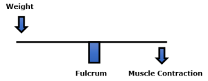
    As a result of their education and examination procedures, chiropractors are legally allowed to provide a number of services to their patients. These include physical therapy, exercise, tissue work, dietary advice, use of supplements, the taking of and the interpretation of x-rays, etc. But the central core of chiropractic clinical practice is the use of mechanical care, and the primary form of mechanical care is specific line-of-drive manipulation (the chiropractic adjustment).

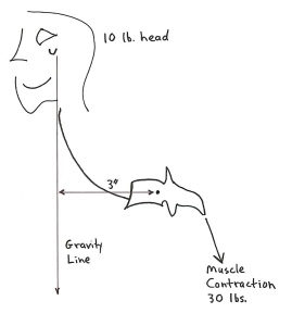
    A typical chiropractic visit involves an assessment of posture and joint motion (possibly with the use of x-rays), helping the chiropractor assess the manner in which his/her patient exists and functions mechanically in a gravity environment. Abnormal findings are usually treated mechanically and primarily with the use of the chiropractic adjustment.

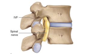
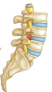
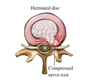
    Early-on, many chiropractors were taught and believed that the physiological basis for their mechanical care was to treat compressive neuropathology (aka the “pinched nerve”). Although some chiropractic patients do suffer from compressive neuropathology, the majority (90+%) of their patients do not suffer from nerve compression (5).

    It is now largely accepted that chiropractic adjustments mechanically activate a neurological sequence of events that close the pain gate (explained later) (5, 6, 7).

    The primary points are that mechanical integrity in a gravity environment is important for how people feel and function, and that chiropractic adjusting influences mechanical integrity in a positive way. Chiropractors are trained to be mechanical providers of care. Ninety-three percent of patients who chose to initially see a chiropractor do so for spinal pain complaints (8). Satisfaction among patients with these complaints was exceptional (8).

    The 2021 Nobel Prize in Physiology or Medicine

    The 2021 Nobel Prize in Physiology or Medicine was awarded to David Julius, PhD, and Ardem Patapoutian, PhD (9, 10). Dr. Julius is a professor and chairman of the department of physiology at the University of California, San Francisco. Dr. Patapoutian is a professor at Scripps Research in La Jolla, California.

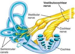
    Their Nobel Prize included explaining the molecular basis for sensing mechanical forces, including how the body senses position and movement.

    Understanding the importance of the neurological sensing of the mechanics of position and movement have been the core interest of the chiropractic profession since 1895. Despite many obstacles and often strong headwinds, the chiropractic profession has held steadfast to its mechanical/neurological roots. For more than a century, chiropractic clinicians and scientists have continued to explore and refine their mechanical applications of care, especially as related to musculoskeletal pain syndromes. The 2021 Noble Prize in Physiology or Medicine supports a century of data used by the chiropractic profession, which is briefly reviewed below.

    •••

    In 1921, Henry Winsor, MD, used 50 cadavers from the University of Pennsylvania and performed autopsies (necropsies) to determine whether there was any connection between spinal mechanical integrity and neurophysiological function (11). Dr. Winsor found, indeed, that spinal mechanical stiffness was common, advances with age, and is associated with deleterious neurological function.

    •••

    In the 1970s, Princeton University educated physiologist Irvin Korr, PhD, re-affirmed that spinal stiffness and/or aberrant motion altered the mechanical proprioceptive input into the central nervous system (12, 13). This aberrant neurological mechanical input would elicit abnormal spinal cord reflexes and abnormal signals to the brain, resulting in a variety of musculoskeletal dysfunctions.

    •••

    In 1985, William H. Kirkaldy-Willis, MD (Professor Emeritus of Orthopedics and director of the Low-Back Pain Clinic at University Hospital, Saskatoon, Canada), used the Gate Theory of Pain to explain the incredible reduction of back and leg pain in 283 subjects from chiropractic spinal adjusting (14).

    The study was a prospective observational study. All 283 patients suffered from chronic low back and/or leg pain; they had also failed prior conservative and/or operative treatment, and they were all totally disabled. For those not suffering from compressive neuropathology, the clinical outcomes were outstanding in 81% of the subjects.

    Dr. Kirkaldy-Willis noted that the Gate Theory of Pain has “withstood rigorous scientific scrutiny.” The Theory indicates that the central transmission of pain can be blocked by increased proprioceptive mechanical input. Spinal articular stiffness reduces proprioception (mechanoreception), “opening” the Pain Gate (increasing pain). Spinal adjusting improves articular motion, improves mechanoreception, and “closes” the Pain Gate (decreasing pain).

    •••

    In 1986 Vert Mooney, MD (orthopedic surgeon from the University of California, San Diego), was elected president of the International Society for the Study of the Lumbar Spine. His Presidential Address was published in the journal Spine the following year (15). In his Presidential Address to the Society, Dr. Mooney unfolds these concepts pertaining to the physiology of back pain:

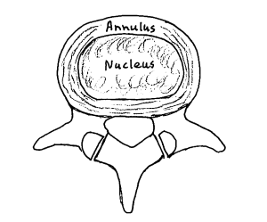
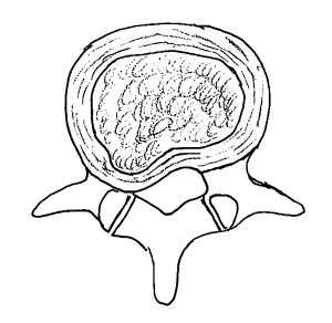
    • Chronic low back pain primarily arises from the intervertebral disc.
    • The intervertebral disc is avascular.
    • An accumulation of inflammatory chemicals in the intervertebral disc initiates the nociceptive (pain) signal to the brain.
    • The dispersion of the discogenic inflammatory nociceptive chemicals can only be achieved by improving the mechanical function of the intervertebral disc.

    Key quotes from Dr. Mooney’s Presidential Address include:

    “Mechanical events can be translated into chemical events related to pain.”

     “Persistent pain in the back with referred pain to the leg is largely on the basis of abnormalities within the disc.”

     “Mechanical activity has a great deal to do with the exchange of water and oxygen concentration [in the disc].” 

     “… research substantiates the view that unchanging posture, as a result of constant pressure such as standing, sitting or lying, leads to an interruption of pressure-dependent transfer of liquid. Actually, the human intervertebral disc lives because of movement.”

     “In summary, what is the answer to the question of where is the pain coming from in the chronic low-back pain patient? I believe its source, ultimately, is in the disc. Basic studies and clinical experience suggest that mechanical therapy is the most rational approach to relief of this painful condition.”

    •••

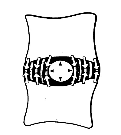
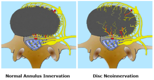
    In 1992, Tuvia Mendel (Louisiana State University Medical Center) documented that the human cervical intervertebral disc is extensively innervated by mechanoreceptors, and that these mechanoreceptors are of considerable clinical importance in pain disorders of the spine (16). Mechanoreceptors were identified in every disc assessed.

    Mr. Mendel notes that these mechanoreceptors provide basic proprioceptive function, specifically the sense of compression, deformation, and alignment. Key quotes from his study include:

    “The presence of neural elements within the intervertebral disc indicates that the mechanical status of the disc is monitored by the central nervous system.”

     “The location of the mechanoreceptors may enable the intervertebral disc to sense peripheral compression or deformation as well as alignment.”

    •••

    In 1994, Robert McLain, MD (then from the Department of Orthopedic Surgery, University of California, Davis, CA), documented that human cervical facet joints were innervated with mechanoreceptors (17). These mechanoreceptors provide basic proprioceptive function; they sense facet capsular tension, pressure, and position. Key quotes from his study include:

     “Encapsulated mechanoreceptors are a consistent finding in normal human cervical facets.”

     “The presence of these receptors in the facet capsule indicate that the mechanical state of the capsule (position, tension, pressure, etc.) is under the surveillance of the central nervous system.”

     “The presence of mechanoreceptive and nociceptive nerve endings in cervical facet capsules proves that these tissues are monitored by the central nervous system and implies that neural input from the facets is important to proprioception and pain sensation in the cervical spine.”

    •••

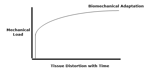
    In 1998, Robert McLain, MD (then from the Department of Orthopedic Surgery, Cleveland Clinic, Ohio), published another study investigating the presence of mechanoreceptors in the facet joints of the thoracic and lumbar spines (18). Similar to his study on cervical spine mechanoreceptors, Dr. McLain established that the thoracic and lumbar facet joints are also innervated with mechanoreceptors. These mechanoreceptors provide basic proprioceptive function, including the sense of motion, tissue distortion, and position. Key quotes from his study include:

    “Ongoing studies of spinal innervation have shown that human facet tissues contain mechanoreceptive endings capable of detecting motion and tissue distortion.”

     “Encapsulated nerve endings are believed to be primarily mechanosensitive and may provide proprioceptive and protective information to the central nervous system regarding joint function and position.”

    •••

     In 1995, Sally Roberts, MD (RJAH Orthopaedic & District Hospital, Oswestry, Shropshire, UK), added to the evidence that human intervertebral discs are innervated with mechanoreceptors (19). She noted that these mechanoreceptors provide basic proprioceptive function, including the maintenance of muscle tone and muscular reflexes. Key quotes from her study include:

    “[Physiologically, these mechanoreceptors] provide the individual with sensation of posture and movement.”

     “In addition to providing proprioception, mechanoreceptors are thought to have roles in maintaining muscle tone and reflexes.”

     “Their presence in the intervertebral disc and longitudinal ligament can have physiologic and clinical implications.”

    •••

    In his 2000 book titled Energy Medicine, James Oschman, PhD, describes a mechanically-based whole-body communication network that functions separately from the nervous system (20). In various parts of the book, Dr. Oschman terms this communication network “tensegrity,” or the “tensegrous matrix,” or the “tensegrous network.” Dr. Oschman amalgamates mechanical integrity and electromagnetic fields into human physiology.

    Dr. Oschman’s book is fascinating and extremely important for providers of mechanically-based care and their patients. He includes a chapter pertaining to gravity and gravitational alignment, as well as a section on the value of chiropractic mechanically-based care. He also includes a section on the mechanics and the practice of orthopedics, as related to musculoskeletal pain syndromes.

    •••

    In 2003, Donald Ingber, MD, PhD (Vascular Biology Program, Departments of Surgery and Pathology, Children’s Hospital and Harvard Medical School), supported the writings of Dr. Oschman above by publishing a study on mechanotransduction and mechanobiology (21). Dr. Ingber notes that mechanical forces are critical regulators of cellular biochemistry and gene expression. He notes that mechanical forces are critical regulators in biology.

    Dr. Ingber states that there is a strong mechanical basis for many generalized medical disabilities, such as lower back pain, which is responsible for a major share of healthcare costs world-wide. Physical interventions can influence cell and tissue function. This would include chiropractic care. Key quotes from his study include:

     “Mechanical forces serve as important regulators at the cell and molecular levels, and they are equally potent as chemical cues.”

    “In vitro and in vivo studies confirm that mechanical forces directly regulate the shape and function of essentially all cell types.”

    •••

    In 2006, Manohar Panjabi, PhD (Department of Orthopaedics and Rehabilitation, Yale University School of Medicine), proposed a new hypothesis to explain chronic back pain (22). He proposed that a single trauma or cumulative microtrauma can cause sub-failure injuries of the ligaments and their embedded mechanoreceptors. These injured mechanoreceptors generate corrupted signals leading to a poor muscle response pattern. This results in abnormal stresses and strains in the ligaments and joints, eventually producing chronic pain.

    Dr. Panjabi emphasizes that the optimal approach to remedy this problem would target the function of the mechanoreceptors. Key quotes from his study include:

    “Abnormal mechanics of the spinal column has been hypothesized to lead to back pain via nociceptive sensors.”

    “Subfailure injuries of the ligaments and embedded mechanoreceptors generate corrupted mechanoreceptor signals.”

     “The hypothesis proposes that the dysfunction of the muscle system over time may lead to chronic back pain via additional mechanoreceptor injury and neural tissue inflammation.”

    •••

    In 2010, Apostolos Dimitroulias, MD (Neurosurgical Department, Medical School, Aristotle University, Thessaloniki, Greece), evaluated the presence of mechanoreceptors in human lumbar (L4–5 and L5–S1) intervertebral discs (23).

    Dr. Dimitroulias concluded that there is an abundant number of mechanoreceptors in the intervertebral discs of the lower lumbar spine in normal human subjects. The role of these mechanoreceptors is the continuous monitoring of position and movement. Abnormal mechanical information will produce muscle spasm, which is an important component of low back pain. A key quote from his study is:

    “These receptors have a key role in the perception of joint position and adjustment of the muscle tone of the vertebral column.”

    •••

    Also, in 2010, James Wang, PhD, (Mechano-Biology Laboratory, Department of Orthopaedic Surgery, University of Pittsburgh School of Medicine) detailed how “Mechanics Rule Cell Biology.” (24) His study continues to support the studies by James Oshman and Donald Ingber above pertaining to the importance of tensegrity and mechanotransduction.

     Dr. Wang notes that cellular mechanotransduction mechanisms convert mechanical force signals into biochemical signals in cells. He notes that “mechanics play a dominant role in cell biology.” He notes that the therapeutic use of mechanics is critical in improving health and function.

    Key quotes from this study include:

    “Mechanical forces are ubiquitous and are known to greatly influence physiology and pathophysiology in humans.”

     “Mechanical forces have a profound effect on tissue homeostasis and pathophysiology.”

    •••

    In her 2017 book Move Your DNA: Restore Your Health Through Natural Movement, biomechanist Katy Bowman summarizes many of the mechanical concepts reviewed in this article (25). She integrates the importance of mechanoreceptors, gravity, alignment, posture, and motion, noting that “our bodies respond to mechanical input.” Key quotes from her book include:

    “We should recognize our lack of health as a sign of a broken (mechanical) environment.”

    “With respect to disease, the human’s internal mechanical environment has been the least-discussed environment of all—a staggering oversight when almost every cell in your body has specialized equipment just to sense the mechanical environment.”

    “You can eat the perfect diet, sleep eight hours a night, and use only baking soda and vinegar to clean your house, but without the loads created by natural movement, all of these worthy efforts are thwarted on a cellular level, and your optimal wellness level remains elusive.”

    “Human diseases are repeatedly explained to us in terms of their chemical or genetic makeup; meanwhile, we’ve completely ignored the load profile that the function of our body depends upon.”

    “Whether out of convenience or ignorance, we have failed to address the habitat [‘habitual position relative to gravity’] in which our genes dwell, and the impact of the way we move on the state of our health.”

    “Movement, position, and resting state of our musculoskeletal system are huge influencers of our mechanical environment.”

    “Movement provides information for the body. Movement is an environmental or epigenetic factor. Our movement environment has been polluted.”

    Katy Bowman’s book is incredibly consistent with the chiropractic perspective and chiropractic clinical practice.

    •••

    Summary

    Mechanical providers of care have helped patients for millennia. Since 1895, in the United States and other countries around the world, the primary providers of mechanical care are chiropractors. The mechanical care delivered by chiropractors primarily influence special nerves that register the manner in which the individual is positioned and moves in a gravity environment. These specialized nerves are called mechanoreceptors. The studies reviewed here predict that the future of healthcare will involve mechanoreceptors and tensegrity. As noted, mechanoreceptors were awarded the Nobel Prize in Physiology or Medicine in October 2021. The status and future of chiropractic is looking very bright.

    REFERENCES

    1. Morgan M; Mutant Message Down Under; 1990.
    2. Anderson R; “Spinal Manipulation Before Chiropractic”; in Haldeman S; Principles and Practice of Chiropractic; Second Edition; Appleton & Lang; 1992.
    3. Pettman E; A History of Manipulative Therapy; The Journal of Manual & Manipulative Therapy; 2007; Vol. 15; No. 3; pp. 165–174.
    4. cce-usa.org; accessed May 3, 2022.
    5. Kirkaldy-Willis WH, Cassidy JD; Spinal Manipulation in the Treatment of Low back Pain; Canadian Family Physician; March 1985; Vol. 31; pp. 535-540.
    6. Vicenzino B, Collins D, Wright A; The Initial Effects of a Cervical Spine Manipulative Physiotherapy Treatment on the Pain and Dysfunction of Lateral Epicondylalgia; Pain; November 1996; Vol. 68; No. 1; pp. 69-74.
    7. Savva C, Giakas G, Efstathiou M; The role of the descending inhibitory pain mechanism in musculoskeletal pain following high-velocity, low amplitude thrust manipulation: a review of the literature; Journal of Back and Musculoskeletal Rehabilitation; 2014; Vol. 27; No. 4; pp. 377-382.
    8. Adams J, Peng W, Cramer H, Sundberg T, Moore C; The Prevalence, Patterns, and Predictors of Chiropractic Use Among US Adults: Results From the 2012 National Health Interview Survey; December 1, 2017; Spine; Vol. 42; No. 23; pp. 1810–1816.
    9. Roland D, Abott B; Nobel Prize in Medicine Awarded for Work on Senses; Wall Street Journal; October 5, 2021.
    10. Zylka MJ; A Nobel Prize for Sensational Research; New England Journal of Medicine; December 16, 2021; Vol. 385; No. 25; pp. 2393-2394.
    11. Winsor H; Sympathetic Segmental Disturbances: The Evidences of the Association, in Dissected Cadavers, of Visceral Disease with Vertebral Deformities of the Same Sympathetic Segments; Medical Times; November 1921; pp. 1-7.
    12. Korr IM; Proprioceptors and Somatic Dysfunction; Journal of the American Osteopathic Association; March 1975; Vol. 74; No. 7; pp. 638-650.
    13. Korr IM; The Spinal Cord as Organizer of Disease Process III: Hyperactivity of Sympathetic Innervation as a Common Factor in Disease; Journal of the American Osteopathic Association; December 1979; Vol. 79; No. 4; pp. 232-237.
    14. Kirkaldy-Willis WH, Cassidy JD; Spinal Manipulation in the Treatment of Low Back Pain; Canadian Family Physician; March 1985; Vol. 31; pp. 535-540.
    15. Mooney V; Where Is the Pain Coming From?; Spine; October 1987; Vol. 12; No. 8; pp. 754-759.
    16. Mendel T, Wink CS, Zimny ML; Neural Elements in Human Cervical Intervertebral Discs; Spine; February 1992; Vol. 17; No. 2; pp. 132-135.
    17. McLain RF; Mechanoreceptor endings in human cervical facet joints; Spine; March 1, 1994; Vol. 19; No. 5; pp. 495-501.
    18. McLain RE, Pickar JG; Mechanoreceptor endings in human thoracic and lumbar facet joints; Spine; January 15, 1998; Vol. 23; No. 2; pp. 168-173.
    19. Roberts S, Eisenstein SM, Menage J, Evans EH, Ashton IK; Mechanoreceptors in intervertebral discs: Morphology, distribution, and neuropeptides; Spine; December 15, 1995; Vol. 20; No. 24; pp. 2645-2651.
    20. Oschman JL; Energy Medicine: The Scientific Basis; Churchill Livingstone; 2000.
    21. Ingber DE; Mechanobiology and Diseases of Mechanotransduction; Annals of Medicine; 2003; Vol. 35; No. 8; pp.564-577.
    22. Panjabi MM; A hypothesis of chronic back pain: Ligament subfailure injuries lead to muscle control dysfunction; European Spine Journal; May 2006; Vol. 15; No. 5; pp. 668-676.
    23. Dimitroulias A, Tsonidis C, Natsis K, Venizelos I, Djau SN. Tsitsopoulos P; An immunohistochemical study of mechanoreceptors in lumbar spine intervertebral discs; Journal of Clinical Neuroscience; June 2010; Vol. 17; No 6; pp. 742-745.
    24. Wang J, Bin Li; Mechanics Rule Cell Biology; Sports Medicine, Arthroscopy, Rehabilitation, Therapy & Technology; 2010; Vol. 2; No. 16.
    25. Bowman K; Move Your DNA: Restore Your Health Through Natural Movement; 2017.

    “Authored by Dan Murphy, D.C.. Published by ChiroTrust® – This publication is not meant to offer treatment advice or protocols. Cited material is not necessarily the opinion of the author or publisher.”

  • The Chiropractic Management  of Discogenic Compressive  Radicular Sciatica

    The Chiropractic Management of Discogenic Compressive Radicular Sciatica

    The low back is the most common body region to suffer from chronic pain (1).

    Low back pain is the second most common reason for primary care physician visits, and it is the most costly medical condition in the United States (2).

    The most common tissue source for low back pain is the intervertebral disc (3, 4, 5, 6).

    The primary clinical syndrome treated by chiropractors is chronic low back pain (7).

    Chiropractic care is proven to be very successful in the treatment of chronic low back pain. Often, chiropractic care is superior to alternative types of management for chronic low back pain, including prescription nonsteroidal anti-inflammatory drugs, physical therapy, exercise, and needle acupuncture (8, 9, 10, 11, 12).

    Long-term follow-up studies on these chronic patients show that chiropractic care obtains stable therapeutic benefits (9, 10, 12).

    Chiropractic care is not only very effective for low back pain, it is also very safe. In some randomized clinical trials, not a single adverse event is reported of the study subjects randomized to receiving chiropractic spinal manipulation (10).

    Definitions/Terminology

     Radiculitis and Radiculopathy

    Between each and every spinal segmental level, a spinal nerve exits from the intervertebral foramen (IVF). The technical terminology for the spinal nerve is the nerve root. Spinal nerve root problems (compression, irritation, inflammation, pathology) are called radiculitis and/or radiculopathy. Radiculitis and Radiculopathy mean it is a nerve root problem.

    Between each and every spinal segmental level, a spinal nerve exits from the intervertebral foramen (IVF). The technical terminology for the spinal nerve is the nerve root. Spinal nerve root problems (compression, irritation, inflammation, pathology) are called radiculitis and/or radiculopathy. Radiculitis and Radiculopathy mean it is a nerve root problem.

    Neuritis and Neuropathy

    A peripheral nerve is made up of more than one nerve root. The technical terminology for a peripheral nerve uses the base neuro. Peripheral nerve problems (compression, irritation, inflammation, pathology) are called neuritis and/or neuropathy. Neuritis and Neuropathy mean it is a peripheral nerve problem.

    Interim Discussion

    The sciatic nerve is a nerve that begins in the lower back and travels through the buttock and down into the back of the leg and then into the foot. It is the longest and widest nerve in the human body. The sciatic nerve is made up of five lumbosacral nerve roots, L4, L5, S1, S2, and S3.

    The sciatic nerve is a nerve that begins in the lower back and travels through the buttock and down into the back of the leg and then into the foot. It is the longest and widest nerve in the human body. The sciatic nerve is made up of five lumbosacral nerve roots, L4, L5, S1, S2, and S3.

    When a nerve is compressed, irritated, or inflamed, it generates symptoms (pain, numbness, tingling, hypersensitivity, burning, achiness, etc.) and/or functional disturbances (weakness, atrophy, etc.).

    The sciatic nerve is a peripheral nerve. When a peripheral nerve causes symptoms and/or signs, it is neuritis or neuropathy. Signs and symptoms attributed to the sciatic nerve are called sciatic neuritis or sciatic neuropathy. However, most people and most health care providers simply refer to it as sciatica.

    If any of the nerve roots that make up the sciatic nerve are compressed, irritated, and/or inflamed, it will generate the signs and/or symptoms of sciatica. In such a circumstance, the technical terminology would be sciatic radiculitis or sciatic radiculopathy.

    Leg Pain, Sciatica

    Some patients with low back pain also suffer from leg pain. When leg pain is present, it often indicates that the pathoanatomical basis for the patient’s symptoms are more complex and that the resolution of the symptoms and signs will also be more complicated.

    Clinicians usually view leg pain in three categories:

    Category 1:

    Sclerogenic pain; also known as Sclerotomic pain or Sclerotogenous pain

    Sclerogenic leg pain occurs as a consequence of an irritation of low back tissues below the deep fascia (deep spinal muscles, facet capsule ligaments, annulus of the intervertebral disc, etc.). It often subjectively presents as a deep dull ache. It is rare for the discomfort to extend below the knee. It is difficult for the patient to precisely locate the pain on the skin; the discomfort is perceived deep to the skin. Lumbar spine vertical compression tests (including Kemp’s) are usually negative. Straight leg raising tests are usually negative. Superficial sensation is usually negative. Myotomal strength tests are usually normal. Deep tendon reflexes are usually normal and symmetrical.

    In general, sclerogenic pain is not dangerous. It is a form of referred pain that occurs as a consequence of a shared neuromere during embryonic development. The neurology of the back and the leg are shared embryologically, which can cause some confusion as to the exact location of the irritation when the electrical signal is sent to the brain: an irritation of low back tissues might be interpreted by the brain as arising from the leg.

    The original descriptions of sclerogenic pain were from the research of JH Kellgren and colleague in 1938 and 1939 (13, 14).

    Chiropractic spinal adjustments (specific line-of-drive manipulations) primarily affect the deep spinal tissues (deep spinal muscles, facet capsule ligaments, annulus of the intervertebral disc, etc.). These are the same tissues that are responsible for the sclerogenic pain referral. Consequently, chiropractic spinal adjusting is very effective in improving and/or resolving both back pain and referred sclerogenic leg pain.

    Category 2:

    Peripheral Neuropathy

    A peripheral neuropathy is NOT a nerve root problem. It is a compressed, irritated, and/or inflamed peripheral nerve. An example of a peripheral neuropathy is piriformis syndrome sciatica.

    A recent review on piriformis syndrome sciatica reminds the reader that the sciatic nerve runs adjacent to the piriformis muscle and may even pierce through the muscle. The authors state (15):

    “Piriformis syndrome is a clinical condition of sciatic nerve entrapment at the level of the ischial tuberosity.”

     “Patients often report pain in the gluteal/buttock region that may ‘shoot,’ burn or ache down the back of the leg (i.e. ‘sciatic’-like pain).”

     “Numbness in the buttocks and tingling sensations along the distribution of the sciatic nerve is not uncommon.”

    When a peripheral neuropathy exists independent from radicular involvement, the treatment is to the peripheral lesion. In the example of piriformis syndrome sciatica, treatment would be to the piriformis muscle and to the hip joint. Pelvic unleveling could also be involved, requiring chiropractic evaluation and management.

     When a peripheral neuropathy exists independent from radicular involvement, the treatment is to the peripheral lesion. In the example of piriformis syndrome sciatica, treatment would be to the piriformis muscle and to the hip joint. Pelvic unleveling could also be involved, requiring chiropractic evaluation and management.

     Category 3:

    Radicular Pain, Radiculitis, Radiculopathy

     Radicular leg pain is often caused by compression of the nerve root, the compression causing nerve root irritation, inflammation, and/or ischemia. This pathology is referred to as compressive radiculopathy.

    The most classic cause of radicular compression is herniation of the intervertebral disc. Other causes include arthritic changes (degenerative joint disease, degenerative disc disease, spondylosis) causing osseous (bone spurs, hypertrophic changes, osteophytes) narrowing of the intervertebral foramen.

    The most classic cause of radicular compression is herniation of the intervertebral disc. Other causes include arthritic changes (degenerative joint disease, degenerative disc disease, spondylosis) causing osseous (bone spurs, hypertrophic changes, osteophytes) narrowing of the intervertebral foramen.

    The chiropractic (low back spinal manipulation) management of discogenic compressive radicular sciatica has a long history that includes publications in a range of medical journals and reference texts. A representative number of these studies are reviewed here.

    •••••••••

    In 1954, a study was published, titled (16):

    Conservative Treatment of Intervertebral Disk Lesions

     The author states:

    “The conservative management of lumbar disk lesions should be given careful consideration because no patient should be considered for surgical treatment without first having failed to respond to an adequate program of conservative treatment.”

     “If after a fair trial of conservative treatment, the pain and disability continue and the symptoms are of sufficient gravity to warrant surgery, the patient is advised that he should be operated upon and the offending disk lesion should be removed.”

    “From what is known about the pathology of lumbar disk lesions, it would seem that the ideal form of conservative treatment would theoretically be a manipulative closed reduction of the displaced disk material.”

     “We limit the use of manipulation almost entirely to those patients who do not seem to be responding well to non-manipulative conservative treatment and who are anxious to have something else done short of operative intervention.”

    ••••••••

    In 1969, a study was published, titled (17):

    Reduction of Lumbar Disc Prolapse by Manipulation

     The authors evaluated patients that presented with an acute onset of low back and buttock pain that did not respond to rest. Diagnostic epidurography showed a clinically relevant disc herniation, along with antalgia and positive lumbar spine nerve stretch tests. These patients were then treated with lumbar spine rotation thrust manipulations. Manipulations were repeated until abnormal symptoms and signs had disappeared. Following the manipulations there was resolution of signs, symptoms, antalgia, and reduction in the size of the protrusions. The authors state:

     “The frequent accompaniment of acute onset low back pain by spinal deformity suggests a mechanical factor, and the accompanying abnormality of straight-leg raise or femoral stretch test suggests that the lesion impinges on the spinal dura matter of the dural nerve sheaths.”

     “The lumbar spine was rotated away from the painful side to the limit of its range, the buttock or thigh of the painful side being used as a lever; a firm additional thrust was made in the same direction. This manoeuver was repeated until abnormal symptoms and signs had disappeared, progress being assessed by repeated examination.”

     “Rotation manipulations apply torsion stress throughout the lumbar spine. If the posterior longitudinal ligament and the annulus fibrosus are intact, some of this torsion force would tend to exert a centripetal force, reducing prolapsed or bulging disc material.” 

     “The results of this study suggest that small disc protrusions were present in patients presenting with lumbago and that the protrusions were diminished in size when their symptoms had been relieved by manipulations.”

    These authors conclude: “it seems likely that the reduction effect [of the disc protrusion] is due to the manipulating thrust used.”

     •••••••••

    Also in 1969, a study was published, titled (18):

    Low Back Pain and Pain Resulting from Lumbar Spine Conditions:
    A Comparison of Treatment Results

    The authors compared the effectiveness of heat/massage/exercise to spinal manipulation in the treatment of 184 patients that were grouped according to the presentation of back and leg pain. This study was then reviewed in their 1990 book, Clinical Biomechanics of the Spine, which made these points (19):

    “A well-designed, well executed, and well-analyzed study.”

    In the group with central low back pain only, “the results were acceptable in 83% for both treatments. However, they were achieved with spinal manipulation using about one-half the number of treatments that were needed for heat, massage, and exercise.”

    In the group with pain radiating into the buttock, “the results were slightly better with manipulation, and again they were achieved with about half as many treatments.”

     In the groups with pain radiation to the knee and/or to the foot, “the manipulation therapy was statistically significantly better,” and in the group with pain radiating to the foot, “the manipulative therapy is significantly better.”

     “This study certainly supports the efficacy of spinal manipulative therapy in comparison with heat, massage, and exercise. The results (80–95% satisfactory) are impressive in comparison with any form of therapy.”

    •••••••••

    In 1977, the third edition of the text book Orthopaedics, Principles and Their Applications was published. The author made these observations (20):

    Treatment of Intervertebral Disc Herniation With Manipulation

    “Manipulation. Some orthopaedic surgeons practice manipulation in an effort at repositioning the disc.  This treatment is regarded as controversial and a form of quackery by many men.  However, the author has attempted the maneuver in patients who did not respond to bed rest and were regarded as candidates for surgery.  Occasionally, the results were dramatic.”

    •••••••••

    In 1987, a study was published, titled (21):

    Treatment of Lumbar Intervertebral Disc Protrusions by Manipulation

    The authors performed a series of eight manipulations on 517 patients with protruded lumbar discs and clinically relevant signs and symptoms. Their outcomes were very good, with 84% achieving a successful outcome and only 9% not responding; 14% suffered a reoccurrence of symptoms at intervals ranging from two months to twelve years. These authors state:

    “Manipulation of the spine can be effective treatment for lumbar disc protrusions.”

     “Most protruded discs may be manipulated. When the diagnosis is in doubt, gentle force should be used at first as a trial in order to gain the confidence of the patient.”

     “During manipulation a snap may accompany rotation. Subjectively it has dramatic influence on both patient and operator and is thought to be a sign of relief.”

     “If derangement of the facets or subluxation of the posterior elements near the protruded disc occurs, the rotation may have caused reduction, giving remarkable relief.”

     “Gapping of the disc on bending and rotation may create a condition favorable for the possible reentry of the protruded disc into the intervertebral cavity, or the rotary manipulation may cause the protruded disc to shift away from pressing on the nerve root.”

    •••••••••

    In 1989, a study was published, titled (22):

    Lumbar Intervertebral Disc Herniation:
    Treatment by Rotational Manipulation

    This was a case study of a patient with an “enormous central herniation lumbar disc” who underwent a course of side posture manipulation. The patient improved considerably with only 2 weeks of treatment. The authors state:

    “It is emphasized that manipulation has been shown to be an effective treatment for some patients with lumbar disc herniations.”

     •••••••••

    In 1993, a “review of the literature” study was published, titled (23):

    Side Posture Manipulation for Lumbar Intervertebral Disk Herniation

    These authors state:

    “The treatment of lumbar disk herniation by side posture manipulation is not new and has been advocated by both chiropractors and medical manipulators.”

     “The treatment of lumbar intervertebral disk herniation by side posture manipulation is both safe and effective.”

    •••••••••

    In 1995, a study was published, titled (24):

    A Series of Consecutive Cases of Low Back Pain
    with Radiating Leg Pain Treated by Chiropractors

    The authors retrospectively reviewed the outcomes of 59 consecutive patients complaining of low back and radiating leg pain were clinically diagnosed as having a lumbar spine disk herniation. Ninety percent of these patients reported improvement of their complaint after chiropractic manipulation. The authors concluded:

    “Based on our results, we postulate that a course of non-operative treatment including manipulation may be effective and safe for the treatment of back and radiating leg pain.”

     •••••••••

     In 2006, a study was published, titled (25):

    Chiropractic Manipulation in the Treatment
    of Acute Back Pain and Sciatica with Disc Protrusion

    The purpose of this study was to assess the short- and long-term effects of spinal manipulations on acute back pain and sciatica with disc protrusion. It is a randomized double-blind trial comparing active and simulated manipulations for these patients. The study used 102 patients. The manipulations or simulated manipulations were done 5 days per week by experienced chiropractors for up to a maximum of 20 patient visits, “using a rapid thrust technique.” Re-evaluations were done at 15, 30, 45, 90, and 180 days. The authors made these observations:

    “Active manipulations have more effect than simulated manipulations on pain relief for acute back pain and sciatica with disc protrusion.”

     “At the end of follow-up, a significant difference was present between active and simulated manipulations in the percentage of cases becoming pain-free (local pain 28% vs. 6%; radiating pain 55% vs. 20%).”

     “Patients receiving active manipulations enjoyed significantly greater relief of local and radiating acute LBP, spent fewer days with moderate-to-severe pain, and consumed fewer drugs for the control of pain.”

    The authors concluded that chiropractic spinal “manipulations may relieve acute back pain and sciatica with disc protrusion.”

    ••••••••

    In 2010, a study was published, titled (26):

    Manipulation or Micro-Diskectomy for Sciatica?

    The purpose of this study was to compare the clinical efficacy of spinal manipulation against micro-diskectomy in patients with sciatica secondary to lumbar disk herniation. It is a randomized clinical trial. All study subjects suffered from sciatic radiculopathy for more than 3 months.

    All study subjects were referred for neurosurgery by their primary care physician after they failed at least 3 months of conservative management including treatment with analgesics, lifestyle modification, physiotherapy, massage therapy, and/or acupuncture. After failing 3 months of traditional conservative care (analgesics, lifestyle modification, physiotherapy, massage, and/or acupuncture), 40 patients were randomized to either surgical micro-diskectomy or to chiropractic spinal manipulation.

    At the 12-week follow-up, 60% of the manipulation group “demonstrated clear improvement in outcomes and continued to complete the 52-week follow-up period.” The authors state:

    “Although 40% of patients referred to spinal manipulative therapy for lumbar disc herniation-induced sciatica may fail to achieve satisfactory relief, the obvious risk and cost profile of operative care argues for serious physician and patient consideration of spinal manipulative therapy before surgical intervention.”

    •••••••••

    In 2014, an interdisciplinary group of physicians, chiropractors, and researchers published a study titled (27):

    Spinal Manipulation and Home Exercise with Advice
    for Subacute and Chronic Back-Related Leg Pain

    This study included 192 patients who were suffering from back-related leg pain for at least 4 weeks. The authors concluded:

    “For leg pain, spinal manipulative therapy plus home exercise and advice had a clinically important advantage over home exercise and advice at 12 weeks.”

     “For patients with subacute and chronic back-related leg pain, spinal manipulative therapy in addition to home exercise and advice is a safe and effective conservative treatment approach, resulting in better short-term outcomes than home exercise and advice alone.”

     •••••••••

    Also in a 2014 study, a group of multidisciplinary researchers and chiropractic clinicians presented a prospective study involving 148 patients with low back and leg pain, titled (28):

    Outcomes of Acute and Chronic Patients with Magnetic Resonance Imaging–Confirmed Symptomatic Lumbar Disc Herniations Receiving High-Velocity, Low-Amplitude, Spinal Manipulative Therapy

    The purpose of this study was to document outcomes of patients with confirmed, symptomatic lumbar disc herniations and sciatica that were treated with chiropractic side posture high-velocity, low-amplitude, spinal manipulation to the level of the disc herniation. The authors made the following statements:

     “The proportion of patients reporting clinically relevant improvement in this current study is surprisingly good, with nearly 70% of patients improved as early as 2 weeks after the start of treatment. By 3 months, this figure was up to 90.5% and then stabilized at 6 months and 1 year.”

     “A large percentage of acute and importantly chronic lumbar disc herniation patients treated with chiropractic spinal manipulation reported clinically relevant improvement.”

     “A large percentage of acute and importantly chronic lumbar disc herniation patients treated with high-velocity, low-amplitude side posture spinal manipulative therapy reported clinically relevant ‘improvement’ with no serious adverse events.”

     “Spinal Manipulative therapy is a very safe and cost-effective option for treating symptomatic lumbar disc herniation.”

    •••••••••

    In 2021, a study was published, titled (29):

    Physical Therapy Referral from Primary Care
    for Acute Back Pain With Sciatica

    This study involved 220 subjects with an acute onset of low back pain and sciatica. Half were given usual care (drugs and a single education session), and half were referred for 6-8 early sessions of exercise with mechanical manual therapy. The manual therapy consisted of mobilization or high-velocity thrust manipulation of the lumbar spine. The authors noted:

     “Patients receiving [early mechanical treatment] were more likely to rate their treatment as successful at 4 weeks and 1 year.”

     Early treatment “hastened functional improvement, indicating that [early mechanical treatment] can be offered to patients as first-line nonpharmacologic care.”

     Early mechanical treatment “for recent-onset low back pain and sciatica resulted in greater improvement in disability and secondary outcomes than usual care across the 1-year follow-up.”

    ••••••••

    Also in 2021, a study was published, titled (30):

    Spinal Manipulation for Subacute and Chronic Lumbar Radiculopathy

    The objective of this study was to evaluate the efficacy of spinal manipulation for the management of subacute and/or chronic lumbar radiculopathy. Forty-four patients, with unilateral radicular low back pain lasting more than 4 weeks, were randomly allocated to a treatment group  [manipulation + physiotherapy] and a control group [physiotherapy only]. Key points from the authors include:

    “Spinal manipulation improves the results of physiotherapy over a period of 3 months for patients with subacute or chronic lumbar radiculopathy.”

    “Minimum side effects, ease of administration, and patient satisfaction are the expected benefits of manipulation.”

    •••••••••

    Conclusions

    It is understood that some patients suffering from discogenic compressive radicular sciatica will require some form of decompressive spinal surgery. The studies presented here support that prior to surgery, spinal manipulation should be tried in an effort to avoid surgery. Spinal manipulation, especially by those expertly trained (chiropractors), is safe and often very effective. Chiropractors are also trained to monitor patient progress for any symptoms or signs that might benefit from a surgical consultation.

    REFERENCES

    1. Wang S; Why Does Chronic Pain Hurt Some People More?; Wall Street Journal; October 7, 2013.
    2. Fritz JM, Lane E, McFadden M, Brennan G, Magel JS, Thackeray A, Minick K, Meier W, Greene T; Physical Therapy Referral from Primary Care for Acute Back Pain with Sciatica: A Randomized Controlled Trial; Annals of Internal Medicine; January 2021; Vol. 174; No. 1; pp. 8-17.
    3. Nachemson A; The Lumbar Spine, An Orthopedic Challenge; Spine; Vol. 1; No. 1; March 1976; pp. 59-71.
    4. Kuslich S, Ulstrom C, Michael C; The Tissue Origin of Low Back Pain and Sciatica: A Report of Pain Response to Tissue Stimulation During Operations on the Lumbar Spine Using Local Anesthesia; Orthopedic Clinics of North America; Vol. 22; No. 2; April 1991; pp. 181-187.
    5. Izzo R, Popolizio T, D’Aprile P, Muto M; Spine Pain; European Journal of Radiology; May 2015; Vol. 84; pp. 746–756.
    6. McGowan JR, Suiter L; Cost-Efficiency and Effectiveness of Including Doctors of Chiropractic to Offer Treatment Under Medicaid: A Critical Appraisal of Missouri Inclusion of Chiropractic Under Missouri Medicaid; Journal of Chiropractic Humanities; December 2019; Vol. 10; No. 26; pp. 31-52.
    7. Adams J, Peng W, Cramer H, Sundberg T, Moore C; The Prevalence, Patterns, and Predictors of Chiropractic Use Among US Adults: Results From the 2012 National Health Interview Survey; Spine; December 1, 2017; Vol. 42; No. 23; pp. 1810–1816.
    8. Kirkaldy-Willis WH, Cassidy JD; Spinal Manipulation in the Treatment of Low back Pain; Canadian Family Physician; March 1985; Vol. 31; pp. 535-540.
    9. Meade TW, Dyer S, Browne W, Townsend J, Frank OA; Low back pain of mechanical origin: Randomized comparison of chiropractic and hospital outpatient treatment; British Medical Journal; June 2, 1990; Vol. 300; pp. 1431-1437.
    10. The Lancet; Chiropractors and Low Back Pain; July 28, 1990; p. 220.
    11. Giles LGF; Muller R; Chronic Spinal Pain: A Randomized Clinical Trial Comparing Medication, Acupuncture, and Spinal Manipulation; Spine; July 15, 2003; Vol. 28; No. 14; pp. 1490-1502.
    12. Muller R, Giles LGF; Long-Term Follow-up of a Randomized Clinical Trial Assessing the Efficacy of Medication, Acupuncture, and Spinal Manipulation for Chronic Mechanical Spinal Pain Syndromes; Journal of Manipulative and Physiological Therapeutics; January 2005; Vol. 28; No. 1; pp. 3-11.
    13. Kellgren JH, Lewis; Observations on referred pain arising from muscle; Clinical Science; 1938; Vol. 3; p. 175.
    14. Kellgren JH; On the distribution of pain arising from deep somatic structures with charts of segmental pain areas; Clinical Science; 1939; Vol. 4; pp. 35-46.
    15. Hicks BL, Lam JC, Varacallo M; Piriformis Syndrome; StatPearls [Internet]; Treasure Island (FL): StatPearls Publishing; January 2022.
    16. Ramsey RH; Conservative Treatment of Intervertebral Disk Lesions; American Academy of Orthopedic Surgeons; Instructional Course Lectures; Vol. 11; 1954; pp. 118-120.
    17. Mathews JA and Yates DAH; Reduction of Lumbar Disc Prolapse by Manipulation; British Medical Journal; September 20, 1969; No. 3; pp. 696-697.
    18. Edwards BC; Low back pain and pain resulting from lumbar spine conditions: a comparison of treatment results; Australian Journal of Physiotherapy; September 1969; Vol. 15; No. 3; pp. 104-110.
    19. White AA, Panjabi MM; Clinical Biomechanics of the Spine; Second edition; JB Lippincott Company; 1990.
    20. Turek S; Orthopaedics, Principles and Their Applications; JB Lippincott Company; 1977; page 1335.
    21. Kuo PP, Loh ZC; Treatment of Lumbar Intervertebral Disc Protrusions by Manipulation; Clinical Orthopedics and Related Research; February 1987; No. 215; pp. 47-55.
    22. Quon JA, Cassidy JD, O’Connor SM, Kirkaldy-Willis WH; Lumbar intervertebral disc herniation: treatment by rotational manipulation; Journal of Manipulative and Physiological Therapeutics; June 1989; Vol. 12; No. 3; pp. 220-227.
    23. Cassidy JD, Thiel HW, Kirkaldy-Willis WH; Side posture manipulation for lumbar intervertebral disk herniation; Journal of Manipulative and Physiological Therapeutics; February 1993; Vol. 16; No. 2; pp. 96-103.
    24. Stern PJ, Côté P, Cassidy JD; A series of consecutive cases of low back pain with radiating leg pain treated by chiropractors; Journal of Manipulative and Physiological Therapeutics; Jul-Aug 1995; Vol. 18; No. 6; pp. 335-342.
    25. Santilli V, Beghi E, Finucci S; Chiropractic manipulation in the treatment of acute back pain and sciatica with disc protrusion: A randomized double-blind clinical trial of active and simulated spinal manipulations; The Spine Journal; March-April 2006; Vol. 6; No. 2; pp. 131–137.
    26. McMorland G, Suter E, Casha S, du Plessis SJ, Hurlbert RJ; Manipulation or Microdiskectomy for Sciatica? A Prospective Randomized Clinical Study; Journal of Manipulative and Physiological Therapeutics; October 2010; Vol. 33; No. 8; pp. 576-584.
    27. Bronfort G, Hondras M, Schulz CA, Evans RL, Long CR, Grimm R; Spinal Manipulation and Home Exercise With Advice for Subacute and Chronic Back-Related Leg Pain: A Trial With Adaptive Allocation; Annals of Internal Medicine; September 16, 2014; Vol. 161; No. 6; pp. 381-391.
    28. Leemann S, Peterson CK, Schmid C, Anklin B, Humphreys BK; Outcomes of Acute and Chronic Patients with Magnetic Resonance Imaging–Confirmed Symptomatic Lumbar Disc Herniations Receiving High-Velocity, Low Amplitude, Spinal Manipulative Therapy: A Prospective Observational Cohort Study With One-Year Follow-Up; Journal of Manipulative and Physiological Therapeutics; March/April 2014; Vol. 37; No. 3; pp. 155-163.
    29. Fritz JM, Lane E, McFadden M, Brennan G, Magel JS, Thackeray A, Minick K, Meier W, Greene T; Physical Therapy Referral from Primary Care for Acute Back Pain with Sciatica: A Randomized Controlled Trial; Annals of Internal Medicine; January 2021; Vol. 174; No. 1; pp. 8-17.
    30. Ghasabmahaleh SH, Rezasoltani Z, Dadarkhah A, Hamidipanah S, Mofrad RK, Sharif Najafi S; Spinal Manipulation for Subacute and Chronic Lumbar Radiculopathy: A Randomized Controlled Trial; The American Journal of Medicine; January 2021; Vol. 134; No. 1; pp. 135−141.

    “Authored by Dan Murphy, D.C.. Published by ChiroTrust® – This publication is not meant to offer treatment advice or protocols. Cited material is not necessarily the opinion of the author or publisher.”

  • Text Neck & Tech Neck

    Text Neck & Tech Neck

    Integrating Posture, Weight, Load, Spinal Degenerative Disease,
    Education of Postural Habits, and Chiropractic Care

    The primary method of public transportation connecting the cities of the San Francisco Bay Area is a train system called BART (Bay Area Rapid Transit). BART passenger boarding platforms are often above ground adjacent to freeways. Driving by on the freeway, one can see dozens to hundreds of BART passengers waiting to board the next train; essentially all of them have their heads down and their necks bent forward while looking at their cell phones.

    Reflecting on advances in technology over the past 120 years reminds us of how much technology has changed our lives. Automobiles, airplanes, space shuttles, drones, telephones, radio, television, computers, and so much more. This discussion will address cell phones.

    The technology of cellular communications is important to everyone. Cellular technology is of particular importance to the chiropractic profession because cell phone use causes postural distortions. These postural distortions can have devastating effects on spinal joints, particularly the joints of the low neck (cervical spine) and upper back (thoracic spine). These spinal joint problems are collectively referred to as accelerated degenerative spinal joint disease or spondylosis (1, 2, 3, 4).

    In addition, the prolonged postures associated with cell phone use are being linked to other health problems, including (5):

    • Muscle fatigue and myofascial pain syndromes (muscle pain problems and headache) (6, 7, 8).
    • Chronic muscle contraction that often results in headaches (9, 10).

    Early on, cell phones had only one function: talking on the phone. Today, cellular devices are often referred to as smart phones because they engage in many other activities (5):

    • Talking on the phone
    • Texting
    • GPS services
    • Wi Fi services
    • Entertainment: videos, games, surfing the web, Facetime, music, radio, podcasts, etc.
    • News, traffic flows, banking, and other business applications.

    Understanding the effects of chronic postural distortions from any cause requires basic knowledge of three key principles of biomechanics:

    • viscoelastic creep
    • weight v. load
    • levers

    Viscoelastic Creep

    Biological systems adapt to prolonged mechanical loads. Tissues literally change in response to these loads; they adapt to become longer or shorter in response to the loads (11). This includes the biomechanical loads caused by the prolonged use of cellular devices. The technical term for this premise is called viscoelastic creep (12).

    Biological systems adapt to prolonged mechanical loads. Tissues literally change in response to these loads; they adapt to become longer or shorter in response to the loads (11). This includes the biomechanical loads caused by the prolonged use of cellular devices. The technical term for this premise is called viscoelastic creep (12).

    Soft tissues (ligaments, cartilage, tendons, muscles, etc.) undergo viscoelastic creep when prolonged loads are applied. The tissues mechanically distort and eventually adapt to the applied loads. This includes the tissue distortions caused by head-down posture and the forward bending of the cervical spine while viewing or working on hand-held cellular devices (13).

    Soft tissues (ligaments, cartilage, tendons, muscles, etc.) undergo viscoelastic creep when prolonged loads are applied. The tissues mechanically distort and eventually adapt to the applied loads. This includes the tissue distortions caused by head-down posture and the forward bending of the cervical spine while viewing or working on hand-held cellular devices (13).

    Weight is self-explanatory. For this discussion, assume that the head weighs 10 lbs. As explained by Katy Bowman in her 2017 book Move Your DNA: Restore Your Health Through Natural Movement, weight is far less important than is load (11). Load is weight multiplied by a lever arm:

    “It’s not the weight that breaks you down, it’s the load created by the way you carry it.”

    “Loads are often oversimplified to ‘weight’ because it makes them easier to understand, but there is much more going on with your sore knee (or foot, or back, or pelvic floor) than your weight.”

    “Weight is not the be-all and end-all of loads. When you want to improve your health, it’s much more important to consider how you carry your weight than to spend hours contemplating the lone data point that is Your Weight.”

    Levers

    Upright posture is a 1st class lever mechanical system (12). The classic example of a 1st class lever mechanical system is a seesaw or teeter totter.

    The fulcrum of a 1st class lever is the place where the forces are the greatest. These forces are dependent upon three factors:

    • The weight of the object that is on one end of the lever
    • The distance the weight is away from the fulcrum (lever arm)
    • The addition of the counterbalancing effort required to remain balanced

    Again, assume the head weighs 10 lbs. If the head’s weight is 10 lbs., and the distance (the lever arm) from the fulcrum was 10 inches, the load on the fulcrum would be 100 lbs. (10 X 10). In order to remain balanced, the effort on the opposite side of the fulcrum would have to also be 100 lbs. The effective load applied to the fulcrum would be 200 lbs. Thus, an actual load of 10 lbs. would have an effective load on the fulcrum of 200 lbs.

    In the spine, the fulcrum of the 1st class lever of upright posture is the vertebral body/disc and the two facet joints. Pertaining to the head/neck complex, the load is the weight of the head multiplied by the distance between the center of mass of the head and the fulcrum (the vertebrae). The effort is the amount of force generated by the contracting muscles on the opposite side of the fulcrum to keep the system balanced.

    Rene Cailliet, MD, nicely explains these concepts in his 1996 book Soft Tissue Pain and Disability (14). Dr. Cailliet notes that when a 10 lb. head is displaced forward with a 3-inch lever arm, the counter balancing muscle contraction on the opposite side of the fulcrum (the vertebrae) would have to increase by 30 lbs. (10 lbs. X 3 inches):

    Recent Lay Articles

    In 2017, Reuters Health published a study titled (15):

    Leaning Forward During Phone Use May Cause ‘Text Neck’

    This article makes these points:

    • Spine surgeons are noticing an increase in patients with neck and upper back pain, likely related to poor posture during prolonged smartphone use.
    • Young patients who should not have back and neck issues are reporting disc hernias and spinal alignment problems.
    • In an x-ray, the neck curve typically is being reversed as people look down at their phones for hours each day.
    • Prolonged abnormal stresses on a growing spinal column may lead to abnormal spinal development with dire long-term spinal health consequences in adulthood.
    • Simple lifestyle changes are suggested to relieve the stress from the “text neck” posture, including holding cell phones in front of the face while texting and using two hands and two thumbs to create a more symmetrical and comfortable position for the spine.
    • People who work on computers or on tablets should use an elevated monitor stand that sits at a natural horizontal eye level.
    • Take frequent rest breaks and/or engage in some physical exercise that can strengthen the neck and shoulder muscles.

     ••••

    In 2018, The New York Post published a study titled (16):

     Tech is Turning Millennials into a Generation of Hunchbacks

     This article profiles the situation of a young man who began suffering from upper-back pain and neck soreness while in his late teens, subsequent to a habit of hunching over his cellular phone. As his symptoms progressed, he developed constant pain, he hunched his shoulders, and the pain caused him to wake up numerous times throughout every night.

    After a decade of suffering, the patient’s chiropractor diagnosed him with “tech neck,” explaining that the cause was the forward/bent neck posture he assumed while using his laptop and iPhone. “Tech neck” is a painful forward head syndrome. It is an increasingly common condition caused by slumping over devices for hours a day, leading to a reversal of the natural neck spinal curve and triggering a physiological imbalance in the upper body.

    The loss or reversal of the normal cervical curve is easily diagnosed with postural x-rays.

    “Undoing the damage is a process that includes breaking bad habits, taking standing breaks and doing exercises such as yoga, foam rolling and stretches that promote good carriage and strengthen core and upper body muscles. Experts also advise patients to hold mobile devices with their elbows at 180 degrees so the screen is in front of their faces.”

    Treatment options included chiropractic, restorative postural traction, and postural and core exercises.

    Medical Literature Studies

    In 2014, a study was published in the journal Surgical Technology International, titled (13):

    Assessment of Stresses in the Cervical Spine
    Caused by Posture and Position of the Head

     In this article, the author notes that billions of people are using cell phone devices, essentially in poor posture. The purpose of this study was to assess the forces incrementally seen by the neck (cervical spine) as the head is tilted forward, worsening forward head posture.

     The author indicates that in 2014, the average person spent 2-4 hours a day with their heads tilted forward reading and texting on their smart phones, amassing 700-1400 hours of excess, abnormal cervical spine stress per year. A high school student may spend an extra 5,000 hours in poor posture per year.

    The author notes:

     “Poor posture invariably occurs with the head in a tilted forward position and the shoulders drooping forward in a rounded position.”

    “The weight seen by the spine dramatically increases when flexing the head forward at varying degrees.”

    “Loss of the natural curve of the cervical spine leads to incrementally increased stresses about the cervical spine. These stresses may lead to early wear, tear, degeneration, and possibly surgeries.”

     ••••

     In 2017, a study was published in the journal Applied Ergonomics, titled (17):

    Texting on Mobile Phones and
    Musculoskeletal Disorders in Young Adults

    The aim of this study was to examine whether texting on a mobile phone is a risk factor for musculoskeletal disorders in the neck and upper extremities in a population of young adults, aged 20-24 years. The study duration was 5 years.

    The authors indicate that young adults have grown up with mobile phones as an ever-present part of their lives. Access to mobile phones was 99% in the group of young adults they studied. In the United States, text messaging is the most frequently used type of communicative medium.

    The authors note that text messaging places a repetitive physical stress on the neck, shoulders, arms and thumbs. The authors concluded:

    “Sitting with the head bent forward while texting was more common among those with musculoskeletal symptoms.”

    “Associations were found between text messaging and reported pain in the neck/upper back, shoulder/upper extremities, and numbness/tingling in the hand/fingers for both men and women.”

    “There were clear associations between the highest category of text messaging and pain in the neck/upper back.”

     “Sustained neck flexion may be a risk factor for developing pain in the neck, shoulder, and upper extremities.”

    “Sitting with the head bent forward without supporting the arms causes a static load in the neck and shoulder muscles, which could explain the reported pain from neck/upper back.”

     “Sustained muscle load and posture are considered to be risk factors for developing musculoskeletal disorders.”

    “Associations with neck and upper extremity pain were found at levels of text messaging as low as 6 texts/day.”

    “The strongest associations with neck/upper back and shoulder pain concerned the highest exposure category (>20 texts/day) in both men and women.”

    These authors believe that the associations between cell phone use and health symptoms are underestimated because they did not include data on how much the participants use their mobile phones for other activities, e.g. gaming.

    ••••

    Also in 2017, a study was published in The Spine Journal, titled (18):

    “Text Neck”
    An Epidemic of the Modern Era of Cell Phones?

    The authors note that cellular/smart phones are everywhere. They are being extensively used by people of all ages. A concern to the modern era physician is the potential musculoskeletal consequence of this extensive cell phone usage, especially texting.

    Extensive cell phone use and associated postures cause spondylotic changes consistent with an aged spine, but they are now being found in younger and younger age groups, including:

    • Disc herniations
    • Kyphotic alignment
    • Abnormal imaging studies

    The authors suggest that the rise in the number of patients with neck and upper back pain is related to accelerated cervicothoracic disc degeneration. These authors propose that there are significant increases in intradiscal pressure during flexion, and that “chronic increased intradiscal pressure likely contributes to disk degeneration [cervical spondylosis] and herniation.”

    These authors also cite a study of over 7,000 young adult subjects (age 20–24) over a 5-year period that showed “persistent neck pain and upper back pain is associated with time spent text messaging.”

    “It now seems rather clear that repetitive texting, or similar activity while using a forward flexed neck position, may lead to neck pain or ‘text neck.’”

     These authors have great concern for the consequences of prolonged cervical spine flexion while texting on the developing (growing) spine. They note that persistent, prolonged forward flexed neck position may lead to anterior ligamentous contractures, accelerating cervical disc degeneration and cervical kyphosis.

    These authors make these recommendations to avoid and/or treat text/tech neck:

    • Patients hold their cell phone up at or near eye level while texting.
    • “For patients who spend significant time working at a computer or tablet we recommend the use of elevated stands for desktop monitors so that the monitor is at a natural horizontal gaze level.”
    • Subjects should perform basic exercises that stretch and strengthen the cervical and upper thoracic soft tissue.

    ••••

    In 2019, a study was published in the journal Current Reviews in Musculoskeletal Medicine, titled (19):

    The Relationship Between Forward Head Posture and Neck Pain:
    A Systematic Review and Meta-Analysis

    The purpose of this study was to determine if forward head posture (FHP) is associated with neck pain. It is a systematic review and meta-analysis that used 15 cross-sectional studies that included 2,339 subjects. This is the first systematic review estimating the relationship between neck pain measures and FHP.

    The craniovertebral angle is the measured angle between the horizon and a line from the tragus of the ear to the spinous process of C7. A decrease in this angle indicates a greater FHP, and it is considered an accurate measure to discriminate FHP severity. “The craniovertebral angle was a significant predictor for lifetime prevalence and doctor visits due to neck pain.”

    The authors note that forward head posture (FHP) is the most common cervical postural fault in the sagittal plane and is found in almost all populations. Increased time texting on mobile phones or using computers may have a long-term impact on neck pain due to prolonged periods of neck flexion. The authors state:

    “Maintaining a high flexion angle of the neck during work leads to an increase in the weight of the head which puts extra load on the spine and leads to changes in ligaments, tendons, and muscles which may progressively cause permanent changes of posture in the form of FHP.”

    “There was a statistically significant difference in FHP between adults with neck pain and asymptomatic adults; i.e., adults with neck pain showed a greater FHP than asymptomatic adults.”

     “The results showed that adults with neck pain show increased FHP when compared to asymptomatic adults and that FHP is significantly correlated with neck pain measures in adults and older adults.”

     “Neck pain was significantly associated with holding the neck in a forward bent posture for a prolonged time.”

     “The results showed that adults with neck pain have significantly more FHP than asymptomatic adults.”

     ••••

    In 2021, a study was published in the International Journal of Environmental Research and Public Health, titled (20):

    Text Neck Syndrome in Children and Adolescents

    The authors present a case report of a 16-year-old female presenting with headache, dizziness, neck pain, vertigo, and ataxia. She had no fever or trauma. All labs were normal. Eye examination was negative. There was no vestibular pathology. MRI (Magnetic Resonance Imaging) of the cervical spine showed a reversal of cervical lordosis and posterior disc protrusion at the C4–C5 level. The doctor’s working diagnosis was “text neck syndrome.”

    The authors note that the improper use of personal computers and cell phones might be related to the development of a complex cluster of clinical symptoms commonly defined as “text neck syndrome.” Children and adolescents spend 5 to 7 hours a day on their smartphones and handheld devices with their heads flexed forward to read and text. The cumulative effects of this exposure cause an alarming excess stress to the cervical spinal structures. The authors state:

    The “text neck syndrome has increased stresses on the cervical spine, which can lead to cervical degeneration along with other developmental, medical, psychological, and social complications.”

    “This clinical condition refers to the onset of cervical spinal degeneration that results from the repeated stress of frequent forward head flexion while we look down at the screens of mobile devices and while we text for long periods of time.”

    “The weight of the head on the spine is dramatically increased when it is flexed forward, and the effects and amount of weight are strongly and progressively enhanced by varying the degrees.”

    “Frequent forward flexion can change the cervical spine curvature, supporting ligaments, tendons, musculature, and the bony segments, commonly causing postural change and pain on the neck and associated areas.”

    The authors make these “text neck” preventative recommendations:

    • Keeping one’s spine in a neutral position as much as possible, avoiding excessive neck flexion.
    • Avoid excessive device usage, and take frequent breaks.
    • Position the device so that it reduces stress both on the head and neck.
    • Limit the time of use of smartphones, tablets, and PCs.

    ••••••••••

    Chiropractors are expertly trained in the ergonomic avoidance of the postures that lead to text/tech neck, advising and coaching thousands of patients on these issues daily. Chiropractic clinical practice offers a variety of treatment approaches that are very effective in the management of the text/tech neck syndrome (21, 22, 23, 24, 25, 26, 27, 28, 29, 30).

    REFERENCES:

    1. Garstang SV, Stitik SP; Osteoarthritis; Epidemiology, Risk Factors, and Pathophysiology; American Journal of Physical Medicine and Rehabilitation; November 2006; Vol. 85; No. 11; pp. S2-S11.
    2. Ruch W; Atlas of Common Subluxations of the Human Spine and Pelvis, Second Edition; Life West Press; 2014.
    3. Uchida K, Nakajima H, Sato R, Yayama T, Mwaka ES, Kobayashi S, Baba H; Cervical Spondylotic Myelopathy Associated with Kyphosis or Sagittal Sigmoid Alignment: Outcome after Anterior or Posterior Decompression; Journal of Neurosurgery: Spine; November 2009; Vol. 11; pp. 521-528.
    4. Grosso M, Hwang R, Mroz T, Benzel, Steinmetz M; Relationship between degree of focal kyphosis correction and neurological outcomes for patients undergoing cervical deformity correction surgery; Journal of Neurosurgery: Spine; June 18, 2013; Vol. 18; No. 6; pp. 537-544.
    5. Davis D; Disconnect: The Truth About Cell Phone Radiation; 2013.
    6. Travell J, Simons D; Myofascial pain and dysfunction, the trigger point manual; New York; Williams & Wilkins; 1983.
    7. Travell J, Simons D; Myofascial pain and dysfunction, the trigger point manual: THE LOWER EXTREMITIES; New York; Williams & Wilkins; 1992.
    8. Simons D, Travell J; Travell & Simons’, Myofascial pain and dysfunction, the trigger point manual: Volume 1, Upper Half of Body; Baltimore; Williams & Wilkins; 1999.
    9. Seletz E; Headache of Extracranial Origin; California Medicine; November 1958; Vol. 89; No. 5; pp. 314-317.
    10. Bogduk N; Anatomy and Physiology of Headache; Biomedicine and Pharmacotherapy; 1995; Vol. 49; No. 10; pp. 435-445.
    11. Bowman K; Move Your DNA: Restore Your Health Through Natural Movement;
    12. White AA, Panjabi MM; Clinical Biomechanics of the Spine, Second Edition; Lippincott; 1990.
    13. Hansraj KK; Assessment of Stresses in the Cervical Spine Caused by Posture and Position of the Head; Surgical Technology International; November 2014; Vol. 25; pp. 277-279.
    14. Cailliet R; Soft Tissue Pain and Disability; 3rd Edition; FA Davis Company; 1996.
    15. Crist C; Leaning forward during phone use may cause ‘text neck’; Reuters Health; April 14, 2017.
    16. Fleming K; Tech is Turning Millennials Into a Generation of Hunchbacks; The New York Post; March 5, 2018.
    17. Gustafsson E, Thoee S, Grimby-Ekman A, Hagberg M; Texting on Mobile Phones and Musculoskeletal Disorders in Young Adults: A Five-year Cohort Study; Applied Ergonomics; January 2017; Vol. 58; pp. 208-214.
    18. Cuéllar JM, Lanman TH; “Text Neck” An Epidemic of the Modern Era of Cell Phones?; The Spine Journal; June 2017; Vol. 17; No. 6; pp. 901–902.
    19. Mahmoud NF, Hassan KA, Abdelmajeed SF, Moustafa IM, Silva AG; The Relationship Between Forward Head Posture and Neck Pain: A Systematic Review and Meta-Analysis; Current Reviews in Musculoskeletal Medicine; December 2019; Vol. 12; No. 4; pp. 562-577.
    20. David D, Giannini C, Chiarelli F, Mohn A; Text Neck Syndrome in Children and Adolescents; International Journal of Environmental Research and Public Health; February 7, 2021; Vol. 18; No. 4.
    21. Leach RA; An evaluation of the effect of chiropractic manipulative therapy on hypolordosis of the cervical spine Journal of Manipulative and Physiological Therapeutics; March 1983; Vol. 66; No. 1; pp. 17-23.
    22. Harrison DD, Jackson BL, Troyanovich S, Robertson G, de George D, Barker WF; The efficacy of cervical extension-compression traction combined with diversified manipulation and drop table adjustments in the rehabilitation of cervical lordosis: a pilot study; Journal of Manipulative and Physiological Therapeutics; September 1994; Vol. 17; No. 7; pp. 454-64.
    23. Troyanovich SJ, Harrison DE, Harrison DD. Structural rehabilitation of the spine and posture: rationale for treatment beyond the resolution of symptoms; Journal of Manipulative and Physiological Therapeutics; January 1998; Vol. 21; No. 1; pp. 37-50.
    24. Harrison DE, Harrison, DD, Haas JW; CBP Structural Rehabilitation of the Cervical Spine; 2002.
    25. Harrison DE, Cailliet R, Harrison DD, Janik TJ, Holland B; A new 3-point bending traction method for restoring cervical lordosis and cervical manipulation: a nonrandomized clinical controlled trial; Archives of Physical Medicine and Rehabilitation; April 2002; Vol. 83; No. 4; pp. 447-w53.
    26. Morningstar MW, Strauchman MN, Weeks DA; Spinal manipulation and anterior headweighting for the correction of forward head posture and cervical hypolordosis: A pilot study; Journal of Chiropractic Medicine; Spring 2003; Vol. 2; No. 2; pp. 51-54.
    27. Harrison DE, Harrison DD, Betz JJ, Janik TJ, Holland B, Colloca CJ, Haas JW; Increasing the cervical lordosis with chiropractic biophysics seated combined extension-compression and transverse load cervical traction with cervical manipulation: nonrandomized clinical control trial; Journal of Manipulative and Physiological Therapeutics; March-April 2003; Vol. 26; No. 3; pp. 139-151.
    28. Ferrantelli JR, Harrison DE, Harrison DD, Stewart D. Conservative treatment of a patient with previously unresponsive whiplash-associated disorders using clinical biomechanics of posture rehabilitation methods; Journal of Manipulative and Physiological Therapeutics; March- April 2005; Vol. 28; No. 3; pp. e1-8.
    29. Oakley PA, Harrison DD, Harrison DE, Haas JW; Evidence-based protocol for structural rehabilitation of the spine and posture: review of clinical biomechanics of posture (CBP) publications. Journal of the Canadian Chiropractic Association; 2000.
    30. Chu EC; Preventing the Progression of Text Neck in a Young Man: A Case Report; Radiology Case Reports; January 18, 2022; Vol. 17; No. 3; pp. 978-982.

    “Authored by Dan Murphy, D.C.. Published by ChiroTrust® – This publication is not meant to offer treatment advice or protocols. Cited material is not necessarily the opinion of the author or publisher.”

  • The Lumbar Discs, Low Back Pain, and Chiropractic Care

    The Lumbar Discs, Low Back Pain, and Chiropractic Care

    A Simple Model

    Approximately half of the adults in America suffer from chronic pain (1). Chronic pain affects every region of the body. The most significantly affected region of the body is the low back (2).

    The largest modern review of the chiropractic profession was published in the journal Spine on December 1, 2017, and titled (3):

    The Prevalence, Patterns, and Predictors
    of Chiropractic Use Among US Adults
    Results From the 2012 National Health Interview Survey

    The data used in this study was from the National Health Interview Survey, which is the principal and reliable source of comprehensive health care information in the United States.

    The authors note that there are more than 70,000 practicing chiropractors in the United States. Chiropractors use manual therapy to treat musculoskeletal and neurological disorders. The authors state:

    “Chiropractic is one of the largest manual therapy professions in the United States and internationally.”

    “Chiropractic is one of the commonly used complementary health approaches in the United States and internationally.”

    “There is a growing trend of chiropractic use among US adults from 2002 to 2012.”

    “Back pain (63.0%) and neck pain (30.2%) were the most prevalent health problems for chiropractic consultations and the majority of users reported chiropractic helping a great deal with their health problem and improving overall health or well-being.”

    “A substantial proportion of US adults utilized chiropractic services during the past 12 months and reported associated positive outcomes for overall well-being…”

    “Our analyses show that, among the US adult population, spinal pain and problems – specifically for back pain and neck pain – have positive associations with the use of chiropractic.”

    “The most common complaints encountered by a chiropractor are back pain and neck pain and is in line with systematic reviews identifying emerging evidence on the efficacy of chiropractic for back pain and neck pain.”

    “Chiropractic services are an important component of the healthcare provision for patients affected by musculoskeletal disorders (especially for back pain and neck pain) and/or for maintaining their overall well-being.”

    Many clinical trials show that chiropractic care and spinal manipulation are safe and effective treatments for back pain (4, 5, 6, 7, 8, 9, 10). As a consequence, back pain clinical guidelines routinely advocate chiropractic care and spinal manipulation for the treatment of back pain (11, 12, 13, 14, 15, 16).

    •••••••••

    Spinal joints bear weight. Weight-bearing joints commonly sustain wear-and-tear changes known as arthritis. The primary weight-bearing joints of the low back are the intervertebral disc (disc). Disc arthritic changes are often termed degenerative disc disease. Degenerative disc disease is often seen and diagnosed using spinal radiographs (x-rays).

    Many healthcare providers, including many who specialize in the treatment of low back pain, attribute low back pain to spinal degenerative disc disease.

    Intervertebral disc injury, abnormal stresses, excessive stresses, and/or prolonged normal stresses accelerate disc degradation. Once the disc matrix is degraded, normal activities of daily living may progressively accelerate the processes of disc degeneration.

    Abnormal disc stresses and degradation generate an accumulation of inflammatory chemicals that depolarizes disc nociceptors (pain receptors). Chemical nociception sensitizes the nociceptors, rendering them more susceptible to mechanical stresses, increasing the perception of pain from mechanical events that otherwise would not be painful.

    With advancing changes in the disc, crosslinking of collagen stiffens the disc matrix, often expressed clinically as a reduced range of movement. (5).

    The resulting spinal stiffness allows the disc to further accumulate inflammatory nociceptive (pain) chemicals while adding to more abnormal cross-linking of the matrix collagen fibers. The positive feedback loop will not self-resolve and interventional help is required.

    Canadian orthopedic surgeon William H. Kirkaldy-Willis, MD, offers a solution (5). He presented a series of 283 chronic, disabled, treatment resistant low back pain patients who were referred to a practicing chiropractor for spinal manipulation. The outcomes were exceptional. Essentially 81% of the patients recovered, especially those who were not suffering from compressive neuropathology.

    Dr. Kirkaldy-Willis notes that in cases of chronic low back pain, there is a shortening of articular connective tissues, and intra-articular adhesions may form. Spinal manipulations can stretch or break these adhesions, restoring mobility. This approach may also remodel the abnormal disc matrix cross linkages (17).

    ••••

    In 2004, a study was published in the journal Spine, titled (18):

    Nutrition of the Intervertebral Disc

    The authors, from Oxford University in the United Kingdom, summarized the information on disc nutrition in relation to intervertebral disc degeneration and disc degenerative disease.

    The health of all body structures and tissues, including the intervertebral disc, is dependent upon the availability of nutrients. Intervertebral disc nutrition is more complicated than other structures in the body because the disc does not have a blood supply. The authors of this study state:

    “The intervertebral disc is the largest avascular tissue in the body, and maintenance of an adequate nutrient supply has long been regarded as essential for preventing disc degeneration.” 

     “There is strong evidence that a fall in [disc] nutrient supply is associated with disc degeneration.”

     “Disc cells require nutrients to stay alive and to function.”

    The delivery of nutrients to the disc and the removal of metabolic waste products must rely on a mechanism other than direct blood flow.

    Disc Nutrition from Vertebral Body Through End Plates

    Disc Nutrition from Vertebral Body Through End Plates

    Above and below the avascular disc is the vertebral body. The vertebral body has an extensive blood supply. The nutrients in the vertebral body are allowed to pass through the porous cartilaginous end plates into the matrix of the intervertebral disc. This process is called diffusion.

    Disc cellular health depends on diffusion through the cartilaginous end plates. Both nutrient delivery and the removal of metabolic wastes depends virtually entirely upon diffusion.

    Things that interfere with disc diffusion reduce disc nutrient supply. Such things include:

    • Sclerosis of the subchondral bone of the vertebral bodies adjacent to the cartilaginous end plates. This is caused by macro injury, repeated micro injury, or from chronic mechanical stresses. Chronic mechanical stress often results from excessive weight, excessive load (weight displaced by a lever arm), postural distortions, and/or alignment problems, including scoliosis. The authors state: “Calcification found in scoliotic discs can impede transport of even small molecules.”
    • Calcification of the cartilaginous endplate. Once again, this is caused by macro injury, repeated micro injury, or from chronic mechanical stresses. The authors state: “Calcification of the endplate can act as a significant barrier to nutrient transport… Nutrients may not reach the disc cells if there is sclerosis of the subchondral bone or if the cartilaginous endplate calcifies.”
    • Disorders that affect the blood supply to the vertebral body. Such issues include arterial disease including atherosclerosis. The authors state: “Disorders that affect the blood supply to the vertebral body such as atherosclerosis of the abdominal aorta are associated with disc degeneration and back pain.”
    • Chronic exposure to vibration. This would include the operation of certain machinery/equipment and of long-haul vehicle driving (truckers).
    • Smoking. Smoking constricts the microcirculation that supplies nutrients to the disc.

    Key summary points from the authors include:

    “The disc is avascular.”

    “It is essential that the nutrient supply to the disc is adequate.”

    “Essential nutrients are supplied to the disc virtually entirely by diffusion.”

    “Loss of nutrient supply can lead to cell death, loss of matrix production, and increase in matrix degradation and hence to disc degeneration.”

    “Injuries to the disc, sclerosis of the subchondral plate, and mechanical environment are all reported to affect the architecture of the capillary bed or the porosity of the subchondral plate with important consequences for delivery of nutrients to the disc.”

    “Fluid movement in and out of the disc and the consequent changes in matrix properties could affect nutrient transport.”

    “Failure of nutrient supply is thought to be a major cause of disc degeneration and a high proportion of degenerate discs do indeed appear to have an impaired nutrient supply.”

    ••••

    In 2021, a study was published in the journal Pain Medicine, titled (19):

    Innervation of the Human Intervertebral Disc:

    The authors, from McGill University and the University of Western Ontario, provided a comprehensive systematic overview of studies that document the topography, morphology, and immunoreactivity of neural elements within the intervertebral disc in humans across the lifespan. It is a comprehensive review of the literature. All studies used were peer reviewed and used human tissues. The age of donors ranged from fetal and infant to adults over 80 years of age. Disc neural elements were studied with histology and immunohistochemistry.

    To integrate the concept of disc innervation and back pain, the authors state:

    “Back pain has a lifetime prevalence of 60–80% in the general population and is the leading cause of years lived with disability worldwide.”

    “More than 85% of patients in the United States experience back pain for which an exact biological cause cannot be reliably identified.”

    “The underlying pathology [for back pain] remains unknown in a significant number of cases.”

    “Changes to the intervertebral disc (IVD) have been associated with back pain, leading many to postulate that the IVD may be a direct source of pain, typically referred to as discogenic back pain.”

    The authors restate that the intervertebral disc is the largest avascular tissue of the body. The disc has three distinct components:

    • The central nucleus pulposus, which is rich in proteoglycans and water. “No neural elements were reported within the nucleus pulposus of control IVD tissues (32/33) across the lifespan, suggesting that it is a largely aneural structure in humans… Neural elements within the nucleus pulposus were identified in degenerated IVDs and/or those from patients reporting back pain… [The] nerve infiltration into the inner annulus fibrosus and/or nucleus pulposus was almost entirely limited to painful or degenerate IVDs.”
    • The peripheral annulus fibrosus. “The annulus fibrosus was the most innervated tissue of the intervertebral disc, as neural elements were described in nearly all of the included articles (32/33).”
    • The vertebral endplates. The endplate serves both to anchor the disc to the vertebra and acts as a permeable barrier for nutrient diffusion.

       Normal Annulus Innervation		     Disc Neoinnervation

    The authors note that nerves are consistently found within the outer layers of the annulus fibrosus. However, with disc disease and disc degeneration, nerve ingrowth into the inner annulus fibrosus and nucleus pulposus would occur. This is termed “neoinnervation.”

    This neoinnervation of the intervertebral disc includes substantial nociceptive (pain producing) afferents. But pain perception requires more than nociceptive innervation. It also requires an environmental stressor to initiate the action potential process. Such an environmental stressor is nearly always inflammation (20).

    Importantly, these authors also found an accumulation of pain initiating inflammatory chemicals in degenerated discs with neoinnervation.

    The authors state:

    “In adults, age-related degenerative changes or spine-related pathologies likely lead to increased infiltration of nerve fibers into the inner annulus fibrosus and nucleus pulposus of the IVD which continue to proliferate with increasing degeneration or disease severity.”

    “It is clear that the human IVD is an innervated structure throughout life and that neural ingrowth is associated with the state of tissue degeneration or damage.”

    The “changes to the IVD that occur with ageing, degeneration, and damage are associated with increased innervation of the IVD and may lead to discogenic back pain.”

    •••••••••

    It is often said, “the structure that produces pain must have a nerve supply.” Decades ago, the majority of studies, reference texts, instructors, and experts maintained that the intervertebral disc was without a nerve supply and hence could not be a source of back pain. It is now understood and accepted that:

    • The disc is extensively innervated.
    • With degenerative changes, the innervation of the disc increases, including innervation to the nucleus pulposus.
    • Painful discs have more nerves as compared to nonpainful discs.
    • Degenerating and diseased discs also accumulate inflammatory chemicals that depolarize disc nociceptors, initiating the perception of pain.

    A SIMPLE CHIROPRACTIC MODEL

    Chiropractors are primarily concerned about spinal motion and alignment integrity. Chiropractors refer to problems of spinal motion and alignment as the “subluxation.”

    A subluxation, in part, reduces or alters segmental motion.

    Reduced/altered motion impairs the nutrient diffusion through the porous cartilaginous end plate into the matrix of the intervertebral disc, to both the annulus fibrosis and the nucleus pulposus.

    Impaired nutrient diffusion into the intervertebral disc enhances disc degeneration and degradation.

    Intervertebral disc degeneration/degradation results in the accumulation of inflammatory chemicals. These inflammatory chemicals:

    • Depolarize disc nociceptors, sending the pain signal to the brain.
    • Accelerate the disc degeneration/degradation process, complicating recovery.
    • Enhance disc “neoinnervation,” complicating recovery.

    The chiropractic adjustment improves segmental motion. This improves disc nutrient diffusion and disperses accumulation of disc inflammatory chemicals, reducing disc nociception. This model is supported by the 1987 Presidential Address of the International Society for the Study of the Lumbar Spine by Vert Mooney, MD (21). Dr. Mooney’s Presidential Address was titled:

    Where Is the Pain Coming From?

    Dr. Mooney notes that “mechanical events can be translated into chemical events related to pain.” An important aspect of disc nutrition and health is the mechanical aspects of the disc related to the fluid mechanics.

    The pumping action of the intervertebral disc maintains its nutrition and biomechanical function. Research substantiates that unchanging posture, as a result of constant pressure such as standing, sitting or lying, leads to an interruption of pressure-dependent transfer of liquid. The fluid content of the disc can be changed by mechanical activity. “Actually, the human intervertebral disc lives because of movement.” He states:

    “In summary, what is the answer to the question of where is the pain coming from in the chronic low-back pain patient? I believe its source, ultimately, is in the disc. Basic studies and clinical experience suggest that mechanical therapy is the most rational approach to relief of this painful condition.”

     “Prolonged rest and passive physical therapy modalities no longer have a place in the treatment of the chronic problem.”

    Summary

    Intervertebral disc injury and degeneration alter the disc matrix in such a way that inflammatory pain-producing chemicals accumulate. It is difficult for the disc to disperse these chemicals because the disc is avascular (has no blood supply). Chronic accumulation of inflammatory chemicals results in chronic low back pain.

    Intervertebral disc injury and degeneration will attempt to heal by producing abnormal cross-links of collagen proteins. These abnormal cross-links further impair motion and the ability to disperse the accumulation of inflammatory chemicals.

    Chiropractic adjustments (specific line-of-drive manipulations) can remodel the disc matrix and improve segmental motion. The improved motion activates a pumping mechanism (through the porous cartilaginous end-plates) that disperses the accumulated inflammatory chemicals. As stated by Dr. Vert Mooney above:

    “Actually, the human intervertebral disc lives because of movement.”

    The result is a unique approach for the management of low back pain that reduces pain and improves function.

    REFERENCES:

    1. Foreman J; A Nation in Pain, Healing Our Biggest Health Problem; Oxford University Press; 2014.
    2. Wang S; Why Does Chronic Pain Hurt Some People More?; Wall Street Journal; October 7, 2013.
    3. Adams J, Peng W, Cramer H, Sundberg T, Moore C; The Prevalence, Patterns, and Predictors of Chiropractic Use Among US Adults; Results From the 2012 National Health Interview Survey; Spine; December 1, 2017; Vol. 42; No. 23; pp. 1810–1816.
    4. Kirkaldy-Willis WH; Managing Low Back Pain; Churchill Livingstone; 1983 and 1988.
    5. Kirkaldy-Willis WH, Cassidy JD; Spinal Manipulation in the Treatment of Low Back Pain; Canadian Family Physician; March 1985; Vol. 31; pp. 535-540.
    6. Meade TW, Dyer S, Browne W, Townsend J, Frank OA; Low back pain of mechanical origin: Randomized comparison of chiropractic and hospital outpatient treatment; British Medical Journal; Volume 300; June 2, 1990; pp. 1431-1437.
    7. Giles LGF, Muller R; Chronic Spinal Pain: A Randomized Clinical Trial Comparing Medication, Acupuncture, and Spinal Manipulation; Spine; July 15, 2003; Vol. 28; No. 14; pp. 1490-1502.
    8. Muller R, Lynton G.F. Giles LGF, DC, PhD; Long-Term Follow-up of a Randomized Clinical Trial Assessing the Efficacy of Medication, Acupuncture, and Spinal Manipulation for Chronic Mechanical Spinal Pain Syndromes; Journal of Manipulative and Physiological Therapeutics; January 2005; Vol. 28; No. 1; pp. 3-11.
    9. Cifuentes M, Willetts J, Wasiak R; Health Maintenance Care in Work-Related Low Back Pain and Its Association With Disability Recurrence; Journal of Occupational and Environmental Medicine; April 14, 2011; Vol. 53; No. 4; pp. 396-404.
    10. Senna MK, Machaly SA; Does Maintained Spinal Manipulation Therapy for Chronic Nonspecific Low Back Pain Result in Better Long-Term Outcome? Randomized Trial; SPINE; August 15, 2011; Vol. 36; No. 18; pp. 1427–1437.
    11. Chou R, Qaseem A, Snow V, Casey D, Cross JT, Shekell, Owens DK; Diagnosis and Treatment of Low Back Pain; Annals of Internal Medicine; Vol. 147; No. 7; October 2007; pp. 478-491.
    12. Chou R, Huffman LH; Non-pharmacologic Therapies for Acute and Chronic Low Back Pain; Annals of Internal Medicine; October 2007; Vol. 147; No. 7; pp. 492-504.
    13. Globe G, Farabaugh RJ, Hawk C, Morris CE, Baker G, DC, Whalen WM, Walters S, Kaeser M, Dehen M, Augat T; Clinical Practice Guideline: Chiropractic Care for Low Back Pain; Journal of Manipulative and Physiological Therapeutics; January 2016; Vol. 39; No. 1; pp. 1-22.
    14. Wong JJ, Cote P, Sutton DA, Randhawa K, Yu H, Varatharajan S, Goldgrub R, Nordin M, Gross DP, Shearer HM, Carroll LJ, Stern PJ, Ameis A, Southerst D, Mior S, Stupar M, Varatharajan T, Taylor-Vaisey A; Clinical practice guidelines for the noninvasive management of low back pain: A systematic review by the Ontario Protocol for Traffic Injury Management (OPTIMa) Collaboration; European Journal of Pain; Vol. 21; No. 2; February 2017; pp. 201-216.
    15. Qaseem A, Wilt TJ, McLean RM, Forciea MA; Noninvasive Treatments for Acute, Subacute, and Chronic Low Back Pain: A Clinical Practice Guideline from the American College of Physicians; For the Clinical Guidelines Committee of the American College of Physicians; Annals of Internal Medicine; April 4, 2017; Vol. 166; No. 7; pp. 514-530.
    16. Oliveira CB, Maher CG, Pinto RZ, et al.; Clinical Practice Guidelines for the Management of Non-Specific Low Back Pain in Primary Care: An Updated Overview; European Spine Journal; November 2018; Vol. 27; No. 11; pp. 2791-2803.
    17. Cyriax J; Textbook of Orthopaedic Medicine, Diagnosis of Soft Tissue Lesions; Bailliere Tindall; Vol. 1; Eighth edition; 1982.
    18. Urban JPG, Smith S, Fairbank JCT; Nutrition of the Intervertebral Disc; Spine; December 1, 2004; Vol. 29; No. 23; pp. 2700–2709.
    19. Groh AMR, Fournier DE, Battie MC, Seguin CA; Innervation of the Human Intervertebral Disc: A Scoping Review; Pain Medicine; June 4, 2021; Vol. 22; No. 6; pp. 1281–1304.
    20. Omoigui S; The Biochemical Origin of Pain: The Origin of All Pain is Inflammation and the Inflammatory Response: Inflammatory Profile of Pain Syndromes; Medical Hypothesis; 2007; Vol. 69; pp. 1169 – 1178.
    21. Mooney V; Where Is the Pain Coming From? Spine; October 1987; Vol. 12; No. 8; pp. 754-759.

    “Authored by Dan Murphy, D.C.. Published by ChiroTrust® – This publication is not meant to offer treatment advice or protocols. Cited material is not necessarily the opinion of the author or publisher.”

  • Cervical Versus Lumbar Intervertebral Discs

    Cervical Versus Lumbar Intervertebral Discs

    The Anatomical, Clinical,
    and Management Distinctions

    The Cervical Disc is
    NOT a Mini Lumbar Disc

    Most of what is understood and taught about the intervertebral discs was ascertained by studying the lumbar discs. The “era of the intervertebral disc” began in 1934 when William Jason Mixter and Joseph Seaton Barr read their paper, Rupture of the Intervertebral Disc with Involvement of the Spinal Canal, to the Annual Meeting of the New England Surgical Society (1). Dr. Mixter was then Chief of Neurosurgery at Massachusetts General Hospital and Professor of Neurosurgery at Harvard Medical School. Dr. Barr was then Orthopedic Surgeon to Outpatients at Massachusetts General Hospital.

    Since 1934, the terminology pertaining to lumbar spinal disc herniations has been confusing, inconsistent, and contradictory. In 2014, the North American Spine Society, the American Society of Spine Radiology, and the American Society of Neuroradiology convened a combined task force to agree upon the nomenclature (2). Their consensus includes:

    The Normal Disc The Normal Disc

    The Bulging Disc

    If any part of the annulus extends beyond the normal disc space, it is considered to be a bulging disc.

    The Protrusion Disc

    In the protrusion disc, the base of the displaced material is greater than the distance the disc material had moved towards the intervertebral foramen and/or the central neural canal.

    The Protrusion Disc

    The Extrusion Disc

     In the extrusion disc, the greatest measure of the displaced disc material is greater than the measure of the base of the displaced material.

     The Extrusion Disc

    The Sequestration Disc

    In the sequestration disc, the disc material has lost all connection with the original disc material.

    This nomenclature of the intervertebral disc is dependent upon a distinct anatomical difference between the nucleus pulposus and the annulus fibrosis.

    The Sequestration Disc

    The nucleus pulposus (“nucleus”) is at the center of the disc. The nucleus is mostly water and functions as a ball-bearing, allowing the vertebrae to bend and twist. The annulus fibrosis (“annulus”) surrounds the nucleus. The annulus fibers are tough, dense, and strong. The annulus fibers surround the nucleus (3, 4).

    With age, the water content of the nucleus diminishes, and the nucleus gradually becomes much more like the annulus in consistency. As such, herniation of the intervertebral discs is much more common in youth and less common as people age.

    The nucleus pulposus (“nucleus”) is at the center of the disc. The nucleus is mostly water and functions as a ball-bearing, allowing the vertebrae to bend and twist. The annulus fibrosis (“annulus”) surrounds the nucleus. The annulus fibers are tough, dense, and strong. The annulus fibers surround the nucleus The nucleus pulposus (“nucleus”) is at the center of the disc. The nucleus is mostly water and functions as a ball-bearing, allowing the vertebrae to bend and twist. The annulus fibrosis (“annulus”) surrounds the nucleus. The annulus fibers are tough, dense, and strong. The annulus fibers surround the nucleus

    Also, with age, acute injury, or repetitive stress, the fibers of the annulus can degrade, allowing for the types of herniations shown above. When the nucleus moves in a posterior (backwards) direction, it may put pressure on the nerve root, the cauda equina, or the spinal cord (3, 4).

    Also, with age, acute injury, or repetitive stress, the fibers of the annulus can degrade, allowing for the types of herniations shown above. When the nucleus moves in a posterior (backwards) direction, it may put pressure on the nerve root, the cauda equina, or the spinal cord

    Although decompressive surgery is occasionally necessary, there is no doubt that spinal manipulation and chiropractic care is an effective conservative treatment for low back disc herniation with compressive neuropathology (5, 6, 7, 8, 9, 10, 11, 12, 13, 14).

    •••••••••

    Academics and clinicians often assume that the anatomy of the cervical intervertebral disc is similar, if not identical, to that of the lumbar spine. The cervical disc is often viewed as a small lumbar disc. Yet, dissension to this began to appear in the orthopedic literature more than 30 years ago.

    In 1988, the journal Spine published a study titled (15):

    Intervertebral Disc Changes
    with Aging of Human Cervical Vertebra:
    From the Neonate to the Eighties

    The authors are from the department of Orthopaedic Surgery, Saitama Medical Center, Japan. They note that the “intervertebral disc is one of the most important structures in the maintenance of spinal function.” Yet, “histologic observations of the aging process of the cervical discs have been reported only in small numbers.”

    Their study of the cervical discs used 155 human cadavers with ages ranging from six days to the ninth decade. The primary cause of death was cardiovascular disease. None of the subjects’ deaths were related to cervical spinal disease. All specimens were stained and evaluated histologically with an optical microscope. Special emphasis was given to the age changes in the nucleus pulposus.

    The age groupings for the specimens were:

    • Less than 1 year of age: 4
    • Ages 2-12: 8
    • Ages 13-19: 4
    • Ages 20-30: 38
    • Ages 40-50: 40
    • Ages 60-90: 61

    The authors’ findings included:

    • In infants <1-year of age, the nucleus was a gelatinous matrix surrounded by dense fibrous tissue.
    • In specimens aged 1 to 12 years, the main cells of the nucleus pulposus “became sparse with aging, and fine fibrous tissue occupied most of the nucleus pulposus.”
    • In the teenage years, most of the nucleus pulposus had been replaced by fibrous tissue.
    • In specimens older than 20 years, “the nucleus pulposus had been replaced by fibrocartilage and dense fibrous tissue.”

    The authors concluded:

    “The infantile type of nucleus pulposus, containing cells originating from the notochord and surrounded by a fine fibrous tissue, was replaced by the adult type with a firmer structure, composed of fibrocartilage and dense fibrous tissue at the first half of the second decade.”

    “The adult nucleus pulposus is composed of fibrocartilage and fiber components, in distinct contrast with the gelatinous nature of the disc of childhood.”

    ••••

    In 1990, the journal Seminars in Arthritis Rheumatism published a study titled (16):

    Anatomy and Physiology of the Cervical Spine

    The primary author, John Bland, MD, was Professor of Medicine and Rheumatology at the University of Vermont College of Medicine (d. 2008, age 90 years).

    Dr. Bland and colleague noted that the lumbar spine had been extensively studied since 1934, but that “the cervical spine had received far less attention.” They state:

    “Anatomic, physiological, biochemical, and biomechanical characteristics of the lumbar spine are often presumed to apply to the cervical spine. The differences are far too extensive to warrant such an assumed correlation.”

    This paper is based on 171 whole human spines they had studied anatomically, physiologically, and histologically. Their findings include:

    “The nuclei pulposi of the cervical intervertebral discs, present at birth, are progressively less evident in adolescence, and by age 40 years have disappeared.”

     “The adult disc is ligamentous-like, ‘dry,’ and composed of fibrocartilage, islands of hyaline cartilage, and tendon-like material, with little or no proteoglycans.”

     “Thus, after age 40 years, it is impossible to clinically herniate the nucleus pulposus as there is none.”

     ••••

    Dr. Bland wrote a book pertaining to the cervical spine titled (17):

    Disorders of the Cervical Spine, Diagnosis and Management

    In the second edition, published in 1994, Dr. Bland had expanded his 171 cervical spine specimen study to 191 specimens. He notes:

    “The nucleus pulposus, present at birth, and remaining until age 9 to 14 years, is made of remnants of primitive notochord and proteoglycan material.”

    “However, in the cervical spine, contrary to events in the lumbar spine, the nucleus pulposus gradually disappears.”

    “In 191 whole human cervical spines, we found no evidence of a gel-like nucleus pulposus after age 45.”

    “The intervertebral disc in the cervical spine is ‘dry,’ more like ligament than ‘disc,’ fibrous, and gradually breaking up in various sized pieces, seemingly a universal, probably physiological, development.”

    ••••

    In 1999, the journal Spine published a study titled (18):

    The Ligaments and Annulus Fibrosus of Human Adult
    Cervical Intervertebral Discs

    The primary author, Susan Mercer, PhD, is from the Department of Anatomy and Structural Biology, University of Otago, Dunedin, New Zealand. The objective of this microdissection study was to determine the morphology of the human adult cervical intervertebral disc and its ligaments. The authors note that “a detailed three-dimensional description of the cervical intervertebral disc and its surrounding ligaments is currently unavailable.” This is concerning because “some studies indicate that the cervical disc is distinctly different from the lumbar intervertebral disc, yet most clinical and anatomic texts appear content with extrapolating data from the lumbar spine.”

    The authors state:

    “Fundamental to any appreciation of the mechanism causing injury to the cervical intervertebral disc is a knowledge of their normal structure.”

    The authors used whole cervical spines from 12 human adult cadavers.

    Using microdissection, the ligaments and the fibrous components of 59 cervical intervertebral discs were resected systematically.

    FINDINGS


    Axial View Cervical Disc
    Viewed From Above

    Anterior

    Axial View Cervical Disc Viewed From Above

    Posterior

    • “The cervical anulus fibrosus does not consist of concentric laminae of collagen fibers as in lumbar discs.”
    • “Instead, it forms a crescentic mass of collagen thick anteriorly and tapering laterally toward the uncinate processes.” It is essentially deficient posteriorly and laterally.
    • “The three-dimensional architecture of the cervical anulus fibrosus is more like a crescentic anterior interosseous ligament than a ring of fibers.”

    KEY COMMENT

    The authors discovered that the “nucleus” consisted of a “deep core of undissectable fibrocartilaginous material.” They state:

    “The development of cervical discs is distinctly different from that of the lumbar discs. The nucleus at birth constitutes no more than 25% of the entire disc, not the 50% as in the lumbar discs.”

    “With aging, the nucleus pulposus rapidly undergoes fibrosis such that by the third decade [20-30 years] there is barely any nuclear material distinguishable.”

    SUMMARY COMMENTS

    “Readers may be startled as the investigators were by the results of this study: the structure of the cervical discs does not match the descriptions they have currently received in literature.”

    “The cervical discs are not like lumbar discs.”

    “Skeptical individuals might argue that the current study was confounded by the use of elderly cadavers and that features observed were the consequence of degenerative or age-related changes. However, all of the specimens exhibited the same morphology including the sample taken from the 39-year-old individual and the two other individuals younger than 50 years of age.”

    “The different anatomy of the cervical annulus fibrosis means that the model devised for lumbar discs cannot be applied to the neck. A separate and new model must be devised for the cervical discs.”

    ••••

    Nikolai Bogduk is a unique individual. As of January 2022, PUBMED, the search engine for the US National Library of Medicine, locates 302 articles using his name. Dr. Bogduk is currently Emeritus Professor of Pain Medicine at the University of Newcastle, NSW, Australia. Dr. Bogduk holds an MD, PhD, and ScD. In addition:

    • He began research into spinal pain in 1972, when essentially nothing was known about the problem.
    • His anatomical studies have become the basis of clinical applications.
    • He developed and tested new diagnostic and surgical procedures for back pain and for neck pain.
    • He is a consultant for the International Spine Intervention Society for various studies of spinal diagnostic and treatment procedures.
    • He developed and tested evidence-based practice guidelines for the management of back pain, neck pain, shoulder pain, knee pain, and pain in the foot, wrist, and elbow.
    • He has been awarded the Volvo Award for Back Pain Research, the Research Prize of the Cervical Spine Research Society, the Award for Outstanding Research of the North American Spine Society, and three times the Research Prize of the Spine Society of Australia.
    • He is on the editorial board of the journal Pain Medicine.
    • He conducted studies on the normal and abnormal biomechanics of the spine, which have now become pertinent to the evaluation of arthroplastic surgery of the spine.
    • He performed anatomical studies and clinical trials to disprove myths in medical practice.
    • He has taught at all academic levels, from tutor to professor, in classical and problem-based learning systems.
    • He has taught undergraduates in all health disciplines.
    • He has taught postgraduate surgeons.
    • He has regularly been a keynote speaker at health profession conferences internationally.
    • He has served on the Academic Boards or Senates of three universities.
    • He has been an Assistant Dean in the Faculty of Medicine.
    • He is on the Editorial Board of Cephalalgia, Journal of the International Headache Society.
    • He is the Editor of Pain Medicine, Official Journal of American Academy of Pain Medicine.
    • He is fluent in English, French, and Russian.

    Dr. Bogduk is the most noted, respected, and influential clinical anatomist of our time, and perhaps of all history.

    In 2012, the journal Radiological Clinics of North America published a study, authored by Dr. Bogduk, titled (19):

    Degenerative Joint Disease of the Spine

    In this article, Dr. Bogduk integrates spinal articular metabolic disturbances, genetic factors, age, and physical loading contributions to spinal degenerative joint disease and to spinal pain syndrome. Importantly, he makes these observations:

    “Cervical disks differ from lumbar disks in their anatomic structure and their expression of degenerative changes.”

    “At a microscopic level, the structure and biomechanics of joints in the cervical spine and lumbar spines differ.”

    “Cervical disks lack a concentric anulus fibrosus; the anulus is well developed only anteriorly, where it serves more as an interosseous ligament, and not as a circumferential constraint around the nucleus.”

    “The nucleus pulposus is relatively small at birth and persists until the second decade of life [10-20 years], but thereafter it gradually disappears, leaving a firm, dry plate of fibrocartilaginous bulges and osteophytes.”

    CONCLUSIONS

    The studies presented here question the ability of the cervical disc to herniate and create compressive neuropathology, especially after the ages 20-40 years. In addition to these findings, there is good evidence that chiropractic spinal adjusting (specific line-of-drive manipulation) is both safe and effective for the management of cervical spine radiculopathy (20).

    REFERENCES

    1. Mixter WJ, Barr JS; Rupture of the Intervertebral Disc with Involvement of the Spinal Canal; New England Journal of Medicine; August 2, 1934; Vol. 211; pp. 210–215.
    2. Fardon DR, Williams LA, Dohring EJ, Rothan SL, Sze GK; Lumbar Disc Nomenclature: Version 2.0: Recommendations of the Combined task forces of the North American Spine Society, the American Society of Spine Radiology, and the American Society of Neuroradiology; Spine Journal; November 1, 2014; Vol. 14; No. 11; pp. 2525-2545.
    3. White AA, Panjabi MM; Clinical Biomechanics of the Spine; Second edition; JB Lippincott Company; 1990.
    4. Kapandji AI; The Physiology of the Joints: The Spinal Column, Pelvic Girdle and Head; Seventh Edition; Handspring Publishing; 2019.
    5. Ramsey RH; Conservative Treatment of Intervertebral Disk Lesions; American Academy of Orthopedic Surgeons, Instructional Course Lectures; Vol. 11; 1954; pp.118-120.
    6. Mathews JA, Yates DAH; Reduction of Lumbar Disc Prolapse by Manipulation; British Medical Journal; September 20, 1969; No. 3; 696-697.
    7. Edwards BC; Low back pain and pain resulting from lumbar spine conditions: A comparison of treatment results; Australian Journal of Physiotherapy; September 1969; Vol. 15; No. 3; pp. 104-110.
    8. Turek S; Orthopaedics, Principles and Their Applications; JB Lippincott Company; 1977; page 1335.
    9. Kuo PP, Loh ZC; Treatment of Lumbar Intervertebral Disc Protrusions by Manipulation; Clinical Orthopedics and Related Research; February 1987; No. 215; pp. 47-55.
    10. White AA, Panjabi MM; Clinical Biomechanics of the Spine; Second edition; JB Lippincott Company; 1990.
    11. Cassidy JD, Thiel HW, Kirkaldy-Willis WH; Side posture manipulation for lumbar intervertebral disk herniation; Journal of Manipulative and Physiological Therapeutics; February 1993; Vol. 16; No. 2; pp. 96-103.
    12. Santilli V, Beghi E, Finucci S; Chiropractic manipulation in the treatment of acute back pain and sciatica with disc protrusion: A randomized double-blind clinical trial of active and simulated spinal manipulations; The Spine Journal; March-April 2006; Vol. 6; No. 2; pp. 131–137.
    13. Leemann S, Peterson KP, Schmid C, Anklin B, Humphreys BK; Outcomes of Acute and Chronic Patients with Magnetic Resonance Imaging–Confirmed Symptomatic Lumbar Disc Herniations Receiving High-Velocity, Low-Amplitude, Spinal Manipulative Therapy: A Prospective Observational Cohort Study with One-Year Follow-Up; Journal of Manipulative and Physiological Therapeutics; March/April 2014; Vol. 37; No. 3; pp. 155-163.
    14. Deyo R, Mirza S; Herniated Lumbar Intervertebral Disk; New England Journal of Medicine; May 5, 2016; Vol. 374; No. 18; pp. 1763-1772.
    15. Oda j, Tanaka H, Tsuzuki N; Intervertebral disc changes with aging of human cervical vertebra. From the neonate to the eighties; Spine; November 1988; Vol. 13; No. 11; pp. 1205-1211.
    16. Bland JH, Boushey DR; Anatomy and Physiology of the Cervical Spine; Seminars in Arthritis and Rheumatism; August 1990; Vol. 20; No. 1; pp. 1-20.
    17. Bland JH; Disorders of the Cervical Spine, Diagnosis and Management; Second Edition; WB Saunders Company; 1994; p. 73.
    18. Mercer S, Bogduk N; The Ligaments and Annulus Fibrosus of Human Adult Cervical Intervertebral Discs; Spine; April 1, 1999; Vol. 24; No. 7; pp. 619-628.
    19. Bogduk N; Degenerative Joint Disease of the Spine; Radiological Clinics of North America; July 2012; Vol. 50; No. 4; pp. 613-628.
    20. Peterson CK, Schmid C, Leemann S, Anklin B, DC; Humphreys BK; Outcomes from Magnetic Resonance Imaging: Confirmed Symptomatic Cervical Disk Herniation Patients Treated with High-Velocity, Low-Amplitude Spinal Manipulation Therapy: A Prospective Cohort Study With 3-Month Follow-Up; Journal of Manipulative and Therapeutics; October 2013; Vol. 36; No. 8; pp. 461-467.

    “Authored by Dan Murphy, D.C.. Published by ChiroTrust® – This publication is not meant to offer treatment advice or protocols. Cited material is not necessarily the opinion of the author or publisher.”

  • Chiropractic Care for  Low Back and Pelvic Pain  During Pregnancy

    Chiropractic Care for Low Back and Pelvic Pain During Pregnancy

    Low back and/or pelvic pain in pregnancy is extremely common.

    A study published in the journal Spine in 1996 indicated that 76% of women reported back pain at some time during pregnancy (1).

    A study published in the Australian and New Zealand Journal of Obstetrics and Gynaecology in 2002 indicated that 35.5% of pregnant women recall having moderate to severe back pain during pregnancy, and 68% of those women “continued to experience recurring low back pain with a self-reported reduction in their health (2).” These authors listed the four most commonly used treatments for pregnancy back pain as bed rest, pain drugs, physiotherapy, and chiropractic.

    A study published in the journal Obstetrics and Gynecology in 2004 indicated that 68.5% of women reported experiencing low back pain during their pregnancy (3). This low back pain caused sleep disturbances in 58% and impaired daily living in 57% of the women. The average pain was moderate in severity. Nearly 30% of respondents stopped performing at least one daily activity because of pain and reported that pain also impaired their performance of other routine tasks.

    Another study published in the journal Spine in 2005 showed the prevalence of low back/pelvic pain during pregnancy was 72% (4). “Low back pain during pregnancy is a common problem that causes hardship in this population.”

    Pregnancy low back and/or pelvic pain is prolonged in a significant number of women after giving birth. A study published in the European Spine Journal in 2002 showed that 20% of all women with back pain during pregnancy continued to have back pain 3 years later (5).

    •••••••••

    For more than a century, studies have assessed the value of chiropractic care and/or spinal manipulation for women with pregnancy back and/or pelvic pain. Accessing the United States National Library of Medicine with the search engine pubmed (www.pubmed.gov) with the following key words located these results (as of 12/1/21):

    • “pregnancy low back pain and chiropractic”              34 articles
    • “low back pain AND pregnancy AND manipulation”     55 articles

    ••••

    In 1911, the Journal of the American Osteopathic Association published a study titled (6):

    Can the Length of Labor be Shortened by Osteopathic Treatment?

    The study involved 223 pregnant women; the findings are summarized below:

    Average Duration of Labor for 223 Women


    First Pregnancy Second+ Pregnancy
    YES spinal manipulation 9 hrs. 54 mins. 6 hrs. 19 mins.
    NO spinal manipulation 21 hrs. 6 mins. 11 hrs. 41 mins.

    The results clearly showed a substantial decrease in the duration of labor in pregnant women who received spinal manipulation throughout their pregnancy compared to those who did not receive spinal manipulation.

    ••••

    In 1918, the Journal of the American Osteopathic Association published another study titled (7):

    Obstetrical Practice

    This study involved 100 pregnant women and also showed a decrease in labor time in women who received regular lumbar spinal manipulation compared to those who did not:

    Average Duration of Labor for 100 Women

    First Pregnancy Second+ Pregnancy
    YES spinal manipulation 9 hrs. 20 mins. 5 hrs.
    NO spinal manipulation 15 hrs. 9 hrs.

    ••••

     In 1982, the Journal of the American Osteopathic Association published another study titled (8):

    Effect of Pressure Applied to the Upper Thoracic (Placebo)
    Versus Lumbar Areas (Osteopathic Manipulative Treatment)
    for Inhibition of Lumbar Myalgia During Labor

    The study involved 500 pregnant women. The results showed that manipulation to the lumbar spine not only decreased pain during labor, but also “reduced the need for major narcotic pain medication.”

    •••

     In 1991, the Family Practice Research Journal published a study titled (9):

    Sacroiliac Subluxation:
    A Common, Treatable Cause of Low-back Pain in Pregnancy

    This study showed that 91% of pregnant patients with sacroiliac dysfunction had alleviation of their low back pain after receiving manipulation.

    ••••

    In 1991, the Journal of Manipulative and Physiological Therapeutics published a study titled (10):

    Back Pain During Pregnancy and Labor

    The authors showed that there was a statistically significant reduction of back pain during labor in women who received spinal manipulative therapy during pregnancy.

    ••••

    In 2005, the Journal of Alternative and Complementary Medicine published a study titled (11):

    Complementary and Alternative Medicine
    for Low-back Pain in Pregnancy:
    A Cross-sectional Survey

    The authors, from Yale University School of Medicine, found that both providers of prenatal health care and pregnant women are likely to use complementary and alternative medicine treatments for pregnancy-induced low back pain. In this study, the most common complementary and alternative medicine therapies recommended for pregnancy low back pain were massage (61%), acupuncture (45%), relaxation (43%), yoga (41%), and chiropractic (37%).

    ••••

    In 2006, the Journal of Midwifery and Woman’s Health published a study titled (12):

    Chiropractic Spinal Manipulation for Low Back Pain of Pregnancy:
    A Retrospective Case Series

    The author retrospectively evaluated chiropractic spinal manipulation for low back pain associated with pregnancy in 17 subjects. The subjects reported significant improvement in pain that they attributed to chiropractic care. No adverse effects were reported in any of the cases. The results suggest that chiropractic treatment was safe and effective in these cases for reducing pain intensity.

    ••••

    In 2008, the journal Midwifery Today with International Midwife published a study titled (13):

    Chiropractic Evaluation and Management of the Pregnant Patient:
    An Update from Recent Literature

    The authors note that many chiropractors work closely with midwives. Fifty-seven percent of midwives recommended complementary and alternative therapies, with chiropractic care being the third most popular choice.

    ••••

    In 2009, the Journal of Manipulative and Physiological Therapeutics published a study titled (14):

    Outcome of Pregnancy-related Lumbopelvic Pain
    Treated
    According to a Diagnosis-based Decision Rule:
    A Prospective Observational Cohort Study

    The authors documented the outcome of pregnancy-related lumbopelvic pain treated with chiropractic care in a prospective observational cohort study. Seventy-three percent of the patients reported their improvement as either “excellent” or “good.” These patients showed clinically significant improvements in pain and disability.

    ••••

    In 2010, the journal American Journal of Obstetrics & Gynecology published a study titled (15):

    Osteopathic Manipulative Treatment of Back Pain
    and Related Symptoms During Pregnancy:
    A Randomized Controlled Trial

    The authors evaluated the efficacy of spinal manipulative treatment of back pain and related symptoms during pregnancy. The authors were from the University of North Texas Health Science Center in Fort Worth. The 144 subjects in the study were in the third trimester of pregnancy. They were randomized into three groups: usual obstetric care and manipulative treatment, usual obstetric care and sham ultrasound treatment (placebo), and usual obstetric care only. Outcomes are summarized in the chart below:

    Results of 144 Patients With Pregnancy Low Back Pain

    Group Outcome
    Usual Obstetric Care

    Only

    Back Pain Increased


    Usual Obstetric Care

    +

    Sham Ultrasound


    Back Pain Unchanged

    Usual Obstetric Care

    +

    Spinal Manipulation


    Back Pain Decreased

    The authors concluded that spinal “manipulative treatment slows or halts the deterioration of back-specific functioning during the third trimester of pregnancy.”

    ••••

    In 2012, the Journal of the American Osteopathic Association published a study titled (16):

    Osteopathic Manipulative Treatment in Pregnant Women

    The author (Chief Resident, Department of Physical Medicine and Rehabilitation, Boston University Medical Center) notes that the “maintenance of the body in its proper alignment improves the body’s function and its ability to maintain health.” Yet, during pregnancy, the maternal body undergoes structural changes to accommodate the growing fetus. These changes have a profound effect on the daily functioning of the pregnant patient.

    Spinal manipulation to the low back and sacroiliac joint can help alleviate some of the discomfort that occurs because of these maternal physiologic changes. Spinal manipulation can ease pain in pregnant women by eliminating somatic dysfunction and maintaining proper structure.

    There is a “statistically significant reduction of degree of back pain during labor in women who received spinal manipulative therapy during pregnancy….Manipulative treatment can alleviate musculoskeletal complaints that arise during pregnancy.”

    Dr. Lavelle notes that there is a “statistically significant reduction of degree of back pain during labor in women who received spinal manipulative therapy during pregnancy.”

    ••••

    In 2012, the journal Chiropractic & Manual Therapies published a study titled (17):

    The Treatment Experience of Patients with Low Back Pain
    During Pregnancy and Their Chiropractors:
    A Qualitative Study

    The abstract of this study states:

    “Chiropractors regularly treat pregnant patients for low back pain during their pregnancy. An increasing amount of literature on this topic supports this form of treatment; however, the experience of the pregnant patient with low back pain and their chiropractor has not yet been explored. The objective of this study is to explore the experience of chiropractic treatment for pregnant women with low back pain, and their chiropractors.”

     The study participants consisted of 11 pregnant patients in their second or third trimester with low back pain and their 12 chiropractors. The interviews consisted of 10 open-ended questions for patients, and eight open-ended questions for chiropractors, asking about their treatment experience or impressions of treating pregnant patients with low back pain.

    Chiropractors were generally open to referring their patients to other professionals, including encouragement to see their midwife or OB-GYN.

    The chiropractors in this study tailored each patient’s treatment plan to the specific needs and particular timing in the patient’s pregnancy: “The chiropractors in this study demonstrated concern regarding patient safety and were vigilant in evaluating for the presence of any contraindications to spinal manipulation.”

    Exercise prescription appeared to be an important component of the treatment program in this study. Most of the chiropractors advised specific stretches or exercises for their patients.

    The comments of the patients and chiropractors lend support to reports in the literature of positive outcomes of chiropractic care for low back pain during pregnancy. In this study, it appeared that these women benefited from chiropractic treatment, including spinal manipulation, soft tissue therapy, and exercise therapy. No adverse events were reported by the pregnant patients or their chiropractors in response to the spinal manipulation received. The article concluded with:

    Chiropractors approach pregnant patients with low back pain from a patient-centered standpoint, and the pregnant patients interviewed in this study who sought chiropractic care appeared to find this approach helpful for managing their back pain symptoms.”

    ••••

    In 2013, the journal American Journal of Obstetrics & Gynecology published a study titled (18):

     A Randomized Controlled Trial Comparing
    a Multimodal Intervention and Standard Obstetrics Care
    for Low Back and Pelvic Pain in Pregnancy

     These authors examined whether a multimodal approach of musculoskeletal and obstetric management was superior to standard obstetric care to reduce pain, impairment, and disability in the antepartum period. This care was provided and directed by chiropractors. It included manual therapy, stabilization exercises, and patient education. This was a prospective study that randomized 169 women to either standard obstetric care alone or to standard obstetric care plus the multimodal chiropractic approach.

    The multimodal group had weekly visits with a chiropractor who provided education, manual therapy, and stabilization exercises. The authors evaluated these patients with three subjective questionnaires and four physical tests to quantify pain, disability, and physical function. The authors state:

    “The goal of manual therapy was to restore joint motion and reduce muscle tension.”

     “Joint mobilization techniques were performed by gently moving hypomobile joints in their restricted directions to help restore proper range of motion.” 

     The multimodal group “demonstrated significant reductions” in both pain and disability from baseline to follow-up evaluation. “The group that received standard obstetric care demonstrated no significant improvements.”

    A multimodal approach to low back and pelvic pain in mid pregnancy benefits patients more than standard obstetric care.”

     “We conclude that a multimodal approach to musculoskeletal low back pain / pelvic pain that is instituted in the late second and early third trimesters of pregnancy benefits patients above and beyond standard obstetrics provider care.”

     “We have shown that a combination of manual therapy, exercise, and patient education reduces pain and disability when applied at 24-33 weeks’ gestation. The benefits derived are both subjective and objective. Patients perceived less pain and disability and an overall global improvement in daily activities. Their physical examinations revealed improved range of motion, stability, and less irritation at the lumbar and pelvic joints.”

    This article supports that pregnant women with low back and/or pelvic pain would benefit from the inclusion of weekly chiropractic care that includes manipulation, mobilization, and patient-specific home exercise coaching, along with the standard management of obstetric care.

    ••••

     In 2013, the journal Canadian Family Physician published a study titled (19):

    Optimizing Pain Relief During Pregnancy Using Manual Therapy

     The authors begin by proposing a question:

    “Many of my pregnant patients have muscle and joint aches, and are reluctant to use analgesics. What is known about chiropractic care during pregnancy?”

    The authors note that musculoskeletal pain is extremely common among pregnant women, with approximately 20% of pregnant women experiencing pelvic girdle pain, and 50%-85% experiencing low back pain. They attribute pregnancy musculoskeletal pain to an anterior shift in a woman’s center of mass in the 2nd and 3rd trimesters of pregnancy, arguing that therefore the root cause of much pelvic pain during pregnancy is mechanical (stemming from the low back or sacral joints) and not hormonal. The authors state:

    “Owing to fears of the potential effects of medications, many women are unsure of what to do about low back and pelvic pain during pregnancy.”

     “Increasing recent evidence attests to the effectiveness and safety of treating [pregnancy low back and pelvic girdle] pain using manual therapy.”

     “Chiropractors, as primary health care professionals, have the ability to identify and diagnose mechanical problems and to alleviate many cases of undue stress or anxiety. Chiropractic doctors are also trained to understand when symptoms are indicative of something more ominous and to refer the patient to the appropriate professional if a non-mechanical issue is suspected.”

     “Massage therapy and chiropractic care, including spinal manipulation, are highly safe and effective evidence-based options for pregnant women suffering from mechanical low back and pelvic pain.”

     “Common sacroiliac joint dysfunction can often cause substantial pelvic pain and can be relieved with a simple adjustment by a chiropractor in minutes.”

     “Women experiencing musculoskeletal pain related to pregnancy can greatly benefit from manual therapies, including spinal manipulation, acupuncture, and massage therapy.”

    ••••

    In 2014, the journal Chiropractic & Manual Therapies published a study titled (20):

    Outcomes of Pregnant Patients with Low Back Pain
    Undergoing Chiropractic Treatment:
    A Prospective Cohort Study with Short Term, Medium Term
    and 1-Year Follow-up

    The authors note, “Low back and pelvic pain in pregnant women is such a common phenomenon that it is often considered a normal part of the pregnancy.” They state that the use of medication to treat pregnancy-related low back pain “is strongly discouraged.”

    The authors assessed 115 patients at baseline and at 1 week, 1 month, 3 months, 6 months, and 1 year after the start of treatment. The results were:

    • 52% of the patients were improved at 1 week
    • 70% of the patients were improved at 1 month
    • 85% of the patients were improved at 3 months
    • 90% of the patients were improved at 6 months

    The authors concluded:

    “Most pregnant patients undergoing chiropractic treatment reported clinically relevant improvement at all time points.”

    “The results of this current study showed that a high proportion of pregnant patients with LBP undergoing chiropractic treatment reported clinically relevant improvement.”

     “No serious adverse events were reported in this study and over 85% of the patients were happy or very happy with their chiropractic treatment.”

    In 2021, the journal Clinical Obstetrics and Gynecology published an article titled (21):

    Chiropractic Care for the Pregnant Body

     The article provides an evidence-based review of the epidemiology of chiropractic use in obstetrics, commonly treated conditions, related physiology of pregnancy, and safety of spinal manipulation. The authors note that low back pain and pelvic pain are prevalent in pregnancy and contribute to significant maternal discomfort in many women.

    Low Back Pain

    Up to 68% of pregnant women report low back pain. In 25% the pain is severe, 17% will also suffer from sciatica, and 8% will become disabled from their low back pain.

    Pelvic Pain

    Pregnancy-related pelvic pain occurs in about 50% of pregnant women.

    Physiology

    Beginning in the first trimester of pregnancy, the hormone relaxin is produced which causes relaxation of the ligaments.  The ligament laxity in the pelvis, hips, and back contributes to development of low back and pelvic pain.

    Ligamentous laxity and the changing center of gravity from the enlarging uterus leads to increasing lumbar lordosis. Increased lumbar lordosis can cause significant stress on the lower back.

    The authors (a physician, an osteopath, and a chiropractor) emphasize the importance of non-pharmacological options for back and pelvic pain in pregnant women. They state:

    “Multiple national physician organizations endorse the use of non-pharmacologic treatment including chiropractic for conditions such as low back pain, including American College of Physicians, American Academy of Family Physicians, and North American Spine Society.” 

    “Non-pharmacologic therapies to relieve pain are increasingly important during pregnancy because of the opioid epidemic.”

    “Non-pharmacologic therapies to relieve pain are even more important during pregnancy to avoid exposure of the fetus to extraneous medications.”

    “Identifying optimal non-pharmacologic therapies to treat musculoskeletal pain in pregnancy is imperative.”

    “Patients and providers can rely on existing data which supports the safety of spinal manipulative therapy in pregnancy and provides a viable treatment option for musculoskeletal pain in pregnancy.”

    “Chiropractic treatment is one of the potential therapies that offers intervention without medications.”

    The authors report that 40% of obstetricians and 57% of midwives recommend chiropractic care during pregnancy. They cite 5 references that demonstrate improvements in low back pain in pregnant patients with spinal manipulation. They state:

    “Reports of serious adverse outcomes after spinal manipulative therapy in pregnancy are exceedingly rare.”  

    ••••

    SUMMARY

    There is ongoing evidence from respected scientific journals, including randomized clinical trials, supporting the use of chiropractic care for pregnancy-related low back and pelvic pain. These studies indicate:

    • Most pregnant women experience low back and/or pelvic pain during pregnancy, especially in the 2nd and 3rd trimesters as a consequence of structural changes in the woman’s body. These structural changes are also related to hormonal changes that are necessary to prepare the female body for the birthing process.
    • Pregnant women with low back and/or pelvic pain tend to respond well when they add chiropractic care to their standard obstetrical care. The chiropractic care is often a combination of spinal manipulation, mobilization, and home exercise instruction.
    • Chiropractic care during pregnancy may reduce labor time. This is beneficial for both the mother and her baby.
    • Chiropractic care of the pregnant woman appears to be quite safe for both the mother and her baby.


    References

    1. Kristiansson P, Svarsudd K, von Schoultz B; Back pain during pregnancy: A prospective study; Spine; March 15, 1996; Vol. 21; No. 6; pp. 702-709.
    2. Stapleton DB, MacLennan AH, Kristiansson P; The prevalence of recalled low back pain during and after pregnancy: A south Australian population survey; Australian and New Zealand Journal of Obstetrics and Gynaecology; November 2002; Vol. 42; No. 5; pp. 482-485.
    3. Wang SM, Dezinno P, Maranets I, Berman MR, Caldwell-Andrews AA, Kain ZN; Low back pain during pregnancy: Prevalence, risk factors and outcomes; Obstetrics & Gynecology; July 2004; Vol. 104; No. 1; pp. 65-70.
    4. Mogren IM, Pohjanen AI; Low back pain and pelvic pain during pregnancy: Prevalence and risk factors; Spine; April 15, 2005; Vol. 30; No. 8; pp. 983-991.
    5. Noren L, Ostgaard S, Johansson G, Ostgaard HC; Lumbar back and posterior pelvic pain during pregnancy: A 3-year follow-up; European Spine Journal; June 2002; Vol. 11; No. 3; pp. 267-271.
    6. Whiting LM; Can the length of labor be shortened by osteopathic treatment?; Journal of the American Osteopathic Association; 1911; Vol. 11; pp. 917-921.
    7. Hart LM; Obstetrical practice; Journal of the American Osteopathic Association; 1918; pp. 609-614.
    8. Guthrie RA, Martin RH; Effect of pressure applied to the upper thoracic (placebo) versus lumbar areas (osteopathic manipulative treatment) for inhibition of lumbar myalgia during labor; Journal of the American Osteopathic Association; December 1982; Vol. 82; No. 4; pp. 247-251.
    9. Daly JM, Frame PS, Rapoza PA; Sacroiliac subluxation: A common, treatable cause of low-back pain in pregnancy; Family Practice Research Journal; June 1991; Vol. 11; No. 2; pp. 149-159.
    10. Diakow PR, Gadsby TA, Gadsby JB, Gleddie JG, Leprich DJ, Scales AM; Back pain during pregnancy and labor; Journal of Manipulative and Physiological Therapeutics; February 1991; Vol. 14; No. 2; pp. 116-118.
    11. Wang SM, DeZinno P, Fermo L, Williams K, Caldwell-Andrews AA, Kain ZN; Complementary and alternative medicine for low-back pain in pregnancy: A cross-sectional survey; Journal of Alternative and Complementary Medicine; June 2005; Vol. 11; No. 3; pp. 459-464.
    12. Lisi AJ; Chiropractic spinal manipulation for low back pain of pregnancy: A retrospective case series; Journal of Midwifery and Woman’s Health; January-February 2006; Vol. 51; No. 1; pp. e7-10.
    13. Zerdecki L, Passmore S; Chiropractic evaluation and management of the pregnant patient: an update from recent literature; Midwifery Today with International Midwife; Autumn 2008; Vol. 87; pp. 28-29, 67-68.
    14. Murphy DR, Hurwitz EL, McGovern EE; Outcome of pregnancy-related lumbopelvic pain treated according to a diagnosis-based decision rule: A prospective observational cohort study; Journal of Manipulative and Physiological Therapeutics; October 2009; Vol. 32; No. 8; pp. 616-624.
    15. Licciardone JC, Buchanan S, Hensel K, King HH, Fulda KG, Stoll ST; Osteopathic manipulative treatment of back pain and related symptoms during pregnancy: a randomized controlled trial; American Journal of Obstetrics & Gynecology; January 2010; 202; No. 1; pp. 43.e1–8.
    16. Lavelle JM; Osteopathic Manipulative Treatment in Pregnant Women; Journal of the American Osteopathic Association; June 2012; Vol. 112; No. 6; pp. 343-346.
    17. Sadr S, Pourkiani-Allah-Abad N, Stuber KJ; The treatment experience of patients with low back pain during pregnancy and their chiropractors: A qualitative study; Chiropractic & Manual Therapies; October 9, 2012; Vol. 20; No. 1.
    18. George JW, Skaggs CD, Thompson PA, Nelson M, Gavard JA, Gross GA; A randomized controlled trial comparing a multimodal intervention and standard obstetrics care for low back and pelvic pain in pregnancy; American Journal of Obstetrics & Gynecology; April 2013; Vol. 208; No, 4; pp 295.e1-7.
    19. Oswald C, Higgins CC, Assimakopoulos D; Optimizing pain relief during pregnancy using manual therapy; Canadian Family Physician; August 2013; Vol. 59; No. 8; pp. 841-842.
    20. Peterson CK, Muhlemann D, Humphreys BK; Outcomes of Pregnant Patients with Low Back Pain Undergoing Chiropractic Treatment: A Prospective Cohort Study with Short Term, Medium Term and 1-Year Follow-up; Chiropractic & Manual Therapies; April 1, 2014; Vol. 22; No. 1.
    21. Conner S, Trudell A, Conner C; Chiropractic Care for the Pregnant Body; Clinical Obstetrics and Gynecology; September 2021; Vol. 64; No. 3; pp. 602–610.

    “Authored by Dan Murphy, D.C.. Published by ChiroTrust® – This publication is not meant to offer treatment advice or protocols. Cited material is not necessarily the opinion of the author or publisher.”

  • Pathoanatomy and Pathophysiology of Chronic Low Back Pain and Chiropractic Care

    Pathoanatomy and Pathophysiology of Chronic Low Back Pain and Chiropractic Care

    Six years later, in 1986, prescribing opiates for chronic pain was further enhanced when physicians Russell Portenoy, MD, and Kathleen Foley, MD, published a small case series (38 subjects) that concluded that chronic opioid analgesic use was safe in patients with no history of drug abuse (5).

    By 2017, America’s opioid crisis had escalated to claiming (killing) 64,000 Americans yearly (6). Yet, it was in the same year (2017) that the first randomized clinical trial to evaluate the effectiveness of opioids for pain was completed and published (7). It appeared in the Journal of the American Medical Association and was titled:

    Effect of Opioid vs Non-opioid Medications
    on Pain-Related Function in Patients
    With Chronic Back Pain or Hip or Knee Osteoarthritis Pain:
    The SPACE Randomized Clinical Trial

    The objective of this study was to compare opioid drugs v. the non-opioid drugs (NSAIDs, acetaminophen) over 12 months on pain-related function, pain intensity, and adverse effects.

    The study involved 234 subjects who were suffering from moderate to severe chronic back pain or hip or knee osteoarthritis pain. Chronic pain was defined as pain nearly every day for 6 months or more. The authors made these comments:

    “Rising rates of opioid overdose deaths have raised questions about prescribing opioids for chronic pain management.”

     “Because of the risk for serious harm without sufficient evidence for benefits, current guidelines discourage opioid prescribing for chronic pain.”

     “Pain intensity was significantly better in the non-opioid group over 12 months.”

     “Adverse medication-related symptoms were significantly more common in the opioid group.”

     “Treatment with opioids was not superior to treatment with non-opioid medications for improving pain-related function over 12 months.”

     “This study does not support initiation of opioid therapy for moderate to severe chronic back pain or hip or knee osteoarthritis pain.”

     “Among patients with chronic back pain or hip or knee osteoarthritis pain, treatment with opioids compared with non-opioid medications did not result in significantly better pain-related function over 12 months.”

     “Opioids caused significantly more medication-related adverse symptoms than non-opioid medications.”

     “Overall, opioids did not demonstrate any advantage over non-opioid medications that could potentially outweigh their greater risk of harms.”

     “Studies have found that treatment with long-term opioid therapy is associated with poor pain outcomes, greater functional impairment, and lower return to work rates.”

    As a consequence of the escalating awareness of the hazards of using opioids for pain and the lack of their effectiveness, physician groups, government agencies, and society began an all-out effort to curtail their use. These efforts increased upon realizing that opioid’s propensity for causing addiction resulted in many Americans transitioning to illegal narcotics (heroin, fentanyl, etc.) Eighty percent of heroin addicts in America were first given a physician prescribed opioid for pain (8).

    Sadly, for a variety of reasons, these curtailment efforts have not worked. Opioid related deaths rose from 64,000 in 2017 to 72,000 in 2019 to more than 93,000 in 2020. The increase from 2019 to 2020 alone represents a 30% increase in a single year (6, 9, 10).

    Adding to the dilemma of using pharmacology to manage pain is an article that appeared in the November 2021 issue of Scientific American, titled (11):

    Painkiller Risks Advil, Tylenol and the Like
    Increasingly Used to Replace Opioids—
    Have Downsides

    The article profiles how the chronic use of nonsteroidal drugs (NSAIDs), such as ibuprofen (Motrin, Advil) are coupled with an increased risk of gastrointestinal bleeding, heart attacks, heart failure, strokes, vascular clotting, fluid retention, and kidney damage.

    Acetaminophen (Tylenol) is found in more than 600 consumer products. “In the United States, acetaminophen poisoning has displaced hepatitis as the most common reason people need a liver transplant.”

    The author states:

    “There is no such thing as a risk-free drug, and that goes for our most trusted painkillers.”

    “Everything we put inside us carries some risk. There’s no free lunch.”

    Acknowledging this crisis, health policy makers have insisted on the vetting of non-drug alternatives for the management of pain, insisting that the assessments should only include approaches that “have sufficient evidence to suggest their potential value” (12). The first such approach they advocated was chiropractic care.

    The largest modern review of the chiropractic profession was published in the journal Spine in December 2017, and titled (13):

    The Prevalence, Patterns, and Predictors
    of Chiropractic Use Among US Adults
    Results
    From the 2012 National Health Interview Survey

    The data for this study was from the National Health Interview Survey, which is the principal and reliable source of comprehensive health care information in the United States.

    The authors note that there are more than 70,000 practicing chiropractors in the United States. Chiropractors use manual therapy to treat musculoskeletal and neurological disorders. The authors state:

    “Chiropractic is one of the largest manual therapy professions in the United States and internationally.”

     “Chiropractic is one of the commonly used complementary health approaches in the United States and internationally.”

     “There is a growing trend of chiropractic use among US adults from 2002 to 2012.”

    “Back pain (63.0%) and neck pain (30.2%) were the most prevalent health problems for chiropractic consultations and the majority of users reported chiropractic helping a great deal with their health problem and improving overall health or well-being.”

     “A substantial proportion of US adults utilized chiropractic services during the past 12 months and reported associated positive outcomes for overall well-being…”

     “Our analyses show that, among the US adult population, spinal pain and problems – specifically for back pain and neck pain – have positive associations with the use of chiropractic.”

     “The most common complaints encountered by a chiropractor are back pain and neck pain and is in line with systematic reviews identifying emerging evidence on the efficacy of chiropractic for back pain and neck pain.”

     “Chiropractic services are an important component of the healthcare profession for patients affected by musculoskeletal disorders (especially for back pain and neck pain) and/or for maintaining their overall well-being.”

    Many other clinical trials show that chiropractic care and spinal manipulation are safe and effective treatments for back pain (14, 15, 16, 17, 18, 19, 20). As a consequence, back pain clinical guidelines routinely advocate chiropractic care and spinal manipulation for the treatment of back pain (21, 22, 23, 24, 25, 26).

    •••••••••

    Spinal joints bear weight. Weight-bearing joints commonly sustain wear-and-tear changes known as arthritis. The primary weight-bearing structure of the low back are the intervertebral disks (disk). Disk arthritic changes are often termed degenerative disk disease. Degenerative disk disease is often seen and diagnosed using spinal radiographs (x-rays).

    Many healthcare providers, including many who specialize in the treatment of low back pain, attribute low back pain to spinal degenerative disk disease. However, such an association may be displaced. The incidence of low back pain is essentially the same in those with x-ray demonstrated degenerative disk disease as those who have no arthritic changes (27, 28, 29, 30, 31, 32). This concept was profiled, in detail, by Nikolai Bogduk, MD, PhD, in an article published in the journal Radiological Clinics of North America, and titled (33):

    Degenerative Joint Disease of the Spine

    Nikolai Bogduk, MD, PhD, is an emeritus professor from the University of Newcastle, Australia. He has authored numerous books, chapters in books, and has 301 articles in PUBMED (as of November 2021).

    Dr. Bogduk began research into spinal pain in 1972 when essentially nothing was known about the spinal pain. His anatomical studies have established the basis for causes of spinal pain. He has performed the anatomical studies and clinical trials to disprove many of the myths in pain-centered medical practice.

    In this study, Dr. Bogduk notes that the most common reason that patients undergo spinal imaging is because of spinal pain. When this imaging shows degenerative disk disease, the provider assumes that the degeneration is the cause of the patient’s pain. Yet, Dr. Bogduk insists that it is not, noting there is NO clinically significant association between degenerative changes and low back pain.

    Dr. Bogduk establishes that spinal degenerative disease is nothing more than a component of the normal aging process, similar in mechanism to grey hair and skin wrinkles. He states:

    “Degenerative changes are normal age changes.”

    “There is no known mechanism whereby degenerative changes can be painful, and the epidemiologic evidence shows that they are not.”

    “Degenerative changes in the lumbar intervertebral disks or zygapophysial joints do not correlate with back pain.”

     “Degenerative changes are not symptomatic.”

     “Degenerative changes are irrelevant to spinal pain.”

    “Degenerative changes have no correlation with neck pain, and no useful correlation with back pain.”

    So, where is low back pain coming from? The evidence presented by Dr. Bogduk is that the most consistent site for low back pain is an intervertebral disk problem called internal disk disruption.

    Internal disk disruption exists independently of spinal arthritic changes. Internal disk disruption is characterized by isolated radial fissures through the annulus fibrosus of lumbar intervertebral disks that do not breach the outer annulus. They are caused by injury to the vertebral end-plates (Modic changes). The vertebral end plates are susceptible to fatigue failure when subjected to repeated compression loads. The end-plate can also be injured from a sudden significant compression injury. The end-plate injury is the cause of the internal disk disruption.

    Internal disk disruption is the most thoroughly studied, putative source of chronic back pain. Internal disk disruption is common. The “radial fissures are neither degenerative nor age changes.” The radial fissures “occur independently of age or degenerative changes.”

    Internal disk disruption is NOT a representative of degenerative changes. Internal disk disruption DOES correlate with back pain.

    Internal disk disruption cannot be seen or diagnosed on plain x-rays or with conventional CT scans. However, MRI scans often display a finding that is highly correlated with internal disk disruption: high-intensity zones. High-intensity zones are a bright white MR signal in the annulus of the disk and represent a fissure/tear; they represent circumferential tears in the annulus. Radial tears that enlarge can become circumferential tears.

    The best diagnostic imaging to diagnose internal disk disruption is CT-diskography. However, there is evidence that diskography may harm the disk and accelerate disk degeneration (34).

    The findings of Modic changes (end-plate fractures), high intensity zones, and a positive pain response to CT diskogram establish a definitive diagnosis of internal disk disruption.

    The fissures of internal disk disruption are graded according to the extent to which they penetrate the annulus:

     Dr. Bogduk notes:

    “Internal disk disruption is characterized by the presence of isolated, radial fissures penetrating from the nucleus pulposus into the annulus fibrosus but without breaching the outer annulus.”

    “High-intensity zones and Modic changes correlate with the disk being painful.”

    Radial fissures are “strongly associated with the affected disk being painful on diskography.”

    “Internal disk disruption is common amongst patients with chronic back pain.”

     The end-plate injury and the tears of the disk matrix generate abnormal disk stresses and accelerate disk degradation. Once the matrix is degraded, normal activities of daily living progressively tear elements of the annulus. Once the nuclear matrix is degraded, the ability of the nucleus to sustain compressive loads is compromised.

    Abnormal disk stresses also generate an inflammatory chemical change that depolarizes disk nociceptors (noxious chemicals). Chemical nociception sensitize the nociceptors, rendering them more susceptible to mechanical nociception.

    With advancing changes, crosslinking of collagen stiffens the disk matrix, “which can be detected biomechanically or expressed clinically as reduced range of movement.” This stiffness “opens” the pain gate, driving another mechanism for back pain (15, 35, 36).

    Eventually the disk will depressurize, dehydrate, and then desiccate. The loss of fluid is reflected as reduced signal intensity on MR imaging.

    The resulting spinal stiffness allows the disk to further accumulate inflammatory nociceptive chemicals while adding to more abnormal cross-linking of matrix collagen fibers. The positive feedback loop will not self resolve and interventional help is required.

    Canadian orthopedic surgeon William H. Kirkaldy-Willis, MD, offers a solution (15). He presented a series of 283 chronic, disabled, treatment resistant low back pain patients who were referred to a practicing chiropractor for spinal manipulation. The outcomes were exceptional. Essentially 81% of the patients recovered, especially those who were not suffering from compressive neuropathology.

    Dr. Kirkaldy-Willis explains his results by using Ronald Melzack’s and Patrick Wall’s 1965 Gate Theory of Pain (35, 36). He notes that this theory has “withstood rigorous scientific scrutiny.” He states:

    “The central transmission of pain can be blocked by increased proprioceptive input.” Pain is facilitated by “lack of proprioceptive input.” This is why it is important for “early mobilization to control pain after musculoskeletal injury.”

     “Increased proprioceptive input in the form of spinal mobility tends to decrease the central transmission of pain from adjacent spinal structures by closing the gate. Any therapy which induces motion into articular structures will help inhibit pain transmission by this means.”

     Dr. Kirkaldy-Willis notes that in cases of chronic low back pain, there is a shortening of articular connective tissues and intra-articular adhesions may form. Spinal manipulations can stretch or break these adhesions, restoring mobility. This approach may also remodel the abnormal disk matrix cross linkages (37).

    When Vert Mooney, MD, was the president of the International Society for the Study of the Lumbar Spine, his Presidential Address emphasized the following concepts (38):

    Where Is the Pain Coming From?

    Wound healing results in scar [fibrosis] formation which is less functional than the original tissue. Yet, early mobilization reduces scar and enhances recovery.

    Degenerative spine disease, like grey hair and wrinkled skin, has an onward march of pathologic changes. The incidence of back pain peaks in the middle years and diminishes in the aged. Consequently, degenerative arthritis of the spine cannot be defined as the major cause of chronic back pain.

    “The gradual symmetric aging process is not painful but the nonsymmetric disruptive process (perhaps with abnormal nutrition due to the asymmetry) is persistently painful.”

    “Persistent pain in the back with referred pain to the leg is largely on the basis of abnormalities within the disk.”

    “In the United States in the decade from 1971 to 1981, the numbers of those individuals disabled from low-back pain grew at a rate 14 times that of the population growth. This is a greater growth of medical disability than any other. Yet this growth occurred in the very decade when there was an explosion of ergonomic knowledge, labor-saving mechanical assistance devices, and improved diagnostic equipment. We apparently could not find the source of pain.”

    “Six weeks to 2 months is usually enough to heal any stretched ligament, muscle tendon, or joint capsule. Yet we know that 10% of back ‘injuries’ do not resolve in 2 months and that they do become chronic.”

     “Anatomically the motion segment of the back is made up of two synovial joints and a unique relatively avascular tissue found nowhere else in the body – the intervertebral disk. Is it possible for the disk to obey different rules of damage than the rest of the connective tissue of the musculoskeletal system?”

    “Mechanical events can be translated into chemical events related to pain.”

    “The fluid content of the disk can be changed by mechanical activity.”

    “Mechanical activity has a great deal to do with the exchange of water and oxygen concentration” [in the disk]. 

    An important aspect of disk nutrition and health is the mechanical aspects of the disk related to the fluid mechanics.

     The pumping action maintains the nutrition and biomechanical function of the intervertebral disk.

     “Research substantiates the view that unchanging posture, as a result of constant pressure such as standing, sitting or lying, leads to an interruption of pressure-dependent transfer of liquid. Actually, the human intervertebral disk lives because of movement.”

    “In summary, what is the answer to the question of where is the pain coming from in the chronic low-back pain patient? I believe its source, ultimately, is in the disk. Basic studies and clinical experience suggest that mechanical therapy is the most rational approach to relief of this painful condition.”

     “Prolonged rest and passive physical therapy modalities no longer have a place in the treatment of the chronic problem.”

    Summary

    Arthritic changes of the spine (disk and facet joints) are normal, essentially universal in the aged, and are non-painful.

    In contrast, injury to the vertebral end-plates and internal disk disruption is not a part of the normal aging process, and they are the primary cause for chronic low back pain.

    Internal disk disruption alters the disk matrix in such a way that inflammatory pain-producing chemicals accumulate. It is difficult for the disk to disperse these chemicals because the disk is avascular (has no blood supply). Chronic accumulation of inflammatory chemicals results in chronic low back pain.

    Additionally, internal disk disruption will attempt to heal by producing abnormal cross-links of collagen proteins. This further impairs motion and the ability to disperse the accumulation of inflammatory chemicals. The reduced motion also opens the pain gate.

    Chiropractic manipulation can remodel the disk matrix and thereby improve segmental motion. The improved motion closes the pain gate and activates a pumping mechanism (through the porous cartilaginous end-plates) that disperses the accumulated inflammatory chemicals. As stated by Dr. Vert Mooney above:

    “Actually, the human intervertebral disk lives because of movement.”

    The results are less pain and improved function.

    REFERENCES 

    1. Foreman J; A Nation in Pain, Healing Our Biggest Health Problem; Oxford University Press; 2014.
    2. Pho, K; USA TODAY, The Forum; September 19, 2011; pg. 9A.
    3. Wang S; Why Does Chronic Pain Hurt Some People More?; Wall Street Journal; October 7, 2013.
    4. Porter J, Jick H; New England Journal of Medicine; January 1980; Vol. 302; No. 2; p. 123.
    5. Portenoy RK, Foley KM; Chronic Use of Opioid Analgesics in Non-malignant Pain: Report of 38 cases; Pain; May 1986; 25; No. 5; pp. 171–186.
    6. Jones MR, Viswanath O, Peck J, Kaye AD, Gill JS, Simopoulos TT; A Brief History of the Opioid Epidemic and Strategies for Pain Medicine; Pain Therapy; June 2018; Vol. 7; No. 1; pp. 13-21.
    7. Krebs EE; Gravely A; Nugent S; Jensen AC; DeRonne B; Goldsmith ES; Kroenke K; Bair MJ; Noorbaloochi S; Effect of Opioid vs Non-opioid Medications on Pain-Related Function in Patients With Chronic Back Pain or Hip or Knee Osteoarthritis Pain: The SPACE Randomized Clinical Trial; Journal of the American Medical Association; March 6, 2018; Vol. 319; No. 9; pp. 872-882.
    8. Irving D; Opioid Rising: How to Stop the World’s Growing Heroin Crisis; October 20, 2015.
    9. Stobbe M; Overdose Deaths Hit Record Number Amid Pandemic; Associated Press; July 15, 2021.
    10. McKay B; U.S. Drug Overdose Deaths Soared Nearly 30%; Wall Street Journal; July 15, 2021.
    11. Wallis C; Painkiller Risks: Advil, Tylenol and the Like—Increasingly Used to Replace Opioids—Have Downsides; Scientific American; November 2021; p. 25.
    12. Abbasi J; Researching Nondrug Approaches to Pain Management; Medical News & Perspectives; An Interview with Robert Kerns, PhD; Journal of the American Medical Association; March 28, 2018; E1.
    13. Adams J, Peng W, Cramer H, Sundberg T, Moore C; The Prevalence, Patterns, and Predictors of Chiropractic Use Among US Adults; Results From the 2012 National Health Interview Survey; Spine; December 1, 2017; Vol. 42; No. 23; pp. 1810–1816.
    14. Kirkaldy-Willis WH; Managing Low Back Pain; Churchill Livingstone; 1983 and 1988.
    15. Kirkaldy-Willis WH, Cassidy JD; Spinal Manipulation in the Treatment of Low Back Pain; Canadian Family Physician; March 1985; Vol. 31; pp. 535-540.
    16. Meade TW, Dyer S, Browne W, Townsend J, Frank OA; Low back pain of mechanical origin: Randomized comparison of chiropractic and hospital outpatient treatment; British Medical Journal; Volume 300; June 2, 1990; pp. 1431-1437.
    17. Giles LGF, Muller R; Chronic Spinal Pain: A Randomized Clinical Trial Comparing Medication, Acupuncture, and Spinal Manipulation; Spine; July 15, 2003; Vol. 28; No. 14; pp. 1490-1502.
    18. Muller R, Lynton G.F. Giles LGF, DC, PhD; Long-Term Follow-up of a Randomized Clinical Trial Assessing the Efficacy of Medication, Acupuncture, and Spinal Manipulation for Chronic Mechanical Spinal Pain Syndromes; Journal of Manipulative and Physiological Therapeutics; January 2005; Vol. 28; No. 1; pp. 3-11.
    19. Cifuentes M, Willetts J, Wasiak R; Health Maintenance Care in Work-Related Low Back Pain and Its Association With Disability Recurrence; Journal of Occupational and Environmental Medicine; April 14, 2011; Vol. 53; No. 4; pp. 396-404.
    20. Senna MK, Machaly SA; Does Maintained Spinal Manipulation Therapy for Chronic Nonspecific Low Back Pain Result in Better Long-Term Outcome? Randomized Trial; Spine; August 15, 2011; Vol. 36; No. 18; pp. 1427–1437.
    21. Chou R, Qaseem A, Snow V, Casey D, Cross JT, Shekell, Owens DK; Diagnosis and Treatment of Low Back Pain; Annals of Internal Medicine; Vol. 147; No. 7; October 2007; pp. 478-491.
    22. Chou R, Huffman LH; Non-pharmacologic Therapies for Acute and Chronic Low Back Pain; Annals of Internal Medicine; October 2007; Vol. 147; No. 7; pp. 492-504.
    23. Globe G, Farabaugh RJ, Hawk C, Morris CE, Baker G, DC, Whalen WM, Walters S, Kaeser M, Dehen M, DC, Augat T; Clinical Practice Guideline: Chiropractic Care for Low Back Pain; Journal of Manipulative and Physiological Therapeutics; January 2016; Vol. 39; No. 1; pp. 1-22.
    24. Wong JJ, Cote P, Sutton DA, Randhawa K, Yu H, Varatharajan S, Goldgrub R, Nordin M, Gross DP, Shearer HM, Carroll LJ, Stern PJ, Ameis A, Southerst D, Mior S, Stupar M, Varatharajan T, Taylor-Vaisey A; Clinical practice guidelines for the noninvasive management of low back pain: A systematic review by the Ontario Protocol for Traffic Injury Management (OPTIMa) Collaboration; European Journal of Pain; Vol. 21; No. 2; February 2017; pp. 201-216.
    25. Qaseem A, Wilt TJ, McLean RM, Forciea MA; Noninvasive Treatments for Acute, Subacute, and Chronic Low Back Pain: A Clinical Practice Guideline from the American College of Physicians; For the Clinical Guidelines Committee of the American College of Physicians; Annals of Internal Medicine; April 4, 2017; Vol. 166; No. 7; pp. 514-530.
    26. Oliveira CB, Maher CG, Pinto RZ, et al.; Clinical Practice Guidelines for the Management of Non-Specific Low Back Pain in Primary Care: An Updated Overview; European Spine Journal; November 2018; Vol. 27; No. 11; pp. 2791-2803.
    27. Boden SD, Davis DO, Dina TS, Patronas NJ, Wiesel SW; Abnormal magnetic-resonance scans of the lumbar spine in asymptomatic subjects. A prospective investigation; Journal of Bone and Joint Surgery; March 1990; Vol. 72; No. 3; pp. 403-408.
    28. Greenberg JO, Schnell; Magnetic resonance imaging of the lumbar spine in asymptomatic adults. Cooperative study–American Society of Neuroimaging; Journal of Neuroimaging; February 1991; Vol. 1; No. 1; pp. 2-7.
    29. Jensen MC, Brant-Zawadzki MN, Obuchowski N, Modic MT, Malkasian G, Ross JS; Magnetic resonance imaging of the lumbar spine in people without back pain; New England Journal of Medicine; July 14, 1994; Vol. 331; Vol. 2; pp. 69-67.
    30. Kanayama M, Togawa D, Takahashi C, Terai T, Hashimoto T; Cross-sectional magnetic resonance imaging study of lumbar disc degeneration in 200 healthy individuals; Journal of Neurosurgery Spine; October 2009; Vol. 11; No. 4; pp. 501-507.
    31. Kalichman L, Kim DH, Li L, Guermazi A, Hunter DJ; Computed tomography-evaluated features of spinal degeneration: prevalence, intercorrelation, and association with self-reported low back pain; Spine Journal; March 2010; Vol. 10; No. 3; pp. 200-208.
    32. Brinjikji W, Luetmer PH, Comstock B, Bresnahan BW, Chen LE, Deyo RA, Halabi S, Turner JA, Avins AL, James K, Wald JT, Kallmes DF, Jarvik JG; Systematic Literature Review of Imaging Features of Spinal Degeneration in Asymptomatic Populations; American Journal of Neuroradiology (AJNR); April 2015; Vol. 36; No. 4; pp. 811–816.
    33. Bogduk N; Degenerative Joint Disease of the Spine; Radiological Clinics of North America; July 2012; Vol. 50; No. 4; pp. 613-628.
    34. Carragee EJ, Don AS, Hurwitz EL, Cuellar JM, Carrino J, Herzog R; Does Discography Cause Accelerated Progression of Degeneration Changes in the Lumbar Disc: A Ten-Year Matched Cohort Study; Spine; October 1, 2009; Vol. 34; No. 21; pp. 2338–2345.
    35. Melzack R, Wall P; Pain Mechanisms: A New Theory; Science; November 19, 1965; Vol. 150; No. 3699; pp. 971-979.
    36. Dickenson AH; Gate Control Theory of Pain Stands the Test of Time; British Journal of Anaesthesia; June 2002; Vol. 88; No. 6; pp. 755-757.
    37. Cyriax J; Textbook of Orthopaedic Medicine, Diagnosis of Soft Tissue Lesions; Bailliere Tindall; Vol. 1; Eighth edition; 1982.
    38. Mooney V; Where Is the Pain Coming From?; Spine; October 1987; Vol. 12; No. 8; pp. 754-759.

    “Authored by Dan Murphy, D.C.. Published by ChiroTrust® – This publication is not meant to offer treatment advice or protocols. Cited material is not necessarily the opinion of the author or publisher.”

  • Whiplash and Women

    Whiplash and Women

    Whip Cracking Mystery Explained

    The snapping of a whip occurs in part because the tip of the whip reaches the speed of sound and creates a sonic boom.

    “Although the loop travels at one speed, some parts of the whip, including the tip in the final stages of motion, travel twice as fast.”

    Professor Alain Goriely
    University of Arizona Department of Mathematics
    Physical Review Letters published by the American Physical Society
    May 23, 2002

    ••••••••••

    “g” is the acceleration due to earth’s gravity, typically 9.81 m/s2 (32.2 ft/s2)

    ••••••••••

    A typical rear-end collision at 8 mph “produced a 2 g acceleration of the [struck] vehicle and a 5 g acceleration of the [occupant’s] head.”

    This is a “magnification of acceleration for the head.”

    The head reaches an acceleration of 2.5 times the vehicle acceleration.

    Clinical Biomechanics of the Spine
    Augustus A. White, MD, D Med Sci
    Professor of Orthopedic Surgery at Harvard Medical School
    Orthopedic Surgeon-in-Chief at Beth Israel Hospital in Boston

    Manohar M. Panjabi, PhD
    Professor of Orthopedics and Rehabilitation and Mechanical Engineering
    Director of Biomechanics Research
    Yale University School of Medicine

    J.B. Lippincott Company
    1990

    ••••••••••

    [Caution: DO NOT TRY THIS!! This discussion is provided for historical reference and biomechanical understanding only]

    An internet search suggests that historically, and in some present-day locations, snakes can be killed by subjecting them to a whipping mechanism. The snake is grabbed by its tail and the grabber subjects the snake to a whipping motion, similar to the motion that one could use to snap a wet towel. The internet posts many comments on this technique, and there are a handful of gruesome videos showing it being performed.

    Some of the internet descriptions or visualizations of these practices report or show the involved snake deceased, with a fractured neck. Others report or show a complete dislocation and inertial decapitation of the snake’s head. The commentary notes that the degree of injury is dependent upon the diameter of the snake’s neck. [This point pertaining to neck diameter and injury is relevant to this discussion].

    Rumor suggests that writer Mark Twain described the technique, its application employed to venomous snakes caught on the banks of the Mississippi River by adolescents. Rumor also claims that Mark Twain labeled the practice as the “whip-snap.”

    •••••

    Prior to the advent of the automobile, whiplash-type neck injuries were reported in the medical journals of the day (1). These injuries were primarily noted to occur following collisions involving trains and were known as “railway spine.”

    The first full-length medical study of “railway spine” was by John Eric Erichsen in 1864. Dr. Erichsen was a Professor of Surgery at University College in London. Erichsen took the view that minor injuries of the head and spine could result in severe disability due to ‘molecular disarrangement’ or anemia of the spinal cord.

    Dr. Erichsen’s first book on the topic was titled Railway and Other Injuries of the Nervous System. The book was considered to be authoritative and was frequently cited in court cases. Erichsen’s second book on the subject was published in 1882, titled On Concussion of the Spine: Nerve Shock and Other Obscure Injuries of the Nervous System in the Clinical and Medico-Legal Aspects.

    In 1883 (2) and 1885 (3), the understanding of “railway spine” advanced with the publication of Injuries of the Spine and Spinal Cord without apparent Mechanical Lesion, and Nervous Shock in their Surgical and Medico-Legal Aspects. The author, Herbert W. Page, MD, was a London surgeon (1845-1925).

    During World War I (1914-1918), a whiplash mechanism injury was recognized in test pilots for the US Navy. Military airplanes were launched from the decks of battleships and cruisers using the catapult method (1):

    “The violent force on the cervical spine in catapulting was great enough to cause a blackout for a few seconds and accidents occurred that were undoubtedly due to the whiplash effect.”

    In their 1982 text The Spine, orthopedic surgeon Richard Rothman and neurosurgeon Frederick Simeone note (5):

    “Acceleration extension injuries of the spine were first recognized as a clinical entity with the introduction of catapult-assisted takeoffs from aircraft. Many pilots developed persisting neck pain of sufficient severity to warrant medical discharge from the service. Some even lost consciousness on takeoff and crashed. It became apparent that the lesion was due to hyperextension of the neck, produced by sudden acceleration.”

    Our modern understanding of these events questions the shortsightedness of using a catapult launch of airplanes that were not equipped with head restraints to protect the pilots from hyperextension injuries.

    In 1919, the Boston Medical and Surgical Journal published an early report on motor vehicle collision injuries, calling them “Neck Injuries.” The author, Herman W. Marshall, MD, presented the most detailed description of the mechanics, pathoanatomy, diagnostics, and management of motor vehicle collision neck injuries of the time. Many of his concepts remain valid today. His article included the necessity of x-rays and he advocated the use of spinal manipulation in treatment (4).

    The term “whiplash” was first used in 1928 to “describe the effects of sudden acceleration-deceleration forces in motor vehicle accidents which resulted in injuries to the cervical spine,” at the symposium of the Western Orthopaedic Association in San Francisco by physician Harold Crowe, MD (1, 6, 7). At this conference, Dr. Crowe presented a report on eight cases of neck injuries resulting from traffic accidents (7).

    The first recorded published use of the word “whip lash” occurred seventeen years later when physician Arthur Davis, MD, used the term in an article titled Injuries of the Cervical Spine (8). [Dr. Davis did not use the word “whiplash” but rather used two words “whip lash”].

    •••••

    The understanding of whiplash injuries advanced during the COVID-19 pandemic. Public fear, government lockdowns, and businesses mandating remote work resulted in far fewer vehicles on the roads and highways. Fewer vehicles on the road allowed for increased speeds, and speeds increased, dramatically. An article from USA Today headlines read (9):

    Rate of Traffic Deaths Rises During Pandemic:
    Drivers, Tempted by Open Roads, Increase Speed

    The article notes that there was “widespread reports of excessive speeding during the pandemic,” stating:

    “The rate of traffic deaths jumped in the first half of 2020, and safety experts blame drivers who sped up on roads left open when COVID-19 pandemic shut down businesses and limited commuting.”

    “Small changes in speed when you’re involved in a crash can readily increase your chances of getting a severe injury.”

    A similar article from the Associated Press headlines was (10):

    Pandemic Set Off Deadly Rise in Speeding that Hasn’t Stopped

    The article states:

    “The number of highway deaths in 2020 was the greatest in more than a decade, even though vehicles were driven fewer miles during the pandemic.”

    “People are flying down the roads.”

    “Tickets issued by the California Highway Patrol for speeding in excess of 100 mph from January to June [2021] were nearly double pre-pandemic levels.”

    The message is clear. Increased speed increases the number of collisions, and these collisions increase rates of both injury and death. Of course, these findings are logical.

    Another interesting finding was presented by researchers from the Insurance Institute for Highway Safety and reviewed in USA Today (11):

    Smaller Cars Means More Injuries for Women

    “The smaller, lighter vehicles that women often drive, and the type of crashes they get into, may explain why they are much more likely to suffer a serious injury in a collision than men.”

    “Women are three times as likely to experience a broken bone, concussion or other moderate injury, and twice as likely to suffer a serious injury.”

    The message here is two-fold:

    • Pertaining to vehicle collisions resulting in injury and/or death, larger vehicles are more protective, and smaller vehicles are more dangerous for its occupants.
    • In similar collisions, injury rates are different for men v. women.

    •••••

    The incidence of whiplash injuries increased significantly in the 1940s. Ian Macnab states (5):

    “Acceleration injuries of the neck were not commonly seen again until the massive invasion by motor vehicles of the urban areas in the late 1940s.”

    By 1968, data began to show that the majority of injuries from motor collisions were in women (12). This was quantified in a study published in the Journal of the American Medical Association, by physicians Charles Schutt, MD and F Curtis Dohan, MD, in an article titled (12):

    Neck Injury to Women in Auto Accidents:
    A Metropolitan Plague

    The primary purpose of this article was to demonstrate the high rate of neck injury incurred in auto accidents by women in metropolitan regions. The authors note that the neck-injury rate for women was 70% higher than that for men. Injuries to other areas, other than the neck, were also higher in women. Their explanation for these findings included:

    • “The higher frequency of acceleration injuries of the neck in women in contrast to men suggests that muscle strength may play a role.” They noted that that slender [necked] women tended to have longer disability.
    • Also, women often occupied the right front passenger seat more frequently, and therefore, they may be less likely to be alerted to an impending collision, through use of the rearview mirror. They concluded that this “awareness factor was an important factor in subsequent injury.”

    In addition, the authors noted that work disability averaged 8 weeks, and that 75% continued to have symptoms more than 6 months after being injured. Yet, neither work disability nor continuing symptoms were related to   pending litigation. The authors noted:

    “These and other data do not support the opinion that prolonged symptoms are commonly due to litigation neurosis or malingering.”

    “Our findings do not support the apparently popular opinion that the symptoms of whiplash injury are often due to malingering and usually to subconscious psychoneurotic mechanisms in people seeking the secondary gain of a large payment from the company insuring the owner of the other car.”

    “It seems probable that many of the patients with whiplash-type injuries may have an organic basis for their supposedly psychoneurotic symptoms.”

    •••

    In 1972, John States and colleagues from the University of Rochester published a pertinent study for the Society of Automotive Engineers (13). The authors showed that women have a greater ratio of head mass to neck cross-sectional area. This is determined by measuring the head circumference (H), then measuring the neck circumference (N). The authors noted that the head/neck ratio (H/N) might explain the increased incidence of whiplash injuries in women. Women have a higher head/neck ratio, indicating a larger head relative to neck size and thus an increased susceptibility to injury in rear-end automobile collisions.

    •••

    In 1975, Richard Snyder and colleagues from the University of Michigan, published a pertinent study for the Highway Safety Research Institute (14). Using mathematical modeling based on anthropometric data, the authors concluded that decreased cervical strength in women may be responsible for their increased incidence of cervical injuries when exposed to rear-end collisions.

    •••

    In the following decades, a number of published studies would note that women are more likely to be injured in a motor vehicle collision than a man, and/or women are more likely to have persistent (long-lasting) neck complaints after a collision:

    February 1995 (15)
    Injuries to the Cervical Spine in Automobile Accidents
    Versicherungsmedizin

    The authors investigated a data pool of 15,000 vehicle-vehicle collisions, noting that “acceleration injuries to the cervical spine occur especially during rear-end collisions.”

    The authors note that acceleration injuries of the cervical spine from rear-end collisions remains significant, despite improvement of protective properties in vehicles. Ironically, the authors claim that there is an increase in the incidence of injuries to the cervical spine that they attribute to the high use of seatbelts. [Shoulder harness seat belts increase the “whip” influence to the cervical spine by restricting the motion of the trunk without restricting the motion of the head]. The authors state:

    “It can be shown that women, front seat occupants and occupants of lighter vehicles have a higher risk of suffering from such injuries.”

    April 1995 (16)
    Scientific Monograph of the Quebec Task Force on Whiplash-Associated Disorders: Redefining “Whiplash” and its Management
    Spine

    In this comprehensive review of the literature, initially evaluating more than 10,000 articles, the authors noted that women are more injured than men in similar motor vehicle collisions.

    1996 (17)
    Gender Patterns in Minor Head and Neck Injuries: An Analysis of Casualty Register Data
    Accident Analysis and Prevention

    These authors note that the “incidence of minor and moderate head and neck injuries was higher among women than men,” noting that women have greater vulnerability to whiplash in injuries. They state:

    “As compared to men, women more often sustained their head and neck injuries as pedestrians and as car passengers, and in rear-end and side collisions.”

    1998 (18)
    Clinical Response of Human Subjects to Rear-End Automobile Collisions
    Archives of Physical Medicine and Rehabilitation

    Most motor vehicle collision research involves only male subjects. This study is different. The authors exposed 42 persons, half were women (twenty-one), to two controlled low-speed rear-end automobile collisions to assess the relation between both gender and impact severity and the presence, severity, and duration of whiplash-associated disorders (WAD). The subjects were young, between 20 to 40 years old (male mean, 26 yrs., female mean, 27 yrs.). Subjects had no medical conditions or history of soft-tissue disease or related syndromes.

    “Given the greater frequency of WAD reported in women, a specific goal of this study was to expose an equal number of male and female volunteer human subjects of a specific age group to two controlled low-speed rear-end automotive collisions (change in velocity of [2.5 m/h and 5 m/h]) to assess the relation of gender and impact severity with the presence, severity, and duration of WAD.”

    “Epidemiologic studies have concluded that women sustain WAD more frequently than do men.”

    Similar to the 1972 study for the Society of Automotive Engineers (13), this study included the anthropometric measures of head and neck circumference to calculate the ratio of head to neck cross-sectional area (using H/N). Again, the authors note:

    “A higher female H/N ratio suggested a larger head relative to neck size and thus an increased susceptibility to WAD in rear-  end automobile collisions.”  

    The authors found that 29% of those exposed to speed change of 2.5 m/h experienced whiplash symptoms, and 38% of those exposed to speed change of 5 m/h experienced whiplash symptoms. They concluded:

    “The duration of symptoms experienced by women was significantly longer when compared with that in men” at 2.5 m/h.

    2001(19)
    Whiplash: Epidemiology, Diagnosis and Treatment
    Ugeskrift for Laeger [Danish]

    These authors note:

    “The association between cause (whiplash mechanism) and effect (symptoms) is poor.”

    “Recent years have seen an increasing number of car occupants with neck complaints following a car collision and a declining number of persons involved in car crashes–a relationship that is not well understood.”

    “Although mostly men are involved in motor vehicle accidents, an equal number of men and women seek emergency care, but it is mostly women who have persistent (long-lasting) neck complaints after a car collision.”

    2003 (20)
    Seat Influences on Female Neck Responses in Rear Crashes: A Reason Why Women Have Higher Whiplash Rates
    Traffic Injury Prevention

    This study addresses seat properties that may explain a reason for the higher injury rates in women. The authors discovered that seat stiffness increases the forward acceleration of the female mass (which is less) in comparison to males (which is greater). As a consequence, they showed that neck displacements are greater in the female than male. The authors state:

    “Since the earliest crash investigations, whiplash has been found to occur more often in women than men.”

    “Neck displacements are greater in women because of a higher ratio of seat stiffness to torso mass.”

    2008 (21)
    Head and Neck Anthropometry, Vertebral Geometry and Neck Strength in Height-matched Men and Women
    Journal of Biomechanics

    The goal of this study was to quantify differences in head and neck geometry and neck strength in male and female subjects. The authors found:

    “Based on 14 matched pairs of men and women, we found that most head and neck anthropometric parameters were significantly smaller in females compared to males.”

    “Female vertebrae between C3 and C7 were significantly smaller than male vertebrae in the anterior-posterior dimension.”

    “Female necks were also significantly weaker than male necks (32% weaker in flexion and 20% weaker in extension).”

    “These results demonstrate that male and female necks are not geometrically similar.”

    “Women have an increased incidence of whiplash injury and neck pain compared to men.”

    2010 (22)
    What Factors Influence Persistent Neck Pain after Whiplash?
    Spine

    The objective of this study was to identify factors that predict a patient’s risk of developing chronic symptoms and disabilities after a whiplash injury. It is a prospective study involving 557 patients who suffered whiplash injury after vehicle collision.

    The authors note that the energy transferred in vehicle collisions “results in bone or soft tissue injuries, which could invariably lead to a variety of clinical symptoms.” In this unselected 557-subject prospective group, 67% of these injuries were to female subjects. The authors state:

    “To be [a] woman is a poor recovery prognostic factor.”

    “Attention must be drawn to the high prevalence (67%) of women who ask for medical assistance after whiplash injury.”

    2014 (23)
    Symptoms, Disabilities, and Life Satisfaction Five Years After Whiplash Injuries
    Scandinavian Journal of Pain

    The primary objective of this investigation was to study neck pain and other symptoms, disability, and life satisfaction five years after whiplash injury. Five years after the emergency department visit, 186 persons aged 18-64 were evaluated.

    The most common symptoms five years after whiplash injury were fatigue (41%), poor memory (39%), and headache (37%). Inability to sustain previous workload (44%) and fatigue at work (43%) were frequently reported disabilities. Only 39% were satisfied with their somatic health and 60% with their psychological health. Compared with healthy controls, the whiplash injured exhibited more symptoms and had lower life satisfaction.

    “Women reported significantly higher pain intensity than men.”

    2015 (24)
    Gender, Age and Ethnicity Influence on Pain levels and Analgesic Use in the Acute Whiplash Injury
    European Journal of Trauma and Emergency Surgery

    The authors evaluated whether gender, age or ethnicity comprise a risk factor for those initial pain levels following whiplash injury. They reviewed 2,538 patients with acute whiplash injury that were treated at the emergency department.

    The initial pain level in acute whiplash injury is the most consistent predictor of transformation to a chronic pain syndrome, and women had significantly higher initial pain levels. Age and ethnicity did not influence pain levels.

    2018 (25)
    Sex-based Differences in Pain Distribution in a Cohort of Patients with Persistent Post-traumatic Neck Pain
    Disability Rehabilitation

    These authors analyzed a cohort of 745 consecutive patients suffering from post-traumatic neck pain. The cohort contained nearly twice as many females as males (64% versus 36%). The authors concluded that being female was a “risk factor for the development of persistent pain after neck trauma.”

    SUMMARY

    The “whiplash” mechanical phenomenon has been observed for centuries, long before the first automobiles appeared on our roads. An early and persistent observation is that when exposed to the same or similar collision forces, females are more greatly injured and more likely to suffer prolonged symptoms than males. A handful of researchers have attempted to discover the explanation for this observation. Proposed mechanisms include:

    • Women, on average, have a smaller neck circumference compared to the circumference of their head as compared to men. This anthropometric difference would subject the female neck to a greater inertial load and result in greater injury.
    • Women, on average, have smaller neck diameter and weaker cervical muscles, ligaments, bones, etc., compared to men. In a given collision, these factors would also subject the female neck to a greater inertial load and result in greater injury.
    • Women, on average, weigh less than men. The forward phase of a rear-end whiplash collision is magnified by the springiness of the seat, which adds to the inertial load to the cervical spine structures. Lighter women are flung forward more greatly than men, increasing their risk for injury.
    • Awareness of an impending collision is an important factor in whiplash injury. Joints are injured when muscles do not adequately protect them. When a collision is not anticipated, forces are fully applied to the joints, bypassing any protection for the muscles. On average, when men and women are in the same vehicle, the man drives and the woman is the front seat passenger. This front seat passenger position often reduces awareness of an impending collision, increasing injury.

    Providers who treat whiplash injuries should be aware of the unique characteristics of injuries to women.

    REFERENCES

    1. Todman D; Whiplash Injuries: A Historical Review; The Internet Journal of Neurology; 2006; Vol. 8; No. 2.
    2. Page HW; Injuries of the Spine and Spinal Cord without apparent Mechanical Lesion, and Nervous Shock in their Surgical and Medico-Legal Aspects; J & A Churchill; 1883.
    3. Page HW; Injuries of the Spine and Spinal Cord without apparent Mechanical Lesion, and Nervous Shock in their Surgical and Medico-Legal Aspects; Second Edition; J & A Churchill; 1885.
    4. Marshall HW; Neck Injuries; Boston Medical and Surgical Journal; January 23, 1919; Vol. 180; No. 4; pp. 93-98
    5. Rothman RH, Simeone FA; The Spine; Second Edition; Vol. 2; Macnab I; Acceleration Extension Injuries of the Cervical Spine; Chapter 10; WB Saunders Company; 1982.
    6. Crowe HE; Injuries to the Cervical Spine; Paper presented at the meeting of the Western Orthopedic Association; San Francisco; 1928.
    7. Crowe H; A New Diagnostic Sign in Neck Injuries; California Medicine; January 1964; Vol. 100; No. 1; pp. 12-13.
    8. Davis AG; Injuries of the Cervical Spine; Journal of the American Medical Association; January 20, 1945; Vol. 127; No. 3; pp. 149-156.
    9. Bomey S; Rate of Traffic Deaths Rises During Pandemic; USA Today; January 29, 2021; pp. 1B-2B.
    10. Sharp D; Pandemic Set Off Deadly Rise in Speeding that Hasn’t Stopped; Associated Press; August 11, 2021.
    11. Smaller Cars Means More Injuries for Women; USA Today; February 12, 2021.
    12. Schutt CH, Dohan FC;Neck Injury to Women in Auto Accidents. A Metropolitan Plague; Journal of the American Medical Association; December 16, 1968; Vol. 206; No. 12; pp. 2689-2692.
    13. States JD, Balcerak JC, Williams JS, Morris AT, Babcock W, Polvino R, Riger P, Dawley RE; Injury frequency and head restraint effectiveness in rear-end impact accidents; Proceedings of the 16th Stapp Car Crash Conference; November 8-10, 1972; Detroit; Warrendale (PA); Society of Automotive Engineers; 1972.
    14. Snyder RG, Chaffin DB, Foust DR; Bioengineering Study of Basic Physical Measurements Related to Susceptibility to Cervical Hyperextension-Hyperflexion Injury; Report No. UM-HSRI-BI-75-6; Ann Arbor (MI): Highway Safety Research Institute; 1975.
    15. Munker H, Langwieder K, Chen E, Hell E; Injuries to the Cervical Spine in Automobile Accidents [article in German]; Versicherungsmedizin; February 1995; Vol. 47; No. 1; pp. 26-32.
    16. Spitzer WO, Skovron ML, Salmi LR, Cassidy JD, Duranceau J, Suissa S, et al.; Scientific Monograph of the Quebec Task Force on Whiplash-Associated Disorders: Redefining “Whiplash” and its Management; Spine; April 15, 1995; Vol. 20; pp. 9S-73S.
    17. Bring G, Bjornstig U, Westman G; Gender Patterns in Minor Head and Neck Injuries: An Analysis of Casualty Register Data; Accident Analysis and Prevention; May 1996; Vol. 28; No. 2; pp. 359-70.
    18. Brault JR, Wheeler JB; Clinical response of human subjects to rear-end automobile collisions; Archives of Physical Medicine and Rehabilitation; 1998, Vol. 79; No. 1; pp. 72-80.
    19. Lonnberg F; Whiplash: Epidemiology, Diagnosis and Treatment [Article in Danish]; April 16, 2001; Ugeskrift for Laeger; Vol. 163; No. 16; pp. 2231-2236.
    20. Viano DC; Seat Influences on Female Neck Responses in Rear Crashes: A Reason Why Women Have Higher Whiplash Rates; Traffic Injury Prevention; September 2003; Vol. 4; No. 3; pp. 228-239.
    21. Vasavada AN, Danarai J, Siegmund GP; Head and Neck Anthropometry, Vertebral Geometry and Neck Strength in Height-matched Men and Women; Journal of Biomechanics; 2008; Vol. 41; No. 1; pp. 114-121.
    22. Cobo EP, and 8 more; What Factors Influence Persistent Neck Pain after Whiplash?; Spine; April 20, 2010; Vol. 35; No. 9; pp. E338-E343.
    23. Styrke J, Sojka, Bjornstig U, Stalnacke BM; Symptoms, Disabilities, and Life Satisfaction Five Years After Whiplash Injuries; Scandinavian Journal of Pain; October 1, 2014; Vol. 5; No. 4; pp. 229-236.
    24. Koren L, Peled E, Trogan R, Norman D, Berkovich Y, Isralit S; Gender, Age and Ethnicity Influence on Pain levels and Analgesic Use in the Acute Whiplash Injury; European Journal of Trauma and Emergency Surgery; June 2015; Vol. 41; No. 3; pp. 287-291.
    25. Westergren H, Larsson J, Freeman M, Carlsson A, Jöud A, Malmström EM; Sex-based differences in pain distribution in a Cohort of Patients with Persistent Post-traumatic Neck Pain; Disability Rehabilitation; May 2018; Vol. 40; No. 9; pp. 1085-1091.

    “Authored by Dan Murphy, D.C.. Published by ChiroTrust® – This publication is not meant to offer treatment advice or protocols. Cited material is not necessarily the opinion of the author or publisher.”

  • Chiropractic Care for Acute Injuries

    Chiropractic Care for Acute Injuries

    In the floor of the main hall of the Jordan Hall of Science at the University of Notre Dame there is a mosaic medallion, which states:

    “Nothing in Biology Makes Sense Except in the Light of Evolution”

    This saying is from Ukrainian-American Geneticist and Evolutionary Biologist, Theodosius Dobzhansky (1900-1975). Dobzhansky originally made this statement in a 1973 article.

    Theodosius Dobzhansky was published widely in books and in peer reviewed scientific journals, including one of the most important books ever written, Genetics and the Origin of Species (1). Understanding the implications of this concept is key to understanding the reason humans have always needed and had available mechanically based healthcare, including chiropractic spinal manipulation.

    •••••

    Pain is a huge problem in America. Recent publications estimate that about half of the adults in America suffer from chronic pain (2). The primary body location for chronic pain is, by far, the low back (3).

    Chiropractors treat spine pain. Recent statistics indicate that 63% of patients that initially seek chiropractic care do so for low back pain; another 30% do so for neck pain (4). Overwhelming evidence shows the substantial benefits of chiropractic spinal adjusting in the management of spinal pain problems (4, 5, 6, 7, 8, 9, 10, 11, 12, 13).

     Almost any intervention will help acute pain: rest, ice, heat, drugs, herbs, homeopathy, manipulation, acupuncture, physical therapy, massage, etc. Yet, the majority of chiropractic patients suffer from chronic pain. The chiropractic degree is “DC,” which stands for Doctor of Chiropractic. Chiropractic instructors often note that DC could stand for “Doctor of Chronic Pain.”

     Chiropractic care is mechanical care. Chiropractors evaluate the ways in which the body lives, exists, and functions in a gravity environment. Problems that are identified are managed by the application of specific line-of-drive forces that are called spinal adjusting. How does mechanical care impact the healing of an acute injury?

    •••••

    Sports in America (and worldwide) are popular for the athletes and fans, and also very big business. All levels of sports (professional, college, amateur, etc.) are coupled with hundreds of billions of dollars yearly. At the center of it all is the health and wellbeing of the athlete.

    Injuries are a reality in all sports and at all levels of participation. The primary concern is the effective and successful management of acute injuries so that the athlete can return to competition. Follow this story from the Sports Pages of the newspaper USA Today “Medical Options Broaden the NFL” (14):

    “Captain Munnerlyn is a believer.”

    “The veteran Minnesota Viking cornerback thought he’d miss up to a month after injuring a hamstring in training camp. Then he visited ‘Dr. Josh,’ who worked on the injury once and left Munnerlyn thinking he’d just witnessed a miracle.”

    “‘It was crazy. I’d never had it done before, but it got me back on the field in a week. Didn’t use anything. No machines, all hands. Wow, is this the Son of God’?”

    “Josh Sandell is not a medical doctor. He’s a licensed chiropractor.”

     The article continues to note how every NFL team now has a chiropractor, and that often the chiropractor is the busiest member of the athlete’s health care team.

    Evolutionary Biology

     The relevance of acute injury care to evolutionary biology is discussed in reference books on pathology, orthopedics, physiology, and immunology (15, 16, 17, 18, 19, 20, 21, 22, 23, 24). This is the basic premise:

    • Throughout history, the primary killer of humans was microbes.
    • Humans evolve mechanisms to increase rates of survival against microbes; consequently, these mechanisms also increased the human ability to reproduce, ensuring the survivability of our species.
    • Adverse microbes damage tissues, resulting in an inflammatory cascade. The inflammatory cascade, in turn, would quickly activate a cellular process of fibrosis. The fibrosis would “wall off” the causative microbes from the adjacent tissues and systems, reducing the spread of the pathogens until the time-delayed adaptive immune response could initiate an elaborate specific response.
    • Many things other than infection activate the acute inflammatory cascade: trauma, repetitive mechanical stress, chemicals, allergies, metabolic imbalances, autoimmunity, etc.
    • The body does not distinguish the different initiators of inflammation from each other, and assumed they are secondary to infection.
    • “Walling off” tissues in the absence of infection serves no selective advantages. Rather, it is deleterious because it creates local mechanical problems. These mechanical problems are biomechanically and neuro-biomechanically harmful to the subject.
    • These adverse biomechanical and neuro-biomechanical tissue changes are improved with various applications of mechanical care. A unique application of mechanical care is the specific line-of-drive adjustments delivered by chiropractors. The chiropractic adjustment has the unique ability to remodel fibrotic tissue changes that exist in the periarticular paraphysiological space, and it accomplishes this without adding to any adverse tissue stress.
    These adverse biomechanical and neuro-biomechanical tissue changes are improved with various applications of mechanical care. A unique application of mechanical care is the specific line-of-drive adjustments delivered by chiropractors. The chiropractic adjustment has the unique ability to remodel fibrotic tissue changes that exist in the periarticular paraphysiological space, and it accomplishes this without adding to any adverse tissue stress.

    Selected supportive quote include:

    “The inflammatory reaction tends to prevent the dissemination of infection. Speaking generally, the more intense the reaction, the more likely the infection to be localized.” (15)

    •••

    “Inflammation is the most common, the most carefully studied, and the most important of the changes that the body undergoes as the result of disease.” (16)

    •••

    “Inflammation is the most common and fundamental pathological reaction.”

    The agents leading to inflammation include “microbial, immunologic, physical, chemical, or traumatic.”

    “The proliferative activity, leading to the production of abundant scar tissue, may in itself be distinctly harmful.” (17)

    •••

    “Inflammation serves to destroy, dilute, or wall-off the injurious agent.”

    “Without inflammation, bacterial infections would go unchecked.”

    “Reparative efforts may lead to disfiguring scars, fibrous bonds that limit the mobility of joints, or masses of scar tissue that hamper the function of organs.” (18)

    •••

    “The excessive reaction of tissues to an injury is conditioned by the overriding needs of a process designed to limit bacterial invasion.  If there is to be only one pattern of response, it must be suited to the graver of the two possible traumas.  However, elaborate preparation for preventing the spread of bacteria is not only pointless after an aseptic injury, but is so excessive as to prove harmful in itself. The principle on which the treatment of post-traumatic inflammation is based is that the reaction of the body to an injury unaccompanied by infection is always too great.” (19)

    •••

    “It is important to realize that the body’s initial reaction to an injury is similar to its reaction to an infection. The reaction is termed inflammation and may manifest macroscopically (such as after an acute injury) or at a microscopic level, with the latter occurring particularly in chronic overuse conditions.” (20)

    •••

    “One of the first results of inflammation is to ‘wall off’ the area of injury from the remaining tissues.”

    “This walling-off process delays the spread of bacteria or toxic products.” (21)

    •••

    “There are two important consequences of being a warm-blooded animal. One is that body fluids make optimal culture media for bacteria. It is to the animal’s advantage, therefore, to heal wounds with alacrity in order to reduce chances of infection.”

    “The prompt development of granulation tissue forecasts the repair of the interrupted dermal tissue to produce a scar.” In addition to providing tensile strength, scars are believed to be a barrier to infectious migration. (22)

    •••

    “When our ancestors became bipedal, descending from the trees and walking the savanna, inflammation was actually an advantage. Inflammation is part of the reaction by the immune system to foreign invaders, which allowed our ancestors to survive stepping on dung, puncturing their feet, eating raw meat filled with pathogens, and sustaining wounds during hunts as well as while fighting with each other.”

    “In all of these situations, mounting a robust inflammatory response protects against life-threatening infection.” (23)

    •••

    Inflammation is defined as “a curative reaction of organisms, and morbid symptoms are no other than the signs of struggle between the mesodermic cells and the microbes.”

    “Pathogens, unlike the healthy cells in our own bodies, don’t like to stay in a particular area. They are built to cross borders, push into virgin tissue, spread, eat, and replicate.”

    “Once inside, the pathogen mingles with our cells, reproduces, makes a colony. At this point, one or more of a number of first-line immune system cells suspect danger. They are the constituents of a fire brigade. This is inflammation.”

    “You need inflammation to protect against invaders.” BUT “In millions of people, excessive immune response is its own chronic disease.”

    Pathogens “move around and through barriers in our bodies more easily that other cells.”

    “The term for one of the key cell types stimulating regeneration of our tissues is fibroblast—highly versatile and hearty cells that proliferate and migrate to the site.”

    “As the fibroblast cells come together, they form connective tissue, a bridge between the new and old tissue. At the wound site, the new tissue takes on a granular quality, hence its name granulation tissue. A kind of a tenacious web forms, a fibrous matrix that protects against invading pathogens.” (24)

    The model presented supports that inflammation is a paradox. Inflammation can directly kill pathogens. Inflammation also triggers a fibrous response that walls-off infection so that the pathogens are less likely to spread and kill the host. Without inflammation, humans would die of infection. A strong inflammatory response is genetically selected, giving humans with such a response a survival advantage. Our ancestors genetically handed down these traits and modern humans possess them. In a world prior to the availability of antibiotics, inflammation, with reactive walling-off fibrosis to contain pathogens, is desirable because it increases host survivability.

    Infection is not the only cause of inflammation. Inflammation is also triggered by trauma, excessive tissue stress, chemicals, immunologic responses, and more. Human bodies cannot distinguish the different causes of inflammation from each other, and therefore they all trigger a fibrous response. “The resolution of inflammation in the body is fibrosis.”

    This fibrous response is necessary when there is an infection, as it is life saving. However in an aseptic sterile injury or tissue stress, the fibrous response is excessive and it creates adverse mechanical deficits. These adverse mechanical deficits create tissue stiffness and limit the mobility of joints. These mechanical deficits impair local biomechanical function, affecting performance, generating pain, and accelerating degenerative changes.

    •••••

    Stages of Healing Following Soft Tissue Injury

    Soft tissues include ligaments, muscles, tendons, discs, and fascia. Essentially all tissues other than bone, but often injuries to the nerves and skin are excluded. For decades it has been understood that injured soft tissues heal over the period of about a year and in three distinct steps. A large range of reference textbooks and published studies are in agreement on these distinct steps (although some materials use slightly different terminology). (22, 25, 26, 27, 28, 29, 30, 31, 32, 33)

    Phase 1:   The Acute Inflammatory or Reaction Phase

    This phase of healing begins immediately after injury and lasts up to about 72 hours. It is characterized by vasodilation, immune system activation of phagocytosis to remove debris, the release of inflammatory prostaglandins and cytokines.

    Phase 2:   The Repair or Regeneration Phase

    This phase begins at about 48 hours and continues for about 6 weeks. This phase is characterized by the synthesis and deposition of collagen, which literally glues the margins of the healing breach together.

    The collagen that is deposited in this phase is not fully oriented in the direction of tensile strength. Rather, is laid down in an irregular, non-physiological pattern.

    Phase 3:   The Remodeling Phase

    This phase mingles with the terminal timing of the regeneration (phase 2). It may last up to 12 months or more.

    It is during this phase that the collagen that is laid down during regeneration (phase 2) improves its alignment, strength, and functional capabilities.

    The Concept of Motion

    When a bone is broken (fracture), healing must take place in the presence of immobilization. In contrast, the healing of injured soft tissues requires controlled motion (19). Uncontrolled motion adds to injury. One cannot fix the injury of an automobile accident by getting involved in another collision. The application of controlled motion has many benefits in enhancing the quality and timing of soft tissue injury healing.

    These statements emphasize the importance of the application of controlled motion for the healing of injured soft tissues:

    “Her (Stearns) main conclusion on the mechanics of the formation of scar tissue was that external mechanical factors, were responsible for the development of the fibrillary network. At first the fibrils developed at random, but later they acquired a definite arrangement, apparently as a direct result of the mechanical factors. Of these factors, movement is obviously the most important and equally obvious it is most effective and least likely to cause pain before the fibrils have developed an abnormal firm attachment to neighboring structures. When free mobility was encouraged from the onset, the fibers in the scar were arranged lengthwise as in a normal ligament. Gentle passive movements do not detach fibrils from their proper formation at the healing breach but prevent their continued adherence at normal sites. The fact that the fibrils rapidly spread in all directions provides sufficient reason for beginning movements at the earliest possible moment; otherwise they develop into strong fibrous scars (adhesions) that so often cause prolonged disability after a sprain.” (34)

    •••

    “When pain is due to bacterial inflammation, Hilton’s advocacy of rest remains unchallenged and is today one of the main principles of medical treatment. When, however somatic pain is caused by inflammation due to trauma, his ideas require modification. When non-bacterial inflammation attacks the soft tissues that move, treatment by rest has been found to result in chronic disability, later, although the symptoms may temporarily diminish. Hence, during the present century, treatment by rest has given way to therapeutic movement in many soft tissue lesions.”

    “Tension within the granulation tissue lines the cells up along the direction of stress. Hence, during the healing of mobile tissues, excessive immobilization is harmful. It prevents the formation of a scar strong in the important direction by avoiding the strains leading to due orientation of fibrous tissue and also allows the scar to become unduly adherent, e.g. to   bone.” (19)

    •••

    “The injured tissues next undergo remodeling, which can take up to one year to complete in the case of major tissue disruption. The remodeling stage blends with the later part of the regeneration stage, which means that motion of the injured tissues will influence their structure when they are healed. This is one reason why it is necessary to consider using controlled motion during the recovery stage. If a limb is completely immobilized during the recovery process, the tissues may emerge fully healed but poorly adapted functionally, with little chance for change, particularly if the immobilization has been prolonged.”

    “Early mobilization, guided by the pain response, promotes a more rapid return to full functional recovery.”

    “It appears that the tensile strength of the collagen is quite specific to the forces imposed on it during the remodeling phase: i.e. the maximum strength will be in the direction of the forces imposed on the ligament.” (20)

    •••

    “Experimental and clinical studies demonstrate that early, controlled mobilization is superior to immobilization for primary treatment of acute musculoskeletal soft-tissue injuries and postoperative management.”

    “The current literature on experimental acute soft-tissue injury speaks strongly for the use of early, controlled mobilization rather than immobilization for optimal heating.”

    Experimentally induced ligament tears in animals heal much better with early, controlled mobilization than with immobilization.

    “The superiority of early controlled mobilization has been especially clear in terms of quicker recovery and return to full activity without jeopardizing the subjective or objective long-term outcome.”

    “The superiority of early controlled mobilization is especially apparent in terms of producing quicker recovery and return to full activity, without jeopardizing the long-term rehabilitative outcome.” (35)

    •••

    “Following this acute inflammatory phase and largely guided by the pain response of the patient, early mobilization is commenced, based upon the premise that the stress of movement on repairing collagen is largely responsible for the orientation and tensile strength of the tendons and ligaments.”

    “The goal of stressing repairing tissues with controlled motion is to induce an adaptive response of functionally stronger connective tissues.”

    “Collagen fiber growth and realignment can be stimulated by early tensile loading of muscle, tendon, and ligament.” (26)

    •••

    Following tendon injury, “micro-angiography, scanning electron microscopy, and light microscopy all demonstrated that the early mobilized group had normal appearance with a smooth surface and were free of adhesions.”

    “These results demonstrated that forces and motion at the repair site provided by controlled early passive mobilization can accelerate healing of the repaired tendons, with reduced adhesions.”

    “Controlled passive mobilization has been demonstrated to augment the quality of repair and stimulate a healing mode based on the tendon’s intrinsic healing capability.” (28)

    •••

    “The large scar tissue mass gradually remodels, likely under the influence of the mechanical environment.”

    “Maturation of the scar tissue requires mechanical loading to continue the remodeling phase of healing.”

    “Depriving healing ligaments of mechanical loading likely has a detrimental impact on healing outcome.” (30)

    •••

    “Mobilization for the treatment of soft tissue damage has also been found to decrease muscle atrophy, osteoporosis, adhesions, and joint stiffness following injury.”

    To improve the quality of ligament healing after injury or surgery, controlled motion “can stimulate repair and restoration of function, and that treatment of ligament injuries with prolonged rest may actually delay recovery and adversely affect the tissue’s ability to repair itself.” (33)

    Summary

    In “light of evolutionary biology,” when inflammation is caused by non-infectious mechanisms, the fibrotic tissue response is excessive, resulting in mechanical harm to the host. This tissue fibrosis is minimized with early persistent controlled mobilization.

    Spinal manipulation is a form of passive controlled motion that mechanically influences more tissue than does either active or passive motions (5). When specifically and carefully applied by a trained provider, such as a chiropractor, spinal manipulation is superior to other therapies at introducing controlled motion into injured soft tissues. The details presented here show value for the introduction of controlled motion in all three phases of soft tissue healing.

    REFERENCES

    1. Dobzhansky T; Genetics and the Origin of Species; Columbia University Press; 1937, 1941, 1951.
    2. Foreman J; A Nation in Pain; Healing Our Biggest Health Problem; Oxford University Press; 2014.
    3. Wang S; Why Does Chronic Pain Hurt Some People More?; Wall Street Journal; October 7, 2013.
    4. Adams J, Peng W, Cramer H, Sundberg T, Moore C; The Prevalence, Patterns, and Predictors of Chiropractic Use Among US Adults; Results From the 2012 National Health Interview Survey; Spine; December 1, 2017; Vol. 42; No. 23; pp. 1810–1816.
    5. Kirkaldy-Willis WH, Cassidy JD; Spinal Manipulation in the Treatment of Low Back Pain; Canadian Family Physician, March 1985; Vol. 31; pp. 535-540.
    6. Meade TW, Dyer S, Browne W, Townsend J, Frank OA; Low back pain of mechanical origin: Randomized comparison of chiropractic and hospital outpatient treatment; British Medical Journal; June 2, 1990; Vol. 300; pp. 1431-1437.
    7. Woodward MN, Cook JCH, Gargan MF, Bannister GC; Chiropractic treatment of chronic ‘whiplash’ injuries; Injury; Vol. 27; No. 9; November 1996; pp. 643-645.
    8. Khan S, Cook J, Gargan M, Bannister G; A symptomatic classification of whiplash injury and the implications for treatment; The Journal of Orthopaedic Medicine; Vol. 21; No. 1; 1999, pp. 22-25.
    9. Giles LGF, Muller R; Chronic Spinal Pain: A Randomized Clinical Trial Comparing Medication, Acupuncture, and Spinal Manipulation; Spine; July 15, 2003; Vol. 28; No.14; pp. 1490-1502.
    10. Muller R, Giles LGF; Long-Term Follow-up of a Randomized Clinical Trial Assessing the Efficacy of Medication, Acupuncture, and Spinal Manipulation for Chronic Mechanical Spinal Pain Syndromes; Journal of Manipulative and Physiological Therapeutics, January 2005; Vol. 28; No. 1; pp. 3-11.
    11. Kirkaldy-Willis WH; Managing Low Back Pain; Churchill Livingstone; 1983.
    12. Cifuentes M, Willetts J, Wasiak R; Health Maintenance Care in Work-Related Low Back Pain and Its Association With Disability Recurrence; Journal of Occupational and Environmental Medicine; April 14, 2011; Vol. 53; No. 4; pp. 396-404.
    13. Senna MK, Machaly SA; Does Maintained Spinal Manipulation Therapy for Chronic Nonspecific Low Back Pain Result in Better Long-Term Outcome? Randomized Trial; SPINE; August 15, 2011; Vol. 36; No. 18; pp. 1427–1437.
    14. Pelissero T; Medical Options Broaden in NFL; USA Today; December 10, 2014; pp. 1C, 5C.
    15. Boyd W; PATHOLOGY: Structure and Function in Disease; Lea and Febiger; 1952.
    16. Boyd W; PATHOLOGY: Structure and Function in Disease; Eighth Edition; Lea & Febiger; Philadelphia; 1970.
    17. Anderson WAD, Scotti TM; Synopsis of Pathology; Ninth Edition; The CV Mosby Company; 1976.
    18. Robbins SL, Cotran RS; PATHOLOGIC BASIS OF DISEASE; Second Edition; WB Saunders Company; Philadelphia; 1979.
    19. Cyriax J; Textbook of Orthopaedic Medicine, Diagnosis of Soft Tissue Lesions; Bailliere Tindall; Vol. 1; eighth edition; 1982.
    20. Roy S, Irvin R; Sports Medicine: Prevention, Evaluation, Management, and Rehabilitation; Prentice-Hall; 1983.
    21. Guyton A; Textbook of Medical Physiology; Saunders; 1986.
    22. Cohen IK, Diegelmann RF, Lindbald WJ; Wound Healing, Biochemical & Clinical Aspects; WB Saunders; 1992.
    23. Bredesen D; The End of Alzheimer’s: The First Program to Prevent and Reverse Cognitive Decline; Avery; 2017.
    24. Richtel M; An Elegant Defense: The Extraordinary New Science of the Immune System; William Morrow; 2019.
    25. Oakes BW; Acute Soft Tissue Injuries: Nature and Management; Australian Family Physician; July 1982; Vol. 10; No. 7 (supplement); pp. 3-16.
    26. Kellett J; Acute soft tissue injuries-a review of the literature; Medicine and Science of Sports and Exercise, American College of Sports Medicine; October 1986; Vol. 18; No. 5; pp. 489-500.
    27. Woo, Savio L.-Y. (ed.); Injury and Repair of the Musculoskeletal Soft Tissues;American Academy of Orthopaedic Surgeons; (1988); pp. 18-21; 106-117; 151-157.
    28. 28) Woo SL-Y, Richard E. Debski, Jennifer Zeminski, Steven D Abramowitch, Serena S Chan Saw, and James A Fenwick;      Injury and Repair of Ligaments and Tendons; Annual Review of Biomedical Engineering; 2000; Vol. 2; pp. 83–118.
    29. Majno G, Jori I; Cells, Tissues, and Disease: Principles of General Pathology; Oxford University Press; 2004.
    30. Hildebrand KA, Gallant-Behm CL, Kydd AS, Hart DA; The Basics of Soft Tissue Healing and General Factors that Influence Such Healing; Sports Medicine Arthroscopic Review; September 2005; Vol. 13; No. 3; pp. 136–144.
    31. Walsh W; Orthopedic Biology and Medicine; Repair and Regeneration of Ligaments, Tendons, and Joint Capsule; Orthopedic Research Laboratory, University of New South Wales, Sydney, Australia; Humana Press; 2006.
    32. Schleip R; Fascia; The Tensional Network of the Human Body; The Scientific and Clinical Applications in Manual and Movement Therapy; Churchill Livingstone; 2012.
    33. Hauser RA, Dolan EE, Phillips HJ, Newlin AC, Moore RE, Woldin BA; Ligament Injury and Healing: A Review of Current Clinical Diagnostics and Therapeutics; The Open Rehabilitation Journal; 2013; No. 6; pp. 1-20.
    34. Stearns ML; Studies on development of connective tissue in transparent chambers in rabbit’s ear; American Journal of Anatomy; Vol. 67; 1940; p. 55.
    35. Kannus P; Immobilization or Early Mobilization After an Acute Soft-Tissue Injury?; The Physician And Sports Medicine; March, 2000; Vol. 26; No. 3; pp. 55-63.

    “Authored by Dan Murphy, D.C.. Published by ChiroTrust® – This publication is not meant to offer treatment advice or protocols. Cited material is not necessarily the opinion of the author or publisher.”

  • Dizziness and the Neck

    Dizziness and the Neck

    All perceptions occur in the brain, specifically in a region of the brain called the cerebral cortex (cortical brain) (1). These perceptions include sight (vision), sound (hearing), hot/cold (temperature), taste, smell, pressure, vibration, positional sense, pain, and more. The cortical perception in this discussion will be referred to as dizziness. Dizziness is a cortical perception occurring in the brain.

    Vertigo” is a term primarily used by health care providers. It is a condition in which somebody feels a sensation of whirling or tilting that causes a loss of balance. Health care providers often use the term vertigo when patients describe sensations of giddiness, unsteadiness, lightheadedness, disorientation, and even perhaps “twisties” (a recently familiar term applied to some gymnasts during the 2021 summer Olympics). The most common lay and professional term for vertigo is dizziness. We will primarily use the word dizziness to encompass these related cortical brain perceptions throughout this presentation.

    Certain brain injuries or strokes can cause the perception of dizziness. However, more commonly, people with dizziness do not have any injury to their brain. Rather, the dizziness perception part of their brain is being sent an electrical signal by a “quarterback.”

    A number of players can send the electrical signal to the quarterback (vestibular nucleus) before the electrical signal is sent to the cortical brain. As noted in the graphic above, these other players include:

•	Labyrinthine Inner Ear (2)
•	Cerebellum (3)
•	Temporomandibular Joint (TMJ) (4, 5, 6)
•	Neck (cervical spine) afferents (7)

    The “quarterback” for dizziness is an area in the lower part (medulla oblongata) of the brainstem, in a tight collection of nerve cells termed the vestibular nuclei (vestibular nucleus is the singular). The vestibular nucleus sends an electrical signal to the cerebral cortex via neurons for the cortical perception of dizziness.

    	A number of players can send the electrical signal to the quarterback (vestibular nucleus) before the electrical signal is sent to the cortical brain. As noted in the graphic above, these other players include:

•	Labyrinthine Inner Ear (2)
•	Cerebellum (3)
•	Temporomandibular Joint (TMJ) (4, 5, 6)
•	Neck (cervical spine) afferents (7)

    A number of players can send the electrical signal to the quarterback (vestibular nucleus) before the electrical signal is sent to the cortical brain. As noted in the graphic above, these other players include:

    • Labyrinthine Inner Ear (2)
    • Cerebellum (3)
    • Temporomandibular Joint (TMJ) (4, 5, 6)
    • Neck (cervical spine) afferents (7)     

    The primary input for the present discussion is that dizziness is being caused by the sensory nerves (afferents) that arise in the neck (cervical spine). The management of such dizziness is to the neck, including spinal manipulation. Many health care providers are not aware that dizziness can be caused by mechanical problems of the cervical spine.

    Relevant to the understanding of dizziness is the neurological anatomy of he ear apparatus. The name of the nerve that innervates the ear is the vestibulocochlear nerve, also known as Cranial Nerve VIII. The vestibulocochlear nerve has two distinct components (8, 9):

    
The Cochlear component:
•	The Cochlear component functions for the perception of sound (hearing).
•	The Vestibular component:
•	The Vestibular component functions for the perception of balance, coordination, posture, space awareness, stability, etc.

    The Cochlear component:

    • The Cochlear component functions for the perception of sound (hearing).
    • The Vestibular component:
    • The Vestibular component functions for the perception of balance, coordination, posture, space awareness, stability, etc.

    As a consequence of this neuroanatomy, most health care professionals and lay persons default to the labyrinthine (inner) ear as the prime driver of dizziness. The provider initially believes that source of the adverse afferent that is causing a patient’s dizziness is the inner ear and/or the vestibular component of the vestibulocochlear nerve. The health care provider will look into the ear canal with an otoscope, and perform a handful of related diagnostic tests. Common differential diagnostics include inner ear infections, inflammation, injury, interrupted blood flow, tumor, etc. Often, these tests are nonrevealing.

    The first study to link problems in the cervical spine to vertigo was published in 1950. Today (August 2021), a search of the National Library of Medicine with PubMed, using the words “cervical vertigo” locates 3,743 citations.

    Review of Pertinent Studies

    In 1955, a study was published in the journal Lancet, titled (10):

    Cervical Vertigo

    The authors state that “the neck plays a larger part in the mechanism of vertigo than is generally thought.” They make a case for cervical vertigo being linked to cervical spine spondylosis and/or to an injury to a spondylotic cervical spine. They state that classically, the patient will have cervical spine symptoms (primarily pain), and objective findings, (especially stiffness). They note that problems in the upper cervical spine are most commonly to be associated with vertigo.

    •••

    In 1983, the journal Manual Medicine published a study titled (11):

    Disequilibrium, Caused by a Functional Disturbance
    of the Upper Cervical Spine:
    Clinical Aspects and Differential Diagnosis

    The author notes that disequilibrium may be “caused by a functional disturbance of the upper cervical spine,” as a consequence of the importance of the proprioceptors at levels C1-C3 level. About 50% of the cervical proprioceptors are found in the joint capsules of C1-C3. The author also notes that the therapy of choice for the functional disturbance of the upper cervical spine is manual therapy, but caution that vascular involvement (vertebral artery) should be ruled out:

    Proprioceptive Cervical Nystagmus v. Vascular Cervical Nystagmus

    ProprioceptiveVascular
    Occurs during body rotationNormally only with maximal neck rotation
    No latent periodYes latent period, from a few seconds to 50 seconds
    Always noted in different directions of cervical motionUsually only in one direction, the direction that increases vertebral artery stenosis

    •••

    In her 1996 chapter titled “Posttraumatic Vertigo”, Dr. Linda Luxon notes that this vertigo can be explained by “disruption of cervical proprioceptive input.” (12) Dr. Luxon notes that dysfunctional upper cervical spinal joints and their capsules can alter the proprioceptive afferent input to the vestibular nucleus resulting in the symptoms of vertigo. Treatment would be to improve the mechanical function of these joints.

    •••

    In 1998, an article appeared in the European Spine Journal titled (13):

    Vertigo in Patients with Cervical Spine Dysfunction

    The authors defined “dysfunction” as a reversible, functional restriction of motion of an individual spinal segment or as articular malfunction presenting with hypomobility. They also note that upper cervical spine dysfunctions can cause vertigo as a consequence of upper cervical spine receptors communicating with the vestibular nuclei. They explain vertigo exists as a consequence of disturbances of proprioception from the neck.

    In this study, the authors present the management and clinical outcome for 50 patients suffering from suspected cervical vertigo. The cervical spine dysfunctions were treated with mobilization and manipulative techniques. The authors concluded that cervical spine dysfunction, especially of the upper cervical spine, is an important cause of vertigo, and that manual/manipulative therapy is the most promising management for these patients.

    •••

    In 2001, an article appeared in the Journal of Neurology, Neurosurgery, and Psychology titled (14):

    Cervical Vertigo

    This article reviews the theoretical basis for cervical vertigo. These authors note that proprioceptive input from the neck participates in the coordination of eye, head, and body posture as well as spatial orientation, and this is the basis of cervical vertigo. Cervical vertigo is when the suspected mechanism is proprioceptive.

    Degenerative or traumatic changes of the cervical spine can induce altered cervical sensory input causing vertigo. Altered cervical proprioceptive input to the vestibular nucleus may result in disorientation and postural imbalance. They state:

    “If cervical vertigo exists, appropriate management is the same as that for the cervical pain syndrome.”

    •••

    In 2002, an article appeared in the Journal of Whiplash & Related Disorders, titled (15):

    A Cross-Sectional Study of the Association Between Pain and Disability
    in Neck Pain Patients with Dizziness of Suspected Cervical Origin

    The authors evaluated 180 consecutive neck pain patients for the presence of cervical vertigo. They note that “increasing evidence suggests that dizziness and vertigo may arise from dysfunctional cervical spine structures.” Cervicogenic dizziness may result fromdisturbed sensory information from dysfunctional joint and neck mechanoreceptors (proprioceptors). They state:

    “Dysfunction or trauma to connective tissues such as cervical muscles and ligaments rich in proprioceptive receptors (mechanoreceptors) may lead to sensory impairment.”

    “Emerging evidence suggests that dizziness and vertigo may commonly arise from dysfunctional cervical spine structures such as joint and neck mechanoreceptors, particularly from trauma.”

    The authors note that dizzy patients also describe their symptoms with “lightheadedness, seasickness, instability.” The basic model presented in this article is that trauma causes “dysfunctional cervical spine structures” resulting in altered “joint and neck mechanoreceptor” function, causing both pain and dizziness.

    •••

    In 2003, a study appeared in the Journal of Rehabilitative Medicine titled (16):

    Dizziness and Unsteadiness Following Whiplash Injury

    These authors note that dizziness and/or unsteadiness are common symptoms of chronic whiplash-associated disorders, and probably secondary to cervical spine joint injury. Their study assessed 102 whiplash subjects and 44 control subjects. The authors concluded:

    “Cervical mechanoreceptor dysfunction is a likely cause of dizziness in whiplash-associated disorder.”

    Dizziness of cervical origin “originates from abnormal afferent activity from the extensive neck muscle and joint proprioceptors, which converges in the central nervous system with vestibular and visual signals, confusing the postural control system.”

    These authors note that the most common words used by patients to describe their symptoms were “lightheaded”, “unsteady” and “off-balance”. Other descriptions were clumsy, giddy, imbalance, motion sickness, falling/veering to one side, imbalance in the dark, vision jiggle (disturbance), faint feeling, might fall. Unsteadiness was the most common description, being stated by 90% of the subjects.

    The authors emphasize the importance of cervical spine mechanoreceptor dysfunction in causing vertigo, dizziness, and unsteadiness.

    •••

    In 2005, a study appeared in the journal Manual Therapy titled (17):

    Manual Therapy Treatment of Cervicogenic Dizziness:
    A Systematic Review

    The authors note, “in some people the cause of their dizziness is pathology or dysfunction of upper cervical vertebral segments that can be treated with manual therapy.” After reviewing the literature on the topic, they state:

    “A consistent finding was that all studies had a positive result with significant improvement in symptoms and signs of dizziness after manual therapy treatment.”

    “Manual therapy treatment of cervicogenic dizziness was obtained indicating it should be considered in the management of patients with this disorder.”

    This same group has continued to publish clinical studies documenting the validity of manual therapy in the treatment of these patients (18, 19, 20).

    •••

    In 2015, an article appeared in the journal Pain Physician titled (21):

    Pathogenesis, Diagnosis, and Treatment of Cervical Vertigo

    The authors detail a syndrome they call “Proprioceptive Cervical Vertigo,” in which abnormal afferent input to the vestibular nucleus from damaged joint receptors in the upper cervical region alter vestibular function resulting in cervical vertigo. They state:

    There are “close connections between the cervical dorsal roots and the vestibular nuclei with the neck receptors (such as proprioceptors and joint receptors), which played a role in eye-hand coordination, perception of balance, and postural adjustments. With such close connections between the cervical receptors and balance function, it is understandable that traumatic, degenerative, inflammatory, or mechanical derangements of the cervical spine can affect the mechanoreceptor system and give rise to vertigo.”

    “Evidence leads to the current theory that cervical vertigo results from abnormal input into the vestibular nuclei from the proprioceptors of the upper cervical region.”

    “Manual therapy is recommended for treatment of proprioceptive cervical vertigo.”

    “Manual therapy is effective for cervical vertigo.”

    These authors also note that cervical vertigo patients usually have pain in the back of the neck and occipital region, sometimes accompanied by neck stiffness. Cervical vertigo is often increased with neck movements or neck pain and decreased with interventions that relieve neck pain. Since cervical vertigo originates from proprioceptive dysfunction of the upper cervical spine, treatment should be to the upper cervical spine. Chiropractic and other manual spinal therapies have been shown to be effective in treating cervical vertigo, typically with around 80% acceptable clinical outcomes.

    •••

    In 2020, an article appeared in the Journal of Exercise Rehabilitation, titled (22):

    Upper Cervical Spine Dysfunction and Dizziness

    The purpose of this study was to investigate the association be­tween the upper part of the cervical spine and dizziness. The author notes that dizziness affects 20%–30% of the general population. He also notes that cervicogenic dizziness may account for as much as 90% of vertigo cases. He indicates that when vestibular and cardiovascular pathologies have been ruled out as causes of dizziness, “cervicogenic dizziness should be suspected.”

    Problems that occur in the ligaments or muscles of the upper cervical spine can cause confusion in proprioception and convey misinformation to the vestibular nucleus. The proprioceptive system is extremely well developed in the cervical zygapophyseal joints, and 50% of all cervical proprio­ceptors are located in the joint capsules from C1–C3.

    Damaged receptors in the upper cervical spine relay abnormal signals to the vestibular nucleus, and result in cervicogenic vertigo. The author states:

    “The sensory mismatch of cervicogenic dizziness may be solved by applying manual therapy to increase stimulation of proprioceptors to the upper cervical spine.”

    •••

    In 2021, an article appeared in the World Journal of Clinical Cases, titled (23):

    Cervical Intervertebral Disc Degeneration and Dizziness

    This study represents a comprehensive review of the literature on the topic of cervicogenic dizziness. The authors note that dizziness is a common complaint encountered in clinical practice, and that cervicogenic dizziness is considered to be one of the most common etiologies of dizziness. Patients with chronic neck pain often suffer from dizziness. The authors state:

    “Abnormal cervical proprioceptive inputs from the mechanoreceptors are transmitted to the central nervous system, resulting in sensory mismatches with vestibular and visual information and leads to dizziness.”

    Disequilibrium originates from “abnormal afferent activity arising in the neck.”

    “Neck pain and dizziness are two common concomitant symptoms of cervical degenerative disease.”

    “Clinical studies have found that patients with cervical degenerative disease is usually accompanied by dizziness.”

    “Cervical degenerative disease is the most common cervical spine disorder in humans,” and that 50%-65% of patients with cervical spondylosis have dizziness.

    Evidence from the studies “suggests that cervicogenic dizziness is caused by cervical disc degeneration.”

    The authors present a discussion on the importance of symmetrical proprioceptive input from the cervical spine to the vestibular nucleus in preventing cervicogenic vertigo. This discussion is particularly important to the chiropractic concepts of symmetry of alignment and symmetry of movement. The authors state:

    “Integration of symmetrical inputs from these afferent systems is essential for normal orientation and balance, and any dysfunction or asymmetry of afferent inputs in these sensory organs can lead to imbalance or dizziness.”

    The authors note that the management of dizziness is the same as for neck pain, namely conservative manual interventions to promote symmetry of alignment and movement. The authors conclude that degenerative cervical discs do not always cause neck pain; degenerative cervical discs do not always cause dizziness; yet, there is “sufficient evidence that degenerated cervical discs are a source of dizziness.”

    SUMMARY

    Hundreds, perhaps thousands of studies have established that mechanical dysfunction of the cervical spine can cause the sensation of dizziness. Cervical spine mechanoreceptors/proprioceptors communicate with the vestibular nucleus as a portion of our human upright balance mechanisms.

    The mechanoreceptor/proprioceptor dysfunction of the cervical spine is determined by those trained in manual diagnostics, static palpation, motion palpation, and/or radiology. These skills are emphasized in chiropractic college education and in post-graduate training courses. Appropriate and safe treatment of upper cervical spine mechanical problems is a skill that is at the core of chiropractic training. Chiropractors are exceptionally trained in the diagnosis and treatment of cervical vertigo.

    REFERENCES

    1. Kandel ER, Schwartz JH, Jessell TM; Principles of Neuroscience; Fourth Edition; McGraw-Hill; 2000.
    2. Epley JM; The canalith repositioning procedure: for treatment of benign paroxysmal positional vertigo; Otolaryngology Head Neck Surgery; September 1992; Vol. 107; No. 3; pp. 399-404.
    3. Baloh R, Honrubia V; Clinical Neurophysiology of the Vestibular System; Third edition; Oxford University Press; 2001.
    4. Jordan P, Ramon Y; Nystagmus and Vertigo Produced by Mechanical Irritation of the Temporomandibular Joint-space; Journal of Laryngology and Otology; August 1965; Vol. 79; pp. 744-748.
    5. Morgan DH; Temporomandibular Joint Surgery. Correction of Pain, Tinnitus, and Vertigo; Dental Radiography and Photography; 1973; Vol. 46; No. 2; pp. 27-39.
    6. Wright EF; Otologic Symptom Improvement Through TMD Therapy; Quintessence International; October 2007; Vol. 38; No. 9; pp. e564-571.
    7. de Jong PT, de Jong JM, Cohen B, Jongkees LB; Ataxia and nystagmus induced by injection of local anesthetics in the Neck; Annals of Neurology; March 1977; Vol. 1; No. 3; pp. 240-246.
    8. Wilson-Pauwels L, Stewart PA, Akesson EJ; Autonomic Nerves: Basic Science, Clinical Aspects, Case Studies; BC Decker, Inc.; 1997.
    9. Wilson-Pauwels L, Akesson EJ, Stewart PA, Spacey SD; Cranial Nerves in Health and Disease; Second Edition; BC Decker, Inc.; 2002.
    10. Ryan GM, Cope S, Leeds MD; Cervical Vertigo; Lancet; December 31, 1955; Vol. 269; No. 6905; pp. 1355-1358.
    11. Hulse M; Disequilibrium, Caused by a Functional Disturbance of the Upper Cervical Spine: Clinical Aspects and Differential Diagnosis; Manual Medicine 1983; No. 1; pp. 18-23.
    12. Luxon L; “Posttraumatic Vertigo” in Disorders of the Vestibular System, edited by Robert W. Baloh and G. Michael Halmagyi; Oxford University Press; 1996.
    13. Galm R, Rittmeister M, Schmitt E; Vertigo in patients with cervical spine dysfunction; European Spine Journal; 1998; Vol. 7; No. 1; pp. 55-58.
    14. Brandt T, Bronstein M; Cervical Vertigo; Journal of Neurology, Neurosurgery, and Psychiatry; July 2001; Vol. 71; No. 1, pp. 8-12.
    15. Humphreys KM, Bolton J, Peterson C, Wood A; A Cross-Sectional Study of the Association Between Pain and Disability in Neck Pain Patients with Dizziness of Suspected Cervical Origin; Journal of Whiplash & Related Disorders; 2002; Vol. 1; No. 2; pp. 63-73.
    16. Treleaven J, Jull G, Sterling M; Dizziness and Unsteadiness Following Whiplash Injury: Characteristic Features and Relationship with Cervical Joint position Error; Journal of Rehabilitative Medicine; January 2003; Vol. 35; No. 1; pp. 36–43.      
    17. Reid SA, Rivett DA; Manual therapy treatment of cervicogenic dizziness: a systematic review; Manual Therapy; February 2005; Vol. 10; No. 1; pp. 4-13.
    18. Reid SA, Rivett DA, Katekar MG, Callister R;Sustained natural apophyseal glides (SNAGs) are an effective treatment for cervicogenic dizziness; Manual Therapy; August 2008; Vol. 13; No. 4; pp. 357-366.
    19. Reid SA, Rivett DA, Katekar MG, Callister R; Comparison of mulligan sustained natural apophyseal glides and maitland mobilizations for treatment of cervicogenic dizziness: a randomized controlled trial; Physical Therapy; April 2014; Vol. 94; No. 4; pp. 466-476.
    20. Reid SA, Callister R, Snodgrass SJ, Katekar MG, Rivett DA;Manual therapy for cervicogenic dizziness: Long-term outcomes of a randomised trial; Manual Therapy; February 2015; Vol. 20; No. 1; pp. 148-156.
    21. Li Y, Baogan Peng B; Pathogenesis, Diagnosis, and Treatment of Cervical Vertigo; Pain Physician; July/August 2015; Vol. 18; pp. E583-E595.
    22. Sung YH; Upper Cervical Spine Dysfunction and Dizziness; Journal of Exercise Rehabilitation; October 27, 2020; Vol. 16; No. 5; pp. 385-391.
    23. Liu TH, Liu YQ, Peng BG; Cervical Intervertebral Disc Degeneration and Dizziness; World Journal of Clinical Cases; March 26, 2021; Vol. 9; No. 9; pp. 2146-2152.

    “Authored by Dan Murphy, D.C.. Published by ChiroTrust® – This publication is not meant to offer treatment advice or protocols. Cited material is not necessarily the opinion of the author or publisher.”