Category: Advanced

  • Neck Pain & Spinal Manipulation: An Update

    Neck Pain & Spinal Manipulation: An Update

    Chronic pain in the United States is an overwhelming, epidemic, problem. Of the 238 million adults in the US, 116 million suffer from chronic pain (1, 2). Quantifying the anatomical regions for American’s chronic pain shows that the fourth most common region of the body afflicted with chronic pain is the neck (3):

    Hip Pain                     07.1%
    Finger Pain                 07.6%
    Shoulder Pain              09.0%
    Neck Pain                 15.1%
    Severe Headache         16.1%
    Knee Pain                   19.5%
    Lower-Back Pain          28.1%

    Neck pain is a public health problem associated with disability, reduced health-related quality of life, and substantial health care system costs. A search of the National Library of Medicine of the United States (February 10, 2016) with PubMed, using the key words “neck pain manipulation” locates 759 articles.

    ••••••••••

    In 2009, Mercer Health and Benefits released a study titled (4):

    Do Chiropractic Services for the Treatment of Low Back and Neck Pain Improve the Value of Health Benefits Plans?
    An Evidence-Based Assessment of Incremental Impact on Population Health and Total Health Care Spending

    A Google Internet search of Mercer states:

    “Mercer is a leading global provider of consulting, outsourcing and investment services. Mercer works with clients to solve their most complex benefit and human capital issues, designing and helping manage health, retirement and other benefits. It is a leader in benefit outsourcing. Mercer’s investment services include investment consulting and multi-manager investment management. Mercer’s 18,000 employees are based in more than 40 countries. The company is a wholly owned subsidiary of Marsh & McLennan Companies, Inc., which lists its stock on the New York, Chicago and London stock exchanges.”

    Physicians Niteesh Choudhry, MD, PhD, and Arnold Milstein, MD, MPH, are the authors this Mercer report.

    Dr. Niteesh Choudhry is from Harvard Medical School where he is an Assistant Professor of Medicine and an Associate Physician in the Division of Pharmaco-epidemiology and Pharmaco-economics. Dr. Arnold Milstein is from Mercer Health and Benefits in San Francisco, California where he is the Medical Director at Pacific Business Group on Health, the largest employer health care purchasing coalition in the US.

    In this groundbreaking, authoritative document, Drs. Choudhry and Milstein note that neck pain is extremely common in the United States, and it consumes large amounts of health care resources. They note that about 14% of the US adult population report neck pain in a year. They make the following points:

    “Low back and neck pain are extremely common conditions that consume large amounts of health care resources.”

    “Chiropractic care, including spinal manipulation and mobilization, are used by almost half of the US patients with persistent back-pain seeking out this modality of treatment.”

    “The peer-reviewed scientific literature evaluating the effectiveness of US chiropractic treatment for patients with back and neck pain suggest that these treatments are at least as effective as other widely used treatments.”

    “Chiropractic care is more effective than other modalities for treating low back and neck pain.”

    “Our findings in combination with existing US studies published in peer-reviewed scientific journals suggests that chiropractic care for the treatment of low back and neck pain is likely to achieve equal or better health outcomes at a cost that compares very favorably to most therapies that are routinely covered in US health benefit plans.”

    “Using data from high-quality randomized controlled European trials and contemporary Unites States based average unit prices payable by commercial insurers, we project that insurance coverage for chiropractic coverage for chiropractic physician care for low back and neck pain for conditions other than fracture and malignancy is likely to drive improved cost-effectiveness of United States care.”

    “For neck pain it is also likely to reduce total United States health care spending.”

    “In combination with the existing United States-based literature, our findings support the value of health insurance coverage of chiropractic care for low back and neck pain at average fees currently payable by Unites States commercial insurers.”

    This article by Mercer Health and Benefits is a unique analysis of the costs and effectiveness of chiropractic care in the management of low back and neck pain as compared to medical care and physiotherapy-led exercise. In the case of neck pain, chiropractic care was the most cost effective service, and its improvement in the quality-adjusted life year showed that if chiropractic care is used in the management of neck pain there would be a savings of $6,035 per person per year. Importantly, these authors indicate that chiropractic care is known to reduce the need for drug treatment.

    ••••••••••

    In 2012, a study was published in the Annals of Internal Medicine, and titled (5):

    Spinal Manipulation, Medication, or Home
    Exercise With Advice for Acute and Subacute Neck Pain
    A Randomized Trial

    The authors note that mechanical neck pain is a common condition that affects an estimated 70% of persons at some point in their lives. This study sought to determine the relative efficacy of chiropractic spinal manipulation therapy (SMT), medication, and home exercise with advice (HEA) for acute and subacute neck pain in both the short and long term. This is a randomized, controlled trial using 272 subjects aged 18 to 65 years who had nonspecific neck pain for 2 to 12 weeks. The intervention was 12 weeks of SMT, medication, or HEA.
    The chiropractic spinal manipulation focused on manipulation of areas of the spine with segmental hypomobility. The primary measurement outcome was participant-rated pain, measured at 2, 4, 8, 12, 26, and 52 weeks after randomization. Secondary measures were self-reported disability, global improvement, medication use, satisfaction, general health status (Short Form-36 Health Survey physical and mental health scales), and adverse events.

    Results: For neck pain, chiropractic spinal manipulation had a statistically significant advantage over medication after 8, 12, 26, and 52 weeks, and HEA was superior to medication at 26 weeks. The authors concluded that for participants with acute and subacute neck pain, chiropractic spinal manipulation was more effective than medication in both the short and long term.

    The home exercise with advice was provided in two 1-hour sessions. The therapists provided instruction, primarily focusing on simple self-mobilization exercise (gentle controlled movement) of the neck and shoulder joints, including neck retraction, extension, flexion, rotation, lateral bending motions, and scapular retraction, with no resistance. Participants were instructed to do 5 to 10 repetitions of each exercise up to 6 to 8 times per day. A booklet (McKenzie R. Treat Your Own Neck. 3rd ed. Waikanae, New Zealand: Spinal Publications; 2002) of prescribed exercises was provided.

    The medication group was provided by a licensed medical physician, with the focus of treatment on prescription medication. The first line of therapy was nonsteroidal anti-inflammatory drugs, acetaminophen, or both.
    Participants who did not respond to or could not tolerate first-line therapy received narcotic medications. Muscle relaxants were also used.

    The authors made these comments:

    “Spinal manipulation therapy was superior to medication at the end of treatment and during follow-up in terms of global improvement, participant satisfaction, and SF-36 –assessed physical function; SMT was also superior to medication in measures of long-term medication use.”

    “The SMT and HEA groups performed similarly on most of the secondary outcomes, although SMT performed better than HEA for satisfaction with care in both the short and long term.”

    “Spinal manipulation therapy and HEA led to similar short- and long-term outcomes, but participants who received medication seemed to fare worse, with a consistently higher use of pain medication for neck pain throughout the trial’s observation period.”

    “Our results suggest that SMT and HEA both constitute viable treatment options for managing acute and subacute mechanical neck pain.”

    Thus, the mechanical approaches to acute/subacute neck pain management were shown not only to be significantly more effective than medication, but also significantly safer.

    Although the printed words in the article suggest that chiropractic spinal adjusting and home exercise/advice are essentially equal in the management of acute and subacute neck pain, a careful review of the measured markers presented in the article show that chiropractic adjustments were nearly always superior to those from home exercise/advice.

    Although the article states several times that the chiropractic adjustments were given over a period of 12 weeks, the actual range of adjustments was 2-23 with a mean of 15.3. This is slightly more than 1 adjustment per week for 12 weeks. In contrast, the home exercise/advice group was seen only 1 or 2 times, but instructed to do neck exercises at home daily. The exercises consisted of 7 isolated maneuvers that required 3 different positions: sitting, supine head supported, and supine head unsupported. Each maneuver required 10 repetitions, and the patient was instructed to repeat all of the maneuvers 6-8 times per day. Performing the exercise maneuvers as prescribed takes approximately 10 minutes per session. As such, the authors are advocating that patients with acute/subacute neck pain exercise 60-80 minutes per day; this is both impractical and unrealistic.

    Considering these issues, chiropractic spinal manipulation was more effective and much more practical than pharmacology and home exercise advise in the treatment of these neck pain subjects.

    ••••••••••

    Another study from 2012 was published in the Journal of Orthopaedic & Sports Physical Therapy, and titled (6):

    Upper Cervical and Upper Thoracic Thrust Manipulation Versus Nonthrust Mobilization in Patients With Mechanical Neck Pain:
    A Multicenter Randomized Clinical Trial

    The authors note that a bout 54% of individuals have experienced neck pain within the last 6 months. The economic burden associated with the management of patients with neck pain is high, second only to low back pain in annual workers’ compensation costs in the United States.

    This study is a randomized clinical trial to compare the short-term effects of upper cervical and upper thoracic high-velocity low-amplitude (HVLA) thrust manipulation to nonthrust mobilization in patients with neck pain. Although upper cervical and upper thoracic HVLA thrust manipulation and nonthrust mobilization are common interventions for the management of neck pain, no studies have directly compared the effects of both upper cervi­cal and upper thoracic HVLA thrust manipulation to nonthrust mobilization in patients with neck pain.

    In this study, 107 neck pain participants were evaluated subjectively and objectively, and then randomized to receive either HVLA thrust manipulation or nonthrust mobilization to the upper cervical (C1-2) and upper thoracic (T1-2) spines (n = 56) or nonthrust mobilization (n = 51).
    The participants were reexamined 48-hours after the initial examination/treatment and again completed the outcome measures. The effects of treatment on disability, pain, C1-2 passive rotation range of motion, and motor performance of the deep cervical flexors were examined.

    The patients with mechanical neck pain who received the combination of upper cervical and upper thoracic HVLA thrust manipulation experienced significantly greater reductions in dis­ability (50.5%) and pain (58.5%) than those of the nonthrust mobilization group (12.8% and 12.6%, respectively) following treatment. In addition, the HVLA thrust manipulation group had significantly greater improvement in both passive C1-2 rotation range of motion and motor performance of the deep cervical flexor muscles as compared to the group that received nonthrust mobilization.

    The authors concluded that the combination of upper cervi­cal and upper thoracic HVLA thrust manipulation is appreciably more effective in the short term than nonthrust mobilization in patients with mechanical neck pain.

    The C1-2 articulation has a high frequency of involvement in patients with neck pain and headaches. Disturbances in joint mobility in the upper thoracic spine may be an un­derlying contributor to musculoskeletal disorders in the cervical spine. Decreased mobility in the cer­vicothoracic junction (C7-T2) is associated with mechanical neck pain.

    The primary outcome measure used in this study was the patient’s perceived level of disability as measured by the Neck Disability Index (NDI). The NDI is the most widely used condition-specific disability scale for patients with neck pain. The NDI has been demonstrated to be a reliable and valid outcome mea­sure for patients with neck pain.

    The authors state:

    “A single session of HVLA thrust ma­nipulation directed to both the upper cervical and upper thoracic spines results in greater improvements in disability, pain, atlantoaxial joint ROM, and motor performance of the deep cervical flexor muscles than nonthrust mobilization directed to the same regions.”

    “We directed treat­ment to the atlantoaxial joints, because the C1-2 articulation has been found to have a high frequency of symptomatic involvement in patients with neck pain and headaches and previous studies have demonstrated that this ar­ticulation is where the majority of cervi­cal rotation occurs.”

    “The results of the current study demonstrated that patients with mechanical neck pain who received the combination of upper cervical and upper thoracic HVLA thrust manipula­tion, experienced greater reduction in pain and disability, showed greater im­provement in passive C1-2 rotation range of motion, and had greater increases in motor performance of the deep cervical flexor muscles, as compared to the group that received nonthrust mobilization at a 48-hour follow-up visit.”

    “The combination of HVLA thrust manipulation procedures direct­ed to both the upper cervical and upper thoracic articulations may enhance the overall outcomes of patients with me­chanical neck pain.”

    Nonthrust mobilization is not worthless; it clearly helped the patients in this study. However, thrust/cavitation manipulations of the same spinal regions (upper cervical and upper thoracic spines) were significantly superior to mobilization in:

    1)     Overall successful outcomes
    2)     Disability reduction
    3)     Pain reduction
    4)     Increased cervical range of motion
    5)     Improvements in motor performance of the deep cervical flexors

    Also, this study indicates that upper cervical and upper thoracic spines are biomechanically functionally linked and that the superior results achieved in this study as compared to other studies is as a consequence of adjusting both regions.

    ••••••••••

    In September 2015, researchers and clinicians from the Orthopedic University Hospital Balgrist, University of Zurich, Switzerland, published a study in the Journal of Manipulative and Physiological Therapeutics, titled (7):

    Prognostic Factors for Recurrences in Neck Pain Patients Up to 1 Year After Chiropractic Care

    This is a prospective cohort study assessing 545 neck pain patients. After a course of chiropractic spinal manipulation, they were followed up for one year regarding recurrence of their neck pain. Nine independent prognostic variables were assessed:

    •       Age
    •       Use of pain medication
    •       Sex
    •       Work status
    •       Duration of complaint
    •       Previous episodes of neck pain
    •       Trauma onset
    •       Numeric Rating Scale (NRS) was used to quantify their pain
    •       Bournemouth Questionnaire was used for neck pain

    These authors note that most people will see a health care provider at least once in their lifetime due to neck pain. In patient populations that do not include chiropractic care, those who have experienced an episode of neck pain are likely to have another episode within the next 1 to 5 years. Overall, for these individuals, the prognosis for a complete recovery from their neck pain is “quite poor.” Those who do not recover from their neck pain within the 3 months after treatment begins tend to suffer from residual neck pain and disability.

    There are known prognostic factors for the onset of neck pain. They include:

    •       Computer work
    •       Heavy physical work
    •       Longer duration of complaint
    •       Older age
    •       Female sex
    •       Previous neck injury

    The results of this study are impressive:

    •       Fifty-four (54) participants (11%) were identified as “recurrent.”
    •       Four hundred ninety one  (491) participants (89%) were not recurrent.
    The authors state:

    “89% of neck pain patients had recovered from their neck pain episode up to 1 year after receiving chiropractic care.”

    This study also found that older age and a previous episode of neck pain were useful predictors of neck pain recurrence within 1 year. This is because an increase in age was associated with recurrence. Patients older than 45 years are twice as likely to experience a neck pain recurrence within 1 year after the start of chiropractic treatment. They state:

    “Having had a previous episode as well as increasing age are increased risk factors for predicting a subsequent new episode of neck pain within a year.”

    These authors concluded:

    “The results of this study suggest that recurrence of neck pain within 1 year after chiropractic intervention is low.”

    This study indicates that chiropractic is both effective in the treatment of neck pain and that its benefits are stable and long lasting.

    ••••••••••

    The years 2000-2010 were designated as The Bone and Joint Decade. In 2008, the Bone and Joint Decade Task Force on Neck Pain and its Associated Disorders (Neck Pain Task Force) published its initial assessment pertaining to neck pain in the journal Spine (8, 9). An international team of experts did the assessment. The study design was to establish “Best Evidence Synthesis.” The researchers identified, critically appraised, and synthesized literature from 1980 through 2006 on noninvasive interventions for neck pain and its associated disorders. The researchers identified 359 articles deemed to be relevant, and 170 (47%) were accepted as scientifically admissible. The group’s conclusion was (9):

    “Our best evidence synthesis suggests that therapies involving manual therapy and exercise are more effective than alternative strategies for patients with neck pain.”

    The manual therapy references included both mobilization and manipulation. Thus, as of a review of the best literature up through 2006 (and published in 2008), mobilization and manipulation were included in the best evidence for the noninvasive treatment of neck pain.

    In 2015, a second international group of experts once again reviewed the literature to further refine the “best evidence” for the treatment of neck pain (10). The authors were from:

    • Canadian Memorial Chiropractic College
    • University of Ontario
    • Mount Sinai Hospital
    • University of Toronto
    • University of Alberta
    • University of Montreal
    • Queen’s University
    • New York University

    The purpose of this study was to update the findings of the Neck Pain Task Force (from 2008) by continuing to examine the effectiveness of manual therapies, passive physical modalities, and acupuncture for the management of whiplash associated disorders or neck associated disorders. The authors performed a systematic review and best evidence synthesis including randomized controlled trials, cohort studies, and case-control studies comparing manual therapies, passive physical modalities, or acupuncture to other interventions, placebo/sham, or no intervention. They searched five databases from 2000 to 2014, screening 8,551 citations, finding 38 studies to be most relevant. The authors state:

    “We also found that there were no serious adverse events reported in randomized clinical trials on manipulation.”
     
    Our review adds new evidence to the Neck Pain Task Force and suggests that mobilization, manipulation, and clinical massage are effective interventions for the management of neck pain.”

    “Our update of the Neck Pain Task Force suggests that mobilization, manipulation, and clinical massage are effective interventions for the management of neck pain.”

    These authors note there is difficulty in comparing different types of studies for many different reasons. Despite such difficulties, they still conclude that manual therapy, both mobilization and manipulation are effective in the treatment of whiplash-associated disorders and other neck pain syndromes, and that both are quite safe (“there were no serious adverse events reported in randomized clinical trials on manipulation.”) In short, the best evidence available, early published in December 2015, concludes that spinal manipulation and manipulation are both safe and effective in the treatment of patients with neck pain.
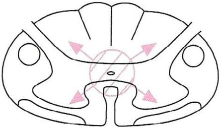
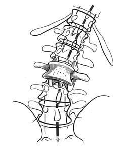
    SUMMARY

    Both acute and chronic neck pain is primarily attributed to the facet joints. This is especially true of those injured in a whiplash-mechanism accident. Chiropractic spinal adjusting (specific joint manipulation) affects many tissues, but primarily the facet joints. Evidence continues to support that spinal manipulation is both effective and safe for patients suffering with neck pain, including patients with neck pain initiated by a whiplash injury. The evidence supports spinal manipulation for both acute and chronic neck pain/. The evidence also shows that spinal manipulation is not only effective, but the benefits are also long-lasting with only a small incidence of recurrence.

    REFERENCES

    1. Foreman J; A Nation in Pain, Healing Our Biggest Health Problem; Oxford University Press; 2014.
    2. Pho, K; USA TODAY, The Forum; September 19, 2011; pg. 9A.
    3. Wang S; Why Does Chronic Pain Hurt Some People More?; Wall Street Journal; October 7, 2013.
    4. Choudhry N, Milstein A; Do Chiropractic Services for the Treatment of Low Back and Neck Pain Improve the Value of Health Benefits Plans? An Evidence-Based Assessment of Incremental Impact on Population Health and Total Health Care Spending; Mercer Health and Benefits; 2009.
    5. Bronfort G, Evans R, Anderson AV, Svendsen KH, Bracha Y, MS; Grimm RH; Spinal Manipulation, Medication, or Home Exercise With Advice for Acute and Subacute Neck Pain: A Randomized Trial; Annals of Internal Medicine; January 3, 2012; Vol. 156; pp. 1-10.
    6. Dunning JR, Cleland JA, Waldrop MA, Arnot C, Young I, Turner M, Sigurdsson G; Upper Cervical and Upper Thoracic Thrust Manipulation Versus Nonthrust Mobilization in Patients With Mechanical Neck Pain: A Multicenter Randomized Clinical Trial; Journal of Orthopaedic & Sports Physical Therapy; January 2012; Volume 42; Number 1; pp. 5-18.
    7. Langenfeld A, Humphreys K, Swanenburg J, Cynthia K. Peterson CK; Prognostic Factors for Recurrences in Neck Pain Patients Up to 1 Year After Chiropractic Care; Journal of Manipulative and Physiological Therapeutics; September 2015; Vol. 38; No. 7; pp. 458-464.
    8. Guzman J, Haldeman S, Carroll LJ, et al. Clinical practice implications of the Bone and Joint Decade 2000-2010 Task Force on Neck Pain and Its Associated Disorders: from concepts and findings to recommendations. Spine (Phila Pa 1976) 2008;33:S199-213.
    9. Hurwitz EL, Carragee EJ, van der Velde G, et al; Treatment of neck pain: noninvasive interventions: results of the Bone and Joint Decade 2000-2010 Task Force on Neck Pain and Its Associated Disorders. Spine (Phila Pa 1976) 2008;33:S123-52.
    10. Jessica J. Wong, Heather M. Shearer, Silvano Mior, Craig Jacobs, Pierre Côté, Kristi Randhawa, Hainan Yu, Danielle Southerst, Sharanya Varatharajan, Deborah Sutton, Gabrielle van der Velde, Linda J. Carroll, Arthur Ameis, Carlo Ammendolia, Robert Brison, Margareta Nordin, Maja Stupar, Anne Taylor-Vaisey; Are manual therapies, passive physical modalities, or acupuncture effective for the management of patients with whiplash-associated disorders or neck pain and associated disorders?; An update of the Bone and Joint Decade Task Force on Neck Pain and its Associated Disorders by the OPTIMa Collaboration; The Spine Journal; December 2015 [epub]
    11. Bogduk N, Aprill C; On the nature of neck pain, discography and cervical zygapophysial joint blocks; Pain; August 1993; Vol. 54; No. 2; pp. 213-217.
    12. Kirkaldy-Willis WH, Cassidy JD; Spinal Manipulation in the Treatment of Low back Pain; Canadian Family Physician; March 1985; Vol. 31; pp. 535-540.

    “Authored by Dan Murphy, D.C.. Published by ChiroTrust® – This publication is not meant to offer treatment advice or protocols. Cited material is not necessarily the opinion of the author or publisher.”

  • Whiplash Injury, Neck Pain AND Spinal Manipulation

    Whiplash Injury, Neck Pain AND Spinal Manipulation

    The understanding of whiplash biomechanics was forever changed in 1997. Researchers from Yale University School of Medicine performed a series of rear-end collisions on human cadavers while imaging their biomechanics with cineradiography (1). The results established that during the earliest phase of collision mechanics, the cervical spine forms an “S” shaped configuration, with flexion of the upper cervical spine and simultaneous significant hyperextension of the lower cervical spine. The tissue distortion noted during this “S” configuration of the cervical spine was of a magnitude that is injurious, especially in the lower cervical spine. As the subjects used in this initial assessment were cadavers, skepticism as to the relevance to live humans in real life collisions remained. This skepticism was mitigated within a few years with follow-up studies.

    In 1999, similar cineradiography studies were performed on live human volunteers (2). The results were the same as those of the cadaver studies, solidifying the concept that the cervical spine undergoes an “S” configuration during a rear-end motor vehicle collision, primarily injuring structures of the lower cervical spine. Essentially all articles published between 1997–1999 regarding whiplash biomechanics cite these studies, noting that the pathology of whiplash primarily occurs during this “S” configuration. A representative article from 2007 states (3):

    “The forward acceleration of the torso deforms the cervical spine into a non-physiologic S-shaped curve, with extension developing between the lower segments and flexion developing between the uppermost segments. Most of the whiplash injury occurs during this deformation phase.”

    “The cervical facet joint is the most common source of chronic neck pain after whiplash injury.”

    “The facet joints are the most common source of chronic neck pain after whiplash injury.”

    Injury to the facet joints and their capsular ligaments as a consequence of the “S” configuration should be emphasized. Findings include (2):

    “The zygapophysial joint is the suspected origin of neck pain after rear-end car collision.”

    “Most whiplash injuries occur during low-speed rear-end collisions and rarely produce morphologic changes such as fracture of the joint. The zygapophysial joint is a synovial joint and has a synovial fold (meniscus), between the articular facets that is innervated with nociceptive receptors. Thus, we hypothesize that facet collisions are likely to impinge on and inflame the synovial folds in the zygapophysial joints, causing neck pain (facet synovial fold impingement syndrome).”

    Many other studies support the premise of whiplash-mechanism facet joint injury and pain (4, 5, 6, 7, 8, 9, 10).

    •••••••••

    The entire December 1, 2011 supplement of the journal Spine is dedicated to whiplash trauma. The issue contains 27 articles by the world’s foremost authorities on whiplash biomechanics, pathology and outcomes, including the physician/clinical anatomist, Nikoli Bogduk, from Australia. Dr. Bogduk’s article in this issue of Spine is titled, “On Cervical Zygapophysial Joint Pain After Whiplash” (9).

    Dr. Bogduk cites 72 references while summarizing the evidence that implicates the cervical zygapophysial joints (facets) as the leading source of chronic neck pain after whiplash trauma. He states that the patho-anatomic basis for neck pain after whiplash is not elusive, but rather well documented and well known. Dr. Bogduk notes that there is convergent validity from

    • whiplash postmortem studies
    • whiplash biomechanics studies
    • whiplash clinical studies

    All of these studies and diverse methods of investigation indicate that the primary source of chronic whiplash pain is injury to the cervical zygapophysial (facet) joints. Dr. Bogduk states:

    “Collectively, these various biomechanics studies, in normal volunteers and in cadavers, predict or produce the same spectrum of lesions as that identified in postmortem studies. In particular, they indicate that the zygapophysial joints can be injured.”

    “The zygapophysial joints are the single, most common source of pain in patients with chronic neck pain after whiplash.”

    There is an extensive amount of evidence indicating that post-whiplash pain syndrome is attributed to injury to the cervical facet joints; no other explanation for whiplash pain has more evidence.

    •••••••••

    Mechanically, spinal manipulation primarily affects the spinal facet joints. This is well accepted and not controversial. In 1985, Canadian Orthopedic Surgeon, WH Kirkaldy-Willis, MD, stated (11):

    “Spinal manipulation is essentially an assisted passive motion applied to the spinal apophyseal and sacroiliac joints.”

    Biological Plausibility

    1. The primary injury from whiplash biomechanics is to the facet joints.
    2. The primary source of both acute and chronic whiplash injury pain is the facet joints.
    3. Spinal adjusting (specific joint manipulation) primarily affects the facet joints.
    4. It would appear to be biologically plausible that spinal adjusting could improve the pain status and recovery time of individuals injured in a whiplash type mechanism.
    5. Studies assessing such biological plausibility should show positive clinical outcomes.

    A few pertinent studies are presented below:

    In 1996, clinicians from the University Department of Orthopaedic Surgery, Bristol, United Kingdom, published a study in the journal Injury, titled (12):

    Chiropractic Treatment of Chronic ‘Whiplash’ Injuries

    The authors note, “The whiplash syndrome is a cause of long-term symptoms for which conventional medicine has failed to discover an effective treatment.” They note that 43% of patients will suffer long-term symptoms following ‘whiplash’ injury, for which no conventional treatment has proven to be effective. Consequently, they performed a retrospective study to determine the effects of chiropractic spinal manipulation [a nonconventional treatment] in a group of 28 patients who were suffering with chronic ‘whiplash’ syndrome. The authors defined “spinal manipulation” as:

    “Spinal manipulation is a high-velocity low-amplitude thrust to a specific vertebral segment aimed at increasing the range of movement in the individual facet joint, breaking down adhesions and stimulating production of synovial fluid.”

    The severity of patient’s symptoms was assessed before and after treatment using the Gargan and Bannister (1990) classification protocol, as follows:

    The Gargan and Bannister Whiplash Classification

    GROUP SYMPTOMS
    A None
    B Nuisance
    C Intrusive
    D Disabling

    The 28 patients in this study had initially been treated with anti-inflammatories, soft collars and physiotherapy. These patients had all become chronic, and were referred for chiropractic at an average of 15.5 months (range was 3–44 months) after their initial injury. At the initial evaluation and prior to chiropractic treatment, 27/28 (96%) of the patients were classified as category C (intrusive) or D (disabling) symptoms.

    Following the chiropractic treatment, 93% of the patients had improved: 16/28 (57%) improved by one symptom group, and 10/28 (36%) improved by two symptom groups. The authors concluded:

    “The encouraging results from this retrospective study merit the instigation of a prospective randomized controlled trial to compare conventional with chiropractic treatment in chronic ‘whiplash’ injury.”

    “The results of this retrospective study would suggest that benefits can occur in over 90% of patients undergoing chiropractic treatment for chronic whiplash injury.”

    •••••••••

    In 1999, the same group of clinicians from the University Department of Orthopaedic Surgery, Bristol, United Kingdom, published a study in the Journal of Orthopaedic Medicine, titled (13):

    A Symptomatic Classification of Whiplash Injury and the Implications for Treatment

    In this study, the author’s objective was to determine which patients with chronic whiplash would benefit from chiropractic treatment. The study was a retrospective review involving 93 consecutive whiplash-injured patients. Once again, the patients were assessed using the Gargan and Bannister classification protocol. The authors note:

    “Conventional treatment of patients with whiplash symptoms is disappointing.”

    “In chronic cases, no conventional treatment has proved successful.”

    All patients underwent spinal manipulation, defined as a “high velocity, low amplitude thrust to a specific vertebral segment.” Patients underwent a mean of 19.3 treatments (range 1-53), over a period of 4.1 months.

    The authors note that their results “confirm the efficacy of chiropractic, with 69 of our 93 patients (74%) improving following treatment.” Importantly, 94% of whiplash patients with neurological signs and/or symptoms in association with neck pain and a restricted range of neck movement, including tingling, numbness, pins and needles in a dermatomal distribution in the arm or hand as well as both hypo- and hyperaesthesia, responded positively to chiropractic spinal manipulation. The authors concluded:

    “The results from this study provide further evidence that chiropractic is an effective treatment for chronic whiplash symptoms.”

    “Chiropractic is the only proven effective treatment in chronic [whiplash] cases.”

    •••••••••

    In 2004, a group of physiotherapists, physicians, and professors from the Rey Juan Carlos University, Spain, published a study in the Journal of Whiplash & Related Disorders, titled (14):

    Manipulative Treatment vs. Conventional Physiotherapy Treatment in Whiplash Injury: A Randomized Controlled Trial

    The authors note that the clinical syndrome of whiplash includes neck pain, restriction of neck motion, dizziness, headaches, tinnitus and blurred vision. Spinal joint dysfunction and myofascial pain syndrome are thought to be one of the greatest causes of musculoskeletal disorders and symptoms in patients diagnosed with whiplash injury.

    The goal of joint manipulation is to restore maximal, pain-free movement of the musculoskeletal system. “Only joints that are found to be hypomobile should be considered as candidates for high velocity low amplitude [manipulative] techniques.”

    The objective of this clinical trial was to compare the results obtained from a manipulative protocol with the results obtained from a conventional physiotherapy treatment in patients suffering from whiplash injury. This is the first controlled experimental trial documenting the effects of the manipulative protocol used in this study. It was a randomized controlled trial using 380 acute whiplash injury (less than 3 months duration) subjects. All subjects were Quebec Task Force classified as grades II and III.

    • Quebec GRADE II = neck complaint and musculoskeletal signs
    • Quebec GRADE III = neck complaint, musculoskeletal signs, and neurologic signs

    The injured subjects were randomly divided into 2 groups:

    Group A (experimental, manipulation group)

    This group was treated with high velocity-low amplitude spinal manipulation and soft tissue manipulation. The manipulation was delivered to the upper cervical spine, the cervical-thoracic junction, thoracic spine, thoracolumbar junction and pelvic girdle. Importantly, although the patients were suffering from neck pain and related symptoms, the manipulation was applies “full spine.” The treatment was weekly.

    Group B (control, physical therapy group)

    This group was treated with a conventional physiotherapy including active exercises, electrotherapy, ultrasound therapy and diathermy. The treatment was daily.

    The outcome measures were the visual analogue scale (VAS), the cervical range of motion (CROM) in flexion and rotation, and number of sessions needed to complete the treatment.

    Analysis of the change in visual analogue scale for pain showed:

    • The manipulation group obtained an average decrease of 40% in head and neck pain in 4 visits. (maximum improvement was 9 ± 1.5 visits)
    • The physical therapy group, obtained a decrease of 19% after 10 visits. (maximum improvement was 23 ± 3.2 visits)

    Analysis of the change in cervical range of motion showed:

    • An improvement of 20° in cervical rotation, and 17.5° in cervical flexion after 4 sessions of spinal manipulation.
    • An improvement of 2.5° in cervical rotation, and in cervical flexion after 10 sessions of physical therapy.
    Spinal Manipulation Physiotherapy
    Number of Visits 4 10
    Decrease in
    Neck/Head Pain
    40% 19%
    Increase ROM
    Rotation
    20° 2.5°
    Increase ROM
    Flexion
    17.5°
    Number of Visits to Maximum Improvement 9 ± 1.5 23 ± 3.2

    The authors note:

    Patients who had received manipulative treatment needed fewer sessions to complete the treatment than patients who had received physiotherapy treatment.”

    Patients of manipulative group needed an average of 9 sessions to complete the treatment, whereas physiotherapy group needed an average of 23 sessions.”

    “Results showed that the manipulative group had more benefits than the physiotherapy group in the VAS and CROM.”

    “Our clinical experience with these [whiplash-injured] patients has demonstrated that manipulative treatment gives better results than conventional physiotherapy treatment.”

    Interestingly and importantly, these authors propose that it is “necessary to manipulate a number of spinal levels” to obtain the best outcomes for the whiplash-injured patient. Thoracic spine joint dysfunction may cause secondary adaptive or maladaptive changes in cervical and sacroiliac joints. Such adaptative changes in the cervical spine may contribute to the resulting headaches and neck pain in these patients. They note that whiplash injury symptoms (head, neck and upper thoracic pain) decrease in response to a thoracic spinal manipulation.

    Also, in a rear-end impact, as the struck vehicle is accelerated forward, the seatback contacts the lumbo-pelvic region, causing compression and extension. “This compression of the lumbo-pelvic region produces a hypomobility in the pelvic girdle that is necessary to manipulate.”

    This study emphasizes the need to use spinal manipulation to multiple spinal regions, including the pelvis, to obtain the best outcomes in the whiplash-injured patient. This is consistent with the perspective of rheumatologist John Bland, MD, from the University of Vermont College of Medicine. Note the following quote from his his book Disorders of the Cervical Spine (15):

    “We tend to divide the examination of the spine into regions: cervical, thoracic, and lumbar spine clinical studies. This is a mistake. The three units are closely interrelated structurally and functionally – a whole person with a whole spine. The cervical spine may be symptomatic because of a thoracic or lumbar spine abnormality, and vice versa! Sometimes treating a lumbar spine will relieve a cervical spine syndrome, or proper management of cervical spine will relieve low backache.”

    This is the first study showing the effectiveness of spinal manipulation in the management of acute whiplash injuries (the prior studies pertained to chronic whiplash injuries). In this study, spinal manipulation was significantly more effective both subjectively and objectively in treating Quebec Task Force whiplash injuries Grades II and III than conventional physiotherapy. These authors concluded:

    “This clinical trial has demonstrated that head and neck pain decrease with fewer treatment sessions in response to a manipulative treatment protocol as compared to a physiotherapy treatment protocol among patients diagnosed with acute whiplash injury.”

    Manipulation is “effective in the management of whiplash injury.”

    “Manipulative treatment is more effective in the management of whiplash injury than conventional physiotherapy treatment.”

    •••••••••

    Recently (September 2015), researchers and clinicians from the Orthopedic University Hospital Balgrist, University of Zurich, Switzerland, published a study in the Journal of Manipulative and Physiological Therapeutics, titled (16):

    Prognostic Factors for Recurrences in Neck Pain Patients

    Up to 1 Year After Chiropractic Care

    This is a prospective cohort study assessing 545 neck pain patients. After a course of chiropractic spinal manipulation, they were followed up for one year regarding recurrence of their neck pain. Nine independent prognostic variables were assessed:

    • Age
    • Use of pain medication
    • Sex
    • Work status
    • Duration of complaint
    • Previous episodes of neck pain
    • Trauma onset
    • Numeric Rating Scale (NRS) was used to quantify their pain
    • Bournemouth Questionnaire was used for neck pain

    These authors note that most people will see a health care provider at least once in their lifetime due to neck pain. In patient populations that do not include chiropractic care, those who have experienced an episode of neck pain are likely to have another episode within the next 1 to 5 years. Overall, for these individuals, the prognosis for a complete recovery from their neck pain is “quite poor.” Those who do not recover from their neck pain within the 3 months after treatment begins tend to suffer from residual neck pain and disability.

    There are known prognostic factors for the onset of neck pain. They include:

    • Computer work
    • Heavy physical work
    • Longer duration of complaint
    • Older age
    • Female sex
    • Previous neck injury

    The results of this study are impressive:

    • Fifty-four (54) participants (11%) were identified as “recurrent.”
    • Four hundred ninety one (491) participants (89%) were not recurrent.

    The authors state:

    “89% of neck pain patients had recovered from their neck pain episode up to 1 year after receiving chiropractic care.”

    This study also found that older age and a previous episode of neck pain were useful predictors of neck pain recurrence within 1 year. This is because an increase in age was associated with recurrence. Patients older than 45 years are twice as likely to experience a neck pain recurrence within 1 year after the start of chiropractic treatment. They state:

    “Having had a previous episode as well as increasing age are increased risk factors for predicting a subsequent new episode of neck pain within a year.”

    These authors concluded:

    “The results of this study suggest that recurrence of neck pain within 1 year after chiropractic intervention is low.”

    This study indicates that chiropractic is both effective in the treatment of neck pain and that its benefits are stable and long lasting.

    •••••••••

    SUMMARY

    Both acute and chronic neck pain is primarily attributed to the facet joints. This is especially true of those injured in a whiplash-mechanism accident. Chiropractic spinal adjusting (specific joint manipulation) affects many tissues, but primarily the facet joints. Evidence continues to support that spinal manipulation is both effective and safe for patients suffering with neck pain, including patients with neck pain initiated by a whiplash injury. The evidence supports spinal manipulation for both acute and chronic neck pain/. The evidence also shows that spinal manipulation is not only effective, but the benefits are also long-lasting with only a small incidence of recurrence.


    REFERENCES

    • Grauer JN, Panjabi MM, Cholewicki J, Nibu K, Dvorak J; Whiplash produces an S-shaped curvature of the neck with hyperextension at lower levels; Spine; November 1, 1997; Vol. 22; No. 21; pp. 2489-94.
    • Kaneoka K, Ono K, Inami S, Hayashi K; Motion analysis of cervical vertebrae during whiplash loading; Spine; April 15, 1999; Vol. 24; No. 8; pp. 763-9.
    • Schofferman J, Bogduk N, Slosar P; Chronic whiplash and whiplash-associated disorders: An evidence-based approach; Journal of the American Academy of Orthopedic Surgeons; October 2007; Vol. 15; No. 10; pp. 596-606.
    • Uhrenholt L, Grunnet-Nilsson N; Hartvigsen J; Spine; Cervical spine lesions after road traffic accidents: a systematic review; September 1, 2002; Vol. 27; No. 17; pp. 1934-1941.
    • Pearson AM, Ivancic PC, Ito S, Panjabi MM; Facet joint kinematics and injury mechanisms during simulated whiplash; Spine; February 15, 2004; Vol. 29; No. 4; pp. 390-397.
    • Bogduk N, Aprill C; On the nature of neck pain, discography and cervical zygapophysial joint blocks; Pain; August 1993; Vol. 54; No. 2; pp. 213-217.
    • Barnsley L, Lord SM, Wallis BJ; Bogduk N; The prevalence of chronic cervical zygapophysial joint pain after whiplash; Spine; January 1, 1995; Vol. 20; No. 1; pp. 20-25.
    • Lord SM, Barnsley L, Wallis BJ; Bogduk N; Chronic cervical zygapophysial joint pain after whiplash. A placebo-controlled prevalence study; Spine; August 1, 1996; Vol. 21; No. 15; pp. 1737-1744.
    • Bogduk N; On Cervical Zygapophysial Joint Pain After Whiplash; Spine December 1, 2011; Vol. 36; No. 25S; pp. S194–S199.
    • Quinn KP, Winkelstein BA; Detection of Altered Collagen Fiber Alignment in the Cervical Facet Capsule After Whiplash-Like Joint Retraction; Annals of Biomedical Engineering; August 2011; Vol. 39; No. 8; pp. 2163–2173.
    • Kirkaldy-Willis WH, Cassidy JD; Spinal Manipulation in the Treatment of Low back Pain; Canadian Family Physician; March 1985; Vol. 31; pp. 535-540.
    • Woodward MN, Cook JCH, Gargan MF, and Bannister GC; Chiropractic treatment of chronic ‘whiplash’ injuries; Injury; Vol. 27; No. 9; November 1996; pp. 643-645.
    • Khan S, Cook J, Gargan M, Bannister G; A symptomatic classification of whiplash injury and the implications for treatment; The Journal of Orthopaedic Medicine; Vol. 21; No. 1; 1999; pp. 22-25.
    • Fernández-de-las-Peñas C, Fernández-Carnero J, Palomeque del Cerro L, Miangolarra-Page JC; Manipulative Treatment vs. Conventional Physiotherapy Treatment in Whiplash Injury: A Randomized Controlled Trial; Journal of Whiplash & Related Disorders; 2004; Vol. 3; No. 2.
    • Bland J; Disorders of the Cervical Spine; WB Saunders Company; 1987; p. 84. [Professor of Medicine, University of Vermont College of Medicine].
    • Langenfeld A, Humphreys K, Swanenburg J, Cynthia K. Peterson CK; Prognostic Factors for Recurrences in Neck Pain Patients Up to 1 Year After Chiropractic Care; Journal of Manipulative and Physiological Therapeutics; September 2015; Vol. 38; No. 7; pp. 458-464.
  • Arnold-Chiari Malformation and Chiropractic

    Arnold-Chiari Malformation and Chiropractic

    Relevant Clinical Background

    A literature search of the United States National Library of Medicine using PubMed (www.pubmed.gov) with the words “Arnold Chiari Malformation” locates 2,995 citations (August 14, 1015).

    Chiropractic clinical practice involves the application of mechanical forces to a patient’s spinal column. Should the patient’s spinal column (spinal canal) have a space occupying entity, spinal manipulation may have to be delivered more cautiously.

    Typical spinal canal space occupying entities include intervertebral disc herniations, tumors, and spinal cord syrinx (a fluid filled cavity within the spinal corn, also known as syringomyelia). These lesions often present with certain symptoms and signs, and their existence is confirmed with diagnostic imaging, usually an MRI. Interestingly, the presence of these entities can also be completely asymptomatic and are found only coincidently. As an example, in 1994, The New England Journal of Medicine published a study where 98 asymptomatic subjects received MRIs of their lumbar spines. Only 36% had normal intervertebral discs at all levels, and 27% had asymptomatic disc protrusions (1).

    Another important space occupying entity that may or may not present with symptomology, is the Arnold-Chiari Malformation.

    What is Arnold-Chiari Malformation?

    At the basic level, Arnold-Chiari Malformation occurs when parts of the brain and/or brainstem slip below the foramen magnum and are found to be in the cervical spinal canal. This compromises the available space for both the cervical spinal cord and for the parts of the brain and/or brainstem that have slipped through the foramen magnum. An important component of this mechanical space occupying compromise is an impairment of the flow of cerebral spinal fluid.

    A frequent and important accompaniment of the Arnold-Chiari Malformation is that of a spinal cord syringomyelia. A syringomyelia, as noted above, is a condition in which a fluid filled cavity is present in the spinal cord and/or medulla. It creates pressure from the inside of the spinal cord, outwards:

    Syringomyelia

    Syringomyelia

    Austrian pathologists Hans Chiari first described the Arnold-Chiari Malformation in 1891, prior to the first x-rays (Roentgen, 1895). German pathologist Julius Arnold’s name was added in 1907. Dr. Chiari described his findings based upon postmortem assessment in infants with varying degrees of congenital hindbrain deformities. Based upon Dr. Chiari’s findings, three Arnold-Chiari Malformation classifications were developed (today there are four):

    Type I
    There is a downward displacement of the cerebellar tonsils through the foramen magnum and into the cervical spinal canal; yet, the fourth ventricle remains in a relatively normal position.

        • Herniation of both cerebellar tonsils 3-5 mm below the plane of the foramen magnum
        • Herniation of one cerebellar tonsil 5 mm or more below the foramen magnum (2)

    Type II
    There is a downward displacement of the cerebellar tonsils and cerebellar vermis into the cervical spinal canal; the brain stem is displaced caudally; there is an elongation of the fourth ventricle.

    Type III
    There is a downward displacement of both the cerebellum and medulla into the cervical spinal canal and the development of a cervical meningocele.

    Arnold-Chiari Type-I Malformation Lateral View

    Arnold-Chiari Type-I Malformation Lateral View

    The degree of tonsillar herniation is measured on the sagittal MRI
    A = Basion, anterior foramen magnum
    B = Opisthion, posterior foramen magnum
    C = tonsillar tip
    Line AB represents the foramen magnum
    Degree of tonsillar herniation is length of perpendicular from AB to C

    Arnold-Chiari Malformation is considered to be developmental or congenital. It is often associated with scoliosis, Klippel-Feil Syndrome, hydrocephalus, and syringomyelia. Adults with Arnold-Chiari Malformation Type-I may be completely asymptomatic, or they may have many dramatic symptoms. It is often asymptomatic and does not present until adulthood. Consequently, Arnold-Chiari Malformation Type-I is often found as an incidental finding on MRIs taken for other reasons. In contrast, Arnold-Chiari Malformation Type-II and Type-III are almost always found in infancy because of significant neurological findings and the presence of meningocele.

    What are the Typical Symptoms of a Patient with Arnold-Chiari Malformation?

    The typical symptoms attributed to the Arnold-Chiari Malformation are:

    • Headache, usually occipital or suboccipital
    • Neck Pain
    • Vertigo / Dizziness / Light Headedness / Disequilibrium
    • Tinnitus
    • Extremity weakness
    • Extremity numbness
    • Ataxia / Gait disturbance
    • Cranial nerve palsies (nystagmus, oscillating vision, dysphagia, photophobia)
    • Apnea
    • Incontinence
    • Fatigue
    • Sleeping Difficulty

    Often, the Valsalva test, or Valsalva types of activities (coughing, sneezing, straining, etc.) aggravate these symptoms. 

    How is Arnold-Chiari Malformation Diagnosed?

    The gold standard for a suspected Arnold-Chiari Malformation is an MRI. Standard MRIs are taken with the patient recumbent. However, as detailed below, there is evidence that in the chronic whiplash trauma patient, upright MRI is significantly superior to recumbent MRI documenting the existence of the lesion (4).

    Also, syrinx/syringomyelia assessment is mandatory. The entire spinal cord (especially the brain stem, cervical cord, and thoracic cord) should be assessed with T1-weighted sagittal MRI images (3).

    Can Chiropractic Spinal Adjusting (Manipulation) be Safely Used to Treat the Symptoms of Arnold Chiari Malformation?

    There is evidence that chiropractic care does not benefit all patients with Arnold-Chiari Type-I Malformation. In 1994, chiropractor Ross McArthur presented two cases of Arnold-Chiari Type-I Malformation in which conservative chiropractic management did not result in improvement in the patient’s clinical presentation and symptomology (3).

    The first case, a 43-year old female had been under chiropractic care for 10 years, including cervical spine adjusting/manipulation. She had been treated for back pain, neck pain and occipital pain, and she always responded well to spinal adjusting.

    Following a fall with head trauma, her symptoms included dizziness, photophobia, diminished smell and taste, increased suboccipital pain, and the Valsalva maneuver increased suboccipital symptoms. X-rays of the cervical spine were normal. Chiropractic care failed to improve symptoms. An MRI was exposed and revealed herniation of the cerebellar tonsils through the foramen magnum and caudal displacement of portions of the cerebellar hemispheres. A diagnosis of Arnold-Chiari Malformation Type-I was made.

    The patient was treated surgically with a posterior fossa craniectomy and a complete laminectomy of the posterior arch of the atlas. The dura was opened and microdissection was used to remove tonsillar adhesions. The presence of these tonsillar adhesions suggested that her Arnold-Chiari Malformation was a long-standing problem. The patient made a full recovery and remained under chiropractic care.

    In this case, chiropractic adjustment/manipulation of her cervical spine did not worsen the patient’s symptoms; they did however fail to result in meaningful improvement.

    In the second case, a 50-year old female presented with spontaneous worsening symptoms in her left arm-forearm-hand, left facial paresthesias, left TMJ symptoms, and left thigh weakness. Examination showed hypertonicity of the anterior scalene and sternocleidomastoid muscles. Cervical spine X-rays were normal.

    Eight chiropractic adjustments of the cervical and thoracic spines over a period of 4 weeks did not improve symptoms, and therefore an MRI was exposed. The MRI showed a typical Arnold-Chiari Malformation with syringomyelia.

    Once again, chiropractic spinal adjusting/manipulation of the cervical spine did not result in meaningful improvement of the patient’s symptoms, but it did not worsen or aggravate her symptoms or clinical presentation.

    •••

    In 1993, Donald Murphy, DC, and colleagues presented two cases where chiropractic high-velocity, low-amplitude adjustments were delivered to patients that had proven Arnold-Chiari Malformation Type I (4). Although the authors do not believe that the patient’s signs and symptoms were related to the Arnold-Chiari Malformation, the presenting symptoms may suggest otherwise. In any case, both patients with known Arnold-Chiari Malformation Type I received typical chiropractic adjustments of the upper cervical spine.

    The first case was a 37-year old male who was asymptomatic until being involved in a rear-end followed by head-on motor vehicle collision. Presenting symptoms include headache, neck pain, photophobia, blurred vision, and right shoulder/scapular pain. Cranial nerve and cerebellar examination was unremarkable. Cervical compression, Soto-Hall, and Valsalva tests all produced local cervical pain. Cervical extension and rotation (vertebral artery tests) were negative for dizziness, nausea, nystagmus, numbness, or paresthesia.

    After 20 chiropractic adjustments over a period of 6 weeks, the patient was 50% improved; yet, an MRI at that time showed an Arnold-Chiari Malformation Type I. The patient remained under chiropractic care, receiving an additional 14 adjustments over the next 6 weeks, at which time signs and symptoms were essentially resolved. The authors state:

    “The patient was treated a total of 34 times and at no time during the treatment program did he note any adverse reactions to high-velocity, low-amplitude adjustments.”

    The second case was a 36-year old female with a 4-year history of chronic severe vertigo and right arm pain. She had already undergone a cervical MRI showing an Arnold-Chiari Malformation Type I. She subsequently presented for chiropractic care.

    The patient had an ataxic gait, could not tandem walk or stand in Romberg’s position, and had nystagmus on all eye movements. Initial chiropractic treatment consisted of 20 visits of low-velocity, low-amplitude mobilization of the upper cervical spine. This was followed by 5 visits of high-velocity, low-amplitude adjustments of the upper cervical spine. These 25 chiropractic visits resulted in improvement but not resolution of her signs and symptoms. The authors state:

    “At no time did she note any lasting adverse effects, even when high-velocity, low-amplitude adjustments were applied to the upper cervical spine.”

    Asymptomatic Arnold-Chiari Malformation Type I is “not necessarily a contraindication to skilled adjustments to the cervical spine, and that viewing it as such would mean possibly denying the patient a form of treatment that could be helpful in the management of a health problem that may be unrelated to the malformation.”

    The authors also make some cautionary comments:

    “In a patient with a known Arnold-Chiari Malformation Type I, certain precautions may be taken to minimize any irritation to the cerebellar tonsils that have herniated through the foramen magnum.”

    Avoid extension of the upper cervical spine during adjustment.

    “Excessively vigorous adjusting to the upper cervical spine should be avoided, since this can potentially irritate neural structures in an already crowded canal.”

    “Neither Occiput-C1 nor C1-C2 were adjusted in these patients, but there does not appear to be any reason why this would create any greater likelihood of complication.”

    •••

    In 2014, chiropractors Adam Sergent and Gregory Cofano published a study in the Journal of Chiropractic Medicine titled (5):

    Chiropractic Care for Headaches and Dizziness of a 34-Year-Old Woman Previously Diagnosed With Arnold-Chiari Malformation Type-I

    This 34-year-old female had a chronic history of headaches, dizziness, nausea, photophobia, and temporary loss of vision aggravated by postural positions while bending forward. These symptoms were often incapacitating. An MRI showed an Arnold-Chiari Malformation Type I, with cerebellar tonsils descending 5 mm into the cervical spinal canal. Neurosurgery was recommended.

    Three years after initial diagnosis, the patient sought chiropractic and expressed her desire to avoid surgery. She was treated for 1 month with 9 visits, using high-velocity, low-amplitude adjustments of the cervical spine of prone diversified manipulation to C4/C5. She reported reduction of her headaches after this treatment.

    Five years later, the patient suffered a symptomatic exacerbation. She was again treated using cervical chiropractic manipulation using prone diversified technique to C4/C5. The dizziness and headache were resolved after 3 visits. At her 3-month follow-up, she continued to be symptom-free. The authors concluded:

     “A patient with headaches and dizziness and a previous diagnosis of Arnold-Chiari Malformation Type I responded positively to chiropractic care.”

    •••

    Also in 2014, Oklahoma orthopedic surgeon Francio Tieppo, published a study in the British Medical Journal Case Reports, titled (6):

    Syringomyelia and Arnold-Chiari malformation associated with neck pain and left arm radiculopathy treated with spinal manipulation

    The abstract from this study makes the following statements:

    “An 18-year-old female patient presented with left dominant neck pain after a motor vehicle collision. Her cervical spine MRI revealed syringomyelia with associated Type I Arnold-Chiari Malformation. Some researchers have reported that these might be considered contraindications to spinal manipulation. Nevertheless, her benign and functional clinical examination suggested otherwise and she underwent four manipulative treatments in 2 weeks. By the end of the treatment plan and after 1-month follow-up, she was asymptomatic, no adverse effects were noted and her outcome assessment score decreased from 56% to 0%. This case illustrates that spinal manipulation may be a useful adjunctive treatment procedure for spinal pain, even in the presence of syringomyelia and Chiari malformation, which may not necessarily be a contraindication to spinal manipulation, when performed by a skilled and well-trained physician.” 

    If Chiropractic Care Does Not Achieve an Acceptable
    Clinical Outcome on the Arnold Chiari Malformation Patient,
    What is the Next Step of Clinical Intervention?

    The standard allopathic management of the patient with subjective Arnold-Chiari Malformation who did not respond favorably to conservative treatment is surgical decompression (4). Specifically, standard surgery is a posterior fossa craniectomy and a complete laminectomy of the posterior arch of the atlas (3).

    Summary

    There is no doubt that asymptomatic chiropractic patients have Arnold-Chiari Malformation Type I. These patients certainly receive routine spinal adjusting to the cervical spine, and no (as of 8.14.15) adverse events have been reported in the literature (PubMed).

    Chiropractic patients with subjective Arnold-Chiari Malformation Type I also present to chiropractors. The evidence presented here suggests that chiropractic spinal adjusting may not help these patients, but cervical spine adjusting is unlikely to worsen the patient’s signs and symptoms.

    Also presented is evidence that patients with known subjective Arnold-Chiari Malformation Type I may benefit from chiropractic spinal adjusting, especially if it is done skillfully and not too vigorously. However, little is known about osseous adjusting of the occiput-atlas-axis on such patients, and caution is advised. Perhaps, the upper cervical spine on such patients should be adjusted with lower force approaches.

    An Interesting Article

    In 2010, Michael Freeman and colleagues published an article in the journal Brain Injury titled (7):

    A Case-control Study of Cerebellar Tonsillar Ectopia (Chiari) and Head/neck Trauma (Whiplash)

    These authors note that Chiari malformation is defined as herniation of the cerebellar tonsils through the foramen magnum, also known as cerebellar tonsillar ectopia. Individuals with existing but quiescent cerebellar tonsillar ectopia may become symptomatic following whiplash trauma. Therefore, the purpose of their study was to assess the frequency of cerebellar tonsillar ectopia in a symptomatic traumatic (whiplash) population versus a symptomatic non-traumatic control population.

    Cervical MRI scans for 1200 neck pain patients were reviewed; 600 whiplash trauma cases and 600 non-trauma controls. Half of the groups were scanned in a recumbent position and half were scanned in an upright position

    600 Chronic Controls

    600 Chronic Whiplash Patients

    Supine MR
    # cerebellar tonsillar ectopia

    5.3%

    9.3%

    Upright MR
    # cerebellar tonsillar ectopia

    5.7%

    23.3%

    In the non-trauma group, cerebellar tonsillar ectopia was found in 5.3% of the recumbent MRIs, and in 5.7% of the upright MRIs. This finding suggests:

    • Around 5% of individuals with non-traumatic neck symptoms have cerebellar tonsillar ectopia on a recumbent MRI.
    • The incidence of cerebellar tonsillar ectopia is not increased when individuals in this group (non-traumatic neck symptoms) have an upright MRI.
    • In the whiplash trauma group, cerebellar tonsillar ectopia was found in 9.8% of the recumbent MRIs, and in 23.3% of the upright MRIs. This finding suggests:
    • Around 10% of individuals with whiplash trauma neck symptoms have cerebellar tonsillar ectopia on a recumbent MRI. This would indicate that twice as many individuals with whiplash trauma neck symptoms have cerebellar tonsillar ectopia on a recumbent MRI as compared to a symptomatic non-traumatic control population. This would suggest that individuals with pre-existing cerebellar tonsillar ectopia who sustain a whiplash injury have an increased risk of sustaining prolonged symptoms.
    • In the whiplash trauma neck symptom group, the incidence of cerebellar tonsillar ectopia is increased significantly (more than doubled) when exposed to an upright MRI. This suggests that the whiplash injury creates some physiological change that increases the incidence of cerebellar tonsillar ectopia.
    • In the chronic whiplash trauma group, the incidence of cerebellar tonsillar ectopia viewed with upright MRI is 4 times greater than in the upright MRI of the control group.
    • In chronic whiplash patients, recumbent MRIs may be inadequate for the assessment of cerebellar tonsillar ectopia as compared to upright MRIs.

    Although Chiari type I malformation is classically considered to be a congenital lesion, these authors note that it can be acquired. These authors note that whiplash trauma is capable of causing a cerebral spinal fluid leak. In the upright position, the reduced volume of cerebral spinal fluid allows the cerebellar tonsils to slip below the foramen magnum, creating the cerebellar tonsillar ectopia finding.

    The standard diagnostic test for cerebral spinal fluid leak is using radioisotope cisternography. The standard treatment is a blood patch. This is procedure in which the patient’s own blood is withdrawn from a vein and injected into the spinal canal. The platelets in the blood will “find” the leak and create a seal, a patch. Symptoms usually quickly resolve, and recurrence is rare.

    These authors make these important points:

    • Typical Chiari type I malformation symptoms include occipital headache, neck pain, upper extremity numbness and paresthesias and weakness; occasionally there may be lower extremity weakness and signs of cerebellar dysfunction.
    •  “Previously quiescent Chiari Type I malformations can become symptomatic as a result of exposure to traumatic injury.” Minor head and neck trauma can cause an asymptomatic Chiari Type I malformation to become symptomatic.
    • Patients with a history of motor vehicle crash-associated neck pain have a “substantially higher frequency” of cerebellar tonsillar ectopia than non-traumatic subjects; 4-times greater when evaluated with an upright MRI scan.
    • “Cerebellar tonsillar ectopia is substantially more prevalent in whiplash-injured neck pain patients than in neck pain patients with no recent history of trauma.”
    • In the trauma group, cerebellar tonsillar ectopia was found 2.5-times more often in the upright MR scan vs. the recumbent MR scan. “Upright position MR imaging appears to increase the sensitivity to cerebellar tonsillar ectopia over recumbent MR imaging by 2.5 times.”
    • “There is clinical evidence that dural leaks are associated with whiplash trauma and chronic symptoms.”
    • It is well established that Chiari type I can be acquired, and this study led the authors to suggest that the increased incidence of cerebellar tonsillar ectopia was caused by the whiplash trauma.
    • The incidence of cerebellar tonsillar ectopia was nearly identical (5.3% v 5.7%) in the control group in both the supine and upright MRI; but the incidence of cerebellar tonsillar ectopia was significantly greater (23.3 v 9.3) in the whiplash-injured group with the upright MRI. This can be explained by reduced cerebral spinal fluid (CSF) as a consequence of a trauma-induced leak.
    • Studies show that there is a substantial and rapid increase in cerebral spinal fluid pressure during simulated whiplash trauma.
    • In this study, neuroradiographic abnormality (cerebellar tonsillar ectopia) was found in approximately 25% of upright whiplash trauma cases. This unrecognized definable pathology may account for a patient’s chronic pain complaints. This suggests that in these cases, chronic whiplash symptoms may not be ascribable to psychosocial factors or litigation status, but rather to organic neurological injury.
    • “Clinicians may want to consider evaluating patients for cerebellar tonsillar ectopia (i.e. upright MRI of the neck and head) when there is a history of whiplash trauma and persisting suboccipital headache in combination with headache worsened by cough or bilateral sensory or motor deficits in the upper extremities.”
    • “In cerebellar tonsillar ectopia patients with headache that is relieved when supine it also may be appropriate to consider radionuclide cisternography to evaluate for the presence of a dural leak.”

    Perhaps all chronic whiplash patients should be assessed with an upright MRI to look for cerebellar tonsillar ectopia.

    REFERENCES 

    • Jensen MC, Brant-Zawadzki MN, Obuchowski N, Modic MT, Malkasian D, Ross JS; Magnetic Resonance Imaging of the Lumbar Spine in People Without Back Pain; N Engl J Med; 1994 Jul 14;331(2):69-73.
    • Elster AD, Chen MYM; Chiari I Malformations: Clinical and Radiological Reappraisal; Radiology; Vol. 183; No. 2; 1992; pp. 347-353.
    • McArthur RA; Arnold-Chiari Type I malformation: a look at two cases in the adult; Journal of the Canadian Chiropractic Association; Vol. 38; No. 4; December 1994; pp. 203-210.
    • Murphy DR, Goldstein D, Katz M; Chiropractic adjustment to the cervical spine and the Arnold-Chiari malformation; Journal of Manipulative Physiological Therapeutics; 1993; Vol. 16; pp. 550-555.
    • Adam Sergent A, Cofano G; Chiropractic Care for Headaches and Dizziness of a 34-Year-Old Woman Previously Diagnosed With Arnold-Chiari Malformation Type 1; Journal of Chiropractic Medicine; September 2014; Vol. 13; No. 3; pp. 192–195.
    • Tieppo Francio V; Syringomyelia and Arnold-Chiari malformation associated with neck pain and left arm radiculopathy treated with spinal manipulation; British Medical Journal Case Reports; November 9, 2014.
    • Freeman MD, Rosa S, Harshfield D, Smith F, Bennett R, Centeno CJ, Kornel E, Nystrom A, Heffez D, Kohles SS; A case-control study of cerebellar tonsillar ectopia (Chiari) and head/neck trauma (whiplash); Brain Injury; July 2010; 24(7–8): 988–994.

    “Authored by Dan Murphy, D.C.. Published by ChiroTrust® – This publication is not meant to offer treatment advice or protocols. Cited material is not necessarily the opinion of the author or publisher.”

  • What Really Is A Chiropractic “Adjustment”?

    What Really Is A Chiropractic “Adjustment”?

    The June 2015 issue of the journal Scientific American has an article by primary care physician Wajahat Z. Mehal, MD, from the Department of Veterans Affairs Medical Center in Connecticut, and Yale University, titled (1):

    Cells on Fire

    In this article, Dr. Mehal notes that inflammation is set in motion by cells of the immune system, and that it is helpful because it kills pathogens and blocks their spread in the body. The inflammatory cascade, initiated by the innate immune response’s macrophages, weakens and immobilizes adverse microbes.

    However, the same inflammatory cascade can occur when no microbes exist, triggered as a consequence of tissue damage and/or excessive tissue stress. This inflammatory response can, in-and-of-itself, become chronic and cause additional tissue damage. In other words, as much as acute inflammation can be beneficial (containing and/or killing pathogens), chronic inflammation can be deleterious, serving no useful purpose.

    Consequently, Dr. Mehal broadly categorized the inflammatory response into two categories:

    1)     Infectious inflammation:

    This is an inflammatory response that is designed to contain and/or kill pathogens.

    This response is critical for individual and species survival.

    2)     Sterile inflammation:

    This is an inflammatory response in which there are no associated pathogens, a response that is triggered by tissue injury and /or excessive tissue stress.

    This response often becomes chronic. As such, this response is excessive and harmful.

    Pathology History

    A decades synopsis of global leading experts, expressed in leading reference texts printed by top medical publishers

    In 1952, William Boyd, MD, Professor Emeritus of Pathology at the University of Toronto, published his reference text, titled (2):

    PATHOLOGY

    Structure and Function in Disease

    In this text, Dr. Boyd states:

    “The inflammatory reaction tends to prevent the dissemination of infection. Speaking generally, the more intense the reaction, the more likely the infection to be localized.”

    In 1970, the eighth edition of Dr. Boyd’s PATHOLOGY text is published

    (3): In chapter 4, titled Inflammation and Repair,Dr. Boyd states:

    “Inflammation is the most common, the most carefully studied, and the most important of the changes that the body undergoes as the result of disease.”

    Dr. Boyd notes that in chronic inflammation, the “only cells that proliferate are the fibroblasts.” Consequently, the chronic inflammatory response is considered to be a “fibroblast reaction,” or “fibrosis.” The lesion of chronic inflammation becomes more and more fibrous as the collagen is laid down. The resulting fibrosis is much more marked than in acute inflammation situations. Also, the “newly-formed fibrous tissue invariably contracts as it becomes older.”

    •••

    In 1976, physicians WAD Anderson, MD, and Thomas Scotti, MD, published the ninth edition of their book titled (4):

    Synopsis of Pathology

    Drs. Anderson and Scotti were Professors of Pathology at the University of Miami School of Medicine. Similar to Boyd, they title chapter 3 of their text Inflammation and Repair,in which they state:

    “Inflammation is the most common and fundamental pathological reaction.”

    The agents leading to inflammation include “microbial, immunologic, physical, chemical, or traumatic.”

    “Chronic inflammation is a process that is prolonged, and proliferation (especially in connective tissues) forms a prominent feature.”

    “The proliferative activity, leading to the production of abundant scar tissue, may in itself be distinctly harmful.”

    “The final healed state is achieved by development of a connective tissue scar.”

    An important premise from Drs. Anderson and Scotti is that in chronic inflammation, “abundant” scar tissue may form, and this connective tissue scar may “itself be distinctly harmful.”

    •••

    In 1979, Harvard Medical School professors Stanley Robbins, MD, and Ramzi Cotran, MD, published the second edition of their book, titled (5):

    PATHOLOGIC BASIS OF DISEASE

    Similar to Boyd, Anderson and Scotti, Robbins and Cotran, title chapter 3 of their text Inflammation and Repair.Robbins and Cotran state:

    “Inflammation serves to destroy, dilute, or wall-off the injurious agent.”

    “Without inflammation, bacterial infections would go unchecked.”

    But, “inflammation itself may be potentially harmful:”

    Chronic inflammation is “generally of longer duration and is associated histologically with the presence of lymphocytes and macrophages and the proliferation of small blood vessels and fibroblasts.”

    Tissues are replaced by “filling the defect with less specialized fibroblastic scar-forming tissue.”

    “Reparative efforts may lead to disfiguring scars, fibrous bonds that limit the mobility of joints, or masses of scar tissue that hamper the function of organs.”


    It is of particular interest to chiropractors that this cascade of inflammation and fibrosis may “limit the mobility of joints.”

    •••

    In 1982, orthopedic surgeon Sir James Cyriax, MD, published the eighth edition of his book titled (6):

    Textbook of Orthopaedic Medicine:
    Diagnosis of Soft Tissue Lesions

    In this text, Dr. Cyriax notes that harmful infections create tissue destruction, resulting in inflammation. Our body recognizes this inflammation and attempts to “wall off” the infectious pathogens by creating a fibrous response. Cyriax states:

    “The excessive reaction of tissues to an injury is conditioned by the overriding needs of a process designed to limit bacterial invasion.  If there is to be only one pattern of response, it must be suited to the graver of the two possible traumas.  However, elaborate preparation for preventing the spread of bacteria is not only pointless after an aseptic injury, but is so excessive as to prove harmful in itself.  The principle on which the treatment of post-traumatic inflammation is based is that the reaction of the body to an injury unaccompanied by infection is always too great.”

    Once again, a link is expressed between infection, inflammation, and excessive-harmful tissue fibrosis.

    •••

    In 1983, physicians Steven Roy and Richard Irvin published their book on sports injury titled (7):

    Sports Medicine:
    Prevention, Evaluation, Management, and Rehabilitation

    In this book, Roy and Irvin state:

    “It is important to realize that the body’s initial reaction to an injury is similar to its reaction to an infection.  The reaction is termed inflammation and may manifest macroscopically (such as after an acute injury) or at a microscopic level, with the latter occurring particularly in chronic overuse conditions.”

    •••

    In 1986, physician and physiologist, Arthur Guyton, MD, published the seventh edition of his book, titled (8):

    Textbook of Medical Physiology

    At the time of publication, Dr. Guyton was Chairman and Professor of Physiology and Biophysics at the University of Mississippi School of Medicine. Dr. Guyton states:

    “One of the first results of inflammation is to ‘wall off’ the area of injury from the remaining tissues.”

    “This walling-off process delays the spread of bacteria or toxic products.”

    Once again, Guyton expresses the concept of a sequential link between infection, inflammation, and fibrosis. This fibrosis, in the absence of inflammation, creates excessive mechanical impairments that are both mechanically and neurologically deleterious to the individual.

    •••

    In 1992, physician I. Kelman Cohen and associates published their book titled Wound Healing, Biochemical & Clinical Aspects (9), in which they state:

    “There are two important consequences of being a warm-blooded animal. One is that body fluids make optimal culture media for bacteria. It is to the animal’s advantage, therefore, to heal wounds with alacrity in order to reduce chances of infection.”

    “The prompt development of granulation tissue forecasts the repair of the interrupted dermal tissue to produce a scar.” In addition to providing tensile strength, scars are believed to be a barrier to infectious migration.

    •••
    For more than half a century, experts in pathology, physiology, orthopedics, sports injuries, and wound healing have suggested the following model:

    Inflammation is a paradox. Inflammation can directly kill pathogens. Inflammation also triggers a fibrous response that walls-off infection so that the pathogens are less likely to spread and kill the host. Without inflammation we would die of infection. All who are alive today had ancestors that could successfully initiate an inflammatory response, kill pathogens, and wall off the pathogens.

    Infection can kill the young before they can reproduce. Hence, a strong inflammatory response is genetically selected, giving those with such a response a survivability advantage. Our ancestors genetically handed down these traits and we possess them. In a world prior to the availability of antibiotics, inflammation, with reactive walling-off fibrosis to contain pathogens, is desirable because it increases host survivability.

    Infections were the primary cause of death for humans for millennias. Infections remained the primary cause of human death until very recent history, only a few decades ago.

    Infection is not the only cause of inflammation. As noted above, inflammation is also triggered by trauma, excessive tissue stress, chemicals, and immunologic responses. Apparently, the body cannot distinguish the different causes of inflammation from each other, and they all trigger a fibrous response. “The resolution of inflammation in the body is fibrosis.”

    This fibrosis response is necesasary when there is an infection, it is life-saving. However in an aseptic sterile injury or tissue stress, the fibrous response is excessive and it creates adverse mechanical deficits. These adverse mechanical deficits create tissue stiffness and limit the mobility of joints. These mechanical deficits impair local biomechanical function, affecting performance, generating pain, and accelerating degenerative changes.

    •••

    SOLUTIONS

    The management of adverse tissue fibrosis creates the pathoanatomical basis for mechanical based health care disciplines, including chiropractic. Abnormal tissue fibrosis can be minimized with early, persistent, controlled motion. Once established, abnormal tissue fibrosis can be improved with the use of a variety of motion applications. Support for the value in using motion to treat soft-tissue injuries has been in the literature for decades. As an example, Beverly Hills neurosurgeon Emil Seletz, associated with the medical school at the University of California, Los Angeles (UCLA), noted in the Journal of the American Medical Association in 1958, the following, with respects to the management of whiplash soft-tissue injuries (10):

    “During injury, hemorrhage within the capsular ligaments gives rise to swelling of the nerves and eventually adhesions between the dural sleeve and the nerve root; these factors give rise to symptoms that may be prolonged for months or even years after the injury.”

    “In reviewing the types of treatment with a number of specialists in this field, it is found that, while therapy naturally varies to suit the individual need, it consists primarily of local heat in the form of hot wet packs and cervical traction, followed by very gentle massage and manual rotations.”

    “The importance of a carefully planned scheme of treatment must be emphasized to the patient, and treatments must be religiously carried out daily during the first two or three weeks (and then about three times weekly), depending, of course, on the individual case.”

    “Delay or faulty treatment leads to adhesions about the facets and scarring about the capsular ligaments, persistent spasm, congestive lymph edema, and fibrosis of muscles, swelling, and eventual adhesions of nerves within the nerve root canals.”

    “The resultant faulty posture in neglected cases enhances the degeneration of the intervertebral disks, as well as spur formation in the lateral co-vertebral articulations, which on the roentgenogram has come to be known as traumatic arthritis.”

    “I cannot too strongly emphasize the urgency of early and persistent therapy, always by a specialist in this field.”

    “Occasionally, a patient is seen with persistent complaints of head, neck, and shoulder pain, who has had on surgical exposure persistent swelling and adhesions of several nerve roots within the dural sleeve of exit.  It is most likely that early, persistent, and adequate therapy by those expertly trained in physical medicine will prevent most patients from developing a surgical condition.”

    On this topic, Cyriax’s comments include a review of the 1940 primary research by ML Stearns (11), stating:

    “Her (Stearns) main conclusion on the mechanics of the formation of scar tissue was that external mechanical factors, were responsible for the development of the fibrillary network into orderly layers.  Within four hours of applying a stimulus, an extensive network of fibrils was already visible around the fibroblasts; during the course of 48 hours this became dense enough to hide the cells almost completely: and in 12 days a heavy layer of fibrils had appeared. At first the fibrils developed at random, but later they acquired a definite arrangement, apparently as a direct result of the mechanical factors.  Of these factors, movement is obviously the most important and equally obvious it is most effective and least likely to cause pain before the fibrils have developed an abnormal firm attachment to neighboring structures.  When free mobility was encouraged from the onset, the fibers in the scar were arranged lengthwise as in a normal ligament. Gentle passive movements do not detach fibrils from their proper formation at the healing breach but prevent their continued adherence at normal sites. The fact that the fibrils rapidly spread in all directions provides sufficient reason for beginning movements at the earliest possible moment; otherwise they develop into strong fibrous scars (adhesions) that so often cause prolonged disability after a sprain.”

    Additionally, Cyriax notes:

    “When pain is due to bacterial inflammation, Hilton’s advocacy of rest remains unchallenged and is today one of the main principles of medical treatment.  When, however somatic pain is caused by inflammation due to trauma, his ideas require modification.  When non-bacterial inflammation attacks the soft tissues that move, treatment by rest has been found to result in chronic disability, later, although the symptoms may temporarily diminish.  Hence, during the present century, treatment by rest has given way to therapeutic movement in many soft tissue lesions.  Movement may be applied in various ways:  the three main categories are:

    (a) Active and resistive exercises:

    (b) Passive, especially forced movement: and

    (c) Deep massage.”

    “Tension within the granulation tissue lines the cells up along the direction of stress. Hence, during the healing of mobile tissues, excessive immobilization is harmful. It prevents the formation of a scar strong in the important direction by avoiding the strains leading to due orientation of fibrous tissue and also allows the scar to become unduly adherent, e.g. to bone.”

    In 1983, sports physicians Steven Roy and Richard Irvin note (7):

    “The injured tissues next undergo remodeling, which can take up to one year to complete in the case of major tissue disruption. The remodeling stage blends with the later part of the regeneration stage, which means that motion of the injured tissues will influence their structure when they are healed. This is one reason why it is necessary to consider using controlled motion during the recovery stage. If a limb is completely immobilized during the recovery process, the tissues may emerge fully healed but poorly adapted functionally, with little chance for change, particularly if the immobilization has been prolonged.  Another reason for encouraging controlled motion is that any adhesions that develop will be flexible and will thus allow the tissues to move easily on each other.”

    In 1986, physician John Kellett notes (12):

    Acute inflammation is beneficial when one has acute infection. However, the “acute inflammatory phase of the body’s response to trauma is apparently of no benefit.”

    “The micropathology of acute soft tissue trauma has been investigated. Healing of ligaments and soft tissue injuries in general has been shown to occur by fibrous repair (scar tissue) and not by regeneration of the damaged tissue.”

    “Early mobilization, guided by the pain response, promotes a more rapid return to full activity.”

    “Early mobilization, guided by the pain response, promotes a more rapid return to full functional recovery.”

    “The collagen is remodeled to increase the functional capabilities of the tendon or ligament to withstand the stresses imposed upon it.”

    “It appears that the tensile strength of the collagen is quite specific to the forces imposed on it during the remodeling phase: i.e. the maximum strength will be in the direction of the forces imposed on the ligament.”

    Dr. Cohen (9) and associates also comment on the value of range of motion exercises in the management of soft tissue injury, stating:

    “During the phase of wound contraction, the active cellular process is locked into position by increasing amounts of rigid collagenous scar. Frequent, gentle exercise can be used to put an extremity joint through a full range of motion and keep the newly developing scar tissue stretched and remodeled. Frequent use of the range of motion exercises is important to keep the developing and contracting scar tissue from becoming a rigid, fixed scar contracture. Range of motion exercises concentrate on remodeling the newly laid collagen before it develops into a rigid scar contracture.”

    In 1994, Halldor Jonsson and associates (13) performed surgical evaluations of 50 patients with chronic whiplash symptoms, showing a “high incidence of discoligamentous injuries in whiplash-type distortions.” The authors noted:

    “The injured spinal segments had become increasingly stiffer over 5 years, which may reflect healing of unrecognized soft tissue injuries.”

    “The most likely source of radicular symptoms is perineural scarring.

    Therefore, patients with neck distortions after traffic accidents should be mobilized early within the limits of pain to prevent scar transformation of hidden injuries.”

    In 2000, Pekka Kannus, MD, Ph.D., published a study in the journal The Physician and Sports Medicine  (14). Dr. Kannus is chief physician and head of the Accident and Trauma Research Center and sports medicine specialist at the Tampere Research Center of Sports Medicine at the UKK Institute in Tampere, Finland. His article titled “Immobilization or Early Mobilization After an Acute Soft-Tissue Injury?” notes:

    Prolonged inflammation may lead to excessive scarring.  Therefore, early, effective treatment seeks to prevent prolonged inflammation and excessive scarring.

    “Experimental and clinical studies demonstrate that early, controlled mobilization is superior to immobilization for primary treatment of acute musculoskeletal soft-tissue injuries and postoperative management.”

    “The current literature on experimental acute soft-tissue injury speaks strongly for the use of early, controlled mobilization rather than immobilization for optimal heating.”

    Experimentally induced ligament tears in animals heal much better with early, controlled mobilization than with immobilization.

    “The superiority of early controlled mobilization has been especially clear in terms of quicker recovery and return to full activity without jeopardizing the subjective or objective long-term outcome.”

    “Controlled experimental and clinical trials have yielded convincing evidence that early, controlled mobilization is superior to immobilization for musculoskeletal soft-tissue injuries. This holds true not only in primary treatment of acute injuries, but also in their postoperative management. The superiority of early controlled mobilization is especially apparent in terms of producing quicker recovery and return to full activity, without jeopardizing the long-term rehabilitative outcome. Therefore, the technique can be recommended as the method of choice for acute soft-tissue injury.”

    Spinal manipulation is a form of passive controlled motion that mechanically influences more tissue than does either active or passive motions (15). Consequently, it is superior to other therapies in remodeling periarticular fibrosis and in reducing intra-articular adhesions. As noted by orthopedic surgeon Kirkaldy-Willis, MD:

    In chronic cases [of back pain], there is a shortening of periarticular connective tissues and intra-articular adhesions may form; manipulations [adjustments] can stretch or break these adhesions.

    “Spinal manipulation is essentially an assisted passive motion applied to the spinal apophyseal and sacroiliac joints.”

    Conclusions

    The discussion and references above support the concept that adverse pathogens cause tissue destruction and subsequent inflammation. The body evolved in a manner to wall-off the area of inflammation by over healing the region with a fibrous response. The fibrous response becomes a physical barrier, reducing the ability of the pathogens to spread to other regions of the body, thereby improving the host’s chances for survival.

    However, when inflammation is caused by non-infectious mechanisms, the same fibrotic tissue response occurs. In such cases, without infectious pathogens, the fibrotic tissue response is excessive, resulting in mechanical harm to the host. This harmful tissue fibrosis is worsened with early immobilization of the affected tissues. This tissue fibrosis is minimized with early persistent controlled mobilization. Established harmful tissue fibrosis is best managed with specific controlled motion. Periarticular and intra-articular adhesions probably respond best to joint manipulation.

    REFERENCES

    1. Mehal WZ; Cells on Fire; Scientific American; June 2015; Vol. 312; No. 6; pp. 45-49.
    2. Boyd W; PATHOLOGY: Structure and Function in Disease; Lea and Febiger, 1952.
    3. Boyd W; PATHOLOGY: Structure and Function in Disease; Eighth Edition; Lea & Febiger; Philadelphia; 1970.
    4. Anderson WAD, Scotti TM; Synopsis of Pathology; Ninth Edition; The CV Mosby Company; 1976.
    5. Robbins SL, Cotran RS; PATHOLOGIC BASIS OF DISEASE; Second Edition; WB Saunders Company; Philadelphia; 1979.
    6. Cyriax, James; Textbook of Orthopaedic Medicine, Diagnosis of Soft Tissue Lesions; Bailliere Tindall; Volume 1; eighth edition; 1982.
    7. Roy, Steven; Irvin, Richard; Sports Medicine: Prevention, Evaluation, Management, and Rehabilitation; Prentice-Hall; 1983.
    8. Guyton A; Textbook of Medical Physiology; Saunders; 1986.
    9. Cohen, I. Kelman; Diegelmann, Robert F; Lindbald, William J; Wound Healing, Biochemical & Clinical Aspects; WB Saunders; 1992.
    10. Seletz E; Whiplash Injuries, Neurophysiological Basis for Pain and Methods Used for Rehabilitation; Journal of the American Medical Association; November 29, 1958; pp. 1750–1755.
    11. Stearns ML; Studies on development of connective tissue in transparent chambers in rabbit’s ear; American Journal of Anatomy; Vol. 67 1940; p. 55.
    12. Kellett J; Acute soft tissue injuries–a review of the literature; Medicine and Science in Sports and Exercise; Oct. 1986;18(5):489-500.
    13. Jonsson H, Cesarini K, Sahlstedt B, Rauschning W; Findings and Outcome in Whiplash-Type Neck Distortions; Spine; Vol. 19; No. 24; December 15, 1994; pp. 2733-2743.
    14. Kannus P; Immobilization or Early Mobilization After an Acute Soft-Tissue Injury?; The Physician And Sports Medicine; March, 2000; Vol. 26; No. 3; pp. 55-63.
    15. Kirkaldy-Willis WH, Cassidy JD; Spinal Manipulation in the Treatment of Low back Pain; Canadian Family Physician; March 1985; Vol. 31; pp. 535-540.
  • Safety Update: Spinal Manipulation and Injury Risk

    Safety Update: Spinal Manipulation and Injury Risk

    Four Recent Studies, Using Unique Assessment Approaches, Assess the Safety of Spinal Manipulation for the Treatment of Musculoskeletal Conditions

    BACKGROUND

    William H. Kirkaldy-Willis, MD, (1914-2006) was a pioneer in the understanding and treatment of spinal problems. In his life, he published 73 articles that are in the United States National Library of Medicine, and he authored four editions of the medical text Managing Low Back Pain. His other noted accomplishments include:

    • President of the East African Association of Surgeons (1959-1960).
    • Professor of Orthopaedic Surgery and Head of the Department at the University Hospital in Saskatoon, Canada, in 1967.
    • President of the Canadian Orthopaedic Research Society (1971-1972).
    • President of the International Society for Study of the Lumbar Spine (1982-1983).
    • President of the North American Spine Society (1986-1987).
    • President of the American Back Society (1988-1991).

    Dr. Kirkaldy-Willis’ greatest contribution to spine care was advancing the understanding of the “degenerative cascade,” including the pathology and pathogenesis of lumbar spondylosis and stenosis, instability of the lumbar spine, and lateral recess spinal nerve entrapment. In addition, he recognized and advocated for the inclusion of chiropractic spinal manipulation and exercise in the management of low back pain (1).

    Thirty years ago, in 1985, Dr. Kirkaldy-Willis was the lead author of a study published in the journal Canadian Family Physician (2), titled:

    “Spinal Manipulation in the Treatment of Low back Pain”

    In this study, Dr. Kirkaldy-Willis notes that spinal manipulation is one of the oldest forms of therapy for back pain, and that it has mostly been practiced outside of the medical profession. He further notes that there has been an escalation of clinical and basic science research on manipulative therapy, which has shown that there is a scientific basis for the treatment of back pain by manipulation.

    Dr. Kirkaldy-Willis discusses how the key to successfully managing chronic low back pain is through the utilization of applied motion. He categorizes applied motion into three groups:

    1)     Active Range of Motion

    This range is achieved through active exercise.

    2)     Passive Range of Motion

    Beyond the end of the Active Range of Motion of any synovial joint, there is a small passive range of mobility. A joint can only move into this zone with passive assistance. Going into this Passive Range of Motion constitutes mobilization, not manipulation.

    3)     Paraphysiological Range of Motion

    At the end of the Passive Range of Motion, an elastic barrier of resistance is encountered. This barrier has a “spring-like end-feel.” When motion separates the articular surfaces of a synovial joint beyond this elastic barrier, the joint surfaces suddenly move apart with a cracking noise. This additional motion can only be achieved after “cracking” the joint and has been labeled the Paraphysiological Range of Motion. This constitutes manipulation. Spinal manipulation is an assisted passive motion applied to the spinal facet joints that creates motion into the Paraphysiological Range. Dr. Kirkaldy-Willis states:

    “Spinal manipulation is essentially an assisted passive motion applied to the spinal apophyseal and sacroiliac joints.”

    At the end of the Paraphysiological Range of Motion, the limit of anatomical integrity is encountered. The facet joint capsular ligaments create the limit of anatomical integrity.

    KEY CONCEPTS:

    • When spinal manipulation moves a joint past the elastic barrier and into the Paraphysiological Range of Motion, is there any injury to the patient?
    • Does spinal manipulation by trained chiropractors cross the limit of anatomical integrity, injuring the facet joint capsular ligaments or other soft tissue structures?

    In his 1985 study, Dr. Kirkaldy-Willis presents the results of a prospective observational study of spinal manipulation in 283 patients with chronic low back and leg pain. All 283 patients in this study had failed prior conservative and/or operative treatment, and they were all totally disabled (“Constant severe pain; disability unaffected by treatment.”) These patients were given a two or three week regimen of daily spinal manipulations by an experienced chiropractor. No patients were made worse by the manipulation, yet many experienced an increase in pain during the first week of treatment. Even with this initial increase in pain, Dr. Kirkaldy-Willis emphasized the importance of continuing with manipulative treatment and not stopping treatment. He states:

    “In most cases of chronic low back pain, there is an initial increase in symptoms after the first few manipulations. In almost all cases, however, this increase in pain is temporary and can be easily controlled by local application of ice.”

    “Patients undergoing manipulative treatment must therefore be reassured that the initial discomfort is only temporary.”

    These outcomes and words imply that chiropractic spinal manipulation is safe and it does not cause any injury. The studies presented below quantify these safety issues surrounding chiropractic spinal manipulation for musculoskeletal syndromes.

    Dr. Kirkaldy-Willis notes that when applying spinal manipulation, there is, as a rule, an initial increase in local symptoms. He explains this finding by noting that in chronic low back pain cases, there is a shortening of the periarticular connective tissues and intra-articular adhesions may form; spinal manipulations can stretch or break these adhesions, causing the symptoms. He states:

    “In most cases of chronic low back pain, there is an initial increase in symptoms after the first few manipulations [probably as a result of breaking adhesions]. In almost all cases, however, this increase in pain is temporary and can be easily controlled by local application of ice.”

    “No patients were made worse by the manipulation, yet many experienced an increase in pain during the first week of treatment. Patients undergoing manipulative treatment must therefore be reassured that the initial discomfort is only temporary.”

    joint ranges of motion

    First Safety Study

    Safety of Chiropractic Manipulation of the Cervical Spine
    A Prospective National Survey

    Spine
    Volume 32(21), October 2007, pp. 2375-2378

    Thiel, Haymo W. DC, PhD; Bolton, Jennifer E. PhD; Docherty, Sharon PhD; Portlock, Jane C. PhD (reference #3)

    This study is a prospective national survey whose objective is to estimate the risk of serious and relatively minor adverse events following chiropractic manipulation of the cervical spine by a sample from the United Kingdom of chiropractors.

    The authors confess that the injury rate for chiropractic cervical spine manipulation is unknown, but is estimated that the injury ranges from 1 in 200,000 to 1 in several million cervical spine manipulations. In order to assess the injury issue, the authors studied 377 chiropractors, 19,722 patients and 50,276 cervical manipulations. This study is the first, large-scale prospective study designed to record serious and minor adverse events following chiropractic manipulation of the neck.

    In this study, manipulation was defined as the application of a high-velocity/low-amplitude or mechanically assisted thrust to the cervical spine. Serious adverse events were defined as referred to hospital and/or severe onset/worsening of symptoms immediately after treatment and/or resulted in persistent or significant disability/incapacity. Minor adverse events reported by patients as a worsening of presenting symptoms or onset of new symptoms, were recorded immediately, and up to 7 days, after treatment.

    “There were no reports of serious adverse events.”

    In agreement with the article by Dr. Kirkaldy-Willis above (2), minor side effects following chiropractic spinal manipulation were more common. The authors state:

    “Relatively minor side effects of cervical spinal manipulation, such as neck pain, stiffness and soreness, headache, and tiredness are common in clinical practice.”

    “Although minor side effects following cervical spine manipulation were relatively common, the risk of a serious adverse event, immediately or up to 7 days after treatment, was low to very low.”

    “Safety of treatment interventions is best established with prospective surveys, and this study is unique in that it is the only prospective survey on such a large scale specifically estimating serious adverse events following cervical spine manipulation.”

    “Although minor side effects were found to be relatively common, the risk of a serious adverse event, immediately and up to 7 days after treatment, was estimated to be low to very low in these consultations.”

    “On this basis, this survey provides evidence that cervical spine manipulation is a relatively safe procedure when administered by registered U.K. chiropractors.”

    “Based on treatment outcomes obtained from 19,722 patients, the risk of a serious adverse event following cervical spine manipulation was estimated to be low to very low; risks of minor side effects, on the other hand, were relatively common.”

    • •••••••••

    Second Safety Study

    Outcomes of Usual Chiropractic:
    The OUCH Randomized Controlled Trial of Adverse Events

    Spine
    September 2013; Vol. 38, No. 20, pp. 1723 – 1729

    Bruce F. Walker, DC, MPH, DrPH; Jeffrey J. Hebert, DC, PhD; Norman J. Stomski, BHSc (hons), PhD, Brenton R. Clarke, PhD; Ross S. Bowden, M. Mathematics; Barrett Losco, M. Chiropractic; Simon D. French, MPH, BAppSc (Chiro), PhD (reference #4)

    These authors note that “Chiropractic therapy is commonly used to manage musculoskeletal conditions in high-income countries.”

    This study is a blinded randomized controlled clinical trial. It’s objective was to establish the frequency and severity of adverse effects from short-term usual chiropractic treatment of the spine when compared with a sham treatment group. The authors used 183 adult subjects with spinal pain, aged 20 to 85 years of age:

    92 received usual chiropractic treatment

    91 received a sham treatment and were told it was chiropractic.

    Each participant received 2 chiropractic treatments: 98% had spinal pain for more than 3 months; 75% had spinal pain for more than 5 years. This means that the study participants were chronic spine pain sufferers.

    The sham chiropractic treatment used in this study included “detuned ultrasound,” and Activator adjusting device on the lowest setting applied randomly through a tongue depressor.

    Real Chiropractic Treatment92 Subjects Sham Chiropractic Treatment91 Subjects“Detuned Ultrasound”“Activator on the lowest setting applied randomly through a tongue depressor”
    # With Adverse Events 42% 33%
    Increased Pain 36% 29%
    Muscle Stiffness 37% 29%
    Headache 9% 17%
    Radiating Discomfort 15% 15%
    Duration < 24 hrs. 41% 51%

    The authors found that the adverse event rate was essentially the same between real chiropractic treatment and sham treatment. They interpret this as meaning that there is no injury risk from real chiropractic treatment. Specifically, they note:

    “Adverse events were common in both the usual chiropractic care and sham groups, but no important differences were seen between the groups and no serious adverse events were reported.”

    “The rate of severe adverse events was not different between the groups.”

    “No serious adverse events were reported,” such as disc injury, cauda equina syndrome, fracture, and stroke.

    “Most adverse events associated with chiropractic treatment are mild, short lasting, and typical of musculoskeletal condition symptoms.”

    Most adverse events attributed to chiropractic have been “benign, transient, and typically consisted of increased pain, muscle stiffness, tiredness, headache, and radiating discomfort.” “Less common events were dizziness, nausea, tinnitus, and impaired vision.”

    “A substantial proportion of adverse events after chiropractic treatment may result from natural history variation and nonspecific effects.”

    “A substantial proportion of adverse events experienced during chiropractic care for spinal pain may be the result of natural symptom fluctuation or from nonspecific effects.”

    Most of the adverse events were benign and transitory.

    “The results of our study suggest that many adverse events experienced after chiropractic treatment result from either natural history variation or nonspecific effects.”

    “Some may view these results as evidence that chiropractic treatment is essentially an entirely benign intervention.”

    In agreement with Dr. Kirkaldy-Willis (2) and Dr. Thiel (3) and colleagues above, chiropractic treatment is associated with minor increases in symptoms. However, this study found similar symptomology in patients receiving sham chiropractic, suggesting that the increased symptoms are probably not secondary to the chiropractic treatment, but rather due to the natural history of their condition. An important comment by the authors is:

    “Some may view these results as evidence that chiropractic treatment is essentially an entirely benign intervention.”

    Third Safety Study

    Risk of Traumatic Injury Associated with Chiropractic Spinal Manipulation in Medicare Part B Beneficiaries Aged 66-99

    Spine
    December 9, 2014 [epub]

    James M Whedon, DC, MS; Todd A Mackenzie, PhD; Reed B Phillips DC, PhD; Jon D Lurie, MD, MS (reference #5)

    The primary authors from this study are from the Dartmouth Institute for Health Policy and Clinical Practice. This is a retrospective cohort study. The objective of this study was to compare the risk of injury to the head, neck or trunk following an office visit for chiropractic spinal manipulation for neuromusculoskeletal pain, as compared to office visits to a primary care physician. The study subjects were aged 66-99 years.

    This study was quite large. It assessed:

    • 13,536,595         primary care office visits
    • 10,532,213         chiropractic office visits

    This is the first nationwide population-based study in the United States on the risk of injury following chiropractic spinal manipulation. It is also the first study of the risks of chiropractic to focus specifically on older adults, aged 66-99 years.

    The cumulative probability of injury was:

    • In the chiropractic cohort:    40 injury incidents per 100,000 subjects
    • In the primary care cohort: 153 incidents per 100,000 subjects

    The authors state:

    “Spinal Manipulation as performed by chiropractors is an effective option for the treatment of certain types of spinal pain and some headaches.”

    “The adjusted risk of injury in the chiropractic cohort was lower as compared to the primary care cohort [by 76%].”

    “Among Medicare beneficiaries aged 66-99 with an office visit risk for a neuromusculoskeletal problem, risk of injury to the head, neck or trunk within 7 days was 76% lower among subjects with a chiropractic office visit as compared to those who saw a primary care physician.”

    “It is unlikely that chiropractic care is a significant cause of injury in older adults. The lower risk [of injury] in the chiropractic cohort may suggest to some that chiropractic care is protective against injury in older adults.”

    “The risk of injury in patients with intervertebral disc disorder with myelopathy was actually reduced, suggesting that this condition is not a risk factor for injury due to chiropractic spinal manipulation.”

    “In conclusion, among Medicare beneficiaries aged 66-99 with an office visit risk for a neuromusculoskeletal problem, risk of injury to the head, neck or trunk within seven days was 76% lower among subjects with a chiropractic office visit as compared to those who saw a primary care physician.”

    The authors did identify factors that increased the risk of injury from chiropractic. Chiropractors are well schooled in these factors, which include:

    • A chronic coagulation defect
    • Inflammatory spondylopathy
    • Osteoporosis
    • Aortic aneurysm and dissection
    • Long-term use of anticoagulant therapy

    The authors suggest that chiropractors use caution in providing spinal manipulation in older patients with these conditions.

    Third Safety Study

    Tissue Damage Markers after a Spinal Manipulation in Healthy Subjects:

    A Preliminary Report of a Randomized Controlled Trial

    Disease Markers

    December 25, 2014

    1. Achalandabaso, G. Plaza-Manzano, R. Lomas-Vega, A. Martínez-Amat, M. V. Camacho, M. Gassó, F. Hita-Contreras, and F.Molina (reference #6)

    The primary author is from the Department of Medicine, Universidad Complutense de Madrid, Spain. This is the first work that focused on the study of spinal manipulation and mechanically induced tissue damage through the analysis of damage biomarkers in blood samples. All the researchers were blinded to the therapist’s intervention.

    These authors note that spinal manipulation is a common form of intervention used by a wide range of practitioners used to relieve pain and disability of the musculoskeletal system. Spine manipulation “presents benefits for patients such as an anti-inflammatory effect, pain relief, and reduction of drug consumption.” However, conceptually, spinal manipulation could be associated with injury because of these factors:

    “The spinal manipulation is frequently defined as a manual procedure that involves a directed impulse to move a joint past its physiologic ROM without exceeding its anatomical limit.”

    “Spinal manipulation is a manual therapy technique frequently applied to treat musculoskeletal disorders because of its analgesic effects.” It involves “a directed impulse to move a joint past its physiologic range of movement (ROM).”

    “In this sense, to exceed the physiologic ROM of a joint could trigger tissue damage, which might represent an adverse effect associated with spinal manipulation. The present work tries to explore the presence of tissue damage associated with spinal manipulation through the damage markers analysis.”

    To assess the possibility of spinal manipulation causing tissue injury, thirty healthy subjects were randomly assigned to:

    • A placebo spinal manipulation (control group; n = 10)
    • A single lower cervical spinal manipulation (n = 10)
    • A thoracic manipulation (n = 10)

    Before and after intervention, each had their blood analyzed for 7 tissue injury biomarkers. “The detection of these proteins in serum and cerebrospinal fluid is a tell-tale of cell breakage produced by tissue damage.”

    Creatine phosphokinase (CPK) [muscle injury]

    Lactate dehydrogenase (LDH) [general tissue damage]

    C-reactive protein (CRP) [systemic marker of inflammation and tissue damage]

    Troponin-I [muscle injury]

    Myoglobin [muscle injury]

    Neuron-specific enolase (NSE) [neuronal damage]

    Aldolase [general tissue damage]

    The thoracic spinal manipulation technique involved a high-velocity, end-range, anterior-posterior force through the elbows to the middle thoracic spine in a supine position with patient’s arms crossed. “There is no evidence of serious adverse events related to thoracic spinal manipulation.”

    The cervical manipulation was a high-velocity, midrange left rotational force to the lower cervical spine while supine, with left rotation and right side bending.

    Control participants were treated following the cervical manipulation protocol with regard to hand contact, but without intention of mobilization, nor application of tissue tension by the treating clinician.

    “Our data show no changes in any of the studied damage markers.”

    “After the analysis of seven tissue damage markers, our data do not show any significant differences in [their] concentrations.”

    “Neither cervical manipulation nor thoracic manipulation did produce significant changes in the CPK, LDH, CRP, troponin-I, myoglobin, NSE, or aldolase blood levels.”

    “Our data suggest that the mechanical strain produced by spinal manipulation seems to be innocuous to the joints and surrounding tissues in healthy subjects.”

    Muscle soreness following spinal manipulation should be “regarded as a minor, and expected, consequence of treatment.”

    “Most adverse events reported by manual therapy patients are thought to be benign and transient and are often unknown to the practitioner unless patients show observable signs (e.g., loss of motion or neurological deficits) or report pain or discomfort.”

    “Lower cervical and thoracic manipulative techniques seem to be safe manual therapies techniques which cause no harm to the health of the subject.”

    SUMMARY

    The four safety studies presented here show that chiropractic spinal manipulation is incredibly safe for the management of musculoskeletal syndromes. Each study looked at the safety issue from a unique perspective, and each reached essentially the same conclusion.

    It is noteworthy that those receiving chiropractic spinal manipulation will experience minor increase in symptoms, which are not indicative of injury or harm. Patients should be told to expect such soreness, so as not to be alarmed.

    REFERENCES

    •  In Memoriam, A Tribute to William Kirkaldy-Willis; Spine; Vol. 31; No. 18; Aug. 15, 2006; pp. 2034-2035.
    • Kirkaldy-Willis WH and Cassidy JD; Spinal Manipulation in the Treatment of Low back Pain; Canadian Family Physician; March 1985, Vol. 31, pp. 535-540.
    • Thiel, Haymo W. DC, PhD; Bolton, Jennifer E. PhD; Docherty, Sharon PhD; Portlock, Jane C. PhD; Safety of Chiropractic Manipulation of the Cervical Spine: A Prospective National Survey; Spine; Volume 32(21), October 2007, pp. 2375-2378.
    • Bruce F. Walker, DC, MPH, DrPH; Jeffrey J. Hebert, DC, PhD; Norman J. Stomski, BHSc (hons), PhD, Brenton R. Clarke, PhD; Ross S. Bowden, M. Mathematics; Barrett Losco, M. Chiropractic; Simon D. French, MPH, BAppSc (Chiro), PhD; Outcomes of Usual Chiropractic: The OUCH Randomized Controlled Trial of Adverse Events; Spine; September 2013; Vol. 38, No. 20, pp. 1723 – 1729.
    • James M Whedon, DC, MS; Todd A Mackenzie, PhD; Reed B Phillips DC, PhD; Jon D Lurie, MD, MS; Risk of Traumatic Injury Associated with Chiropractic Spinal Manipulation in Medicare Part B Beneficiaries Aged 66-99; Spine; December 9, 2014 [epub].
    • Achalandabaso, G. Plaza-Manzano, R. Lomas-Vega, A. Martínez-Amat, M. V. Camacho, M. Gassó, F. Hita-Contreras, and F. Molina; Tissue Damage Markers after a Spinal Manipulation in Healthy Subjects: A Preliminary Report of a Randomized Controlled Trial; Disease Markers; December 25, 2014.


  • How Much Treatment is Necessary? Understanding Measurement Outcomes

    How Much Treatment is Necessary? Understanding Measurement Outcomes

    The cost of healthcare is extremely high in the United States. According to the California HealthCare Foundation, these are the most recent numbers (1):

    United States Health Care Spending

    2012

    2014 (projected)

    Total Health Spending

    $2.8 Trillion

    $3.1 Trillion

    Health Care as
    Share of GDP

    17.2%

    18.3%

    Per CapitaSpending

    $8,915

    $9,697

    Growth in Total Spending

    3.0%

    5.2%

    In 2012 (the most recently available data), approximately 44% of the United States costs of Health Care were paid by government (Federal and State) agencies. This would amount to approximately $1.23 trillion.

    This escalation in Health Care spending, especially by government agencies, has mandated an approach for cost containment, hopefully without compromising the quality of health care. The contemporary approach in this effort is called “Evidenced-Based Medicine.”

    Evidence Based Medicine has been around for centuries, but its proponents have become increasingly organized in the 1990s. As an example, in 1996, the British Medical Journal published an editorial titled (2):

    Evidence Based Medicine: What it is and What it isn’t

    This article describes Evidence Based Medicine as:

    “Evidence based medicine is the conscientious, explicit, and judicious use of current best evidence in making decisions about the care of individual patients. The practice of evidence based medicine means integrating individual clinical expertise with the best available external clinical evidence from systematic research.”

    The accepted delivery of healthcare in the United States is increasingly becoming “Evidence Based.” A necessary component for the implementation of Evidence Based Medicine is the development and use of Clinical Practice Guidelines. As stated in the journal Health Affairs in 2005 (3):

    “One common implementation of Evidence Based Medicine involves the use of clinical practice guidelines during medical decision making to encourage effective care.”

    Evidence Based Medicine is not without criticism. A common criticism is that practice guidelines take the “doctoring” out of being a doctor. Doctors are increasingly being judged by Evidence Based Practice Guidelines. Doctors were (are) often questioned about their treatment approach to a particular patient when the approach varied from Evidence Based Practice Guidelines. Again, in 2005, the journal Health Affairs published a study titled (3) The Promises And Pitfalls Of Evidence-Based Medicine, which states:

    “While advocates welcome the stronger scientific foundation of such guidelines, critics fear that they will lead to ‘cookbook medicine’.”

    Another criticism of Evidence Based Practice Guidelines is that sometimes, “evidence” will “slip through the cracks.” As an example, apparently Evidence Based Practice Guidelines pertaining to the management of Low Back Pain, worldwide, advocate the use of the drug acetaminophen (the primary ingredient in Tylenol) as the first line treatment for acute low back pain. Astonishingly, this advice had never been subjected to the “gold standard” of evidence, the “double-blind, randomized controlled clinical trial.” Such a trial was completed last year, and published (November 1, 2014) in the journal The Lancet, and titled (4):

    Efficacy of Paracetamol [Tylenol] for Acute Low-back Pain:
    A Double-blind, Randomised Controlled Trial

    The authors for this study are from the Sydney Medical School, Sydney, Australia. The study was a multicenter, double-blind, randomized, placebo controlled trial involving 1,652 patients with acute low-back pain. The authors made the following points:

    • Low-back pain is the leading cause of disability worldwide.
    • Guidelines for acute low-back pain universally recommend paracetamol [Tylenol] as the first-line analgesic; No direct evidence supports this universal recommendation.
    • Regular paracetamol is the recommended first-line analgesic for acute low-back pain; however, no high-quality evidence supports this recommendation.
    • There was no difference between treatment groups for time to recovery in this study.

    “Our findings suggest that regular or as-needed dosing with paracetamol does not affect recovery time compared with placebo in low-back pain, and question the universal endorsement of paracetamol in this patient group.”

     “Although guidelines endorse paracetamol for acute low-back pain, this recommendation is based on scarce evidence.”

     “Neither regular nor as-needed paracetamol improved recovery time or pain intensity, disability, function, global change in symptoms, sleep, or quality of life at any stage during a 3-  month follow up.”

    The results of this study “suggest that simple analgesics such as paracetamol might not be of primary importance in the management of acute low-back pain, and the universal recommendation in clinical practice guidelines to provide paracetamol as a first-line treatment should be reconsidered.”

     “Our results convey the need to reconsider the universal endorsement of paracetamol in clinical practice guidelines as first-line care for low-back pain.”

    A follow-up Comment made in The Lancet pertaining to this study stated (5):

    “Do patients with acute low-back pain need paracetamol?”

    The findings of the Paracetamol for Low-Back Pain Study, show that paracetamol was not more effective than placebo in patients with acute low-back pain.

    “In a well-designed and large clinical trial in Australia, 1652 patients with acute low-back pain were randomly assigned to receive paracetamol in regular doses, paracetamol as needed, or placebo.” The investigators reported no differences in the primary outcome (time to recovery) in any of the groups.

    “Nor were differences recorded in secondary outcomes (eg, pain intensity, disability, symptom change, and function) between the three study groups.”

    “This study is the first randomised clinical trial to assess paracetamol versus placebo for patients with acute low-back pain, and its results could have a substantial effect on the management of patients with low-back pain. Worldwide, national clinical guidelines recommend paracetamol as the first choice for prescribed analgesics for acute low-back pain.”

    Evidence Based Guidelines for the chiropractic management of spinal problems began in 1992 with the publication of Guidelines for Chiropractic Quality Assurance and Practice Parameters: Proceedings of the Mercy Center Consensus Conference (6). These Guidelines are often referred to as the “Mercy Guidelines,” named after the location of the conference, the Mercy Conference Center in Burlingame, CA.

    In 1989, the United States federal government established the Agency for Health Care Policy and Research.  At that time the message was clear – either the health professions developed their own guidelines or third parties would impose them. The task of attempting to do something for the chiropractic profession was taken up by the Congress of Chiropractic State Associations or COCSA. Through a slow and detailed process, 35 chiropractors participated in developing a consensus document on chiropractic quality assurance and parameters of practice. The group had broad support from chiropractic colleges and organizations.

    Despite being 23 years old, the Mercy Guidelines set the stage for more contemporary guidelines. The Mercy Guidelines make the following statements:

    • “These recommendations do not give a ‘cookbook’ approach to the duration of care or number of treatments.”
    • “They are NOT designed as a prescriptive or cookbook procedure for determining the absolute frequency and duration of treatment/care for any specific case.”
    • Note: statistical descriptions of treatment frequency such as mean/median/mode, should NOT be used as a standard to judge care administered to an INDIVIDUAL patient.”

    Today, there are a number of guidelines for the management of spinal problems, including the recent, comprehensive, and authoritative Clinical Guidelines for the Diagnosis and Treatment of Low Back Pain that was published in the October 2007 issue of the journal Annals of Internal Medicine. An extensive panel of qualified experts constructed these clinical practice guidelines. These experts performed a review of the literature on the topic and then graded the validity of each study. The literature search for this guideline included studies from MEDLINE (1966 through November 2006), the Cochrane Database of Systematic Reviews, the Cochrane Central Register of Controlled Trials, and EMBASE. This project was commissioned as a joint effort of the American College of Physicians and the American Pain Society. The specific document pertaining to the chiropractic profession is titled (7):

    Non-pharmacologic Therapies for Acute and Chronic Low Back Pain:
    A Review of the Evidence for an American Pain Society
    And American College of Physicians Clinical Practice Guideline

    This article is probably the most comprehensive review of the literature concerning non-drug therapies used in the treatment of low back pain. It was prepared for the American Pain Society and the American College of Physicians Clinical Practice Guideline. These authors note that there are many non-pharmacologic therapies available for treatment of low back pain. They therefore assessed the benefits and harms of acupuncture, back schools, psychological therapies, exercise therapy, functional restoration, interdisciplinary therapy, massage, physical therapies (interferential therapy, low-level laser therapy, lumbar supports, shortwave diathermy, superficial heat, traction, transcutaneous electrical nerve stimulation, and ultrasonography), spinal manipulation, and yoga for acute or chronic low back pain (with or without leg pain). This article has 188 references.

    Importantly, the Co-chairs and members of the American College of Physicians/American Pain Society Low Back Pain Guidelines Panel included one chiropractor, Donald R. Murphy, DC, DACAN.

    The following chart summarizes the guideline’s recommendations for the treatment of acute, sub-acute, and chronic low back pain. Importantly, the only treatment approved for all three stages of low back pain is spinal manipulation.

    The Following Chart Summarizes The Treatment Benefit For Low Back Pain 

      Acute Subacute Chronic
           
    Manipulation yes yes yes
           
    Massage insufficient insufficient yes
           
    Acupuncture no no yes
           
    Exercise Therapy no no yes
           
    Yoga no no yes
           
    Back Schools no no no
           
    Psychological Therapies no no no
           
    Interdisciplinary Rehabilitation no no yes
           
    Interferential Therapy no no no
           
    Low-Level Laser Therapy no no yes
           
    Lumbar Supports no no no
           
    Shortwave Diathermy no no no
           
    Superficial Heat yes no no
           
    Traction no no no
           
    TENS no no no
           
    Ultrasound no no no

    A central theme in Evidence Based Guidelines is that treatment should be reasonable and necessary. To establish that treatment is reasonable and necessary, the treating clinician should be using measurement outcomes.

    Measurement Outcomes measure the health status of a patient before treatment, and then again after treatment (or a series of treatments) is/are performed. If the post treatment measurement outcome shows improvement, it is classically interpreted that the treatment was both reasonable and necessary. When measurement outcomes stop improving, it means that the patient’s clinical status has reached maximum improvement, or that a different clinical approach may be warranted.

    There are literally hundreds of measurement outcomes available for health care providers. Each discipline of health care has developed recognized measurement outcomes that best serve their disciplines and patient’s needs. Chiropractors and other musculoskeletal disciplines often use the same measurement outcomes. These same measurement outcomes are often used in musculoskeletal research. For example, note:

    • The number one ranked musculoskeletal journal in the world is the journal Spine. Spine is the official journal of publication for the world’s top thirteen orthopedic societies. In 2003, Spine published a study using the “gold standard” randomized clinical trial, comparing the benefits of prescription non-steroidal anti-inflammatory drugs (NSAIDs) to needle acupuncture to chiropractic spinal manipulation (adjusting) in patients suffering from chronic back and neck pain (8).
    • The study evaluated patient progress using standard and accepted measurement outcomes:
      • Oswestry Back Pain Disability Index (ODI)
      • Neck Disability Index (NDI)
      • Visual analog scales (VAS)
    • Using these measurement outcomes, it was shown that needle acupuncture was twice as effective than the drugs in improving the patient’s pain and disability. Chiropractic spinal adjusting was better than five times more effective than the drugs. Also, importantly, only those receiving chiropractic spinal adjusting showed a long-term clinical benefit at the one-year follow-up assessment (9).

    Drugs

    Acupuncture

    Chiropractic Adjustments

    Asymptomatic within 9 weeks

    5%

    9.4%

    27.3%

    Another research example using measurement outcomes is a study published in the journal Public Library of Science ONE (PloS One) in 2011 (10). The authors, from Harvard’s Medical School, used Positron Emission Tomography (PET) imaging to assess residual inflammation on patients suffering from chronic whiplash injuries. The authors used standard measurement outcomes to assess the patient’s clinical status:

    • Neck Disability Index (NDI)
    • Visual analog scales (VAS)

    The authors showed that chronic whiplash patients do have regions of chronic inflammation that are consistent with their complaints, and that matched normal controls do not have these inflammatory regions. They conclude that their findings support an anatomical basis for chronic whiplash pain.

    Clinically, measurement outcomes are primarily used for two purposes:

    • To show that the treatment given was reasonable and necessary.
    • To determine when the patients clinical status has reached maximum improvement.

    Essentially, if measurement outcomes show progressive improvement in a patient’s clinical status, it means that the treatment given was reasonable and necessary, and that continued treatment is warranted. Additional treatment is warranted as long as the patient’s clinical status continues to improve.

    In contrast, if measurement outcomes no longer show improvement, it is often interpreted as meaning that the patient’s clinical status had reached maximum improvement, and additional scheduled treatment may not be warranted. As an example, California chronic pain specialist, Jerome Schofferman, MD, used standard measurement outcomes to determine the recovery rate of people injured in motor vehicle accidents (11). Patients were treated until they became pain free, or until they reached maximum improvement, as assessed with measurement outcomes. Maximum improvement was determined to be when there was no significant change in symptoms and measurement outcomes for approximately 8 weeks.

    Interestingly, the mean duration of treatment was 29 weeks (7 months 1 week); the range of treatment was 8 weeks (2 months) to 108 weeks (2 years and 1 month).

    Secondary reasons to use measurement outcomes include:

    • To document long-term or permanent symptoms and/or disabilities for a particular patient.
    • To help apportion treatment, residual symptoms and permanent disabilities.

    Reading the literature and a number of practice guidelines, the most important measurement outcomes for chiropractors are these:

    • Quantify the symptoms with the Visual Analog Scale (VAS).
    • Use a standard Pain Drawing, ideally large enough for the patient to mark individual fingers and toes.
    • The Oswestry Back Pain Disability Index (ODI).
    • The Neck Disability Index (NDI).
    • Samples of the Visual Analog Scale and Pain Drawing are attached.
    • The frequency of the use of measurement outcomes is at the discretion of the treating doctor, but here are a few suggestions:
    • All four of these should be used as a component of initial patient intake. The exception is that there is no need to do the Oswestry if the patient has no low back complaints, and there is no need to do the Neck Disability Index if the patient has no neck complaints.
    • For patients under active care, these measurement outcomes should be completed at least once per month.
    • For patients under maintenance care, these measurement outcomes should be completed at least once every 10-12 patient visits.
    • At the discretion of the treating doctor, the Visual Analog Scale of the patient’s major symptoms may be done every visit.
    • At the discretion of the treating doctor, the Pain Drawing may also be done every visit.

    Measurement outcomes are now a standard component of contemporary clinical practice, including for chiropractors. Using measurement outcomes are an invaluable tool for the modern health care provider. Failure to use measurement outcomes invites criticism and conflict. Future health providers will be increasingly judged by their use of measurement outcomes and their results.

    REFERENCES

    • Wilson KB; Health Care Costs 101: US Health Care Spending; California HealthCare Foundation; www.chcf.org.
    • Sackett DL, Rosenberg WMC, Gray JAM, Haynes RB, Richardson WE; Editorials: Evidence based medicine: what it is and what it isn’t; British Medical Journal; January 13, 1996; 312:71.
    • Timmermans S, Mauck    A; The Promises And Pitfalls Of Evidence-Based Medicine; Health Affairs; January 2005; Vol. 24; No. 1; pp. 18-28.
    • Williams CM, Maher CG, Latimer J, McLachlan AJ, Hancock MJ, Day RO, Lin CW; Efficacy of paracetamol for acute low-back pain: a double-blind, randomised controlled trial. Lancet. 2014 Nov 1;384(9954):1586-96.
    • Koes BW, Enthoven WT; Do patients with acute low-back pain need paracetamol?; The Lancet; July 24, 2014 (published online).
    • Haldeman S, Chapman-Smith D, Peterson DM; Guidelines for Chiropractic Quality Assurance and Practice Parameters: Proceedings of the Mercy Center Consensus Conference; Aspen publications; 1992.
    • Chou R, Huffman LH; Non-pharmacologic Therapies for Acute and Chronic Low Back Pain: A Review of the Evidence for an American Pain Society And American College of Physicians Clinical Practice Guideline; Annals of Internal Medicine; October 2007, Volume 147, Number 7, pp. 492-504.
    • Giles LGF, Muller R; Chronic Spinal Pain: A Randomized Clinical Trial Comparing Medication, Acupuncture, and Spinal Manipulation; Spine July 15, 2003; 28(14):1490-1502.
    • Muller R, Giles LGF; Long-Term Follow-up of a Randomized Clinical Trial Assessing the Efficacy of Medication, Acupuncture, and Spinal Manipulation for Chronic Mechanical Spinal Pain Syndromes; Journal of Manipulative and Physiological Therapeutics; January 2005, Vol. 28; No. 1; pp. 3-11.
    • Linnman C, Appel L, Fredrikson M, Gordh T, Soderlund A, Langstrom B, Engler H; Elevated [11C]-D-Deprenyl Uptake in Chronic Whiplash Associated Disorder Suggests Persistent Musculoskeletal Inflammation; Public Library of Medicine (PLoS) ONE; April 6, 2011, Vol. 6 No. 4, pp. e19182.
    • Schofferman J, Wasserman S; Successful treatment of low back pain and neck pain after a motor vehicle accident despite litigation; Spine; May 1, 1994;19(9):1007-10.

    Visual Analog Scale
    Visual Analog Scale
    pain diagram

  • Whiplash Injury, Neck Pain, and Chiropractic

    Whiplash Injury, Neck Pain, and Chiropractic

    The Importance of Understanding the Relationship Between Vehicle Crush and Patient Injury 

    The headline and front-page photographs were stunning and unbelievable. On Saturday, November 22, 2014, the front page of the San Francisco Chronicle newspaper led with this headline (1):

    11-Story Plummet: What Onlookers Saw

    “The first inkling that office workers scurrying along Montgomery Street had that something was wrong came when they looked up to see a blue blur falling from the sky. Some thought it was debris—but then it got closer, and the crowd began to scream and scatter. It was a man, plummeting 11 stories towards them.”

    “With a thump and a crash of shattering glass, the middle – aged window washer smashed into a moving car just after 10 a.m. Friday morning. He lay wincing and miraculously alive …on the crumpled roof of a green Toyota Camry.”

    Peter Melton, from the California Department of Industrial Relations, noted:

     “It seems pretty clear the cushioning of the car he fell onto kept him alive.”

    ••••••••••

    The universe and our world are under the control of the principles of math, physics, chemistry, etc. The principles of inertia have likewise always been with us, but they were not officially acknowledged through publication until Isaac Newton wrote, in the year 1687, the book Mathematical Principles of Natural Philosophy.

    Inertia is the resistance of a physical object to any change in its state of motion or rest. As often stated, an object in motion will remain in motion unless an outside force acts upon that object. Likewise, an object at rest will remain at rest unless an outside force acts upon that object.

    When the laws of inertia are applied to a whiplash injury, it is quickly pointed out that different parts of a single object can have different inertias, depending upon how the object’s mass is distributed. Specifically the human body (a single object) can have different inertias between the trunk and the head. In a rear-end motor vehicle collision, the struck vehicle, its seat, and trunk of the occupant are quickly propelled forward, while the head, having its own inertial mass, will remain at rest. The head remains still, while the body is moved forward, under the head. This gives the appearance that the head is extending upon the trunk, the so-called “hyperextension” phase of a rear-end motor vehicle collision.

    Hyperextension As Trunk Is Pushed Under Head

    Hyperextension As Trunk Is Pushed Under Head

    Under these circumstances, the most vulnerable body part to injury is not the trunk nor the head, but rather the part of the body that balances these two larger inertial masses to each other, the neck. Because of the large inertial masses of the trunk and the head, the neck is historically very vulnerable to “inertial injury.” In this context, an “inertial injury” means that there is no direct blow or contact injury to the neck.

    A horrible example of a neck inertial injury is the injury sustained in “shaken baby syndrome.” The violent shaking of the baby’s trunk causes the baby’s head to inertially “bounce” on the neck, often leading to catastrophic neck injuries. The neck and spinal cord can sustain horrific injuries, even though there is no direct blow to the head or neck of the child; these are inertial injuries.

    Cervical spine (neck) inertial injuries caused by motor vehicle collisions are the rule, and their occurrence is not controversial. Yet, because there are many pain-producing tissues in the neck (discs, facet capsules, ligaments, muscles, tendons, nerves, bone, skin, etc.), research has been conducted to determine if there is one particular tissue in the neck that might be responsible for the predominance of whiplash mechanism induced cervical spine inertial injury pain. Alas, one such tissue has been identified. It is the facet joint capsular ligaments (2, 3, 4, 5, 6, 7, 8). In fact, in 2002, Nikolai Bogduk, MD, PhD, states of the Uhrenholt study (4) in the following, attached, Point Of View:

    This study has “harvested the best available evidence concerning the possible pathology of whiplash.”

    The credibility of these injuries is enhanced because different lines of investigation, using totally independent methods, point to the same conclusion. “This constitutes convergent validity.”

    “In the case of whiplash, postmortem studies, biomechanics studies, and clinical studies converge.”

    “Postmortem studies point to lesions in the zygapophysial [facet] joints.”

    “Biomechanical studies show how these joints can be injured to produce the lesions seen at mortem.”

    “Clinical studies have shown that zygapophysial [facet] joint pain is common in patients with chronic neck pain after whiplash.”

    “All three lines of investigation point to the same culprit,” the facet joint.

    ••••••••••

    Chiropractic spinal adjusting (manipulation) affects the facet joints. As described by Canadian Orthopedic Surgeon and Professor, William Kirkaldy-Willis, MD (9):

    “Spinal manipulation is essentially an assisted passive motion applied to the spinal apophyseal [facet] and sacroiliac joints.”

    There are mechanical neurophysiological explanations as to how spinal manipulation inhibits pain. The most accepted of these involve the use of the Gate Theory by Ronald Melzack and Patrick Wall (10, 11), established more than 50 years ago. This Gate Theory has survived the test of time (12). As described by R. Kirkaldy-Willis (9):

    Melzack and Wall proposed the Gate Theory of Pain in 1965, and this theory has “withstood rigorous scientific scrutiny.”

    “The central transmission of pain can be blocked by increased proprioceptive input.” Pain is facilitated by “lack of proprioceptive input.” This is why it is important for “early mobilization to control pain after musculoskeletal injury.”

    The facet capsules are densely populated with mechanoreceptors. “Increased proprioceptive input in the form of spinal mobility tends to decrease the central transmission of pain from adjacent spinal structures by closing the gate. Any therapy which induces motion into articular structures will help inhibit pain transmission by this means.”

    ••••••••••

    A number of important studies have documented the remarkable benefit of cervical spine (neck) joint manipulation (adjustment) for neck pain including for chronic whiplash mechanism neck pain. A few such studies include these (13, 14, 15, 16):

    In 1996, the journal Injury published a study titled (13):

    Chiropractic treatment of chronic ‘whiplash’ injuries

    The authors of this study are from the University Department of Orthopaedic Surgery, Bristol, UK. The authors retrospectively evaluated the effects of chiropractic in a group of 28 patients who had been referred with chronic ‘whiplash’ syndrome. The 28 chronic whiplash patients in this study were treated by a chiropractor using “specific spinal manipulation, proprioceptive neuromuscular facilitation, and cryotherapy.” The treatment was evaluated by an independent orthopedic surgeon, M. Woodward, who was blinded as to the treatment. The results showed that following chiropractic treatment, 93% of the patients had improved. The authors stated:

    “The results of this retrospective study would suggest that benefits can occur in over 90% of patients undergoing chiropractic treatment for chronic whiplash injury.”

    •••

    In 1999, a second article pertaining to the chiropractic management of chronic whiplash appeared in the Journal of Orthopedic Medicine, and is titled (14):

    A symptomatic classification of whiplash injury and the implications for treatment

         In this study, the authors retrospectively evaluated 93 consecutive patients seen in chiropractic clinics for chronic whiplash symptoms. All patients underwent spinal manipulation, a high velocity, low amplitude thrust to a specific vertebral segment by a licensed chiropractor. These authors made the following points and conclusions:

    “Conventional treatment of patients with whiplash symptoms is disappointing.”

    “In chronic cases, no conventional treatment has proved successful.”

     “Chiropractic is the only proven effective treatment in chronic [whiplash] cases.”

    “Our results confirm the efficacy of chiropractic, with 69 of our 93 patients (74%) improving following treatment.”

     “The results from this study provide further evidence that chiropractic is an effective treatment for chronic whiplash symptoms.”

    •••

    In 2002, physical therapist Jan Hoving and colleagues published a randomized clinical trial in the treatment of acute neck pain involving physician care v. exercise v. manual manipulative therapy. The article was published in the Annals of Internal Medicine and titled (15):

    Manual Therapy, Physical Therapy, or Continued Care by a General Practitioner for Patients with Neck Pain A Randomized, Controlled Trial

    In this study, “Manual Therapy” was defined as:

    “Orthopedic manipulative (manual) therapy is a specialization within physical therapy and provides comprehensive conservative management for pain and other symptoms of neuro-musculo-articular dysfunction in the spine and extremities.”

    These authors made the following points and conclusions:

    “At 7 weeks, the success rates were 68.3% for manual therapy, 50.8% for physical therapy, and 35.9% for continued [physician] care.”

    “Manual therapy scored consistently better than the other two interventions on most outcome measures.”

    “In daily practice, manual therapy is a favorable treatment option for patients with neck pain compared with physical therapy or continued care by a general practitioner.”

    “Primary care physicians should consider manual therapy when treating patients with neck pain.”

    “The success rates for manual therapy were statistically significantly higher than those for physical therapy.”

    “Patients receiving manual therapy had fewer absences from work than patients receiving physical therapy or continued [physician] care.”

    “In our study, mobilization, the passive component of the manual therapy strategy, formed the main contrast with physical therapy or continued care and was considered to be the most effective component.”

    •••

    In 2012, Gert Bronfort, DC, PhD, and colleagues from Northwestern Health Science University, published a study in the Annals of Internal Medicine, titled (16):

    Spinal Manipulation, Medication, or Home Exercise With Advice for Acute and Subacute Neck Pain A Randomized Trial

    This study sought to determine the relative efficacy of spinal manipulation therapy (SMT), medication, and home exercise with advice (HEA) for acute and subacute neck pain in both the short and long term. This was a randomized, controlled trial using 272 subjects aged 18 to 65 years who had nonspecific neck pain for 2 to 12 weeks. The intervention was 12 weeks of SMT, medication, or HEA.

    The primary measurement outcome was participant-rated pain, measured at 2, 4, 8, 12, 26, and 52 weeks after randomization. Secondary measures were self-reported disability, global improvement, medication use, satisfaction, general health status (Short Form-36 Health Survey physical and mental health scales), and adverse events. The main conclusion from these authors is:

    “For [neck] pain, SMT had a statistically significant advantage over medication after 8, 12, 26, and 52 weeks.”

    “For participants with acute and subacute neck pain, SMT was more effective than medication in both the short and long term. However, a few instructional sessions of HEA resulted in similar outcomes at most time points.”

    “Our results suggest that SMT and HEA both constitute viable treatment options for managing acute and subacute mechanical neck pain.”

    This study clearly shows the superiority of chiropractic spinal manipulation over pharmacologic management of patients suffering from acute and subacute neck pain. However, the findings that chiropractic spinal manipulation and home exercise with advice (HEA) achieved similar results on these patients requires discussion.

    The printed words in the article suggest that chiropractic spinal adjusting and home exercise/advice are essentially equal in the management of acute and subacute neck pain. Yet, a careful review of the measured markers presented in the article show that chiropractic adjustments were nearly always superior to those from home exercise/advice. As examples, nine markers are listed for “Portion With Absolute Reduction In Pain”: spinal adjusting was superior in 8 of 9 of the listed markers. Six markers are listed for “Pain Score”: spinal adjusting was superior in 5 of the 6 markers listed.

    Additionally, a careful review of the charts presented in the article show that during the randomization, nearly twice as many of the chiropractic group (29.7%) had trauma initiated neck pain compared to the home exercise/advice group (16.5%). Trauma triggered neck pain is always more difficult to manage in both the short and long term as compared to non-trauma triggered neck pain. It appears that the chiropractors had a “tougher” patient draw as compared to the home exercise /advice group. This finding was not discussed in the text of the article.

    Although the article states several times that the chiropractic adjustments were given over a period of 12 weeks, the actual range of adjustments was 2-23 with a mean of 15.3. This is slightly more than 1 adjustment per week for 12 weeks. In contrast, the home exercise/advice group was seen only 1 or 2 times, but instructed to do neck exercises at home daily. The exercises consisted of 7 isolated maneuvers that required 3 different positions: sitting, supine head supported, and supine head unsupported. Each maneuver required 10 repetitions, and the patient was instructed to repeat all of the maneuvers 6-8 times per day. Performing the exercise maneuvers as prescribed takes approximately 10 minutes per session. As such, the authors are advocating that patients with acute/subacute neck pain exercise 60-80 minutes per day. This is both impractical and unrealistic.

    •••

    Back to the “Protection of Crush”

    Can a patient involved in a vehicle-to-vehicle collision sustain a crushing type of injury? Yes, of course. However, outside of the emergency department, such injuries are quite rare. Rather, chiropractors primarily treat whiplash inertial injuries, as noted above.

    The inertial injuries sustained by the occupant of a struck motor vehicle is dependent upon the rate of struck vehicle acceleration, and distance such vehicle moved (17). Consequently, anything that reduces the forward acceleration of the struck vehicle will reduce inertial injuries to the occupants of that vehicle. Interestingly, one factor that reduces the acceleration of the struck vehicle is the “crushing” of that vehicle.

    World-class pole vaulters routinely clear a bar that is approximately 20 feet high.

    What goes up must come down. To protect the athlete, the landing pit is constructed of foam. Imagine what the consequences would be if the landing pit were made of concrete; the landing athlete would no doubt be injured.

    pole vaulter

    The foam in the landing pit protects the athlete because it “crushes” when the athlete lands on it. It is precisely this crushing that dissipates the energy from the falling athlete, protecting her/him from injury. In contrast, concrete would not crush and disperse the energy, and the athlete would be injured.

    These injury-sparing principles apply to motor vehicle collisions. When a struck vehicle crushes, it dispersed energy that otherwise would accelerate the vehicle, increasing occupant inertial injuries. This concept has been recognized for decades and is often published. As an example, in 1982, Ian MacNab, MD, succinctly states (17):

    “The amount of damage sustained by the car bears little relationship to the force applied.  To take an extreme example: If the car was stuck in concrete, the damage sustained [crush] might be very great but the occupants would not be injured because the car could not move forward, whereas, on ice, the damage to the car could be slight but the injuries sustained might be severe because of the rapid accelerations permitted.”

    The point is that vehicle “crush” is protective of the occupant because it dispersed energy that would otherwise increase the vehicle’s acceleration. In the opening story, the crushing of the roof of the falling window washer was certainly responsible for saving his life. Of course, this all changes if the vehicle is crushed so severely that the occupant also sustains a “crushing” injury.

    Other studies that support this “crushing” principle include:

    • In 1986, the Journal of Musculoskeletal Medicine publish a study titled (18):

    Objective Findings for the Diagnosis “Whiplash”

    The amount of damage to the automobile bears little relationship to the force applied to the cervical spine of the occupants.  The acceleration of the occupant’s head depends upon the force imparted, the moment of inertia of the struck vehicle, and the amount of collapse of force dissemination by the crumpling [crushing] of the vehicle.

    • In 1989, the journal of the Society of Automotive Engineers, published a study titled (19):

    Whiplash in Low Speed Vehicle Collisions

    “…experimental results indicate that some vehicles can withstand a reasonable high speed impact without significant structural damage [crush]. The resulting occupant motions are marked by a lag interval, followed by a potentially dangerous acceleration up to speeds greater that of the vehicle. 

    As the vehicle becomes stiffer [less crush], the vehicle damage costs are reduced as less permanent deformation takes place.  However, the occupant experiences a more violent ride which increases the potential for injury. 

    …the average acceleration experienced by the occupant in the elastic [no crush] vehicle would be approximately twice that of the plastic [crushed] vehicle.  This theory implies that vehicles which do not sustain damage [crush] in low speed impacts can produce correspondingly higher dynamic loadings on their occupants than those which plastically deform under the same of more severe impact conditions.”

    • In 1993, the journal Trial Talk published a study titled (20):

    The Physics, Biomechanics and Statistics of Automobile Rear Impact Collisions

    “The absence or presence of vehicle damage is not a reliable indicator of injury potential in rear impacts.  Based upon the principle of conservation of energy, any energy which does not go into damaging [crushing] the vehicle must be converted into kinetic energy, the source of injuries.”

    • In 1997, the journal of the Society of Automotive Engineers published a study titled (21):

    Lack of Relationship Between Vehicle Damage and Occupant Injury

    Using a mathematical analysis and examples from a pole vaulter and high performance racing car crashes, this article explains vehicle crush actually reduces injury to occupants. The author notes that racing cars are designed with state-of-the-art crash engineering. When racing cars are in a collision, they appear to almost shed their body structure and crush in almost every direction. This design is to dissipate energy in a collision and reduce injurious peak G forces to the occupant. The results are very low driver injury rates, even though the collisions involve very high speeds.

    “… the body of the racing car is sacrificed to prevent driver injury or death.”

    • In 2005, the Journal of Neurology, Neurosurgery, and Psychiatry published a study titled (22):

    Whiplash Following Rear-end Collisions: A Prospective Cohort Study

    “It is surprising that it has not been possible to relate estimated striking speeds to early whiplash or to any measure of neck pain severity either early on or at 1 year.”

    In this study, driving a large car and being struck increased the risk of neck pain.  This “seems counterintuitive.” “Large cars are less likely to deform [crush] and therefore more of the energy of the collision was transmitted to the occupants.”

    SUMMARY

    • Although it is counter-intuitive, vehicle crushing actually protects vehicle occupants from inertial injuries. This is important because inertial injuries are the most common injury seen in clinical practice. In contrast, lack of vehicle damage can occur in the presence of significant vehicle acceleration and accompanying inertial injury to the vehicle’s occupants.
    • The primary inertial injury sustained in those who experience vehicle-to-vehicle collisions is to the facet joint capsules of the cervical spinal joints.
    • Chiropractic spinal adjusting is proven to be effective in the majority of patients with facet joint injuries and pain syndromes.

    Addendum

         On December 6, 2014, the newspaper San Francisco Chronicle published an update on the clinical status of the window-washer (23). The article noted that the 58 year-old man had plunged 130 feet and had fractured his pelvis and his right arm, but that he did not suffer any major head injury. He is breathing on his own and he is talking. The man who was driving the vehicle the window-washer fell on said:

    “God wanted me to be there just at the moment the poor man fell.”

         Absent landing on a passing vehicle and crushing its roof, the window-washer would not have survived.

    •••••

    The Chiropractic Impact Report™ is a monthly publication by myself, Dan Murphy, DC. I am a 1978 graduate of Western States Chiropractic College in Portland, OR. I have managed about 10,000 whiplash-injury cases. In the past 32 years, I have taught more than 500 12-hour post graduate continuing education classes pertaining to whiplash and spinal trauma, including 21 years of coordinating a year-long certification program in spine trauma, certified through the International Chiropractic Association. Additionally, I am board certified in chiropractic orthopedics (DABCO), and I am on the faculty at Life Chiropractic College West in Hayward, CA (28 years).

    The purpose of The Chiropractic Impact Report™ is to keep you updated as to relevant academic concepts pertaining to whiplash-injured patients. The hope is that the information is useful in terms of enhanced understanding, as well as helping the personal injury attorney deal with insurance claim adjusters and adverse medical experts.

    The chiropractor sending you this Report is well versed and trained in these concepts, and can be a valuable asset in personal injury cases in terms of both academics and treatment. Additionally, these expert chiropractors have access to a monthly phone consultation with me to discuss any pertinent issues that they may be facing on a particular case. I hope that you find this Report and the referring chiropractor a valuable resource.

    Sincerely,

    Daniel J. Murphy DC, DABCO

    REFERENCES

    • Aleaziz H, Gagan K; 11-story plummet: what onlookers saw; San Francisco Chronicle; November 22, 2014, A1 and A9.
    • Bogduk N, Aprill C. On the nature of neck pain, discography and cervical zygapophysial joint blocks; Pain; August 1993;54(2):213-7.
    • Kaneoka K, Ono K, Inami S, Hayashi K; Motion analysis of cervical vertebrae during whiplash loading. Spine. 1999 Apr 15;24(8):763-9.
    • Uhrenholt L, Grunet-Nilsson N, Hartvigsen J; Cervical spine lesions after road traffic accidents: a systematic review; Spine;2002 Sep 1;27(17):1934-41.
    • Pearson AM, Ivancic, Ito S, Panjabi MM; Facet joint kinematics and injury mechanisms during simulated whiplash; Spine; 2004 Feb. 15; 29(4):390-7.
    • Barnsley L, Lord SM, Wallis BJ, Bogduk N; The prevalence of chronic cervical zygapophysial joint pain after whiplash. Spine. 1995 Jan 1;20(1):20-5.
    • Lord SM, Barnsley L, Wallis BJ, Bogduk N; Chronic cervical zygapophysial joint pain after whiplash. A placebo-controlled prevalence study; Spine;1996 Aug 1;21(15):1737-44.
    • Bogduk N; On Cervical Zygapophysial Joint Pain After Whiplash; Spine December 1, 2011; Volume 36, Number 25S, pp S194–S199.
    • Kirkaldy-Willis WH, Cassidy JD; Spinal Manipulation in the Treatment of Low back Pain; Canadian Family Physician; March 1985, Vol. 31, pp. 535-540.
    • Melzack R, Wall PD; On the nature of cutaneous sensory mechanisms; Brain; 1962 Jun;85:331-56.
    • Melzack R, Wall PD; Pain mechanisms: a new theory. Science. 1965 Nov 19;150(3699):971-9.
    • Dickenson AH; Gate Control Theory of pain stands the test of time; British Journal of Anaesthesia, Vol. 88; No. 6; June 2002; pp. 755-757.
    • Woodward MN, Cook JCH, Gargan MF, and Bannister GC; Chiropractic treatment of chronic ‘whiplash’ injuries; Injury; Volume 27; Issue 9; November 1996; pp. 643-645.
    • Khan S, Cook J, Gargan M, Bannister G; A symptomatic classification of whiplash injury and the implications for treatment; The Journal of Orthopaedic Medicine 21(1) 1999; pp. 22-25.
    • Hoving JL, Koes BW, de Vet HCW, van der Windt DAWM, Assendelft WJJ, van Mameren H, Devillé WLJM, Pool JJM, Scholten RJPM, Bouter LM; Manual Therapy, Physical Therapy, or Continued Care by a General Practitioner for Patients with Neck Pain A Randomized, Controlled Trial; Annals of Internal Medicine; May 21, 2002; Vol. 136; No. 10; pp. 713-722.
    • Bronfort G, Evans R, Anderson AV, Svendsen KH, Bracha Y, Grimm RH; Spinal Manipulation, Medication, or Home Exercise With Advice for Acute and Subacute Neck Pain: A Randomized Trial; Annals of Internal Medicine;  January 3, 2012; Vol. 156; pp. 1-10.
    • MacNab I; “Acceleration Extension Injuries of the Cervical Spine” Chapter 10 in The Spine by Rothman and Simeone; Saunders; 1982; p. 649.
    • Carroll C, McAfee P, Riley L, Objective findings for the diagnosis “whiplash”, J Musculoskeletal Medicine, March, 1986.
    • Emori RI, Horiguchi J, Whiplash in Low Speed Vehicle Collisions, SAE, Feb, 1990, p. 108.
    • Smith JJ, The Physics, Biomechanics and Statistics of Automobile Rear Impact Collisions, Trial Talk, June 1993, 10-14.
    • Robbins, MC, Lack of relationship between vehicle damage and occupant injury; Society of Automobile Engineers, February 1997, #970490, pp. 117-9.
    • Pobereskin LH, Whiplash following rear end collisions: a prospective cohort study; Journal of Neurology, Neurosurgery, and Psychiatry, August 2005;76:1146-1151.
    • Aleaziz H; Man still in hospital after falling 11 stories; San Francisco Chronicle; December 6, 2014, C4.
  • Lumbar Spine Degenerative Disc Disease and Chiropractic Spinal Adjusting Pathophysiology, Safety, Effectiveness

    Lumbar Spine Degenerative Disc Disease and Chiropractic Spinal Adjusting Pathophysiology, Safety, Effectiveness

    Introduction

    A pioneering study was published in The New England Journal of Medicine in 1994 (1). Researchers from Hoag Memorial Hospital in Newport Beach, California, performed magnetic resonance imaging (MRI) examinations on 98 people who did not have back pain. Sixty-four percent of the 98 asymptomatic subjects were found to have abnormal lumber spine discs. The pathologies included bulging, protrusions, an extrusion and other annular defects (disruption of the outer fibrous ring of the disk); all secondary to disc degenerative disease. The age of the afflicted subjects was from 20 to 80 years (mean age, 42.3 years), and the pathologies increased with age. The authors concluded:

    “These findings support the contentions that annular defects are generally associated with disk degeneration and that such defects are frequently asymptomatic.”

    This study indicates that the majority of chiropractic patients, including patients who do not have back pain, have abnormalities of the intervertebral disc. Chiropractors are routinely adjusting the low backs of patients who have intervertebral disc pathology.

    Anatomy

    The spinal column is made up of 24 segments (vertebrae). The low back (lumbar spine) is made up of 5 segments (L1, L2, L3, L4, L5). Total spinal column motion is dependent upon the additive motion between the individual segments. Individual segmental motion is largely dependent upon the integrity of the intervertebral disc. Picture from Kapandji (2):

    Total spinal column motion is dependent upon the additive motion between the individual segments

    The unique anatomy of the intervertebral disc allows for both cushioning (like a shock absorber) and motion. The disc has two distinct regions:

    •       The nucleus pulposus:

    The nucleus is mostly water and functions as a ball bearing.

    •       The annulus fibrosis:

    The annulus is composed of tough protein fibers and cells.

    Above and below each intervertebral disc is the vertebral body. The vertebral body is quite vascular (it has a well developed blood supply). Between the disc and the vertebral body is a porous cartilaginous end plate.

    The intervertebral disc has no inherent blood supply. Nutrients for the cells of the intervertebral disc must travel from the vertebral body through the porous cartilaginous end plate (see Physiology below) (from reference #2).

    The intervertebral disc has no inherent blood supply. Nutrients for the cells of the intervertebral disc must travel from the vertebral body through the porous cartilaginous end plate (see Physiology below) (from reference #2).

    Physiology

    The adult human has approximately 75 trillion cells (3). The health and function of each cell depends upon a steady supply of nutrients (glucose, oxygen, vitamins, minerals, amino acids, etc.). For most of the cells of the human body, the delivery of these nutrients is via the blood supply. The blood is pumped to the cells by the heart, beginning with the larger arteries and eventually to the minute capillaries. The capillaries are adjacent to most of the cells of the body.

    Unfortunately, the intervertebral disc is void of both arteries and capillaries. This makes the cells of the disc vulnerable to dysfunction, disease, and death. This lack of inherent vascular nutrition is the reason the intervertebral disc has increased risk for disease and degeneration (disc degenerative disease); it is also why disc injuries are notoriously slow to recover and heal.

    So how do the cells of the intervertebral disc obtain the nutrients required to maintain health and function?

    The answer is through motion. Anything that impairs the movement of the vertebral motor unit (two adjacent vertebrae with an intervertebral disc in between), impairs the supply of nutrients to the avascular disc.

    As Vert Mooney, MD, noted in his Presidential Address to the International Society for the Study of the Lumbar Spine (4):

    “Mechanical activity has a great deal to do with the exchange of water and oxygen concentration” in the disc. 

    An important aspect of disc nutrition and health is the mechanical aspects of the disc related to the fluid mechanics. The pumping action maintains the nutrition and biomechanical function of the intervertebral disc.

    Thus, “research substantiates the view that unchanging posture, as a result of constant pressure such as standing, sitting or lying, leads to an interruption of pressure-dependent transfer of liquid. Actually the human intervertebral disc lives because of movement.”

    Pathophysiology

    Aging is a multifactorial phenomenon involving many variables, including time, genetics, injuries, chronic stress, nutrition, habits, etc. The aging of joints is classically called arthritis or osteoarthritis or degenerative joint disease. Degenerative joint disease of the joints of our spinal column is known as spondylosis or spondyloarthritis. The aging of the intervertebral disc is classically called degenerative disc disease.

    In 2006, Susan Garstag, MD, and Todd Stitik, MD, wrote an informative article on joint degeneration (5). They attribute joint degeneration (osteoarthritis) to a number of factors, categorizing their list to local factors and systemic factors:

    Local Factors include:

    • Altered joint biomechanics
    • Malalignment
    • Muscle weakness
    • Impaired proprioception neurology

    Systemic Factors include:

    • Age
    • Genetic Factors
    • Nutritional Factors, especially inflammatory profile and free radical burden (generators of oxidative stress)

    Drs. Garstag’s and Stitik’s comments linking osteoarthritis to malalignment, muscle weakness, impaired proprioception, and altered joint biomechanics is particularly interesting to chiropractors, as these are all components of what chiropractors call the subluxation. Traditional chiropractic teaches that untreated joint subluxations lead to osteoarthritic changes.

    Recent concepts pertaining to aging in general indicate that the aging of all of our cells is the life-long process of glycation (also known as glycosylation). This concept is nicely described by Nora Gedgaudas in 2011 (6):

    “Aging is now being understood by people researching longevity as essentially a gradual process of glycation of all tissues, including the brain.” p. 126

    Technically, glycation is the abnormal attachment of sugars to proteins. When a sugar attaches to a protein, the function of the protein is altered. As noted above, the proteins of the intervertebral disc are suppose to allow for the necessary motion to pump the nutritious fluids from the vertebral bodies through the cartilaginous end plates into the avascular disc. When the disc proteins are glycated with sugars, the disc becomes stiff, reducing movement, and impairing the pumping of fluid into the disc. The consequence is that the disc becomes dry (desiccated), sick, and degenerated (degenerative disc disease). This process of intervertebral disc glycation was nicely documented in an article published earlier this year by Michael Adams and colleagues from the University of Bristol, UK (7).

    This process of glycation is directly linked to one’s consumption of sugars, especially fructose (6). Sadly, sugar consumption is addicting, and food scientists both know this and exploit it (8, 9). As a result, sugar has become the most successful food additive in human history. When sugar is added to food or drink, net sales (and profits) increase. If sugar is removed, sales decline. Consequently, essentially everything possible now contains added sugar (10). Sadly, this trend increases both glycation and cellular aging, including that of the intervertebral disc (6, 7).

    When the proteins of the intervertebral disc become glycated, the disc itself becomes stiffer. This increased stiffness reduces motion, impairing the pumping of nutritious fluid to the cells of the disc, contributing to disc degenerative disease (8).

    Another problem with glycation, is that glycated proteins increase the production of free radicals by a factor of about 50X. This is relevant in light of the osteoarthritis factors described by Drs. Garstag and Stitik above. Recall, a driving factor of osteoarthritis is one’s free radical burden and production of oxidative stress. Glycated intervertebral disc proteins and their substantially increased production of free radicals accelerate disc degenerative disease (7).

    Thus, glycated disc proteins drive disc degeneration through two mechanisms, both of which were described by Drs. Garstag and Stitik above (5):

    • Reducing disc motion, impairing disc nutrition.
    • Increase production of free radicals, accelerating oxidative stress.

    As the cells of one’s disc (or any other tissue) degenerate, the aging can be quantified. This is done by measuring the length of the telomeres on the end of the chromosomes of the disc cells. This process was awarded the 2009 Nobel Prize in Medicine/Physiology, and is nicely described by Michael Fossel, MD, PhD, and colleagues, in 2011 (11).

    Essentially, the ends of our chromosomes have a repeating sequence of nucleotide bases that are collectively referred to as the telomere. Each time the cell divides, the telomere shortens a little. When most of the telomere disappears, the cell dies. Consequently, telomere length has been proposed as a marker of biological aging. As noted, this concept was awarded the 2009 Nobel Prize in Medicine/Physiology to Elizabeth Blackburn, PhD, from the University of California, San Francisco, and colleagues.

    This concepts of telomere shortening is true when applied to disc degenerative disease (7). As noted by Dr. Fossell and colleagues, the main drivers of cellular aging are oxidation, inflammation, and glycation (11).

    The point of this discussion, and as noted with MRIs in 1994, the majority of people, and consequently the majority of chiropractic patients, have disc degenerative disease of varying degrees.

    Safety

            Is it safe to manipulate a low back spinal segment if that segment has disc degenerative disease?

    For six decades, studies have shown that lumbar spinal manipulation is both safe and effective in the treatment of patients with low back pain, with or without leg pain, that is secondary to pathology of the intervertebral disc (12, 13, 14, 15, 16, 17, 18). Importantly, there is primary research detailing the biomechanical reason for the safety of spinal manipulation for both normal and degenerated lumbar spine intervertebral discs, noted below. It is important to understand that “torsion” is a common component of lumbar spinal manipulation.

            In 1981, MA Adams, PhD, and WC Hutton, MSc, published an important study in the journal Spine, titled (19):

    The Relevance of Torsion to the Mechanical Derangement of the Lumbar Spine

            In their cadaver experiments, these authors noted that the limit of spinal segmental rotation was not created by the disc, but rather by the facet joint. During rotational stress, the compression facet is the first structure to yield at the limit of torsion, and this occurs after about 1-2° of rotation. “Much greater angles are required to damage the intervertebral disc, so torsion seems unimportant in the etiology of disc degeneration and prolapse.” Important statements in this article include:

    “Because of the protection offered by the compression facet, the intervertebral disc is subjected to relatively small stresses and strains in the physiologic range of torsion. By the time the facets are damaged, the disc is rotated only about one-third to one-tenth of its maximum angle and is bearing a small fraction of the torque required to rupture it.”

    “Except in cases of extreme trauma and as a sequel to crushing of the apophyseal joints, axial rotation can play no major part in the mechanical derangement of the intervertebral disc in life.”

    Two years later, in 1983, Adams and Hutton publish an additional cadaver studies in journal Spine, titled (20):

    The Mechanical Function of the Lumbar Apophyseal Joints

    Based upon their experiments, they conclude that the facet joints “prevent excessive movement from damaging the discs: the posterior annulus is protected in torsion by the facet surfaces and in flexion by the capsular ligaments.” They note that the facets only allow at most 2° of rotation, and also note that the disc will completely recover from all rotational stresses that are less then 3°. These authors state:

    “In flexion, as in torsion, the apophyseal joints protect the intervertebral disc.”

    “The function of the lumbar apophyseal joints is to allow limited movement between vertebrae and to protect the discs from shear forces, excessive flexion, and axial rotation.”

    In another follow-up article in 1995, Adams and Dolan published a study in the journal Clinical Biomechanics titled (21):

    Recent Advances in Lumbar Spinal Mechanics and their Clinical Significance

    Once again, these authors note that rotational loading of a spinal motor unit will always damage the facet joints “long before the disc.” If the facet joints are removed, rotational forces will damage the disc if subjected to rotational loads between 10-20 degrees.

    These authors also note “severely degenerated discs cannot be made to prolapse, presumably because the nucleus is too fibrous to exert a hydrostatic pressure on the annulus.”

    The information presented by Adams, Hutton, and Dolan, indicates that for most patients suffering from a lumbar disc disease, including intervertebral disc prolapse, manipulation of the lumbar spine is both safe and often effective. These authors also specifically applied their experiments to discs that were already degenerated. Even in the presence of disc degenerative disease, these authors were able to show that rotational and torsional spinal motions did not injure these discs. They found that even in the presence of disc degeneration that the facet joints protected the disc from injury.

    Outcomes

    Is the manipulation of a spinal segment with disc degenerative disease helpful to the patient with low back pain?

    A preliminary review of the literature to address this question was published in 2005 in the Journal of Manipulative and Physiological Therapeutics, and titled (22):

    High-velocity low-amplitude spinal manipulation for symptomatic lumbar disk disease: A systematic review of the literature

    The authors of this study performed a systematic review of the literature, including the Cochrane Central Register of Controlled Trials, Medline, Cumulative Index to Nursing and Allied Health Literature, and Mantis. Sixteen studies met the inclusion criteria, representing 203 total subjects. The authors concluded:

    “The reviewed evidence supports the hypothesis that high velocity low amplitude spinal manipulation may be effective in the treatment of symptomatic lumbar disc disease and does not support the hypothesis that high velocity low amplitude spinal manipulation is inherently unsafe in symptomatic lumbar disc disease cases. It appears that patients with lumbar disk pathology do undergo manipulative treatment in practice. Consequently, this should be an area of research importance.”

    A more recent study looking at the benefits of spinal manipulation for subjects suffering from degenerative disc disease was recently published (September 2014). The article was published in the journal Archives of Physical Medicine and Rehabilitation, and titled (23):

    Short-Term Effect of Spinal Manipulation on Pain Perception,

     Spinal Mobility, and Full Height Recovery in Male Subjects With Degenerative Disk Disease: A Randomized Controlled Trial

    The authors point out that lumbar intervertebral disk disease is one of the main causes for low back pain. Disk degeneration is the most common pathology in the adult spine and accounting for approximately 90% of surgery cases. Subjects with symptomatic disk disorders incur the highest health care expenditure among those with other low back pain diagnoses. In agreement with above studies, degenerative disc disease is an extremely common pathology.

    Disc degeneration results in structural disruption and loss of the hydrostatic capacity of the nucleus.  Thus, disc degeneration eventually ends up leading to a loss of disc height with a reduction of spinal range of motion.

    The purpose of the study was to evaluate, in subjects with lumbar disc degeneration, the immediate effect of a lumbosacral high-velocity low-amplitude spinal manipulation on four established variables that assess spinal function:

    A) The subjects’ height via stadiometer:

    The cumulative effect of the intervertebral disc loss of fluid in response to mechanical stress may reduce the subject’s measured height, spinal shrinkage. The stadiometer measures height variations and the amount of intervertebral disc compression caused by pressure on the spinal column. The stadiometer is a noninvasive method that has proven validity and is easier to use and less costly than MRI.

    B) Self perceived LBP (using a visual analog scale [VAS]):

    The VAS is an effective, sensitive, and appropriate tool to measure acute and chronic pain.

    C) Neural mechanosensitivity:

    Neural mechanosensitivity was assessed by means of the passive straight-leg raise (SLR) test. The point of initial appearance of pain or discomfort was the test endpoint, and was measured with a goniometer. The test is considered an easy-to use tool, with a reliability of 87%.

    D) Spinal mobility in flexion:

    Spine mobility in flexion was measured as finger-to-floor distance (FFD). This test is considered easy to conduct and has a high degree of inter-examiner reliability (96-98%).

    The study was a randomized, double-blind, controlled clinical trial that evaluated 40 men with an average age of about 40 years. All subjects had degenerative lumbar disc disease at L5-S1. Twenty of the men were randomally assigned to receive a side-posture L5-S1 manipulation. The spinal manipulation was a standard side-posture very fast thrust with a short range of motion. The other 20 men were given a single placebo intervention. The placebo maneuver (sham) was also side-posture set up but involved no manipulative thrust.

    Measured Outcome Improvements in Men with Degenerative Disc Disease Following a Single Side-Posture Spinal Adjustment

    (rounded)

    Measurement

    Outcome Improvement

    Height (stadiometer)

    4 ± 1 mm

    Pain (VAS)

    Reduced by 46%

    Neural Tension (SLR)

    Increased by 14° ± 9°

    Spinal Mobility (FFD)

    Increased by 4 ± 2 cm

    Results

    A)     Height Change Stadiometry:

    • “A significant height change was found in the TG:” (3.98 ± 1.46 mm).
    • The increase in paravertebral muscle activity in chronic low back pain is associated with greater compressive loads. The impact of spinal manipulation on diminishing paravertebral hyperactivity may produce changes in stadiometry (improved height).

    B)     Visual Analog Scale (VAS):

    “The lumbosacral spinal manipulation achieved an immediate reduction in self perceived low back pain.” Pain perception decreased by 46% in in the manipulation group in relation to baseline.

    C)     Neural Tension Straight Leg Raise (SLR):

    The passive straight-leg raising test increased with spinal manipulation by 14° ± 9° (rounded).

    D)    Spinal Mobility Finger-to-Floor Distance (FFD):

    Spinal mobility during the finger-to-floor distance test was increased in the manipulation group by 4 ± 2 cm (rounded). It appeared that spinal manipulation modulates the somatosensory system, which inhibits the paravertebral muscle hyperactivity and improves spine function.

    The authors of this study concluded:

    “Ahigh-velocity, low-amplitude spinal manipulation in the lumbosacral joint performed on men with degenerative disk disease immediately improves self-perceived pain, spinal mobility in flexion, hip flexion during the passive SLR test, and subjects’ full height.”

    “A side-lying spinal manipulation technique in the lumbosacral region decreases self-perceived low back pain in the short-term and produces an immediate improvement in spinal mobility in flexion, the subject’s height, and hip flexion mobility during the passive straight-leg raising test in men with disc degeneration.”

    The intragroup comparison indicated a significant improvement in all variables in the spinal manipulation group compared to the control group. “In the between-group comparison of the mean differences from pre- to post intervention, there was statistical significance for all cases.”

    “Only the mechanical effect from the thrust appears to be a key element to the effectiveness of the spinal manipulation.”

    “Spinal Manipulation has been demonstrated to decrease pain and improve function in symptomatic lumbar disc degeneration.”


    Summary

    Spinal mechanical neurophysiological function is dependent upon the integrity of the intervertebral disc. The adult intervertebral disc is avascular; adult disc health depends upon motion that pumps nutrients and fluid from the adjacent vertebral bodies through the cartilaginous end plates into the disc. Any impairment in spinal segmental motion negatively impacts disc health.

    The universal mechanism of cellular aging is glycation, the process of attaching sugars to proteins. The rate of glycation is linked to sugar consumption, inflammation, and oxidative stress. When the intervertebral disc is glycated it becomes stiff, reducing motion, impairing the nutritious fluid pump, accelerating degenerative disc disease. Consequently, disc degeneration is a ubiquitous adult finding. Disc degeneration is found even in asymptomatic subjects. Disc degeneration makes such individuals more susceptible to back injury and pain, and impairs recovery rates. More that half of adults have some form of low back disc degeneration.

    Spinal adjusting (manipulation) at segmental levels of disc degeneration is safe. Not only it is safe, it is beneficial. The improved neuro-biomechanics that occurs as a consequence of spinal adjusting (manipulation), increases height, improves range of motion, relieves stress in the nervous system, and significantly improves pain syndrome. 

    REFERENCES 

    • Jensen MC, Brant-Zawadzki MN, Obuchowski N, Modic MT, Malkasian D, Ross JS; Magnetic resonance imaging of the lumbar spine in people without back pain; New England Journal of Medicine; 1994 Jul 14;331(2):69-73.
    • Kapamdji IA; The Physiology of the Joints; Volume 3; The Trunk and the Vertebral Column; Churchill Livingstone; 1974.
    • Guyton A; Textbook of Medical Physiology; Saunders; 1981; p. 2.
    • Mooney V; Where Is the Pain Coming From?; Spine; Vol. 12; No. 8; 1987’ pp. 754-759.
    • Garstang SV, Stitik TP; Osteoarthritis: Epidemiology, Risk Factors, and Pathophysiology; American Journal of Physical Medicine and Rehabilitation November 2006, Vol. 85, No. 11, pp. S2-S11.
    • Gedgaudas N; Primal Body, Primal Mind: Beyond the Paleo Diet for Total Health and a Longer Life; Healing Arts Press; 2011.
    • Adams MA, Lama P, Zehra U, Dolan P;Why Do Some Intervertebral Discs Degenerate, When Others (in the Same Spine) Do Not?; Clinical Anatomy; April 19, 2014 [epub].
    • Kessler DA; The End of Overeating: Taking Control of the Insatiable American Appetite; Rodale; 2009.
    • Moss M; Salt, Sugar, Fat: How the Food Giants Hooked Us; Random House; 2013.
    • Lustig R; Fat Chance: Beating the Odds Against Sugar, Processed Food, Obesity, and Disease; Hudson Street Press; 2013.
    • Fossel M, Blackburn G, Woynarowski; The Immortality Edge: Realize the Secrets of Your Telomeres for a Longer, Healthier Life; Wiley; 2011.
    • Ramsey RH; Conservative Treatment of Intervertebral Disk Lesions; American Academy of Orthopedic Surgeons, Instructional Course Lectures; Volume 11, 1954, pp.118-120.
    • Mathews JA and Yates DAH; Reduction of Lumbar Disc Prolapse by Manipulation; British Medical Journal September 20, 1969, No. 3, 696-697.
    • Edwards BC; Low back pain and pain resulting from lumbar spine conditions: a comparison of treatment results; Australian Journal of Physiotherapy; 15:104, 1969.
    • White AA, Panjabi MM; Clinical Biomechanics of the Spine; Second edition, JB Lippincott Company, 1990.
    • Turek S; Orthopaedics, Principles and Their Applications; JB Lippincott Company; 1977; page 1335.
    • Kuo PP and Loh ZC; Treatment of Lumbar Intervertebral Disc Protrusions by Manipulation; Clinical Orthopedics and Related Research. No. 215, February 1987, pp. 47-55.
    • Cassidy JD, Thiel HW, Kirkaldy-Willis WH; Side posture manipulation for lumbar intervertebral disk herniation; Journal of Manipulative and Physiological Therapeutics; February 1993;16(2):96-103.
    • Adams MA and Hutton WC, MSc; The Relevance of Torsion to the Mechanical Derangement of the Lumbar Spine; Spine; Volume 6, Number 3, May/June 1981, pp. 241-248.
    • Adams MA and Hutton WC, MSc; The Mechanical Function of the Lumbar Apophyseal Joints; Spine; Volume 8, Number 3, April 1983, pp. 327-330.
    • Adams MA and Dolan P; Recent advances in lumbar spinal mechanics and their clinical significance; Clinical Biomechanics; Volume 10, Number 1, 1995, pp. 3-19.
    • Lisi AJ, Holmes EJ, Ammendolia C; High-velocity low-amplitude spinal manipulation for symptomatic lumbar disk disease: a systematic review of the literature; J Manipulative Physiol Ther. 2005 Jul-Aug;28(6):429-42.
    • Vieira-Pellenz F, Oliva-Pascual-Vaca A, Rodriguez-Blanco C, Heredia-Rizo AM, Ricard F, Almazán-Campos G; Short-term effect of spinal manipulation on pain perception, spinal mobility, and full height recovery in male subjects with degenerative disk disease: a randomized controlled trial; Arch Phys Med Rehabil; 2014 Sep;95(9):1613-9.
  • On-Site Chiropractic Services

    On-Site Chiropractic Services

    Anheuser-Busch is the world’s leading brewing company. One of their wholesalers, Brewers Distributing, is located in Peoria, IL. Many of their employees spend most of their time moving around heavy cases and kegs of beer. “Having noticed a large number of workers compensation cases due to lifting-related injuries, the company decided to introduce several workplace wellness programs to prevent these injuries from occurring in the first place. One of these programs involved weekly, on-site chiropractic visits, free of charge to any employee.” The chief financial officer at Brewers Distributing believes that on-site chiropractic has given his employees the greatest benefit.

    The on-site chiropractic services at Brewers Distributing are provided by a young, new graduate chiropractor, who is also establishing his practice in Peoria. He works on-site at Brewers Distributing on Thursdays. The cost to the employee for this on-site chiropractic care is free (the doctor is paid by Brewers Distributing). If an employee needed help outside of that weekly window of time, they can visit the doctor in his office.

    The strategy at Brewers Distributing is that by treating minor musculoskeletal problems their employees develop, they are preventing such problems from developing into a serious injury that would need to be covered by workers’ compensation. The view is that it is a win-win-win situation, a strategy that benefits the employee, which reduces costs, which benefits the employer.

    The author of the article, Annie Locke, notes (1):

    “While some business owners and administrators might dismiss such a program as too expensive, it has actually saved Brewers a significant amount of money. In the two years since it was implemented, the number of employee sick days has declined by 22 percent, while the accident rate has been cut in half. Consequently, the company’s workers’ compensation costs have experienced a dramatic reduction, with premiums declining by more than 25 percent.”

    “With that in mind, Brewers has encouraged others to adopt similar programs. At a recent industry conference, the company’s on-site chiropractic program was recognized as a ‘best practice’ among the hundreds of attendees. Katie Waddington, human resources manager, presented information about the program at the conference. ‘A lot of people at first think, Oh my goodness, that costs to have a chiropractor in your facility!’ But when you really look at the numbers… it makes sense. We have the return on investment and the statistics to prove it.’”

    ••••••••••

    One hundred sixteen million (116,000,000) Americans suffer from chronic pain (2). Their pain is distributed as follows (3):

    Low Back Pain 28.1%

    Knee Pain 19.5%

    Severe Headache/Migraine 16.1%

    Neck Pain  15.1%

    Shoulder Pain  09.0%

    Finger Pain 07.6%

    Hip Pain 07.1%

    Back pain, neck pain and severe headache/migraine comprise some of the most prevalent chronic medical problems in America. Back and neck pain are found among approximately 25% of the US population aged 18 to 44 years and 33% of the US population aged 45 to 64 years. Severe headaches or migraines are found in approximately 20% of the population aged 18 to 44 years and 15% aged 45 to 64 years (4):


    Back Pain
    and
    Neck Pain

    Severe Headache
    or
    Migraine

    18-44 years

    25%

    20%

    45-64 years

    33%

    15%

    The direct costs in the United States for the treatment of back and neck pain are escalating (5):

    • A 65% total cost increase between 1997-2005.
    • A 171% increase in drug costs between 1997-2005.

    Back problems are the second most common cause of disability in the United States, accounting for tens of billions of dollars in lost wages (6).

    Chiropractic care has a proven record as an effective treatment for back pain, neck pain, and headache. A few such examples are briefly presented here:

    • A clinical trial including 283 patients with chronic, serious, disabling, and treatment resistant low back pain with side-posture chiropractic spinal manipulation. The chiropractic spinal manipulation achieved an acceptable clinical outcome in 81% of the subjects who were suffering from referred pain syndromes, and in 41% of those who were suffering from compressive neuropathology. The authors concluded: “the physician who makes use of this [manipulation] resource will provide relief for many back pain patients.” (7)
    • A clinical trial including 28 patients with chronic, treatment resistant neck pain subsequent to a whiplash-type injury. These patients had failed to improve with anti-inflammatory drugs, and were referred for specific chiropractic spinal manipulation. Ninety-three percent of the patients showed meaningful clinical improvement. The authors concluded: “The results of this retrospective study would suggest that benefits can occur in over 90% of patients undergoing chiropractic treatment for chronic whiplash injury.” (8)
    • A randomized clinical trial (1990) compared chiropractic to hospital outpatient management for patients with mechanical low back pain. The study involved 741 patients aged 18-85, with a 2-year follow-up period. The authors concluded: “chiropractic almost certainly confers worthwhile, long-term benefit in comparison with hospital outpatient management. The benefit is seen mainly in those with chronic or severe pain.” (9)
    • A nine-week randomized clinical trial (2003) comparing prescription NSAID medication, needle acupuncture, and chiropractic spinal manipulation for patients with chronic neck and back pain (10). The chiropractic spinal manipulation was better than five times more effective than the drugs and nearly three times more effective that needle acupuncture. Importantly, no chiropractic subjects suffered from any adverse side effects, and the musculoskeletal benefits largely remained stable at the one-year follow-up (11).
    • A randomized double-blinded clinical trial (2004) of patients with sub-acute low back pain showed that patients receiving chiropractic adjustments experienced significantly more pain relief compared with those receiving sham adjustments or muscle relaxants (12).
    • A comprehensive literature review (2007) found that spinal manipulation was beneficial for patients with chronic mechanical neck pain at 6 weeks, 12 weeks, and up to 2 years after treatment (13).
    • A systematic literature review (2011) of controlled clinical trials found evidence supporting the use of spinal manipulation for migraine and cervicogenic headache (14).
    • A hazard ratios study (2011) comparing chiropractic, physical therapy, and physician care in 894 work related low back pain cases, followed for one year. The patients under chiropractic care were significantly less likely to have a recurrence of low back pain. The chiropractic patients also had shorter periods of disability (they returned to work earlier), and had “fewer surgeries, used fewer opioids, and had lower costs for medical care than the other provider groups.” (15)
    • A randomized clinical trial (2012) comparing chiropractic spinal manipulation, medication, or home exercise with advice for patients with acute and sub-acute neck pain. The study assessed 272 subjects aged 18 to 65 years with a follow-up of one year. The outcomes showed chiropractic spinal manipulation to be significantly more effective and safer than medications, and significantly less time consuming than engaging in exercise/advice (16).

    ••••••••••

    Chiropractic care offered at an on-site health center has the potential to reduce the economic costs and the clinical burden of musculoskeletal conditions.

    Searching the National Library of Medicine using the “PubMed” search engine with the key words “on-site chiropractic” two pertinent articles are found, and reviewed below.

    Value of Chiropractic Services at an On-Site Health Center
    Journal of Occupational and Environmental Medicine
    August 2012; Vol. 54; No. 8; pp. 917-921

    Curt A. Krause, DC, Lisa Kaspin, PhD, Kathleen M. Gorman, MPH, Ross M. Miller, MD, MPH (17)

    Chiropractic care has been demonstrated to be an effective treatment for back pain, neck pain, and headache. This study is a retrospective claims analysis and clinical evaluation that was performed to assess the influence of on-site chiropractic services on health care utilization and outcomes. It may be the first study to evaluate the impact of chiropractic when offered at an on-site [work place] health center.

    The on-site health centers’ mission was to enhance patients’ health, promote patient satisfaction, increase productivity, and decrease absenteeism by delivering convenient and excellent care in a high-tech work place environment. Utilization of on-site chiropractic services offers patients more convenient care than they would receive off-site. Given the convenience and quality of care provided by on-site health centers, it was hypothesized that on-site chiropractic care would be more beneficial than off-site clinic care.

    To determine the effectiveness of the on-site chiropractic care, patients were asked to complete functional-assessment questionnaires appropriate to their care:

    • Headache Disability Index (HADI)
    • Neck Pain Disability Index (NPDI)
    • Oswestry Low Back Pain Questionnaire (OLBQ)

    Patients with headache, neck pain, and low back pain who were treated with chiropractic on-site showed significant improvement in both pain and functional status:

    Efficacy of On-site Chiropractic Care in Functional Status

    Questionnaire

    Percent Improvement

    Headache Disability Index
    (HADI)

    19%

    Neck Pain Disability Index
    (NPDI)

    15%

    Oswestry Low Back Pain Questionnaire
    (OLBQ)

    15%

    Research has shown that functional improvements resulting from chiropractic care increase the ability to perform work-related activities.

    In claims for musculoskeletal injuries treated by medical doctors and chiropractors, treatment by chiropractors, resulted in fewer lost workdays and lower workers’ compensation payment. There is evidence that patients with chiropractic coverage will directly substitute chiropractic care for medical care.  Chiropractic patients have lower utilization of ancillary medical services.

    Patients treated at the off-site chiropractic office were significantly more likely to have a physical therapy visit and an outpatient physician visit. Patients in the on-site chiropractic group had lower odds of having a physical therapy visit and also had fewer physical therapist visits.  In this study, “patients receiving chiropractic care on-site were less likely to have a physical therapy visit and outpatient [physician] visit.”

    These authors made these following points:

    Chiropractic care offered at an on-site health center could reduce the economic and clinical burden of musculoskeletal conditions.”

    “Patients treated off-site were significantly more likely to have physical therapy and outpatient visits. In addition, the average total number of health care visits, radiology procedures, and musculoskeletal medication use per patient with each event were significantly higher for the off-site group.”

    These results suggest that chiropractic services offered at on-site health centers may promote lower utilization of certain health care services, while improving musculoskeletal function.”

    “The average total numbers of health care visits, radiology procedures, and musculoskeletal medication use per patient with each event were significantly higher for the off-site group.”

    “Significant reductions in all functional assessment measures were observed, suggesting that the cohort experienced substantial improved functional status for all the three musculoskeletal conditions.” “Further evidence of the on-site chiropractic care’s effectiveness is the change in patient-    reported functional status after treatment. That is, over the study period, there was a statistically significant decrease in the average HADI, NPDI, and OLBQ scores.” “These findings confirm that on-site chiropractic care successfully improved patients’ daily functioning.”

    “Compared with alternatives, including physician visits, hospitalizations, and surgery, chiropractic care is considered a cost-effective treatment.”

    “[Treatment with] on-site chiropractic services was associated with lower utilization of certain health care services, as well as improved functional outcomes.”

    The results of this study support the value of chiropractic services offered at on-site health centers.”

    In this study, “the average total numbers of health care visits, radiology procedures, and musculoskeletal medication use per associate with each event were significantly lower for the on-site [chiropractic] group.”

    These authors suggest that on-site chiropractic care is used as an alternative to off-site physical therapy and physician care. “Lower health care utilization among the on-site group may also be related to the characteristics of effective chiropractic care.”

    There is evidence that chiropractic care is less invasive and more conservative than alternative treatments. “Patients with chiropractic coverage seemed to be avoiding more surgeries, hospitalizations, and radiographic imaging procedures.”

    The improved functional status found in this analysis indicates potential for reduced indirect costs, including absenteeism and productivity losses, with on-site chiropractic services.

    This unique study highlights the potential benefits for all [employees, employers, costs benefits] concerned by offering on-site chiropractic services for employees,

    ••••••••••

    Impact of Chiropractic Services at an On-Site Health Center
    Journal of Occupational and Environmental Medicine
    September 2014; Volume 56; No. 9; pp. 990–992

    Sylvia L. Kindermann, MPH; Qingjiang Hou, MS; Ross M. Miller, MD, MPH (18)

    The objective of this study was to compare the influence of employer-sponsored, on-site chiropractic care against community-obtained care on health care utilization. It was a retrospective claims analysis study, using 876 on-site and 759 off-site participants.

    Doctors of chiropractic promote wellness and injury prevention. Chiropractic care has been demonstrated to deliver effective treatment for the symptoms of musculoskeletal conditions. On-site chiropractic care has been shown to deliver substantial value through convenience of access, high quality of care and delivery, and lowered overall costs.

    Employer-sponsored worksite clinics could control costs and increase workplace productivity while providing high quality and convenient care to their employees.

    These authors make these background points:

    • Musculoskeletal conditions are the primary cause of physical disability in the United States.
    • About 50% of US adults have back pain, arthritis, osteoporosis, or bodily injury in excess of 3 months’ duration annually.
    • Seventeen percent of US workers have absenteeism as a result of musculoskeletal conditions yearly.
    • Neck pain inhibits about 14% of workers from successfully completing their jobs.
    • In 2006, the average direct cost of treatment for musculoskeletal conditions was $576 billion, and indirect costs added an additional $373 billion, primarily in wage losses; a total cost of $949 billion.

    In this study, the authors found:

    • “The mean number of chiropractic services and physical therapy visits per member were both significantly higher in the off-site group.”
    • “Patients receiving chiropractic care on-site were significantly less likely to have radiology [diagnostic imaging, including MRI, ultrasound, and x-rays] testing.”
    • The off-site group received more radiology services overall (55.5% vs 38.2%) including magnetic resonance imaging, ultrasound, and radiograph; had higher outpatient and emergency department utilization; and demonstrated greater use of chiropractic care and physical therapy.
    [numbers rounded]

    On-Site Chiropractic Care

    Off-site Chiropractic Care

    Diagnostic Imaging

    (X-ray, MRI, Ultrasound)

    38%

    56%

    (X-ray)

    27%

    46%

    (MRI)

    12%

    15%

    (Ultrasound)

    11%

    16%

    Repeated Diagnostic Imaging

    19%

    30%

    Outpatient Utilization

    30%

    47%

    Emergency Department Visits

    13%

    19%

    These authors state:

    On-site chiropractic care has “demonstrated significant improvements in headache, neck pain, and low back pain functional status in patients utilizing on-site services over a short time frame while still showing lower utilization and cost outcomes than community-based care.”

    “This study demonstrates that users of on-site chiropractic services have lower health care utilization than those who obtain their care at off-site community care centers.”

    “The results of this study support the value of chiropractic services offered at on-site health centers in comparison with chiropractic services provided off-site. Future research into potential indirect and direct cost savings would supplement this study and further demonstrate the advantages of on-site chiropractic care.”

    “Compared with off-site care, on-site chiropractic services are associated with lower health care utilization. These results support the value of chiropractic services offered at on-site health centers.”

    SUMMARY

    These studies suggests that all work places should offer on-site chiropractic services because:

    • It is convenient for the employee, enhancing employee satisfaction.
    • It is very effective in reducing the employees pain and disability.
    • It reduces costs.
    • It is extremely safe.
    • It reduces worker absenteeism.
    • It reduces the use of drugs.
    • It reduces hospitalization.
    • It reduces visits to physical therapists and to physicians.
    • It reduces the utilization of a number of diagnostic imaging procedures.
    • It increases employee productivity.
    • It appears that on-site chiropractic services reduce costs, benefits the employee, and benefits the employer.

    REFERENCES 

    1. Locke A; Saving Backs… and Costs; On-site Chiropractic Care Can Improve Employee Health While Cutting Overall Costs; Peoria Magazines, InterBusiness Issues; September 2014; pp. 83-84.
    2. Pho K; Pain Management: Education is Key; USA Today; September 19, 2011.
    3. Where it Hurts; Wall Street Journal; October 7, 2013.
    4. US Department of Health and Human Services; Centers for Disease Control and Prevention, National Center for Health Statistics; 2010; Severe headache or migraine, low back pain, and neck pain among adults 18 years of age and over, by selected characteristics; Available at: http://www.cdc.gov/nchs/data/hus/hus10.pdf#126.
    5. Martin BI, Deyo RA, Mirza SK, et al.; Expenditures and health status among adults with back and neck problems; Journal of the American Medical Association; 2008; Vol. 299; pp. 656–664.
    6. Centers for Disease Control and Prevention; Prevalence and most common causes of disability among adults—United States, 2005; Morb Mortal Weekly Report; 2009; Vol. 58; p. 421.
    7. Kirkaldy-Willis KH, Cassidy JD; Spinal Manipulation in the Treatment of Low back Pain; Canadian Family Physician; March 1985, Vol. 31, pp. 535-540.
    8. Woodward MN, Cook JCH, Gargan MF, Bannister GC; Chiropractic treatment of chronic ‘whiplash’ injuries; Injury; Volume 27; Issue 9; November 1996; pp. 643-645.
    9. Meade TW, Dyer S, Browne W, Townsend J, Frank AO; Low back pain of mechanical origin: Randomized comparison of chiropractic and hospital outpatient treatment; British Medical Journal; Volume 300, June 2, 1990, pp. 1431-7,
    10. Giles LGF, Muller R; Chronic Spinal Pain: A Randomized Clinical Trial Comparing Medication, Acupuncture, and Spinal Manipulation; Spine; July  15, 2003; Vol. 28; No. 14; pp. 1490-1502.
    11. Muller R, Giles LGF; Long-Term Follow-up of a Randomized Clinical Trial Assessing the Efficacy of Medication, Acupuncture, and Spinal Manipulation for Chronic Mechanical Spinal Pain Syndromes; Journal of Manipulative and Physiological Therapeutics; January 2005; Volume 28; Number 1; pp. 3-11.
    12. Hoiriis KT, Pfleger B, McDuffie FC, et al; A randomized clinical trial comparing chiropractic adjustments to muscle relaxants for subacute low back pain; J Manipulative Physiol Ther; 2004; Vol. 27; pp.388–398.
    13. Vernon H, Humphreys K, Hagino C; Chronic mechanical neck pain in adults treated by manual therapy: a systematic review of change scores in randomized clinical trials; J Manipul Physiol Ther; 2007; Vol. 30; pp. 215–227.
    14. Bryans R, Descarreaux M, Duranleau M, et al; Evidence-based guidelines for the chiropractic treatment of adults with headache; J Manipul Physiol Ther; 2011; Vol. 34; pp. 274–289.
    15. Cifuentes M, Willetts J, Wasiak R; Health Maintenance Care in Work-Related Low Back Pain and Its Association With Disability Recurrence; Journal of Occupational and Environmental Medicine; April, 2011; Vol. 53; No. 4; pp. 396-404.
    16. Bronfort G, Evans R, Anderson AV, Svendsen KH, Bracha Y; Grimm RH; Spinal Manipulation, Medication, or Home Exercise With Advice for Acute and Subacute Neck Pain: A Randomized Trial; Annals of Internal Medicine; January 3, 2012; Vol. 156; pp. 1-10,
    17. Krause CA, Kaspin L; Gorman KM; Miller RM; Value of Chiropractic Services at an On-Site Health Center; Journal of Occupational and Environmental Medicine; August 2012; Vol. 54; No. 8; pp. 917-921.
    18. Kindermann SL, Hou Q, Miller RM; Impact of Chiropractic Services at an On-Site Health Center; Journal of Occupational and Environmental Medicine; September 2014; Volume 56; No. 9; pp. 990–992.
  • Cervical Artery Dissection AND Cervical Spine Manipulation

    Cervical Artery Dissection AND Cervical Spine Manipulation

    DEFINITIONS

    Scientific research articles use words that have important meanings in interpreting the value of a study. Such important words include:

    Correlation / Association
    Causation
    Reverse Causation

    We will use the words correlation and association as being synonymous, or as being the same.

    Correlation (association) (1):

    There is a relationship between two or more variables.

    Correlations can be positive or negative. In a positive correlation, as one variable goes up, so does the other. As an example, as one increases marijuana smoking, relationship trouble increases.

    In a negative correlation, as one variable goes up, another goes down. As an example, as one increases marijuana smoking, grade point average goes down.

    Causation (1):

    Causation means that the changes in one variable directly caused changes in the other. In biomedical research, this is often hard to show because of the difficulty in controlling all of the variables. Unless properly controlled for, there could be other variables affecting this relationship that the researchers don’t know about. A simple example of causation would be that if one exercises vigorously, one’s heart rate increases.

    It is impossible to establish 100% causality. Most research indicates a correlation between variables, not causation.

    In biomedical research, the closest one can come to establishing causation is by dong a randomized clinical trial. In order to be able to claim causation, the researchers have to split the participants into different groups, and assign them the behavior they want to study. It’s this random assignment to conditions that makes experiments suitable for the discovery of causality. Unlike in association studies, random assignment assures (if everything is designed correctly) that it’s the behavior being studied, and not some other random effect, that is causing the outcome.

    The only study design involving humans that does rise to the level of demonstrating cause and effect is a randomized trial. In this design, study subjects are assigned an exposure (or a control condition) at random, irrespective of any other exposures in their lives, and all such other exposures are then assumed to even out between the treated group and the control group of subjects (and this can be demonstrated). As a result, the only difference between the groups is whether they receive the exposure under study or the control condition. This approach is a true experiment. Any difference in outcome seen between the control and the experimental group should be due to the one factor or variable that differs.

    In 2014, Nina Teicholz states (2):

    “To establish cause and effect with any reliability, investigators must almost always undertake a type of research called the clinical trial.”

    It is much more difficult to prove causation than it is to prove a correlation. Under the best analysis circumstances, correlation is not the same as causation. “Association does not imply causation!” (3)

    Reverse Causation

    Reverse Causality is cause and effect in reverse. The effects precede the cause. In the example of positive correlation from above, if one increases marijuana smoking, relationship trouble increase. An example of reverse causality would be that people with relationship troubles smoke more marijuana.

    Reverse causality refers to a direction of cause-and-effect contrary to a common presumption. Reverse causality is cause and effect in reverse. That is to say the effects precede the cause. The problem is when the assumption is A causes B when the truth may actually be that B causes A.

    Another example of reverse causality is that when a whiplash injured patient seeks compensation (ask for compensation, hires a lawyer, etc.)(A), they have worse health outcomes and slower recovery rates (B). However, the concept of reverse causality is that slower recovery following a whiplash injury (B) leads individuals to fie a claim, seek legal advice, and litigate (A). (4)

    As another example, there is a positive correlation between the number of firemen fighting a fire and the size of the fire. However, this doesn’t mean that bringing more firemen will cause the size of the fire to increase.

    •••••

    For decades, health care providers have noticed a correlation between certain cervical spine manipulations and dissections of either the internal carotid artery and/or the vertebral artery (cervical artery dissections). To date, the most extensive review of the literature pertaining to the relationship between cervical spine manipulation and cervical artery dissection was published in the journal Stroke, August 7, 2014 (5). Stroke is the official journal of the American Heart Association. The title of the article is:

    Cervical Arterial Dissections and Association
    With Cervical Manipulative Therapy:
    A Statement for Healthcare Professionals From the
    American Heart Association/American Stroke Association

    The American Heart Association Stroke Council’s Scientific

    Statements Oversight Committee and the American Heart Association’s Manuscript Oversight Committee assigned thirteen experts to review the literature and write this paper. Their end product is endorsed by the American Association of Neurological Surgeons and Congress of Neurological Surgeons. “At the American Heart Association’s invitation, the American Chiropractic Association designated a representative to participate in the development of this paper but who elected not to be named.” This paper lists 236 references.

    Key points from this publication include:

    • At least 38% of US adults and 12% of children use some form of alternative medicine, including chiropractic and osteopathic manipulations, mainly for back, neck, and joint pain.
    •  “Chiropractic is the largest alternative medical profession in the United States and the third largest clinical profession granting doctorates behind medicine and dentistry.”
    • Cervical artery “dissections can be either spontaneous or traumatic.”
    • Patients with cervical artery dissection may present with unilateral headaches, posterior cervical pain, cranial nerve palsies, oculosympathetic palsy (Horner’s syndrome), or pulsatile tinnitus.
    • Cervical artery dissection primarily occurs in young and middle-aged patients.
    • Cervical artery dissection is most prevalent in the upper cervical spine and can involve the internal carotid artery or vertebral artery.
    • “Current biomechanical evidence is insufficient to establish the claim that cervical manipulation causes cervical artery dissection.”
    • “The underlying pathogenesis responsible for spontaneous cervical artery dissections is unknown.”
    • Traumatic cervical artery dissections can range from the severe, such as that which might occur in a high-speed motor vehicle crash, to the more subtle, such as coughing, sneezing, or countless sporting activities such as heavy lifting, golf, tennis, and yoga.
    • Other factors associated with cervical artery dissection include:
      • Major and minor cervical trauma
      • Arterial hypertension
      • Young age
      • Current use of oral contraceptives
      • Migraine
      • Fibromuscular dysplasia
      • Ultrastructural connective tissue abnormalities
      • Vascular subtype of Ehlers-Danlos syndrome
      • Marfan syndrome
      • Turner syndrome
      • Williams syndrome
      • Hereditary hemochromatosis
      • Osteogenesis imperfecta
      • Hyperhomocysteinemia
      • Long styloid process length
      • Autosomal-dominant polycystic kidney disease
      • Infections
      • Vessel redundancies (coils, kinks, loops), especially if bilateral
    • It is “plausible that cervical manipulation could exacerbate the symptoms of the VAD and possibly increase the risk of stroke.”
    • When a patient has neck pain or headache with focal neurological symptoms after any minor trauma, or following cervical manipulation, they should be evaluated immediately for possible cervical artery dissection.
    • “Patients with neck pain and without neurological symptoms after any trauma should be informed about the potential risks and benefits of receiving cervical manipulation, and practitioners should carefully consider cervical artery dissection prior to performing cervical manipulation.”
    • The vertebral artery (VA) between C1-C2, where most of cervical spine rotation occurs, “is most susceptible to injury.” “Dissections thought to be associated with cervical manipulation have a clear VA predominance.”
    • “Current biomechanical evidence is insufficient to establish the claim that spinal manipulation causes cervical artery dissection, including data from a canine model showing no significant changes in VA lesions before and after cervical manipulation.”
    • “The presence of high cervical osteophytic disease or other anatomic variations may predispose to or increase the likelihood of VA injury during extension and rotation of the head.”
    • “With the increasing use of noninvasive imaging, cervical artery dissection is being diagnosed in many patients who present with subtle manifestations.” In fact, cervical artery dissection symptoms may remain asymptomatic. Asymptomatic cervical artery dissections are frequently encountered.
    • The typical patient with internal carotid artery dissection presents with pain on one side of the head, face, or neck accompanied by a partial Horner syndrome. Although Horner’s syndrome has long been recognized as a manifestation of internal carotid artery dissection, it is found in fewer than half of the patients.
    • Following the onset of pain of an internal carotid artery dissection, the median time to the appearance of neurological symptoms is on average 9 days with a range of 1–90 days.
    • The typical patient with VAD presents with pain in the back of the neck or head, and these symptoms are usually initially interpreted as musculoskeletal in nature. The patient will usually have an occipital headache, but there have been cases of hemicranial or frontal headache, and the neck pain or headache can be bilateral. “The median interval between the onset of neck pain and the appearance of other symptoms is about 2 weeks.”
    • “In the absence of prospective cohort or randomized studies, the current best available evidence suggests that cervical artery dissection, especially [vertebral artery dissection] VAD, may be of a low incidence but could be a serious complication of cervical manipulation. Although these studies suggest an association, it is very difficult to determine causation because patients with VAD commonly present with neck pain, which may not be diagnosed prior to any cervical manipulation. Because patients with VAD commonly present with neck pain, it is possible that they seek therapy for this symptom from providers, including cervical manipulation practitioners, and that the VAD occurs spontaneously, implying that the association between cervical manipulation and VAD/vertebrobasilar artery stroke is not causal.”
    • Diagnostic tests for cervical artery dissection include:
      • Duplex Ultrasonography
      • Cat Scan (CT)
      • Cat Scan Angiography (CTA)
      • MRI
      • MRI Angiogram
      • Digital Subtraction Angiography
    • There is no gold standard diagnostic test for cervical artery dissection.
    • “The clear majority of patients with cervical artery dissections have good outcomes.”
    • “Although the incidence of cervical artery dissections in cervical manipulation patients is probably low, and causality difficult to prove, practitioners should both strongly consider the possibility of cervical artery dissections and inform patients of the statistical association between cervical artery dissections and cervical manipulation, prior to performing manipulation of the cervical spine.”

    These authors note that the majority of the literature associating cervical manipulation with vertebral artery stroke is from case reports/case series, surveys, or expert opinions. “Given the very low incidence of cervical artery dissection, the best study design that has been used to date to determine whether cervical manipulation may cause cervical artery dissection is the case-control study.” These authors could only find 4 acceptable case-control studies evaluating cervical artery dissection and cervical manipulation. They were clearly most impressed with the article by Cassidy et al., noting (6):

    “Cassidy et al analyzed every case of vertebrobasilar artery territory distribution ischemic stroke in the province of Ontario, Canada, over a 9-year period in a population-based case-control and case-crossover design.

    There were 818 cases in 100 million person-years of analysis. They evaluated the association between VA territory stroke and chiropractic visits, as well as seeing a primary care physician.

    For those <45 years of age, 8 cases (7.8%) had consulted a chiropractor within 7 days of the index date compared with 14 of controls (3.4%).

    They found an association between chiropractic visits and VA strokes. However, the risk was similar to the risk of VA stroke after seeing a primary care physician. This led the authors to conclude that chiropractic care does not appear to pose an excess risk of VA stroke and to suggest that headache or neck pain from VAD causes people to seek care from either chiropractic or medical physicians.”

    This study by Dr. David Cassidy also made these points (6):

    1. “Vertebrobasilar artery stroke is a rare event in the population.”
    2. “We found no evidence of excess risk of vertebral artery stroke associated with chiropractic care.”
    3. “Neck pain and headache are common symptoms of vertebral artery dissection, which commonly precedes vertebral artery stroke.”
    4. “The increased risks of vertebral artery stroke associated with chiropractic and primary care physician visits is likely due to patients with headache and neck pain from vertebral artery dissection seeking care before their stroke.”
    5. Most cases of vertebral arterial dissection occur spontaneously.
    6. “Because patients with vertebrobasilar artery dissection commonly present with headache and neck pain, it is possible that patients seek chiropractic care for these symptoms and that the subsequent vertebral artery stroke occurs spontaneously, implying that the association between chiropractic care and vertebral artery stroke is not causal.”
    7. Cervical manipulation “is unlikely to be a major cause” of these rare vertebral artery stroke events.
    8. “Our results suggest that the association between chiropractic care and vertebral artery stroke found in previous studies is likely explained by presenting symptoms attributable to vertebral artery dissection.”
    9. “There is no acceptable screening procedure to identify patients with neck pain at risk of vertebral artery stroke.”

    •••••

    This article from the American Heart/Stroke Association and published in the journal Stroke suggests there is an association (correlation) between cervical manipulation and cervical artery dissection. The authors emphasize that the evidence suggests association only, NOT causation, and at the most, the incidence in quite low. The authors indicate that the artery at greatest risk for dissection is the vertebral artery, and it is most vulnerable to dissection between C1-C2, especially when subjected to the combination of extension-rotation-thrust maneuvers. Importantly, for decades, chiropractors have been taught not to perform such maneuvers on patients. The incidence of cervical artery dissection is so rare that the vast majority of chiropractors will never see a single case in their career.

    The authors of the Stroke study also make a strong argument for reverse causality. Relying on the Cassidy study (6), they present evidence that since spontaneous cervical artery dissections cause neck and head symptoms, such individuals present to health care providers to evaluate and treat such symptoms. In other words, the cervical artery dissection is spontaneous, not being caused by cervical manipulation.

    Another important issue in evaluating the literature pertaining to cervical artery dissection and cervical manipulation, is that much of the published literature on the topic considers “chiropractic” and “manipulation” to be synonymous. Chiropractors are extensively trained in the science and art of manipulation, while lay practitioners often are not. Alan Terrett has shown that often, in the literature, when an untrained person manipulates a patient and causes an injury, the literature inappropriately labels the manipulator as being a chiropractor (7). The list of discovered manipulators included:

    • A Blind Masseur
    • An Indian Barber
    • A Wife
    • A Kung-Fu Practitioner
    • Self Manipulation
    • A medical doctor
    • An osteopath
    • A naturopath
    • A physical therapist

    Dr. Terrett concluded:

    “This study reveals that the words chiropractic and chiropractor commonly appear in the literature to describe spinal manipulative therapy, or practitioner of spinal manipulative therapy, in association with iatrogenic complications, regardless of the presence or absence of professional training of the practitioner involved.”

    “The words chiropractic and chiropractor have been incorrectly used in numerous publications dealing with spinal manipulative therapy injury by medical authors, respected medical journals and medical organizations.”

    “In many cases, this is not accidental; the authors had access to original reports that identified the practitioner involved as a non-chiropractor. The true incidence of such reporting cannot be determined.”

    “Such reporting adversely affects the reader’s opinion of chiropractic and chiropractors.”

    “It has been clearly demonstrated that the literature of medical organizations, medical authors and respected, peer-reviewed, indexed journals have, on numerous occasions, misrepresented the facts regarding the identity of a practitioner of manual therapy associated with patient injury.”

    “Such biased reporting must influence the perception of chiropractic held by the reader, especially when cases of death, tetraplegia and neurological deficit are incorrectly reported as having been caused by chiropractic.”

    “Because of the unwarranted negative opinion generated in medical readers and the lay public alike, erroneous reporting is likely to result in hesitancy to refer to and underutilization of a mode of health care delivery.”

    With respects to risk associated with cervical manipulation, there is particular concern pertaining to vertebral artery dissection. All chiropractors are well aware of the issue. Vertebral artery dissection is extensively discussed in both chiropractic undergraduate and post graduate continuing educational programs. Entire books are written on the subject and are a part of core curriculum at chiropractic colleges (8). Chiropractors are well schooled on the pertinent anatomy, signs/symptoms, clinical presentations, examination findings, and procedures that may possibly be associated with increased risk.

    In 2002, the Journal of Neurology (9) published a study titled:

    “Hyperhomocysteinemia

    A potential risk factor for cervical artery dissection following chiropractic manipulation of the cervical spine”

    In this study, the authors note that the risk factors used by chiropractors to screen patients at risk for cervical artery dissection from manipulation are usually unrevealing. Therefore, “the population at risk cannot be identified a priori” using standard risk factor screening.

    In 2002, Dr. Scott Haldeman from the Department of Neurology, University of California, Irvine, and colleagues, published a study titled (10):

    “Unpredictability of cerebrovascular ischemia associated
    with cervical spine manipulation therapy:
    a review of sixty-four cases after cervical spine manipulation”

    The study, published in Spine, was a retrospective review of 64 medicolegal records describing cerebrovascular ischemia after cervical spine manipulation. The authors note, that up to the publication of their article in 2002, only about 117 cases of post-manipulation cerebrovascular ischemia had been reported in the English language literature.

    The authors further indicate that proposed risk factors for cerebrovascular ischemia secondary to spinal manipulation include age, gender, migraine headaches, hypertension, diabetes, birth control pills, cervical spondylosis, and smoking, and that it is often assumed that these complications may be avoided by clinically screening patients and by pre-manipulation positioning of the head and neck to evaluate the patency of the vertebral arteries.

    After an extensive review, these authors conclude:

    “This study was unable to identify factors from the clinical history and physical examination of the patient that would assist a physician attempting to isolate the patient at risk of cerebral ischemia after cervical manipulation.”

    “Cerebrovascular accidents after manipulation appear to be unpredictable and should be considered an inherent, idiosyncratic, and rare complication of this treatment approach.”

    In 2004, the American Academy of Orthopedic Surgeons published a monograph titled Neck Pain (11). The second to last chapter in the monograph, chapter 7, is titled:

    “Manual Therapy Including Manipulation
    For Acute and Chronic Neck Pain”

    The editor of the monograph is Jeffery Fischgrund, MD, from the Department of Orthopaedic Surgery at William Beaumont Hospital in Royal Oaks, Michigan. With respect to the safety of spinal manipulation, the authors make the following comments:

    “Major complications from manual therapies are extremely rare but, nonetheless, have been a source of much discussion.”

    “Estimates of vertebral artery dissections or stroke rates associated with cervical manipulation have ranged from 1 per 400,000 to 1 per 10 million manipulations.”

    “An estimate of 1 per 5.85 million manipulations, based on 1988 to 1997 medical record and chiropractic malpractice data from Canada, reflects the experience of practitioners of manipulation.”

    “No serious complications from spinal manipulation or other chiropractic forms of manual treatment have been reported from any of the published clinical trials involving manipulation or mobilization for neck pain.”

    “It should be noted that complications rates from medications, surgery, and most other neck pain treatments for which data are available are estimated to be higher than those from manual and manipulative therapies.”

    In 2012, Walter Herzog and colleagues from the University of Calgary, Canada, published a study titled (12):

    Vertebral artery strains during high-speed,  low amplitude cervical spinal manipulation

    These authors subjected 12 human cadavers (2 embalmed and 10 fresh) to rotational manipulations performed by 3 licensed chiropractors, while measuring the strain on the cadaver’s vertebral arteries. The study is extensive, performing a total of 3,034 segment strains. Their results include:

    “The VA is never really strained during spinal manipulative treatments [SMT] but that the VA is merely taking up slack as the neck and head are moved during SMT, but that there is no stress and thus no possibility for microstructural damage.”

    “The results from this study demonstrate that average and maximal VA strains during high-speed low-amplitude cervical spinal manipulation are substantially less than the strains that can be achieved during ROM testing for all vertebral artery segments.”

    “We conclude that cervical spinal manipulations, as tested here, are safe from a mechanical point of view for normal, healthy VA.”

    “We conclude from this work that cervical SMT performed by trained clinicians does not appear to place undue strain on VA, and thus does not seem to be a factor in vertebro-basilar injuries.”

    •••••

    All therapeutic interventions have risk. The risk of cervical artery dissection from cervical manipulation is extremely low, and may be nonexistent. Associations between cervical artery dissection and cervical manipulation may in fact be an example of reverse causality, meaning the patient with neck/head symptoms my be presenting themselves to a chiropractic office already in spontaneous dissection. Direct causation between cervical manipulation and cervical artery dissection cannot be established because a randomized clinical trial of the risk has never been done and will never be done.

    If there is a risk of cervical artery dissection from cervical manipulation, it is clear that such risk is greatest when the manipulation is performed by an untrained individual. Those well trained in the science and art of spinal manipulation, like chiropractors, know to avoid the supposed most risky maneuvers, specifically extension-rotation-thrust of the upper cervical spine. In addition, chiropractors are well schooled in the signs and symptoms of spontaneous cervical artery dissection and the need for an appropriate referral. They are also aware of the genetic, biochemical, congenital and life-style factors that may increase the risk of a cervical artery dissection. Should a suspected vascular event occur following a spinal manipulation, chiropractors are aware of the need for an immediate emergency intervention.

    Despite the uncertainty of the relationship between cervical manipulation and cervical artery dissection, informing each patient of the potential risk is prudent.

    REFERENCES 

    1. Jaffe, A; All About Addiction: Correlation, causation, and association – What does it all mean???; Psychology Today; March 30, 2010.
    2. Teicholz N; The Big Fat Surprise, Why Butter, Meat, and Cheese Belong in a Healthy Diet; SIMON & SCHUSTER, 2014, pg. 72.
    3. Dallal GE; Little Handbook of Statistics: An Overview of Statistical Methods; Published Online, 2012.
    4. Spearing NM, Connelly LB; Is compensation “bad for health”? A systematic meta-review; Injury; January 8, 2010.
    5. Biller J, Sacco RL, Albuquerque FC, Demaerschalk BM, Fayad P, Long PH, Noorollah LD, Panagos PD, Schievink WI, Schwartz NE, Shuaib A, Thaler DE, Tirschwell DL; on behalf of the American Heart Association Stroke Council; Cervical Arterial Dissections and Association With Cervical Manipulative Therapy: A Statement for Healthcare Professionals From the American Heart Association/American Stroke Association; Stroke; August 7, 2014. [epub]
    6. Cassidy JD, Boyle E, Cote P, He Y, Hogg-Johnson S, Silver FL, Bondy SJ. Risk of vertebrobasilar stroke and chiropractic care: results of a population-based case-control and case-crossover study. Spine (Phila Pa 1976). 2008;33:S176–S183.
    7. Terrett AG; Misuse of the literature by medical authors in discussing spinal manipulative therapy injury; Journal of Manipulative and Physiological Therapeutics; 1995 May;18(4):203-10.
    8. Terrett AGJ; Current Concepts in Vertebrobasilar Complications Following Spinal Manipulation; Second Edition, NCMIC Group, 2001.
    9. Pezzini A, Del Zotto E, Padovani A; Hyperhomocysteinemia: A potential risk factor for cervical artery dissection following chiropractic manipulation of the cervical spine; Journal of Neurology, October 2002, Vol. 249, Issue 10, pp. 1401-1403.
    10. Haldeman S, Kohlbeck FJ, McGregor M; Unpredictability of cerebrovascular ischemia associated with cervical spine manipulation therapy: a review of sixty-four cases after cervical spine manipulation; Spine; 2002 Jan 1;27(1):49-55.
    11. Fischgrund, JS; Neck Pain, 2004.
    12. Herzog W, Leonard TR, Symons B, Tang C, Wuest S; Vertebral artery strains during high-speed, low amplitude cervical spinal manipulation; Journal of Electromyography and Kinesiology; October 2012; Vol. 22; No. 5; pp. 740-746.