Category: Advanced

  • Mechanical Pain and Chiropractic Care

    Mechanical Pain and Chiropractic Care

    The medical profession primarily views pain as a consequence of an inflammatory problem (1, 2, 3, 4). With the realization that inflammation, for the most part, is a chemical event, focus on the inflammatory chemistry of pain has become a priority of drug companies and for the physicians that prescribe their products. Often these efforts target inflammatory eicosanoids (prostaglandins, leukotrienes, etc.) and/or inflammatory cytokines (interleukins) (5, 6).

    However, a huge problem attributed to the use of anti-inflammatory chemistry drugs for chronic pain has been documented: these drugs are toxic, and their regular use causes significant health problems and even death. A few examples include:

    KIDNEY DAMAGE

    Risk of Kidney Failure Associated with the Use of Acetaminophen, Aspirin, and Nonsteroidal Antiinflammatory Drugs:
    New England Journal of Medicine; December 22, 1994 (7)

    GASTROINTESTINAL BLEEDING

    Gastrointestinal Toxicity of Nonsteroidal Anti-inflammatory Drugs:
    The New England Journal of Medicine; June 17, 1999 (8)

    HEART ATTACK / STROKE

    NSAID Use and the Risk of Hospitalization for First Myocardial Infarction in the General Population:
    European Heart Journal; May 26, 2006 (9)

    Cardiovascular Safety of Non-steroidal Anti-inflammatory Drugs
    British Medical Journal; January 11, 2011 (10)

    Editorial: Cardiovascular Safety of NSAIDs
    British Medical Journal; January 11, 2011 (11)

    Duration of Treatment with Nonsteroidal Anti-Inflammatory Drugs and Impact on Risk of Death and Recurrent Myocardial Infarction in Patients with Prior Myocardial Infarction:
    Circulation; May 21, 2011 (12)

    Non-steroidal Anti-inflammatory Drugs and the Risk of Atrial Fibrillation:
    British Medical Journal Open; April 8, 2014 (13)

    DEMENTIA / ALZHEIMER’S

    Risk of Dementia and Alzheimer’s Disease with Prior Exposure to NSAIDs in an Elderly Community-based Cohort:
    Neurology; June 2, 2009 (14)

    HEARING LOSS

    Analgesic Use and the Risk of Hearing Loss in Men:
    The American Journal of Medicine; March 2010 (15)

    ERECTILE DYSFUNCTION

    Regular Nonsteroidal Anti-Inflammatory Drug Use and Erectile Dysfunction:
    Journal of Urology; April 2011 (16)

    Ibuprofen Alters Human Testicular Physiology to Produce a State of Compensated Hypogonadism:
    Proceedings of the National Academy of Sciences; January 23, 2018 (17)

    SURGICAL HEALING

    Nonsteroidal Anti-inflammatory Drugs in the Acute Post-operative Period Are Associated with an Increased Incidence of Pseudarthrosis, Hardware Failure, and Revision Surgery Following Single-level Spinal Fusion:
    Spine; August 1, 2023 (18)

    AUTISM

    Prenatal and Postnatal Exposure to Acetaminophen in Relation to Autism Spectrum and Attention‑deficit and Hyperactivity Symptoms in Childhood:
    Meta‑analysis in Six European Population‑based Cohorts:
    European Journal of Epidemiology; October 2021 (19)

    Paracetamol [acetaminophen] Use During Pregnancy: A Call for Precautionary Action:
    Nature Reviews Endocrinology; December 2021; (20)

    A Systematic Review of the Link Between Autism Spectrum Disorder and Acetaminophen: A Mystery to Resolve:
    Cureus; July 18, 2022 (21)

    In 2003, an article published in the journal Spine stated (22):

    “There is insufficient evidence for the use of NSAIDs to manage chronic low back pain.”

    “Gastrointestinal toxicity induced by NSAIDs is one of the most common serious adverse drug events in the industrialized world.”

    In 2006, an article published in the journal Surgical Neurology stated (23):

    “The use of NSAID medications is a well-established effective therapy for both acute and chronic nonspecific neck and back pain.”  

    “Extreme complications, including gastric ulcers, bleeding, myocardial infarction, and even deaths, are associated with their use.”

    “More than 70 million NSAID prescriptions are written each year, and 30 billion over-the-counter NSAID tablets are sold annually.”

    “5% to 10% of the adult US population and approximately 14% of the elderly routinely use NSAIDs for pain control.”

    “Selling NSAIDs is a multibillion-dollar industry.” 

    “Almost all patients who take the long-term NSAIDs will have gastric hemorrhage, 50% will have dyspepsia, 8% to 20% will have gastric ulceration, 3% of patients develop serious gastrointestinal side effects, which results in more than 100,000 hospitalizations, an estimated 16,500 deaths.”

    “NSAIDs are the most common cause of drug-related morbidity and mortality reported to the FDA and other regulatory agencies around the world.”

    “[One author referred to the] chronic systemic use of NSAIDs to ‘carpet-bombing,’ with attendant collateral end-stage damage to human organs.”

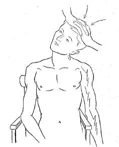
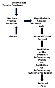
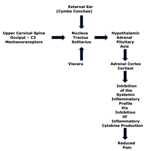
    Historically, chiropractic care primarily targets mechanical pain. As the name suggests, mechanical pain is pain that is primarily mechanical in nature rather than inflammatory in nature. A common metaphor is the pain associated with having one’s finger caught in a slammed door. The best solution for this mechanical pain is not the consumption of a pain drug, but rather for someone to mechanically open the door, releasing the finger. Chiropractic mechanical based care (spinal manipulation, or specific line-of-drive adjustments) is common and successful for mechanical-based pain syndromes, with high levels of patient satisfaction (24, 25). In fact, a recent study (October 2023) documented that chiropractic care was the most commonly chosen care by patients suffering from new-onset neck pain (26).

    With advances in the understanding of spine pain syndromes, it became understood that there was an overlap between mechanical pain and chemical-inflammatory pain; as different as these etiologies for these two types of pain are, they often co-exist.

    An early example of this understanding was published by Vert Mooney, MD, from the University of California, San Diego. When Dr. Mooney was the president of the International Society for the Study of the Lumbar Spine, his Presidential Address emphasized the relationship between mechanical pain and chemical-inflammatory pain. His address was published in the journal Spine, and titled (27).

    Where Is the Pain Coming From?

    Wound healing results in scar (fibrosis) formation which is less functional than the original tissue. Yet, early mobilization reduces scarring and enhances recovery.

    Dr. Mooney notes that spinal healing and spinal degenerative disease create spinal stiffness. Spinal stiffness has the greatest impact on the chemistry of the intervertebral disc because the disc is avascular. It is difficult to disperse any inflammatory chemicals that accumulate within the disc because of its lack of blood supply, especially in the presence of spinal stiffness. Dr. Mooney explains these concepts as follows:

    “Mechanical events can be translated into chemical events related to pain.”

    “The fluid content of the disk can be changed by mechanical activity.”

    “Mechanical activity has a great deal to do with the exchange of water and oxygen concentration [in the disk].” 

    An important aspect of disk nutrition and health is the mechanical aspects of the disk related to the fluid mechanics.

    The pumping action maintains the nutrition and biomechanical function of the intervertebral disk.  

    “Research substantiates the view that unchanging posture, as a result of constant pressure such as standing, sitting or lying, leads to an interruption of pressure-dependent transfer of liquid. Actually, the human intervertebral disk lives because of movement.”

    “In summary, what is the answer to the question of where is the pain coming from in the chronic low-back pain patient? I believe its source, ultimately, is in the disk. Basic studies and clinical experience suggest that mechanical therapy is the most rational approach to relief of this painful condition.”

    “Prolonged rest and passive physical therapy modalities no longer have a place in the treatment of the chronic problem.”

    The relationship between mechanical pain and chemical-inflammatory pain was significantly advanced by Manohar M. Panjabi, PhD, from Yale University School of Medicine. Dr. Panjabi explained his hypothesis in the European Spine Journal in 2006 in an article titled (28):

    A Hypothesis of Chronic Back Pain:
    Ligament Sub-failure Injuries Lead to Muscle Control Dysfunction

    Dr. Panjabi understands that a sub-failure injury of the spinal ligament is an injury caused by stretching of the tissue beyond its physiological limit, but less than to its failure point. These sub-failure injuries of ligaments are incomplete injuries, often tearing only a few fibers. These sub-failure injuries to ligaments “disrupt and/or injure the embedded mechanoreceptors.”

    Injured muscles heal relatively quickly due to abundant blood supply and therefore are not the main cause of chronic back pain. In contrast, ligament injuries heal poorly and therefore lead to tissue degeneration over time. Tissue degeneration further causes aberrant mechanical function, resulting in tissue stress, inflammation, and pain. Dr. Panjabi notes:

    “Abnormal mechanics of the spinal column has been hypothesized to lead to back pain via nociceptive sensors.”

    “The role played by the injury to the mechanoreceptors embedded in the ligaments of the spinal column has not been explored by any hypothesis.”

    A summary of Dr. Panjabi’s hypothesis is presented below:

    Dr. Panjabi's Hypothesis

    Dr. Panjabi’s Explanation

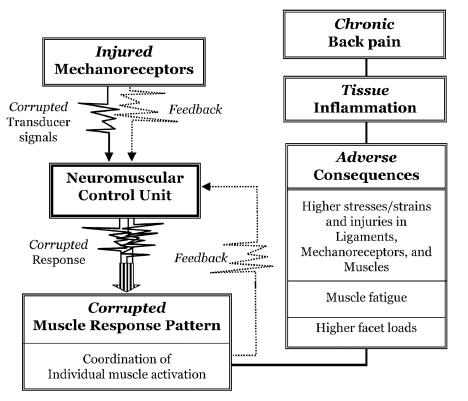
    “The injured mechanoreceptors send out corrupted transducer signals to the neuromuscular control unit, which finds spatial and temporal mismatch between the expected and received transducer signals, and, as a result, there is muscle system dysfunction and corrupted muscle response pattern is generated.”

    “Consequently, there are adverse consequences: higher stresses, strains, and even injuries, in the ligaments, mechanoreceptors, and muscles.”

    “There may also be muscle fatigue, and excessive facet loads.”

    “These abnormal conditions produce neural and ligament inflammation, and over time, chronic back pain.”

    In 2011, Nikolai Bogduk, MD, PhD, presented a paper in the journal Spine, titled (29):

    On Cervical Zygapophysial Joint Pain After Whiplash

    Nikolai Bogduk is a unique individual. As of December 2023, PUBMED, the search engine for the U.S. National Library of Medicine, locates 306 articles using his name. Dr. Bogduk is Emeritus Professor of Pain Medicine at the University of Newcastle, NSW, Australia. Dr. Bogduk holds an MD, PhD, and ScD. He began his research into spinal pain in 1972. Presently, Dr. Bogduk is the most noted, respected, and influential clinical anatomist of our time, and perhaps of all history.

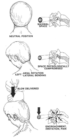
    In this article, Dr. Bogduk explains the mechanism of rear-end collision “whiplash” injuries. He notes that the cervical spine undergoes a “highly abnormal” “S” shaped deformation with extension of the lower cervical spine and flexion of the upper cervical spine. This abnormal mechanical configuration primarily injures the facet joints.

    Dr. Bogduk presents the “convergent validity” mechanistic evidence that implicates the cervical facet joints as the leading source of chronic neck pain after whiplash. He reviews data from:

    • Whiplash postmortem studies
    • Whiplash biomechanics studies
    • Whiplash clinical studies

    In his explanations, Dr. Bogduk references the mechanical work of Dr. Panjabi. In other words, he is looking at the mechanical explanation for chronic whiplash pain.

    In 2021, an article was published in the journal Pain Medicine, titled (30):

    The Prevalence of “Pure” Lumbar Zygapophysial Joint Pain in Patients with Chronic Low Back Pain

    In this study, the authors use zygapophysial joint (Z joint) synonymously for facet joint. Two hundred and six patients with possible lumbar facet joint pain underwent controlled diagnostic blocks to determine the prevalence of “pure” lumbar facet joint pain. All patients attributed their back pain to some form of injury. The authors note:

    Prior studies indicate that the joints that are most often painful are L4–L5 and L5–S1. This finding was confirmed in this study.

    “The prevalence of ‘pure’ lumbar Z joint pain was 15% (10–20%).”

    It is relevant to acknowledge that chiropractic spinal adjustments primarily influence the facet (Z) joints. It is also important to know that 63% of patients who seek chiropractic care do so for the management of chronic back pain (25).

    In 2017, a study from researchers at the University of Pennsylvania published a study in the Journal of Biomechanical Engineering, titled (31):

    The Interface of Mechanics and Nociception in Joint Pathophysiology

    This study reviews the mechanical, chemical, and neurological mechanisms contributing to synovial joint pain. The authors note that chronic joint pain is a widespread problem that frequently occurs with aging and trauma. The authors emphasize the importance of the mechanical pain model by stating:

    General Concepts

    “Synovial joints permit complex motion and often sustain loads several times greater than the weight of the human body.”

    “Repeated mechanical loading above the normal physiological conditions contribute to the initiation of acute tissue damage and degeneration.”

    “Trauma and repeated loading can induce structural and biochemical changes in joints, altering their microenvironment and modifying the biomechanics of their constitutive tissues, which themselves are innervated.”

    “Peripheral pain sensors can become activated in response to changes in the joint microenvironment and relay pain signals to the spinal cord and brain where pain is processed and perceived.”

    “The local mechanical environment of joints can be substantially altered after injury or degeneration, which can produce dysfunctional nociceptive signaling.”

     “Innervated by mechanoreceptors, disruption of the collagen fibers that make up the facet capsular ligament can initiate nociceptive signaling and lead to both acute and chronic pain.”

    “Both traumatic and repeated mechanical disruption can induce structural changes to joint tissues and inflammatory cascades, which separately activate pain fibers via a modified biomechanical and biochemical environment.”

    “These changes in neuronal signaling can lead to persistent pain even in the absence of any on-going tissue damage or input from peripheral nociceptive fibers.”

    “The facet capsular ligament is innervated by mechanoreceptors that encode movement and stretch, and also by nociceptors that encode nociceptive stimuli.”

     “Disruption of the facet capsule and other innervated tissues in the facet joint can elicit pain.”

     “Excessive stretch of the facet capsular ligament induces persistent pain and modifications in neuronal signaling.”

    “[Complete facet capsule rupture] does not initiate chronic pain, [but it can lead to] long-term alterations in the mechanical properties of that joint due to tissue scarring and/or degenerative cascades.”

    “Mechanical loading exceeding the physiologic range of a given joint can lead to persistent pain that arises from many structures in that joint.”

    Degenerated Joints

    Degenerative joint pathologies, such as OA, are initiated by a combination of applied mechanical loads and biological cascades within the joints’ tissues.

    Both trauma and age contribute to joint degeneration.

    “Degenerated joint tissues respond differently to loading than healthy counter-parts.”

    Degenerated joint tissues have lower thresholds for mechanical injury.

    Joint degeneration alters the biomechanical environment of innervated joint tissues, lowering thresholds for painful injury.

    Joint degeneration changes joint biomechanics, inflammatory status, and pain, and increases nociceptive signaling.

    The Cervical Spine

    “In the neck, the facet capsular ligament is particularly vulnerable to excessive spinal motions because of the potential for ligament stretch to activate the nociceptors innervating that ligament.”

    “Neck pain is most often induced by trauma and can involve damage, like loading and stretching of the facet capsular ligament, to the bilateral facet joints that provide articulations between spinal levels.”

    Neck trauma can produce peak strains in the C6/C7 facet capsule, sufficient to produce localized altered collagen fiber kinematics and sustained pain.

    Physiologic strains of 10–15% only activate mechanoreceptors, whereas strains greater than 25% produce sustained nociceptive “after-discharge,” and induce morphological changes in axons, including local swelling.

    “Facet joint …. pain syndromes are complex disorders with mechanical, molecular, and nociceptive contributors.”

    Large modulation in the biomechanics of joints may not be required for pain to be initiated, “especially if there are altered joint kinematics.”

    “This may explain why patients who undergo moderate loading of the neck develop persistent pain symptoms.”

    This study, in particular, emphasizes the relationship between altered mechanical function, the accumulation of inflammatory chemicals, and the genesis of pain syndromes. It is very supportive of the models that are taught in chiropractic education and used in chiropractic clinical practice. It explains the benefits and necessity for mechanical-based care for the management of pain syndromes.

    SUMMARY

    An ongoing theme from the studies presented here is that inflammatory chemicals increase the incidence of pain. Traditional medical practice often uses anti-inflammatory drugs to help people with pain syndromes, but frequently these drugs are not very effective and they are associated with many dangerous side-effects, including death, especially if chronically consumed.

    Patients with unresolved spinal pain syndromes often shop for alternatives in an effort to be helped. The top alternative with good outcomes, low costs, high patient satisfaction, long-term benefits, and an extremely high safety profile is chiropractic care (22, 32, 33, 26, 34, 35). An explanation for these outcomes, supported here, is that ongoing inflammation and pain can be driven by aberrant mechanical function. Chiropractic care targets the aberrant mechanical function with spinal adjusting and follow-ups, like exercise. This reduces both inflammation and pain.

    REFERENCES

    1. Omoigui S; The Biochemical Origin of Pain; S.O.T.A. Technologies, Inc.; Hawthorne, CA; 2003.
    2. Omoigui S; The Biochemical Origin of Pain: The Origin of All Pain is Inflammation and the Inflammatory Response: Inflammatory Profile of Pain Syndromes; Medical Hypothesis; 2007; Vol. 69; No. 6; pp. 1169–1178.
    3. Foreman J; A Nation in Pain; Healing Our Biggest Health Problem; Oxford University Press; 2014.
    4. Zhang Q, Bang S, Chandra S, Ji RR; Inflammation and Infection in Pain and the Role of GPR37; International Journal of Molecular Science; November 20, 2022; Vol. 23; No. 22; Article 14426.
    5. Kang JD, Georgescu HI, McIntyre-Larkin L, Stefanovic-Racic M, Donaldson WF, Evans CH; Herniated Lumbar Intervertebral Discs Spontaneously Produce Matrix Metalloproteinases, Nitric Oxide, Interleukin-6, and Prostaglandin E2; Spine; February 1, 1996; Vol. 21; No. 3; pp. 271-277.
    6. Kang JD , Stefanovic-Racic M, McIntyre LA, Georgescu HI, Evans CH; Toward a Biochemical Understanding of Human Intervertebral Disc Degeneration and Herniation: Contributions of Nitric Oxide, Interleukins, Prostaglandin E2, and Matrix Metalloproteinases; Spine; May 15, 1997; Vol. 22; No. 10; pp 1065-1073.
    7. Perneger TV, Whelton PK, Klag MJ; Risk of Kidney Failure Associated with the use of Acetaminophen, Aspirin, and Nonsteroidal Anti-inflammatory Drugs; New England Journal of Medicine; December 22, 1994; Vol. 331; No. 25; pp. 1675-1679.
    8. Wolfe MM, Lichtenstein DR, Singh G; Gastrointestinal Toxicity of Nonsteroidal Anti-inflammatory Drugs; New England Journal of Medicine; June 17, 1999; Vol. 340; No. 24; pp. 1888-1899.
    9. Helin-Salmivaara A, Virtanen A, Vesalainen R, Gronroos JM, Klaukka T, Idanpaan-Heikkila JE, Huupponen R; NSAID Use and the Risk of Hospitalization for First Myocardial Infarction in the General Population: A Nationwide Case-control Study from Finland; European Heart Journal; July 2006; Vol. 27; No. 14; pp. 16567-1663.
    10. Trelle S, Reichenbach S, Wandel S, Hildebrand P, Tschannen B, Peter M Villiger PM, Egger M; Cardiovascular Safety of Non-steroidal Anti-inflammatory Drugs: Network meta-analysis; British Medical Journal; January 11, 2011; Vol. 342; Article c7086.
    11. Editorial: Cardiovascular safety of NSAIDs; British Medical Journal; January 11, 2011; Vol. 342; Article c6618.
    12. Olsen AMS, Fosbol E, Lindhardsen J, Folke F, Charlot M, Selmer C; Duration of Treatment with Nonsteroidal Anti-Inflammatory Drugs and Impact on Risk of Death and Recurrent Myocardial Infarction in Patients With Prior Myocardial Infarction: A Nationwide Cohort Study; Circulation; May 21, 2011; Vol. 123; No. 20: pp. 2226-2235.
    13. Krijthe BP, Heeringa J, Hofman A, Franco OH, Stricker BH; Non-steroidal anti-inflammatory drugs and the risk of atrial fibrillation: A population-based follow-up study; British Medical Journal Open; April 8, 2014; Vol. 4; No. 4; e004059.
    14. Breitner JCS, Haneuse A, Walker R, Dublin S, Crane PK, Gray SL, Larson EB; Risk of Dementia and Alzheimer’s Disease with Prior Exposure to NSAIDs in an Elderly Community-based Cohort; Neurology; June 2, 2009; Vol. 72; No. 22; pp. 1899-1905.
    15. Curhan SG, Eavey R, Shargorodsky J, Curhan GC; Analgesic Use and the Risk of Hearing Loss in Men; The American Journal of Medicine; March 2010; Vol. 123; No. 3; pp. 231-237.
    16. Gleason JM, Slezak JM, Jung H, Reynolds K, van den Eeden SH, Haque R, Quinn VP, Loo RK, Jacobsen SJ; Regular Nonsteroidal Anti-Inflammatory Drug Use and Erectile Dysfunction; Journal of Urology; April 2011; Vol. 185; pp. 1388-1393.
    17. Kristensen DM, Desdoits-Lethimonier C, Mackey AL, and 16 more; Ibuprofen Alters Human Testicular Physiology to Produce a State of Compensated Hypogonadism; Proceedings of the National Academy of Sciences (PNAS, USA); January 23, 2018; Vol. 114; No. 4; pp. E715-E724.
    18. Lindsay SE, MD, Philipp T, Ryu WHA, Wright C, Yoo J; Nonsteroidal Anti-inflammatory Drugs in the Acute Post-operative Period Are Associated with an Increased Incidence of Pseudarthrosis, Hardware Failure, and Revision Surgery Following Single-level Spinal Fusion; Spine; August 1, 2023; Vol. 48; No. 15; pp. 1057–1063.
    19. Alemany S, Avella‑Garcia C, Liew Z, Garcia‑Esteban R, and 22 more; Prenatal and Postnatal Exposure to Acetaminophen in Relation to Autism Spectrum and Attention‑deficit and Hyperactivity Symptoms in Childhood: Meta‑analysis in Six European Population‑based Cohorts; European Journal of Epidemiology; October 2021; Vol. 36; No. 10; pp. 993-1004.
    20. Bauer AZ, Swan SH, Kriebel D, Liew Z, Taylor HS, Bornehag CG, Andrade AM, Olsen J, Jensen RH, Mitchell RT, Skakkebaek NE, Jégou B, Kristensen DM: Paracetamol [acetaminophen] Use During Pregnancy: A Call for Precautionary Action: Nature Reviews Endocrinology; December 2021; Vol. 17; No. 12; pp. 757-766.
    21. Khan FY, Kabiraj G, Ahmed MA, Adam M, Mannuru SP, Ramesh V, Shahzad A, Chaduvula P, Khan S; A Systematic Review of the Link Between Autism Spectrum Disorder and Acetaminophen: A Mystery to Resolve; Cureus; July 18, 2022; Vol. 14; No. 7; Article e26995.
    22. Giles LGF, Muller R; Chronic Spinal Pain: A Randomized Clinical Trial Comparing Medication, Acupuncture, and Spinal Manipulation; Spine; July 15, 2003; Vol. 28; No. 14; pp. 1490-1502.
    23. Maroon JC, Bost JW; Omega-3 Fatty Acids (fish oil) as an Anti-inflammatory: An Alternative to Nonsteroidal Anti-inflammatory Drugs for Discogenic Pain; Surgical Neurology; April 2006; Vol. 65; pp. 326–331.
    24. Kirkaldy-Willis WH, Cassidy JD; Spinal Manipulation in the Treatment of Low back Pain; Canadian Family Physician; March 1985; Vol. 31; pp. 535-540.
    25. Adams J, Peng W, Cramer H, Sundberg T, Moore C; The Prevalence, Patterns, and Predictors of Chiropractic Use Among US Adults; Results From the 2012 National Health Interview Survey; Spine; December 1, 2017; Vol. 42; No. 23; pp. 1810–1816.
    26. Fenton JJ, Fang SY, Ray M, Kennedy J, Padilla K, Amundson R, Elton D, Haldeman S, Lisi AJ, Sico J, Wayne PM, Romano PS; Longitudinal Care Patterns and Utilization Among Patients with New-Onset Neck Pain by Initial Provider Specialty; Spine; October 15, 2023; Vol. 48; No 20; pp. 1409–1418.
    27. Mooney V; Where Is the Pain Coming From?; Spine; October 1987; Vol. 12; No. 8; pp. 754-759.
    28. Panjabi MM: A Hypothesis of Chronic Back Pain: Ligament Sub-failure Injuries Lead to Muscle Control Dysfunction; European Spine Journal; May 2006; Vol. 15; No. 5; pp. 668-676.
    29. Bogduk N; On Cervical Zygapophysial Joint Pain After Whiplash; Spine; December 1, 2011; Vol. 36; No. 25S; pp. S194–S199.
    30. MacVicar J, MacVicar AM, Bogduk N; The Prevalence of “Pure” Lumbar Zygapophysial Joint Pain in Patients with Chronic Low Back Pain; Pain Medicine; February 4, 2021; Vol. 22; No.1; pp. 41–48.
    31. Sperry MM, Ita ME, Kartha S, Zhang S, Yu YH, Winkelstein B; The Interface of Mechanics and Nociception in Joint Pathophysiology: Insights form te Facet and Temporomandibular Joints; Journal of Biomechanical Engineering; February 2017; Vol. 139; No. 2; pp. 0210031-02100313.
    32. Muller R, Giles LGF; Long-Term Follow-up of a Randomized Clinical Trial Assessing the Efficacy of Medication, Acupuncture, and Spinal Manipulation for Chronic Mechanical Spinal Pain Syndromes; Journal of Manipulative and Physiological Therapeutics; January 2005; Vol. 28; No. 1; pp. 3-11.
    33. Keeney BJ, Fulton-Kehoe D, Turner JA, Wickizer TM, Chan KCG; Franklin GM; Early Predictors of Lumbar Spine Surgery after Occupational Back Injury: Results from a Prospective Study of Workers in Washington State; Spine; May 15, 2013; Vol. 38; No. 11; pp. 953-964.
    34. Kim S, Kim G, Kim H, Park J, Lee J, and nine more; Safety of Chuna Manipulation Therapy in 289,953 Patients with Musculoskeletal Disorders: A Retrospective Study; Healthcare; February 2, 2022; Vol. 10; No. 2; Article 294.
    35. Chu E, Trager RJ, Lee L, Niazi IK; A Retrospective Analysis of the Incidence of Severe Adverse Events Among Recipients of Chiropractic Spinal Manipulative Therapy; Scientific Reports; January 23, 2023; Vol. 13; No. 1; Article 1254.
  • Safety of Mechanical Care

    Safety of Mechanical Care

    People become healthcare providers because they are interested, often passionately, about helping others with their health. Most healthcare providers are horrified at the thought that anything they did or failed to do could end up harming a patient. Yet, every type of health care is associated with some risks of potential problems. These problems can range from an annoyance to soreness to disability or even to death.

    In acknowledging the existence of risks or problems associated with healthcare interventions, healthcare licensing bodies, governments, and associations have established “standards of care” for each healthcare discipline. These “standards of care” include informing patients about known or statistical risks/complications/harms that have been documented to occur following the healthcare intervention that is being proposed.

    For decades, top universities and top medical journals have been documenting errors and the consequences of those errors that occur as a consequence of traditional medical healthcare in the United States. These studies often emphasize the hospital setting where such errors are more easily quantified. Sadly, the early data is scary. It comes with warnings about how improvements are imperative. Yet, recent data (2023) shows that not much progress, if any, has been made.

    ••••

    In 1994, the Journal of the American Medical Association published an article titled (1):

    Error in Medicine

     The author, Lucian Leape, MD, is from the Harvard School of Public Health. This study only looked at hospital error. Dr. Leape notes and cites:

    • A 1964 study reported that 20% of patients admitted to a university hospital medical service suffered iatrogenic injury and that 20% of those injuries were serious or fatal. [Annals of Internal Medicine]
    • A 1981 study found that 36% of patients admitted to a university medical service in a teaching hospital suffered an iatrogenic event, of which 25% were serious or life threatening. More than half of the injuries were related to use of medication. [New England Journal of Medicine]
    • A 1991 study found that 64% of cardiac arrests at a teaching hospital were preventable and that inappropriate use of drugs was the leading cause of the cardiac arrests. [Journal of the American Medical Association]
    • A 1991 study of hospitalized patients from New York found that 4% suffered an injury that prolonged their hospital stay or resulted in measurable disability. [New England Journal of Medicine]

    Dr. Leape notes that if the data from these studies were applied to the entire U.S. population, “180,000 people die each year as a result of iatrogenic injury, the equivalent of three jumbo jet crashes every 2 days.” Dr. Leape states:

    “For years, medical and nursing students have been taught Florence Nightingale’s dictum—first, do no harm.”

    “Yet, evidence from a number of sources, reported over decades, indicates that a substantial number of patients suffer treatment-caused injuries while in the hospital.”

    “Autopsy studies have shown high rates (35% to 40%) of missed diagnoses causing death.”

    ••••

    In 1998, the Journal of the American Medical Association published a study titled (2):

    Incidence of Adverse Drug Reactions in Hospitalized Patients
    A Meta-analysis of Prospective Studies

    The authors of this study were from the University of Toronto. Their study evaluated 39 prospective studies from U.S. hospitals. The authors defined serious Adverse Drug Reactions (ADRs) as:

    “Serious ADRs were defined as those that required hospitalization, were permanently disabling, or resulted in death.”

    Importantly:

    • The authors “excluded errors in drug administration, noncompliance, overdose, drug abuse, therapeutic failures, and possible ADRs.”
    • The authors investigated “injuries incurred by drugs that were properly prescribed and administered.”
    • “Possible ADRs” were excluded from this study.

    Findings:

    The authors found that, overall, 2,216,000 U.S. hospitalized patients had serious ADRs and 106,000 had fatal ADRs, making these reactions between the fourth and sixth leading cause of death yearly in the United States.

    Comments by the Authors:

    “The incidence of serious and fatal ADRs in US hospitals was found to be extremely high.”

    “We have found that serious ADRs are frequent and more so than generally recognized.”

    “Their [fatal ADRs] incidence has remained stable over the last 30 years.”

    “[This study on ADRs, which excluded medication errors, showed] that there are a large number of serious ADRs even when the drugs are properly prescribed and administered.”

    “The costs associated with ADRs may be very high.”

    “It is important to note that we have taken a conservative approach, and this keeps the ADR estimates low by excluding errors in administration, overdose, drug abuse, therapeutic failures, and possible ADRs. Hence, we are probably not overestimating the incidence of ADRs.”

    A critical understanding from the data in this study is that it only assessed patients in the hospital. Problems outside of the hospital, such as outpatient care, long-term care centers, home, etc. were not included. Yet, the average of 106,000 hospitalized patients died and 2,216,000 had a serious reaction to the right drug being given for the correct diagnosis and in the correct dose, yearly.

    ••••

    In 1999, an editorial was published in the British Medical Journal (BMJ), titled (3):

    Reducing Errors in Medicine:
    It’s Time to Take this More Seriously

    Drs. Berwick and Leape make this analogy:

    “Ladies and gentlemen, welcome aboard Sterling Airline’s Flight Number 743, bound for Edinburgh. This is your captain speaking. Our flight time will be two hours, and I am pleased to report both that you have a 97% chance of reaching your destination without being significantly injured during the flight and that our chances of making a serious error during the flight, whether you are injured or not, is only 6.7%. Please fasten your seatbelts, and enjoy the flight. The weather in Edinburgh is sunny.”

    “Would you stay aboard? We doubt it.”

    “In health care it is a totally different story. With the rising complexity and reach of modern medicine have come startling levels of risk and harm to patients. One recent study in two of the most highly regarded hospitals in the world discovered serious or potentially serious medication errors in the care of 6.7 out of every 100 patients, and the Harvard Medical Practice Study, which reviewed over 30,000 hospital records in New York state, found injuries from care itself (‘adverse events’) to occur in 3.7% of hospital admissions, over half of which were preventable and 13.6% of which led to death. If these figures can be extrapolated to American health care in general then over 120,000 Americans die each year as a result of preventable errors in their hospital care.”

    “Studies in Australia, Israel, the United Kingdom and elsewhere, suggest levels of error and hazard in patient care that are no lower than in America.”

    ••••

    In 1999, the lay press printed a study titled (4):

    HMO Chief: Patients are at Risk:
    Blunders take 400,000 Lives Every Year, Kaiser Head Says

    This publication quotes Dr. David Lawrence, who was chief executive of Kaiser Permanente, in a speech to the national Press Club. Kaiser is the U.S.’s largest HMO. Dr. Lawrence states:

    “Medical accidents and mistakes kill 400,000 people a year, ranking behind only heart disease and cancer as the leading cause of death.” 

    “Mistakes alone kill more people each year than tobacco, alcohol, firearms, or automobiles.”

    “[Patients] continue to believe in the myth of Marcus Welby, the unbridled benefits of technology and the assumption that competence and safety are spread evenly and consistently throughout the health care system.”

    “If passengers were asked to fly with a commercial airline organized like most health care, they wouldn’t get on the plane.”

    ••••

    In 2000, a study was published in the Journal of the American Medical Association titled (5):

    Is US Health Really the Best in the World?

    The author, Barbara Starfield, MD, MPH, is from Johns Hopkins School of Hygiene and Public Health.

    Dr. Starfield notes that there is accumulating information concerning the deficiencies of US medical care; 20% to 30% of patients receive contraindicated care.  The US ranks behind Japan, Sweden, Canada, France, Australia, Spain, Finland, the Netherlands, the United Kingdom, Denmark, and Belgium.

    The U.S. health care system itself appears to be linked to “poor health through its adverse effects.” For example, Dr. Starfield cites:

    • 7,000 deaths/year from medication errors in hospitals
    • 12,000 deaths/year from unnecessary surgery
    • 20,000 deaths/year from other errors in hospitals
    • 80,000 deaths/year from nosocomial infections in hospitals
    • 106,000 deaths/year from non-error, adverse effects of medications

    These total to 225,000 deaths per year from iatrogenic causes. These estimates are for deaths only and do not include adverse effects that are associated with disability or discomfort. If other estimates are used, the deaths due to iatrogenic causes would range from 230,000 to 284,000. Dr. Starfield states:

    “In any case, 225,000 deaths per year constitutes the third leading cause of death in the United States, after deaths from heart disease and cancer.”

    “Recognition of the harmful effects of health care interventions, and the likely possibility that they account for a substantial proportion of the excess deaths in the United States compared with other comparably industrialized nations, sheds new light on imperatives for research and health policy.”  

    “The US population does not have anywhere near the best health in the world. Of 13 countries in a recent comparison, the United States ranks an average of 12th (second from the bottom) for 16 available health indicators.”

    ••••

    In 2009, a study published in the Journal of the American Medical Association titled (6):

    Exploring the Harmful Effects of Health Care

    The authors Charles M. Kilo, MD, MPH and Eric B. Larson, MD, MPH, were from The Trust for Healthcare Excellence in Portland, OR.

    These authors are quite cynical, suggesting that the benefits of the U.S. healthcare system may not outweigh the harms that it causes. There is evidence that about one-third of medical spending is for services that do not improve health or the quality-of-care, and may make things worse. The U.S. healthcare delivery can cause direct adverse physical and emotional effects. The authors state:

    “Although health care’s objective should be to improve health, its primary emphasis has been on producing services.”

    “The benefits that US health care currently deliver may not outweigh the aggregate health harm it imparts.”

    “Higher-intensity care generally does not improve survival, and complications of medical care accounted for 1.1 million hospitalizations in 2006, costing nearly $42 billion.”

    “Exaggerated fears and ‘medicalizing’ normal phenomena are as harmful as unrealistic expectations and are fostered frequently by marketing hype and sometimes inadvertently by health care clinicians.”

    “The possibility that health care might cause net harm is increasingly important given the sheer magnitude of the modern health care enterprise.”

    ••••

     In 2011, a study was published in the journal Health Affairs titled (7):

    ‘Global Trigger Tool’ Shows That Adverse Events in Hospitals
    May Be Ten Times Greater Than Previously Measured

    The authors, from the University of Utah, explore the best method for estimating the incidence of adverse events in hospitals so that patient safety outcomes against such events can be optimized. They found that the adverse event detection methods commonly used to track patient safety in the United States fared very poorly compared to other methods and missed 90% of the adverse events.

    Overall, the authors found that adverse events occurred in one-third of hospital admissions. Because some admissions suffered from more than one adverse event, the rate of adverse events rose to essentially half of admissions. The authors state:

    “Our study suggests that despite sizable investments and aggressive promotional efforts by local hospitals, these reporting systems fail to detect most adverse events.”

    “Our study also detected far more adverse events in hospitalized patients than have been found in prior studies.”

    “Despite more than a decade of focus on improving patient safety in the United States, the current rates of adverse events among inpatients at three leading hospitals are still quite high at 33.2 percent of hospital admissions for adults.”

    “The true rates are likely to be higher still, given the consistent finding that direct observational studies reveal higher rates of adverse events than retrospective studies because not all adverse events are documented in the patient record.”

    “Our findings indicate that two methods commonly used by most care delivery organizations and supported by policy makers to measure the safety of care—enhanced voluntary reporting systems and the use of the Agency for Healthcare Research and Quality’s Patient Safety Indicators—fail to detect more than 90 percent of the adverse events that occur among hospitalized patients.” 

    “Despite almost ten years since the Institute of Medicine report on patient safety, rates of adverse events in hospital patients are still high.” 

    ••••

    In 2016, an article was published in the British Medical Journal (BMJ) titled (8):

    Medical Error:
    The Third Leading Cause of Death in the United States

    The authors are from the Department of Surgery, Johns Hopkins University School of Medicine. They analyzed the scientific literature on medical error to identify its contribution to U.S. deaths. Their primary concern  is that “medical error is not included on death certificates or in rankings of cause of death.” The annual list of the most common causes of death in the United States, compiled by the Centers for Disease Control and Prevention (CDC), has a major limitation as it relies on assigning an International Classification of Disease (ICD) code to the cause of death, and ICD code does not include error. The authors state:

    “Currently, deaths caused by [medical] errors are unmeasured.”

    “If medical error was a disease, it would rank as the third leading cause of death in the US.”

    “Medical error is the third biggest cause of death in the US and therefore requires greater attention.”

    “Medical error leading to patient death is under-recognized in many other countries, including the UK and Canada.” 

    “The system for measuring national vital statistics should be revised to facilitate better understanding of deaths due to medical care.”

    ••••

    In 2023, an article was published in the BMJ (British Medical Journal) Quality & Safety, titled (9):

    Burden of Serious Harms from Diagnostic Error in the USA

    The authors are from Johns Hopkins School of Medicine and Harvard Medical School. The goal of this research was to estimate the total number of serious misdiagnosis-related harms (i.e., permanent disability or death) occurring annually in the USA across all care settings (ambulatory clinic, emergency department, and inpatient). This is the first national estimate of permanent morbidity and mortality resulting from diagnostic errors across all clinical settings, including both hospital-based and clinic-based care each year in the USA alone. Importantly, this study, “considered only false negative diagnoses (i.e., initially missed or delayed) and associated harms.” The authors note:

    “Diagnostic errors cause substantial preventable harms worldwide.”

    “An estimated 795,000 Americans become permanently disabled or die annually across care settings because dangerous diseases are misdiagnosed.”

    “This manuscript provides the first robust, national annual US estimate for serious misdiagnosis-related harms (nearly 800,000 combined deaths (~371,000) or permanent disabilities (~424,000)) across care settings (ambulatory clinic, emergency department, and inpatient).”

    “Across clinical settings (ambulatory clinics, emergency department, and inpatient), we estimate that nearly 800,000 Americans die or are permanently disabled by diagnostic error each year, making it the single largest source of serious harms from medical mistakes.” 

    “The number of affected patients is large, and this makes diagnostic error a pressing public health concern.”

    “Total annual diagnostic errors in the USA likely number in the tens of millions.”

    “Measured error and harm rates in primary care and emergency departments are similar in the USA, the UK, and Western Europe.”

    Chiropractic Care

    In 2022, a study was published in the journal Healthcare, and titled (10):

    Safety of Chuna Manipulation Therapy in 289,953 Patients
    with Musculoskeletal Disorders

    This study was from medical facilities in South Korea. It involved a form of high-velocity low amplitude spinal adjustment (specific line-of-drive manipulation). This type of traditional joint manipulation is similar to that in chiropractic. This type of manual therapy has been incorporated into the Korean health care system and has been administered in 16.4% of inpatients and 83.6% of outpatients with musculoskeletal disorders in Korean medicine hospitals specializing in spine and joint diseases.

    The authors make these observations:

    “Manual therapy is performed in various forms by chiropractors, osteopaths, and physical therapists across the world, including the United States, Europe, and Australia.”

    “The use of spinal manipulation has increased in recent decades in Western countries, as has the popularity of chiropractic therapy among American adults.”

    “The UK National Institute for Health and Clinical Excellence guidelines now recommend manual therapy for treating persistent or subacute lower back pain.”

    This study was also massive. In total, the authors assessed 2,682,258 manipulation procedures that were performed on 289,953 patients from 14 different facilities. The authors state:

    “In this study, 289,953 patients and more than 2.5 million cases of CMT were reviewed, making it a rare, very wide-ranging, and reliable investigation of severe adverse events.”

    Adverse events were graded as follows:

    MILD: asymptomatic or mild symptoms

    MODERATE: minimal, local, or non-invasive intervention indicated

    SEVERE: severe or medically significant but not immediately life-threatening; hospitalization or prolongation of hospitalization indicated

    LIFE-THREATENING: urgent intervention indicated

    FATAL: death

    The authors state:

    “Our analysis of 289,953 patients and 2,682,258 cases of CMT indicates that both mild–moderate and severe AEs are rare after CMT.”

    “Adverse events of any level of severity were very rare after CMT.”

    “There were no instances of carotid artery dissection or spinal cord injury.”

    No life-threatening or fatal events were identified. Eleven rib fractures were noted on elderly patients with osteoporosis, but all recovered without residuals.

    ••••

    In 2023, a study was published in the journal Scientific Reports, and titled (11):

    A Retrospective Analysis of the Incidence of Severe Adverse Events Among Recipients of Chiropractic Spinal Manipulative Therapy

    This study examined the incidence and severity of adverse events (AEs) in 54,846 patients who received 960,140 chiropractic spinal manipulations. Data originated from 30 chiropractic clinics using 38 different chiropractors. All patients received spinal manipulative therapy (SMT) administered via manual thrust (i.e., a hands-on impulse applied to the spinal joints). The authors concluded:

    “In this study, severe spinal manipulative therapy-related adverse events were reassuringly very rare.”

    “There were no adverse events related to stroke or cauda equina syndrome.” 

    “There were no cases of stroke, transient ischemic attack, vertebral or carotid artery dissection, cauda equina syndrome, or spinal fracture.”

    “No adverse events were identified that were life-threatening or resulted in death.”

    “No adverse events were reported to be permanent.”

    Once again, two rib fractures were identified, both occurring in elderly patients with a history of osteoporosis. Both patients recovered without residuals.

    ••••

    Chiropractic care is in a unique position. These last two robust studies document the extreme safety of chiropractic care. When a patient fails to respond to chiropractic care, medical referral is standardly done by chiropractors with the intention of benefiting the patient with the best of both approaches to healthcare. Often the chiropractic input is invaluable in helping to reduce missed diagnoses.

    REFERENCES

    1. Leape L; Error in Medicine; Journal of the American Medical Association; December 21, 1994; Vol. 272; No. 23; pp. 1851-1857.
    2. Lazarou, BH Pomeranz BH, PN Corey PN: Incidence of Adverse Drug Reactions in Hospitalized Patients: A Meta-analysis of Prospective Studies; Journal American Medical Association; April 15, 1998; Vol. 279; No. 15; pp. 1200-1205.
    3. Berwick D, Lucian L; Reducing Errors in Medicine; It’s Time to Take this More Seriously; British Medical Journal (BMJ); July 17, 1999; Vol. 318; pp. 136-137.
    4. Rosenblatt RA; “HMO Chief: Patients are at risk: Blunders take 400,000 lives every year, Kaiser head says;” LOS ANGELES TIMES, Oakland Tribune; July 15, 1999.
    5. Starfield B; Is US Health Really the Best in the World?; Journal of the American Medical Association; July 26, 2000; Vol. 284; No. 4; pp. 483-485.
    6. Kilo KM, Larson EB; Exploring the Harmful Effects of Health Care; Journal of the American Medical Association; July 1, 2009; Vol. 302; No. 1; pp. 89-91.
    7. Classen DC, Resar R, Griffin F, Federico F, Frankel T, Kimmel N, Whittington JC, Frankel A, Seger A, James BC; ‘Global Trigger Tool’ Shows That Adverse Events in Hospitals May Be Ten Times Greater Than Previously Measured; Health Affairs; April 2011; Vol. 30; No. 4; pp. 581-589.
    8. Makary MA; Medical Error: The Third Leading Cause of Death in the United States; British Medical Journal (BMJ); May 3, 2016; Vol. 353; Article i2139.
    9. Newman-Toker DE, Nassery N, Schaffer AC, Yu-Moe CW, Clemens GD, Wang Z, Zhu Y, Tehrani SAS, Fanai M, Hassoon A, Siegal D; Burden of Serious Harms from Diagnostic Error in the USA; BMJ [British Medical Journal] Quality & Safety; July 17, 2023; Epub [accessed December 6, 2023]
    10. Kim S, Kim G, Kim H, Park J, Lee J, and nine more; Safety of Chuna Manipulation Therapy in 289,953 Patients with Musculoskeletal Disorders: A Retrospective Study; Healthcare; February 2, 2022; Vol. 10; No. 2; Article 294.
    11. Chu E, Trager RJ, Lee L, Niazi IK; A Retrospective Analysis of the Incidence of Severe Adverse Events Among Recipients of Chiropractic Spinal Manipulative Therapy; Scientific Reports; January 23, 2023; Vol. 13; No. 1; Article 1254.

    “Authored by Dan Murphy, D.C.. Published by ChiroTrust® – This publication is not meant to offer treatment advice or protocols. Cited material is not necessarily the opinion of the author or publisher.”

  • The Importance of the Initial Provider for Spine Care

    The Importance of the Initial Provider for Spine Care

    Considerations for Costs, Outcomes, Surgery, Injections, and Imaging

    Introduction

    Healthcare providers of various disciplines tend to believe in and offer patients the intervention of their specialty:

    • Medical doctors tend to prescribe medicines.
    • Surgeons tend to offer surgery.
    • Acupuncturists tend to offer needles.
    • Massage therapists tend to offer massage.
    • Physical therapists tend to offer physical therapy.
    • Chiropractors tend to offer adjustments (specific line-of-drive manipulation).
    • Etc.

    Often, the initial provider for an ailment is the only provider ever seen for that ailment. It is quite rare for a patient to switch provider types. These biases can affect clinical outcomes and costs. Fortunately, these biases are being understood by our healthcare system. Their findings are quite good for the chiropractic profession.

    Low Back Considerations

    In 1977, Samuel Turek, MD, was a clinical professor in the Department of Orthopedics and Rehabilitation at the University of Miami School of Medicine. That year, the third edition of his book, Orthopaedics, Principles and Their Applications was published. It was an incredible work, comprising 1,574 pages (1).

    In his book, Dr. Turek has a section titled:

    Treatment of Intervertebral Disc Herniation with Manipulation

    This section states:

    “Manipulation. Some orthopaedic surgeons practice manipulation in an effort at repositioning the disc. This treatment is regarded as controversial and a form of quackery by many men. However, the author has attempted the maneuver in patients who did not respond to bed rest and were regarded as candidates for surgery. Occasionally, the results were dramatic.”

    Technique. The patient lies on his side on the edge of the table facing the surgeon, and the uppermost leg is allowed to drop forward over the edge of the table, carrying forward that side of the pelvis. The uppermost arm is placed backward behind the patient, pulling the shoulder back. The surgeon places one hand on the shoulder and the other on the iliac crest and twists the torso by pushing the shoulder backward and the iliac crest forward. The maneuver is sudden and forceful and frequently is associated with an audible and palpable crunching sound in the lower back. When this is felt, the relief of pain is usually immediate. The maneuver is repeated with the patient on the opposite side.”

    “The patient should be cautioned beforehand that the manipulation may make his symptoms worse and that this is an attempt to avoid surgery.”

    ••••

    In 1985, William H. Kirkaldy-Willis, MD, was a Professor Emeritus of orthopedics and director of the Low-Back Pain Clinic at the University Hospital, Saskatoon, Canada. That year, he published an article titled (2):

    Spinal Manipulation in the Treatment of Low Back Pain

    In this article, Dr. Kirkaldy-Willis presents the results of 283 patients who were suffering from chronic low back and leg pain and treated with chiropractic spinal adjusting (specific line-of-drive manipulation). All 283 patients in this study had failed prior conservative and/or operative treatment, and they were all totally disabled (“constant severe pain; disability unaffected by treatment”).

    Patient outcomes were graded as “good” if they achieved these results:

    • Symptom-free with no restrictions for work or other activities.
    • Mild intermittent pain with no restrictions for work or other activities.

    81% of the patients with referred pain syndromes achieved the “good” result. 48% of the patients with nerve compression syndromes, primarily subsequent to disc lesions and/or central canal spinal stenosis, achieved the “good” result. No patients were made worse by the chiropractic joint adjusting. The authors made these statements:

    “Spinal manipulation, one of the oldest forms of therapy for back pain, has mostly been practiced outside of the medical profession.”

    “Over the past decade, there has been an escalation of clinical and basic science research on manipulative therapy, which has shown that there is a scientific basis for the treatment of back pain by manipulation.”

    “Most family practitioners have neither the time nor inclination to master the art of manipulation and will wish to refer their patients to a skilled practitioner of this therapy.”

    “The physician who makes use of this [manipulation] resource will provide relief for many back pain patients.”

    ••••

    In 2013, Benjamin J Keeney, PhD, was from the Department of Orthopaedics at Dartmouth Medical School. That year, Dr. Keeney and colleagues published a study titled (3):

    Early Predictors of Lumbar Spine Surgery after Occupational Back Injury:
    Results from a Prospective Study of Workers in Washington State

     This was a prospective population-based cohort study of 1,885 subjects, designed to identify early predictors of lumbar spine surgery within 3 years after occupational back injury from the state of Washington. The authors note, “Reducing unnecessary spine surgeries is important for improving patient safety and outcomes and reducing surgery complications and health care costs.”

    Study subjects were assessed using the Roland Morris Disability Questionnaire (RMDQ). The RMDQ has been shown to be predictive of chronic work disability, longer duration of sick leave, chronic pain, and other measures of function.

    Study findings include:

    “Those with greater injury severity and those whose first provider seen for the injury was a surgeon also had significantly higher odds of surgery, after adjusting for all other variables.”

    “42.7% of workers who first saw a surgeon had surgery, in contrast to only 1.5% of those who saw a chiropractor.”  

    “There was a very strong association between surgery and first provider seen for the injury, even after adjustment for other important variables.”

    “It is possible that these findings indicate that who you see is what you get.”

    “In Washington State worker’s compensation, injured workers may choose their medical provider. Even after controlling for injury severity and other measures, workers with an initial visit for the injury to a surgeon had almost nine times the odds of receiving lumbar spine surgery compared to those seeing primary care providers, whereas workers whose first visit was to a chiropractor had significantly lower odds of surgery [by 78%].”

    “Approximately 43% of workers who saw a surgeon had surgery within 3 years, in contrast to only 1.5% of those who saw a chiropractor.”

    ••••

    In 2016, William B. Weeks, MD, PhD, was a professor at the Geisel School of Medicine at Dartmouth. That year, Dr. Weeks was the lead author on an article titled (4):

    The Association Between Use of Chiropractic Care and Costs of Care Among Older Medicare Patients with Chronic Low Back Pain and Multiple Comorbidities

    Dr. Weeks and colleagues wanted to determine whether use of chiropractic manipulation was associated with lower healthcare costs among multiple-comorbid Medicare beneficiaries with chronic low back pain. Their analysis assessed 72,326 patients. These authors note:

    “Medicare reimbursements during the chronic LBP treatment episode were lowest for patients who used [chiropractic manipulation] alone; these patients had higher rates of healthcare use for low back pain but lower rates of back surgery in the year following the treatment episode.”

    “Expenditures were greatest for patients receiving medical care alone.”

    “Patients who used only [chiropractic manipulation] had the lowest annual growth rates in almost all Medicare expenditure categories.”

    “This study found that older multiple-comorbid patients who used only [chiropractic manipulation] during their chronic LBP    episodes had lower overall costs of care, shorter episodes, and lower cost of care per episode day than patients in the other treatment groups.”

    “These findings support initial [chiropractic manipulation] use in the treatment of, and possibly broader chiropractic management of, older multiple-comorbid  chronic LBP patients.”

    ••••

    In 2022, Robert James Trager, DC, was a clinical chiropractor and a researcher at the University Hospitals Cleveland Medical Center, Ohio. That year, Dr. Trager and colleagues published a study titled (5):

    Association Between Chiropractic Spinal Manipulation and Lumbar Discectomy in Adults with Lumbar Disc Herniation and Radiculopathy

    The authors of this study assessed matched cohorts of 5,785 patients with a mean age of 37 years.

    The authors note that it is common for patients with lumbar disc herniations and radiculopathy to receive chiropractic care or undergo surgery to remove herniated disc material, a procedure called discectomy. Prior studies have found that patients who initiate care for low back pain with a chiropractor have significantly reduced odds of having discectomy.

    In this study, the relative odds for discectomy were significantly reduced in the chiropractic cohort compared with the cohort receiving other care over 1-year (by 69%) and 2-year follow-up (by 77%). This study shows that patients initially receiving chiropractic care for lumbar disc herniation with radiculopathy have reduced odds of discectomy over 1-year and 2-year follow-up.

    Neck Considerations

    In 1996, MN Woodward was a researcher at the University Department of Orthopaedic Surgery, Bristol, UK. That year he was the lead author in an article titled (6):

    Chiropractic Treatment of Chronic ‘Whiplash’ Injuries

    The 28 patients in this study had initially been treated with anti-inflammatory drugs, soft collars, and physiotherapy. These patients had all become chronic and were referred for chiropractic at an average of 15.5 months (range was 3–44 months) after their initial injury.

    Following chiropractic care, 93% of the patients had improved. These authors state:

    “The results of this retrospective study would suggest that benefits can occur in over 90% of patients undergoing chiropractic treatment for chronic whiplash injury.”

    ••••

    In 2002, Jan Lucas Hoving, PT, PhD, was in the Department of Clinical Epidemiology, Cabrini Hospital, Victoria, Australia. That year he was the lead author of an article titled (7):

    Manual Therapy, Physical Therapy, or Continued Care by a General Practitioner for Patients with Neck Pain:A Randomized Controlled Trial

    In this study, the authors compared the effectiveness of manual therapy, physical therapy, and care by a general practitioner (pharmacology) in the treatment of neck pain. They used a randomized controlled trial design. The study involved 183 patients.

    The authors defined “manual therapy” as:

    “According to the International Federation of Orthopedic Manipulative Therapies, ‘Orthopedic manipulative (manual) therapy is a specialization within physical therapy and provides comprehensive conservative management for pain and other symptoms of neuro-musculo-articular dysfunction in the spine and extremities.’”

    These authors concluded:

    “Manual therapy scored consistently better than the other two interventions on most outcome measures.”

    “In daily practice, manual therapy is a favorable treatment option for patients with neck pain compared with physical therapy or continued care by a general practitioner.”

    “The success rates for manual therapy were statistically significantly higher than those for physical therapy.”

    “Manual therapy scored better than physical therapy on all outcome measures…”

    “Range of motion improved more markedly for those who received manual therapy or physical therapy than for those who received continued [physician/pharmacology] care.”

    “The postulated objective of manual therapy is the restoration of normal joint motion, which was achieved, as indicated by the relatively large increase in the range of motion of the cervical spine.”

    “Patients receiving manual therapy had fewer absences from work than patients receiving physical therapy or continued care.”

    “Manual therapy seems to be a favorable treatment option for patients with neck pain.”

    “Primary care physicians should consider manual therapy when treating patients with neck pain.”

    ••••

    In 2013, Cynthia K. Peterson, RN, DC, was a professor of chiropractic medicine and radiology at the Orthopaedic University Hospital Balgrist, Zürich, Switzerland. That year she was the lead author in an article titled (8):

    Outcomes from Magnetic Resonance Imaging:
    Confirmed Symptomatic Cervical Disk Herniation Patients Treated with High-Velocity, Low-Amplitude Spinal Manipulation Therapy:
    A Prospective Cohort Study With 3-Month Follow-Up

     The purpose of this study was to investigate outcomes of patients with cervical radiculopathy from cervical disk herniation (CDH) who were treated with chiropractic spinal manipulative therapy. The authors used 50 patients with a mean age of 44 years. Treatments were repeated 3 to 5 times per week for the first 2 to 4 weeks and carried on 1 to 3 times per week thereafter until the patient was asymptomatic. They were evaluated at baseline, 2-weeks, 1-month, and 3-months.

    All patients in this study had neck pain and moderate to severe arm pain in a dermatomal pattern with sensory, motor, or reflex changes corresponding to the involved nerve root. They also had magnetic resonance imaging-proven CDH at the corresponding spinal segment.

    “High-velocity, low-amplitude spinal manipulations were administered by experienced doctors of chiropractic.” These manipulations were delivered to the segmental level of the CDH. The goal of the manipulation was to produce an audible release, which was achieved in most cases. The outcomes were as follows:

    • “By 2 weeks after the first treatment, 55.3% of all patients reported that they were significantly improved and none reported being worse.”
    • “At 1 month, 68.9% were significantly improved.”
    • By 3 months, 85.7% were significantly improved with no patients being worse.
    • “There were no adverse events in this cohort of patients.”

    The authors noted that the presence of cervical radiculopathy was not a negative predictor of outcomes for these chiropractic patients with CDH. The authors concluded:

    “Most patients in this study with MRI-proven symptomatic CDHs who were treated with high-velocity, low-amplitude spinal manipulation reported clinically significant improvement at all time points, particularly at 3 months.”

    “Patients with symptomatic MRI-confirmed cervical disk herniations treated with SMT to the level of herniation reported high levels of clinically relevant improvement at 2 weeks, 1 month, and 3 months after the first treatment.”

    The authors also noted that chiropractic spinal manipulation for CDH patients with radiculopathy was 2-3 times superior to published studies of patients with CDH with radiculopathy who received 2 cervical nerve root blocks with a corticosteroid and anesthetic.

    ••••

    In 2017, Maggie E Horn, DPT, MPH, PhD, was an assistant professor from the Department of Orthopaedic Surgery at Duke University. That year she was the lead author in a study titled (9):

    Influence of Initial Provider on Health Care Utilization in Patients Seeking Care for Neck Pain

    The authors note:

    “It is imperative to evaluate the difference in health care process and outcomes in patients initially consulting with non-pharmacological providers (ie, chiropractors [DCs] and physical therapists [PTs]) and pharmacological providers (ie, specialists [such as physiatrists and neurologists]) in comparison to PCPs.”

    “These specific provider types were included in the analysis because they are the most common providers consulted for neck pain.”

    This study looked at a cohort of 1,702 patients seeking care for a new episode of neck pain who consulted a primary care provider (PCP), physical therapist (PT), chiropractor (DC), or specialist:

    PCP 44%
    DC 23%
    PT 17%
    Specialist (physiatrists, neurologist) 16%

    The health care utilization was assessed at 14 days, 30 days, and 1 year from the initial visit between various providers. The health care utilization assessed included:

    • Imaging (MRI, computed tomography, radiography)
    • Opioids
    • Surgery (spinal arthrodesis, discectomy, laminectomy, or fusion)
    • Injections (including nerve blocks)

    Based upon these results, the authors make the following important observations:

    “Physical therapists and chiropractors primarily treat neck pain with exercise therapy and manual therapy, which has been found to have good effectiveness in treating nonspecific neck pain.” 

    “[In contrast], primary care providers’ first line of treatment often includes medication, imaging, specialist referral, or a combination of those factors.”  

    “Initial consultation from either a chiropractor or physical therapist decreases the patient’s odds of being prescribed an opioid at 30 days or within any time in the 1-year follow-up period.”

    Compared with initial consultation with a primary care provider, the odds of undergoing advanced imaging (MRI or computed tomography) within 1 year “was reduced when the initial provider was a chiropractor and increased when the initial provider was a specialist or a physical therapist.”

    “When patients in the sample initially consulted with a chiropractor, the odds of MRI use decreased compared with consulting with a primary care provider.”

    “We found that initial consultation with a non-pharmacological provider, such as a chiropractor or physical therapist, is associated with a decrease in the downstream utilization of health care services, and importantly a decrease in opioid use 30 days and 1 year after the initial consultation.”

    “Initiating care with a specialist was associated with an increase in the odds of receiving spinal injections and undergoing MRI and radiography and had the highest percentage of patients undergoing surgery.”

    “Initially consulting with a specialist for a new episode of neck pain appears to escalate the level of care patients with neck pain receive.” 

    “These findings support that initiating care with a non-pharmacological provider for a new episode of neck pain may present an opportunity to decrease opioid exposure (chiropractor and physical therapist) and advanced imaging and injections (chiropractor only).”

    “Stronger alignment of physical therapists and chiropractors as front-line providers by health care systems may be needed in light of the widespread [drug] addiction, which has been identified as a public health epidemic.”

    Pertaining to chiropractors and the use of spinal x-rays, these authors make these observations:

    “Radiographic studies have been a longstanding mainstay of chiropractic practice.”

    “Radiography is routinely ordered as part of a [chiropractic] treatment plan and is often performed at the initial visit.”

    “It is plausible that the use of radiography may have paradoxically shielded patients from undergoing more advanced imaging such as MRI.”

    “When a provider orders imaging, this can alleviate patients’ concern about serious pathology, despite a lack of evidence for clinical utility in routine care of patients with neck pain.”

    ••••

    In 2023, Joshua J. Fenton, MD, MPH, was in the Department of Family Medicine at the University of California, Davis. This year, Dr. Fenton published a study titled (10):

    Longitudinal Care Patterns and Utilization Among Patients with New-Onset Neck Pain by Initial Provider Specialty

    This study was a retrospective cohort design involving 777,326 patients, aged 18 to 89 years. Its objective was to compare utilization patterns for patients with new-onset neck pain by initial provider specialty.

    Utilization was assessed during a 180-day follow-up period, including subsequent neck pain visits at 30, 90, and 180 days, diagnostic imaging, and therapeutic interventions. Findings from this study include:

    “The most common initial provider specialty was chiropractor (45.2%), followed by primary care (33.4%).”

    “Compared to patients with PCP or specialist physician initial providers, patients with chiropractor initial providers had substantially lower rates of imaging and invasive therapeutic interventions, including injections and surgery, during a 180-  day follow-up period.”

    “As starting with a chiropractor was associated with lower rates of invasive therapeutic interventions and surgery, our study suggests initial care for new-onset neck pain by chiropractors is likely associated with lower longer-term care intensity and costs.”

    “Even after extensive adjustment for sociodemographic and clinical characteristics, patients who started with an orthopedic surgeon, … had substantially higher rates of therapeutic injection and major surgery within 180 days of follow-up than patients who started with a chiropractor.”

    In this large study, patients who initially saw a chiropractor for their neck pain were 40% less likely to have injections compared to patients who initially saw an orthopedic surgeon. Similarly, patients who initially saw a chiropractor for their neck pain were 97% less likely to have major surgery compared to patients who initially saw an orthopedic surgeon.

    The authors note that both the health systems and medical providers,  including specialists, and the patients themselves, would benefit from higher use and referrals to chiropractors for patients suffering from neck pain.

    ••••

    SUMMARY

    These studies show the incredible value of chiropractic care to both patients and to our healthcare delivery system. Chiropractic care should be the first option for patients with low back and/or neck pain. Chiropractic care is safe, outcome effective, and cost effective for these musculoskeletal complaints.

    REFERENCES

    1. Turek S; Orthopaedics, Principles and Their Applications; JB Lippincott Company; 1977; p. 1335.
    2. Kirkaldy-Willis WH, Cassidy JD; Spinal Manipulation in the Treatment of Low Back Pain; Canadian Family Physician; March 1985; Vol. 31; pp. 535-540.
    3. Keeney BJ, PhD, Fulton-Kehoe D, PhD, Turner JA, Wickizer TM, Chan KCG, Franklin GM; Early Predictors of Lumbar Spine Surgery after Occupational Back Injury: Results from a Prospective Study of Workers in Washington State; Spine; May 15, 2013; Vol. 38; No. 11; pp. 953-964.
    4. Weeks, WB, Leininger B, Whedon JM, Lurie JD, MS, Tosteson TD, Swenson R, O’Malley AJ, Goertz CM, The Association Between Use of Chiropractic Care and Costs of Care Among Older Medicare Patients with Chronic Low Back Pain and Multiple Comorbidities; Journal of Manipulative and Physiological Therapeutics; February 2016; Vol. 39; No. 2; pp. 63-75.
    5. Trager RJ, Daniels CJ, Perez JA, Casselberry RM, Dusek JA: Association Between Chiropractic Spinal Manipulation and Lumbar Discectomy in Adults with Lumbar Disc Herniation and Radiculopathy: Retrospective Cohort Study Using United States’ Data; BMJ Open; December 16, 2022; Vol. 12; No. 12; Article e068262.
    6. Woodward MN, Cook JCH, Gargan MF, Bannister GC; Chiropractic Treatment of Chronic ‘whiplash’ Injuries; Injury; November 1996; Vol. 27; No. 9; pp. 643-645.
    7. Hoving JC, Koes BW, de Vet HCW, van der Windt DAWM, Assendelft WJJ, Mameren H, Devillé WLJM; Manual Therapy, Physical Therapy, or Continued Care by a General Practitioner for Patients with Neck Pain; A Randomized Controlled Trial; Annals of Internal Medicine; May 21, 2002; 136; No. 10; pp. 713-722.
    8. Peterson CK, Schmid C, Leemann S, Anklin B, Humphreys BK; Outcomes from Magnetic Resonance Imaging: Confirmed Symptomatic Cervical Disk Herniation Patients Treated With High-Velocity, Low-Amplitude Spinal Manipulation Therapy: A Prospective Cohort Study With 3-Month Follow-Up; Journal of Manipulative and Physiological Therapeutics; October 2013; Vol. 36; pp. 461-467.
    9. Horn ME, George SZ, Fritz JM; Influence of Initial Provider on Health Care Utilization in Patients Seeking Care for Neck Pain; Mayo Clinic Proceedings: Innovations, Quality & Outcomes; October 19, 2017; Vol. 1; No. 3; pp. 226-233.
    10. Fenton JJ, Fang SY, Ray M, Kennedy J, Padilla K, MPP, Amundson R, David Elton D, Haldeman S, Lisi AJ, DC, Sico J, Wayne PM, Romano PS; Longitudinal Care Patterns and Utilization Among Patients with New-Onset Neck Pain by Initial Provider Specialty; Spine; October 15, 2023; Vol. 48; No 20; pp. 1409–1418.
  • Upper Cervical Spine Chiropractic Care

    Upper Cervical Spine Chiropractic Care

    The Vagus Nerve and Musculoskeletal Pain

    By a wide margin, people go to chiropractors for the management of musculoskeletal pain. Recent formal reviews of the chiropractic profession indicate that 93% of chiropractic patients initially seek chiropractic care for spinal pain complaints: 63% for low back pain and 30% for neck pain. Patient satisfaction with their chiropractic care is exceptionally high (1).

    A central tenant to the understanding of pain is that pain is linked to inflammation. This was nicely reviewed in the journal Medical Hypothesis in 2007 in an article titled (2):

    The Biochemical Origin of Pain:
    The Origin of All Pain is Inflammation
    and the Inflammatory Response

    The author, Sota Omoigui, MD, the medical director of the Los Angeles Pain Clinic, states:

    “Every pain syndrome has an inflammatory profile consisting of the inflammatory mediators that are present in the pain syndrome.”

    “The key to treatment of Pain Syndromes is an understanding of their inflammatory profile.”

    “Our unifying theory or law of pain states: the origin of all pain is inflammation and the inflammatory response.”

    “Irrespective of the type of pain whether it is acute or chronic pain, peripheral or central pain, nociceptive or neuropathic pain, the underlying origin is inflammation and the inflammatory response.”

    “Activation of pain receptors, transmission and modulation of pain signals, neuro-plasticity and central sensitization are all one continuum of inflammation and the inflammatory response.”

    “Irrespective of the characteristic of the pain, whether it is sharp, dull, aching, burning, stabbing, numbing or tingling, all pain arises from inflammation and the inflammatory response.”

    The most common treatment of inflammatory pain is consumption of drugs, specifically non-steroidal anti-inflammatory drugs (NSAIDs). However, when these drugs (NSAIDs) are taken for chronic pain, there are many serious side effects. In 2003, the journal Spine stated (3):

    “Adverse reactions to non-steroidal anti-inflammatory (NSAID) medication have been well documented.”

    “Gastrointestinal toxicity induced by NSAIDs is one of the most common serious adverse drug events in the industrialized world.”

    “The newer COX-2-selective NSAIDs are less than perfect, so it is imperative that contraindications be respected.”

    “[There is] insufficient evidence for the use of NSAIDs to manage chronic low back pain, although they may be somewhat effective for short-term symptomatic relief.”

    In 2006, the journal Surgical Neurology states (4):

    Blockage of the COX enzyme inhibits the conversion of arachidonic acid to the very pro-inflammatory prostaglandins that mediate the classic inflammatory response of pain.

    “More than 70 million NSAID prescriptions are written each year, and 30 billion over-the-counter NSAID tablets are sold annually.”

    “5% to 10% of the adult US population and approximately 14% of the elderly routinely use NSAIDs for pain control.” 

    Almost all patients who take the long-term NSAIDs will have gastric hemorrhage, 50% will have dyspepsia, 8% to 20% will have gastric ulceration, 3% of patients develop serious gastrointestinal side effects, which results in more than 100,000 hospitalizations, an estimated 16,500 deaths, and an annual cost to treat the complications that exceeds 1.5 billion dollars.

    “NSAIDs are the most common cause of drug-related morbidity and mortality reported to the FDA and other regulatory agencies around the world.”

    One author referred to the “chronic systemic use of NSAIDs to ‘carpet-bombing,’ with attendant collateral end-stage damage to human organs.”

    In May 2022, an article was published pertaining to the use of anti-inflammatory drugs (NSAIDs or steroids) and the future incidence of chronic low back pain. It was published in the journal Science Translational Medicine, titled (5):

    Acute Inflammatory Response Via Neutrophil Activation Protects Against the Development of Chronic Pain

    The authors note that treating the inflammation and pain of low back pain (LBP) with NSAIDs or steroid drugs would significantly increase the risk of suffering from chronic low back pain. The authors state:

    “Early treatment with a steroid or nonsteroidal anti-  inflammatory drug (NSAID) led to prolonged pain despite being analgesic in the short term.”

    “Drugs that inhibit inflammation might interfere with the natural recovery process, thus increasing the odds for chronic pain.”

    “Results indicate the importance of the up-regulation of the inflammatory response at the acute stage of musculoskeletal pain as a protective mechanism against the development of chronic pain.”

    The acute treatment of inflammation with either a steroid or a NSAID, “although both effectively reducing pain behavior during their administration—greatly prolonged the resolution of neuropathic, myofascial, and especially inflammatory pain states.”

    “Individuals with acute back pain were at greater risk [76%] of developing chronic back pain if they reported NSAID usage than if they were not taking NSAIDs, adjusting for age, sex, [and] ethnicity.”

    The use of anti-inflammatory drugs for pain management has many concerns. These drugs are not very effective. They are associated with multiple serious side-effects in a linear fashion (the more one takes, the greater the risks of suffering side-effects). These drugs for acute back pain may significantly increase the risk of becoming a chronic back pain sufferer.

    Traditional Chiropractic Care

    Chiropractic care is known for using joint adjusting (specific line-of-drive manipulation) for the management of spine pain syndromes. Chiropractors are also legally allowed to use a variety of non-drug based anti-inflammatory modalities, like an ice pack.

    However, one should ask: If treating the inflammation and pain of low back pain was as simple as using an ice pack, why would society need a chiropractor?

    It has been known for decades that chronic low back pain arises primarily from the intervertebral disc (IVD) (6, 7, 8, 9, 10, 11, 12). Anatomically, the IVD is located deep within the human body. When viewing the human body from the side, in a normal weight person, the IVD is located near the center of the lumbar spine (low back).

    The depth of the location of the IVD makes it improbable for a topical application of ice to be very effective. Yet, a different therapeutic model and approach to the management of chronic LBP is offered by Vert Mooney, MD (8).

    Dr. Vert Mooney, MD, was a renowned and respected orthopedic surgeon. He was trained at Columbia University College of Physicians and Surgeons (medical school) and at the University of Pittsburgh (Orthopedic Surgery Resudency). He died in 2009.

    Dr. Mooney was a founding member of the North American Spine Society (NASS), and he served as NASS President from 1987-1988. He also received the Lifetime Achievement Award in Lumbar Spine Research from the International Society for the Study of the Lumbar Spine in 2008. His area of specialty was advocating for non-surgical approaches to the management of acute and chronic lumbo-pelvic pain and the promotion of interdisciplinary approaches to the prevention, diagnosis, and treatment of lower back spinal problems.

    In his 1986 Presidential Address to the International Society for the Study of the Lumbar Spine, Dr. Mooney made these comments, published in the journal Spine, in an article titled (8):

    Where Is the Pain Coming From?

    “In the United States in the decade from 1971 to 1981, the numbers of those individuals disabled from low-back pain grew at a rate 14 times that of the population growth. This is a greater growth of medical disability than any other. Yet this growth occurred in the very decade when there was an explosion of ergonomic knowledge, labor-saving mechanical assistance devices, and improved diagnostic equipment. We apparently could not find the source of pain.”

    “Anatomically the motion segment of the back is made up of two synovial joints and a unique relatively avascular tissue found nowhere else in the body – the intervertebral disc. Is it possible for the disc to obey different rules of damage than the rest of the connective tissue of the musculoskeletal system?”

    “Mechanical events can be translated into chemical events related to pain.”

    “Mechanical activity has a great deal to do with the exchange of water and oxygen concentration” in the disc. 

    An important aspect of disc nutrition and health is the mechanical aspects of the disc related to the fluid mechanics.

    The pumping action maintains the nutrition and biomechanical function of the intervertebral disc. Thus, “research substantiates the view that unchanging posture, as a result of constant pressure such as standing, sitting or lying, leads to an interruption of pressure-dependent transfer of liquid. Actually, the human intervertebral disc lives because of movement.”

    “The fluid content of the disc can be changed by mechanical activity.” 

    “In summary, what is the answer to the question of where is the pain coming from in the chronic low-back pain patient? I believe its source, ultimately, is in the disc. Basic studies and clinical experience suggest that mechanical therapy is the most rational approach to relief of this painful condition.”

    This model and approach to the management of low back pain has supported the chiropractic perspective for decades:

    • The intervertebral disc (IVD) is the primary source of chronic low back pain.
    • Like any other tissue in the body, the IVD will accumulate meaningful (pain producing) amounts of inflammatory chemicals. This will occur as a consequence of injury, chronic stress, excessive weight, age, etc. Reducing these inflammatory chemicals with drugs is very problematic: they do not work very well, they are coupled with many side effects (some are very serious including death (13)), and they increase the risk of chronicity.
    • The disc is anatomically unique: it does not have a blood supply. This anatomical fact explains why chronic discogenic low back pain is not much improved with anti-inflammatory drugs. The chemicals cannot penetrate effectively into the disc because of the lack of blood supply.
    • As nicely explained above by Dr. Mooney, the only way to disperse the inflammatory chemicals of chronic back pain is through motion. Above and below the avascular IVD are very porous cartilaginous end- Motion literally pumps the inflammatory chemicals through the porous cartilaginous end-plates into the very vascular vertebral bodies, dispersing their accumulation.
    • Chronic back pain patients have reduced segmental mobility. Chiropractors have the unique ability to locate the level and direction of this lack of mobility and to improve it with the delivery of the spinal adjustment. The chiropractic approach is exceptionally effective and accomplished without adverse events (15). Chiropractic care for back pain is highly effective, has high levels of patient satisfaction, routinely out performs pain drugs in randomized clinical trials, and carries none of the risks associated with pain drugs.

    The Vagus Nerve and Systemic Inflammation

     A method of categorizing the peripheral nervous system is by distinguishing between cranial nerves and spinal nerves. Spinal nerves exit between the spinal column vertebrae. Cranial nerves exit from the skull. For the most part, cranial nerves originate in the brain or brainstem. There is one exception to this, but it is irrelevant to this discussion. The most important cranial nerve is the vagus nerve, also known as cranial nerve X (ten).

    The word “vagus,” from Latin, means “wandering.” The vagus nerve is so named because it meanders throughout the neck, chest, and abdomen, innervating most of the viscera. As we will discuss, this includes the viscera responsible for controlling one’s systemic inflammatory profile.

    The vagus nerve is both a motor (output, efferent) and a sensory (input, afferent) nerve. Of the 160,000 fibers in the vagus nerve, 80% are sensory and 20% are motor.

    The August 26, 2023 issue of the journal New Scientist featured the relevance and importance of the vagus nerve (16). The article asks:

    “What diseases are affected by inflammation?
    Pretty much every chronic illness.”

    The article then details how the vagus nerve mediates systemic inflammation. This includes the inflammation associated with chronic musculoskeletal syndromes.

    In 2019, an article was published in the journal Frontiers in Neuroscience (17). The authors are truly an international group, located in Austria, Germany, Spain, Italy, Belgium, Switzerland, Lithuania, and Poland. This article profiles the ability of the vagus nerve to modulate nociceptive (pain) processing. The authors note that the antinociceptive effects of vagus nerve stimulation have been shown in numerous clinical trials.

    Their explanation for the value of vagus nerve optimization is that the body’s response to infection and/or injury is the same. Both initially activate the innate immune system’s key player, the macrophage. Macrophages “clean-up” the area of infection or injury by producing pro-inflammatory proteins called cytokines. After the initial “clean-up” is completed, the production of these inflammatory cytokines must be halted, and the vagus nerve coordinates this shutdown with a systemic anti-inflammatory cascade through the adrenal gland. This is depicted in a graph below.

    In 2021, researchers from Sorbonne University and Saint-Antoine Hospital, both in Paris, France, published an article in the journal Joint Bone Spine, titled (18):

    Vagus Nerve Stimulation in Musculoskeletal Diseases

    In this article, the authors also emphasize the anti-inflammatory and analgesic functions of the vagus nerve. They specifically review studies pertaining to the pain of arthritis, fibromyalgia, migraine headache, and inflammatory bowel disease. They also use the macrophage-cytokine-adrenal systemic inflammatory model, as noted below:

    macrophage-cytokine-adrenal systemic inflammatory model

    The Nucleus Tractus Solitarius is the sensory nucleus of the vagus nerve, located in the medulla.

    Upper Cervical Chiropractic Care

    Chiropractic education holds that the mechanoreceptors of the upper cervical spine, occiput through C3, influence the vagus nerve. Hence, upper cervical chiropractic adjustments and postural improvements will activate vagus nerve driven reduction of the systemic inflammatory profile, reducing pain throughout the body. This pathway is added to the above graphic, below:

    vagus nerve driven reduction of the systemic inflammatory profile pathway

    This perspective was significantly advanced in 2007 when a study was published in the Journal of Neuroscience, titled (19):

    The Neurochemically Diverse Intermedius Nucleus of the Medulla as a Source of Excitatory and Inhibitory Synaptic Input to the Nucleus Tractus Solitarii

    The authors are biologists from the University of Leeds in the United Kingdom. Thy explain these points:

    • The autonomic nervous system has two branches: the sympathetic nervous system and the parasympathetic nervous system.
    • The nucleus tractus solitarius of the medulla is the principal sensory nucleus of the vagus
    • The vagus nerve is the primary contributor to the parasympathetic nervous system.
    • The parasympathetic nervous system must be in balance with the sympathetic nervous system. Increasing parasympathetic tone will decrease sympathetic tone, and vice versa. Increased parasympathetic tone is anti-inflammatory. Increased sympathetic tone is inflammatory.
    • Upper cervical mechanoreceptors input to the sensory nucleus of the vagus nerve (nucleus tractus solitarius), increasing parasympathetic tone and decreasing sympathetic

    These authors state:

    “[There are] interactions between the somatic and autonomic nervous systems.”

    “[Somatic] sensory afferent input to the spinal cord, is then relayed to the nucleus tractus solitarius (NTS).”

    These spinal somatic reflex circuits include the tonic neck reflexes involved in postural control.

    “[The nucleus tractus solitarius] has been shown to receive afferent inputs from neck muscles.”

    “Additional evidence for the involvement of the suboccipital muscle group in the cervico-sympathetic reflex comes from changes in blood pressure associated with chiropractic manipulations of the C1 vertebrae, which would result in altering the length of fibers in the suboccipital muscle group.”

    These concepts were further supported in 2009 when this same group published a study in the Journal of Chemical Neuroanatomy, titled (20):

    The Intermedius Nucleus of the Medulla:  A Potential Site for the Integration of Cervical Information and the Generation of Autonomic Responses

    In this publication, the authors add:

    “[The nucleus tractus solitarius is] heavily influenced by the position of the head relative to the trunk.”

    “Changes in the positioning of the head relative to the trunk, or sensory information arising from the neck musculature, have been clinically implicated in the control of [vagus nerve activity].”

    “[This pathway from the neck musculature to the vagus nerve might be behind the] changes in heart rate and blood pressure observed following upper cervical chiropractic manipulations and autonomic disturbances observed in whiplash patients.”

    Lastly, in 2015, these authors add more support for these concepts with a publication in the journal Brain Structure & Function, in an article titled (21):

    Neck Muscle Afferents Influence Oromotor
    and
    Cardiorespiratory Brainstem Neural Circuits

    In this article, the authors add to the details of how the mechanoreceptors and proprioceptors of the upper cervical spine influence the vagus nerve.

    In summary, proprioceptive information from the joints and muscles of the upper cervical spine play an important role in modulating the vagus nerve and one’s systemic inflammatory profile. This can suppress pain at any location in the body. These studies add to the support of upper cervical spine chiropractic care for musculoskeletal pain syndromes.

    REFERENCES

    1. Adams J, Peng W, Cramer H, Sundberg T, Moore C; The Prevalence, Patterns, and Predictors of Chiropractic Use Among US Adults; Results From the 2012 National Health Interview Survey; Spine; December 1, 2017; Vol. 42; No. 23; pp. 1810–1816.
    2. Omoigui S; The Biochemical Origin of Pain: The Origin of All Pain is Inflammation and the Inflammatory Response: Inflammatory Profile of Pain Syndromes; Medical Hypothesis; 2007; Vol. 69; No. 6; pp. 1169–1178.
    3. Giles LGF, Muller R; Chronic Spinal Pain: A Randomized Clinical Trial Comparing Medication, Acupuncture, and Spinal Manipulation; Spine; July 15, 2003; Vol. 28; No. 14; pp. 1490-1502.
    4. Maroon JC, Bost JW; Omega-3 Fatty acids (fish oil) as an anti-inflammatory: An alternative to nonsteroidal anti-inflammatory drugs for discogenic pain; Surgical Neurology; April 2006; Vol. 65; No. 4; pp. 326–331.
    5. Parisien M, Lima LV, Dagostino C, El-Hachem N, and 16 more; Acute Inflammatory Response Via Neutrophil Activation Protects Against the Development of Chronic Pain; Science Translational Medicine; May 11, 2022; Vol. 14; Article eabj9954.
    6. Nachemson AL; The Lumbar Spine, an Orthopedic Challenge; Spine; March 1976; Vol. 1; No. 1; pp. 59-71.
    7. Bogduk N, Tynan W, Wilson AS; The nerve supply to the human lumbar intervertebral discs; Journal of Anatomy; 1981; Vol. 132; No. 1; pp. 39-56.
    8. Mooney V; Where Is the Pain Coming From?; Spine; Vol. 12; No. 8; 1987; pp. 754-759.
    9. Kuslich S, Ulstrom C, Michael C; The Tissue Origin of Low Back Pain and Sciatica: A Report of Pain Response to Tissue Stimulation During Operations on the Lumbar Spine Using Local Anesthesia; Orthopedic Clinics of North America; April 1991; Vol. 22; No. 2; pp. 181-187.
    10. Ozawa T, Ohtori S, Inoue G, Aoki Y, Moriya H, Takahashi; The Degenerated Lumbar Intervertebral Disc is Innervated Primarily by Peptide-Containing Sensory Nerve Fibers in Humans; Spine; October 1, 2006; Vol. 31; No 21; pp. 2418-2422.
    11. DePalma MJ, Ketchum JM, Saullo T; What is the source of chronic low back pain and does age play a role?; Pain Medicine; February 2011; Vol. 12; No. 2; pp. 224-233.
    12. Izzo R, Popolizio T, D’Aprile P, Muto M; Spine Pain; European Journal of Radiology; May 2015; Vol. 84; No. 5; pp. 746–756.
    13. Wolfe MM, Lichtenstein DR, Singh G; Gastrointestional Toxicity of Nonsteroidal Anti-inflammatory Drugs; New England Journal of Medicine; June 17, 1999; Vol. 340; No. 24; pp. 1888-1899.
    14. Kapandji IA; The Physiology of the Joints; Volume 3; The Trunk and the Vertebral Column; Churchill Livingstone; 1974.
    15. Kirkaldy-Willis WH, Cassidy JD; Spinal Manipulation in the Treatment of Low back Pain; Canadian Family Physician; March 1985; Vol. 31; pp. 535-540.
    16. Wade G; Your Body’s Secret Superhighway: The Vagus Nerve Influences Everything from Your Heartbeat to Your Immune System; New Scientist; August 26, 2023; pp. 40-43.
    17. Kaniusas E, Kampusch S, Tittgemeyer M, Panetsos F, Gines RF, Papa M, Kiss A, Podesser B, Cassara AM, Tanghe E, Samoudi AM, Tarnaud T, Joseph W, Marozas V, Lukosevicius A, Ištuk N, Šarolić A, Lechner S, Klonowski W, Varoneckas G, Széles JC; Current Directions in the Auricular Vagus Nerve Stimulation I: A Physiological Perspective; Frontiers in Neuroscience; August 9, 2019; Vol. 13; article 854.
    18. Courties A, Berenbaum F, Sellam J; Vagus Nerve Stimulation in Musculoskeletal Diseases; Joint Bone Spine; May 2021; Vol. 88; No. 3; article 105149.
    19. Edwards IJ, Dallas ML, Poole SL, Milligan CJ, Yanagawa Y, Szabo G, Erdelyi F, Deuchars SA, Deuchars J; The Neurochemically Diverse Intermedius Nucleus of the Medulla as a Source of Excitatory and Inhibitory Synaptic Input to the Nucleus Tractus Solitarii; Journal of Neuroscience; August 1, 2007; Vol. 27; No. 31; pp. 8324-8333.
    20. Edwards IJ, Deuchars SA, Deuchars J; The Intermedius Nucleus of the Medulla: A Potential Site for the Integration of Cervical Information and the Generation of Autonomic Responses; Journal of Chemical Neuroanatomy; November 2009; Vol 38; No. 3; pp. 166-175.
    21. Edwards IJ, Lall VK, Paton JF, Yanagawa Y, Szabo G, Deuchars SA, Deuchars J; Neck Muscle Afferents Influence Oromotor and Cardiorespiratory Brainstem Neural Circuits; Brain Structure and Function; 2015; Vol. 220; No. 3; pp. 1421-1436.

    “Authored by Dan Murphy, D.C.. Published by ChiroTrust® – This publication is not meant to offer treatment advice or protocols. Cited material is not necessarily the opinion of the author or publisher.”

  • Thoracic Outlet Syndrome and Chiropractic Care

    Thoracic Outlet Syndrome and Chiropractic Care

    The Importance of Double Crush and Postural Distortions

    Background

    Nerves emanating from the neck (cervical spine) travel into the arm (shoulder, arm, forearm wrist, hand, fingers). When a patient presents with radiating symptoms (pain, tingling, numbness, etc.) into the arm, default thinking is that the patient has a neck problem. Therefore, the initial examination, imaging, and other diagnostic endeavors target the neck and the cervical spine nerves. The technical terminology for cervical nerve root problems is radiculitis and/or radiculopathy.

    Not all radiating arm symptoms are radicular (attributed to the spinal nerve root). The nerves that travel down the arm must also travel through anatomical locations where these nerves may become compressed, irritated, or inflamed. These locations are collectively termed the thoracic outlet. The thoracic outlet has three locations, described below. The technical terminology for nerve problems emanating from the thoracic outlet is neuritis or neuropathy. The distinction is the prefix “radiculo,” meaning nerve root. The prefix “neuro” means peripheral nerve. Thoracic outlet nerve involvement is “neuro.”

    In contrast to the cervical spine, thoracic outlet problems may, in addition to affecting the nerves, compromise blood flow through the subclavian artery and subclavian vein.

    Thoracic Outlet Anatomy

     There are three anatomical locations for the thoracic outlet syndrome:

    • Scalene Triangle: Compression, irritation, or inflammation occurs as the brachial plexus traverses the space between the middle and anterior scalene muscles and the first rib.
    • Costoclavicular Space: Compression, irritation, or inflammation occurs in the space between the clavicle and the first rib.
    • Pectoralis Minor Space: Compression, irritation, or inflammation occurs in the area formed from the pectoralis minor muscle anteriorly and the rib cage posteriorly.

    Denver thoracic outlet surgeon Richard Sanders, MD, defines thoracic outlet syndrome (TOS) as (1):

    “TOS is neurovascular symptoms in the upper extremity due to pressure on the nerves and vessels in the thoracic outlet area.  The specific structures compressed are usually the nerves of the brachial plexus and occasionally the subclavian artery or subclavian vein.”

    The Double Crush Nerve Concept

    In 1973, physicians Adrian Upton and Alan McComas introduced a concept suggesting that undiagnosed cervical spine problems could increase the incidence of extremity peripheral entrapment syndromes. They referred to this as the (2):

    Double Crush in Nerve-Entrapment Syndromes

    Drs. Upton and McComas published their initial study on this Double Crush Nerve-Entrapment Syndrome in the journal The Lancet, on August 18, 1973 (2).

    Drs. Upton and McComas performed a comprehensive electromyographic study of 115 patients with carpal-tunnel syndrome and lesions of the ulnar nerve at the elbow. In 81 cases (70%) they found electrophysiological evidence, often supported by clinical symptoms, of associated neural lesions in the neck. They concluded that the association between carpel-tunnel syndromes, elbow ulnar nerve lesions, and electrophysiological abnormalities of the cervical spine were not “fortuitous, but rather the result of serial constraints of axoplasmic flow in nerve fibers.”

    Upton and McComas describe these elements supporting a nerve root lesion increasing the incidence of peripheral neuropathies:

    • Radiological evidence of cervical spondylosis.
    • Complaints of neck pain and stiffness.
    • A previous history of neck injury, commonly of the hyperextension whiplash type sustained in a rear-end motor vehicle accidents.
    • “Clinical evidence of sensory abnormality corresponding to a dermatomal rather than a peripheral nerve distribution.”
    • Electromyographic evidence of denervation of other muscles that are supplied by the nerve root.

    Drs. Upton and McComas state:

    “Most patients with carpal tunnel syndromes or ulnar neuropathies not only have compressive lesions at the wrist or elbow, but they also have evidence of damage at the level of the cervical roots.”

    “Treatment, rather than being directed at a single site, should be applied to all vulnerable points along the course of the nerve—i.e., to both the neck and to the wrist or elbow, depending on the nerve involved.”

    Since the original publication on Double Crush Syndrome in 1973, many studies have continued to support its validity (3, 4, 5, 6, 7, 8, 9, 10, 11).

    There is evidence for a Double Crush component to thoracic outlet syndrome (2, 3, 4). Consequently, appropriate management of cervical spine mechanical problems is essential in the overall management of thoracic outlet syndrome. Studies (3) indicate that the primary Double Crush component of thoracic outlet syndrome is discogenic. Conservative treatment duration for patients with traumatic neurogenic thoracic outlet syndrome can exceed 4 months, and even exceed 1 year if the cervical discs are involved. Treatment of the spinal problems in such patients will improve clinical outcomes.

    Types of Thoracic Outlet Syndrome

    There are two primary types of thoracic outlet syndrome (1, 20):

    • Neurogenic Thoracic Outlet Syndrome: Neurogenic thoracic outlet syndrome (nTOS) comprises more than 95% of cases.
    • Vascular Thoracic Outlet Syndrome: Vascular thoracic outlet syndrome comprises less than 5% of cases.

    There are two types of vascular TOS:

    • Arterial Thoracic Outlet Syndrome (aTOS), with involvement of the subclavian artery.
    • Venous Thoracic Outlet Syndrome (vTOS), with involvement of the subclavian vein.

    The vascular symptoms of thoracic outlet syndrome are more often arterial than venous. Arterial thoracic outlet syndrome is compression or blockage of the of the subclavian artery. The artery can also be afflicted by aneurysm, thrombosis, or emboli. Arterial thoracic outlet syndrome accounts for a large percentage of the serious disabilities resulting from thoracic outlet syndrome. Most cases of arterial thoracic outlet syndrome are associated with a bony abnormality, usually a complete cervical rib. Also, incomplete cervical ribs are usually attached to the first rib by a dense fibrous band that can exert pressure against an arterial wall similar to that imposed by a cervical rib.

    Arterial thoracic outlet syndrome symptoms include coldness, pallor, Raynauld’s phenomenon (color changes), claudication, and gangrene of fingertips (from emboli).

    The incidence of venous obstruction in all thoracic outlet syndrome patients is very small. Venous thoracic outlet syndrome symptoms include swelling, cyanosis, and aching.

    In 2022, an article was published in the Journal of Osteopathic Medicine, titled (20):

    Thoracic Outlet Syndrome: A Review for the Primary Care Provider

    The authors state:

    “All patients presenting with signs and symptoms of either arterial or venous compression should be evaluated urgently to prevent limb ischemia and/or other complications from developing.”

    “MR and CT angiography are the preferred modalities to identify the area of compression and guide surgical treatment in vascular forms of TOS.”

    Importantly, the authors state that vascular thoracic outlet syndrome patients may require urgent anti-coagulation therapy and/or surgery to prevent long-term complications and disabilities.

    Incidences of Thoracic Outlet Syndrome

    • A 2020 study of male high school baseball players found that 33% ha symptomatic TOS (15).
    • 95% of TOS is neurogenic (nTOS).
    • 3% of TOS is venous vascular (vTOS).
    • 1% of TOS is arterial vascular (aTOS).

    Etiologies of Thoracic Outlet Syndrome

    • Congenital: cervical or first rib which may be coupled with fibrous bands.
    • Traumatic: whiplash injuries, motor vehicle accidents, sports injuries, etc.
    • Functional: the most encountered, from vigorous, repetitive upper-extremity activity.

    Symptoms of Thoracic Outlet Syndrome

    “The most common symptoms are pain and paresthesias of the head, neck, and upper extremities” (20). Typically, symptoms are distributed as follows:

    • Paresthesias – 98% (58% involve all five fingers, 26% involve the fourth and fifth fingers, 14% involve the first to third fingers)
    • Trapezius pain – 92%
    • Supraclavicular pain – 76%
    • Chest pain – 72%
    • Shoulder and arm pain – 88%
    • Occipital headaches – 76%
    • Upper-extremity pain – 99%
    • Exacerbation of symptoms by arm elevation – 97%
    • Localized supraclavicular or subcoracoid tenderness on palpation – 96%
    • Upper-extremity and/or hand paresthesias – 94%

    Mechanisms of Thoracic Outlet Syndrome

    Scalene muscle injury, secondary to cervical spine trauma, is the most common etiology of thoracic outlet syndrome. In some studies, trauma was the cause of TOS in 86% of patients, and automobile accidents were the most common type of injury (1).

    Risk factors for the development of post-traumatic thoracic outlet syndrome include a longer neck, cervical kyphotic malalignment, and sloping shoulders (13). An assessment of the biomechanics of cervical risk factors can be done radiographically.

    Pathology of Thoracic Outlet Syndrome

    Anything that causes swelling and/or fibrosis of the scalene muscles can elicit symptoms of thoracic outlet syndrome (1, 14, 16). Since 1990, histochemical studies have demonstrated fiber changes as well as scar tissue in both anterior and middle scalene muscles of thoracic outlet syndrome patients. Histological studies show increased connective tissue in the scalenes of most thoracic outlet syndrome patients. Trauma caused inflammation, fibrosis, and contracture of the anterior scalene muscle, which compresses the brachial plexus and subclavian artery to produce thoracic outlet syndrome symptoms.

    Diagnosis of Thoracic Outlet Syndrome

    Dr. Sanders notes that the history is more important than the physical examination in making a diagnosis of thoracic outlet syndrome (1). Yet, in 2016, the Journal of Vascular Surgery stated that arriving at a diagnosis of nTOS is best achieved through the use of the Upper Limb Tension Test (ULTT) and the Elevated Arm Stress Test (EAST).

    Roos Test
    Elevated Arm Stress Test = EAST

    • Sit at attention
    • Arms abducted 90°, parallel to horizon
    • Elbows flexed 90°, the surrender position
    • Fingers are slowly opened and closed for 3 minutes

    Elvey Test
    Upper Limb Tension Test = ULTT
    Brachial Plexus Tension Test = BPTT

    There are a number of variations of the BPTT test. It can be done either supine or seated. These steps are done in sequence, progressively increasing nerve tension:

    • Arm abducted 90°
    • Elbow flexed 90°
    • Wrist and fingers extended
    • Forearm supinated
    • Elbow extended
    • Contralateral lateral flexion of neck

    Posture and Thoracic Outlet Syndrome

    Postural distortions will create chronic muscle contractions. Postural distortions of the head-neck-shoulder regions will influence the scalene muscles and the pectoralis minor muscles, contributing to thoracic outlet syndrome (20).

    • A common TOS postural distortion is an anterior-inferior shoulder, “which shortens the sternocleidomastoid and the scalene and pectoral muscles, and elevates the upper ribs, narrowing the costoclavicular space.”
    • “Muscle imbalance, such as hypertrophied pectoralis minor or hypertrophied scalene and sternocleidomastoid muscles, can directly contribute to the entrapment of the brachial plexus, subclavian artery, and vein.”
    • “Pelvic alignment affects posture, including head, neck, and upper thoracic spine alignment, thus indirectly contributing to the function of the thoracic outlet.”
    • “An anterior pelvic tilt due to tight hip flexors may cause an increased lordosis of the lumbar spine and a compensatory kyphosis of the thoracic spine. This may contribute to the shoulders being rolled forward with shortening of the pectoralis minor muscles and tightening of the cervical muscles, ultimately entrapping the cervical brachial plexus and vasculature.”
    • “Evaluating and treating overall posture and addressing muscle imbalances may help minimize entrapment and ease the symptoms of TOS.”

    Traditional Chiropractic Connections

    The scalene and the pectoralis minor muscles are innervated by cervical nerve roots:

    Anterior Scalene       C5-C8

    Middle Scalene           C3-C8

    Posterior Scalene       C6-C8

    Pectoralis Minor         C8-T1

    Cervical spine nerve root problems will affect these muscles, contributing to thoracic outlet syndrome.

    • “Cervical spine somatic dysfunctions can create facilitation of the nerve roots and affect the tone of the cervicobrachial musculature” (20).
    • “Extension somatic dysfunctions of the upper thoracic spine are often associated with TOS symptoms of upper-extremity numbness” (20).

    The Sympathetic Nervous System

    Local increased sympathetic tone reduces blood flow through the subclavian artery, leading to thoracic outlet symptoms. It has been shown that chiropractic spinal adjusting will improve mechanical function and result in decreased sympathetic tone, improving blood flow (18, 19). The following statements review the importance of chiropractic spinal adjusting in an effort to inhibit aberrant sympathetic tone (20):

    “Thoracic vertebral somatic dysfunctions can create facilitation of the sympathetic innervation of the upper extremities, leading to increased muscular tone and vasoconstriction.”

    “Raynaud’s in the setting of nTOS is thought to be caused by an overactive sympathetic nervous system response triggered by irritation of sympathetic nerve fibers that run along the nerve roots of C8 and/or T1 and the lower trunk of the brachial plexus.”

    “This anatomical relationship provides an opportunity for the use of manipulative treatment modalities that aim to decrease sympathetic tone to the upper limb.”

    Adjunctive Treatment

    Tissue texture changes and restrictions in the cervical spine from C2-C7, T1, 1st rib, thoracic inlet, clavicle, and scalene muscles should be evaluated. The initial treatment for neurogenic TOS is always conservative. Surgery for nTOS is considered only if improvement fails to occur.

    The best conservative approach to nTOS is a combination of spinal manipulation, other manual modalities, and a home exercise regimen. These approaches may also be utilized:

    • “Evaluating the whole body and treating individual somatic dysfunctions that may be directly or indirectly contributing to the neurovascular compression helps alleviate nTOS symptoms” (20).
    • Daily stretching of the TOS muscles, including the pectoralis minor, scalenes, and other muscles of the neck and shoulder.
    • Postural correction through spinal adjusting, blocking, traction, stretching, and exercise.
    • “Techniques such as high-velocity/low-amplitude, muscle energy, counter-strain, myofascial release… can all correct dysfunctions that are present and contributing to the TOS symptoms” (20).
    • Trigger point injections may offer relief to some patients.
    • Acupuncture may help to alleviate pain of nTOS.

    Surgery for thoracic outlet syndrome should not be performed until the patient has failed to improve while on conservative management for at least a few months. Dr. Richard Sanders states (1):

    “Surgery is a last resort; conservative treatment should be tried first.” 

    “If symptoms do not improve with several months of conservative management, the patient regards the symptoms as disabling, and all other treatable conditions have been excluded, the patient must either live with the symptoms or undergo surgery. With surgery not an ideal solution, if there is persistent nerve compression, surgery is the only alternative.”

    “Recurrent symptoms develop in 15-20% of patients who have received operations for thoracic outlet syndrome.”

    “The almost constant finding at reoperations for thoracic outlet syndrome is the presence of scar tissue around the nerves of the plexus.”

    “Scar tissue lies not only around the entire neurovascular bundle, but also around the individual nerves comprising the plexus.  Presumably it is the maturation and contraction of this scar tissue that produces brachial plexus compression.”

    The late James Cyriax, MD, from the Department of Orthopedic Medicine, St. Thomas’s Hospital in London, and Professor of Orthopaedic Medicine, University of Rochester Medical School, notes in his 1983 text Illustrated Manual of Orthopaedic Medicine, the value of transverse friction in the treatment of post-traumatic muscle fibrosis (21).

    REFERENCES

    1. Sanders R; Thoracic Outlet Syndrome: A Common Sequela of Neck Injuries; Sanders; 1991.
    2. Upton A, McComas A; The Double Crush in Nerve-Entrapment Syndromes; The Lancet; August 18, 1973; pp. 359-362.
    3. Buerger AA, Tobis JS, editors; Approaches to the Validation of Manipulation Therapy; Thomas; 1977.
    4. Hurst LC, Weissberg D, Carroll RE; The relationship of the double crush to carpal tunnel syndrome (an analysis of 1,000 cases of carpal tunnel syndrome); Journal of Hand Surgery [British]; June 1985; Vol. 10; No. 2; pp. 202-204.
    5. Osterman AL; The double crush syndrome; Orthopedic Clinics of North America; January 1988; Vol. 19; No. 1; pp. 147-155.
    6. Dellon AL, Mackinnon SE; Chronic nerve compression model for the double crush hypothesis; Annals of Plastic Surgery; March 1991; Vol. 26; No. 3; pp 259-264.
    7. Raps SP, Rubin M; Proximal median neuropathy and cervical radiculopathy: double crush revisited; Electromyography and Clinical Neurophysiology; June 1994; Vol. 34; No. 4; pp. 195-196.
    8. Zahir KS, Zahir FS, Thomas JG, Dudrick SJ; The double-crush phenomenon: an unusual presentation and literature review; Connecticut Medicine; September 1999; Vol. 63; No. 9; pp. 535-538.
    9. Pierre-Jerome C, Bekkelund SI; Magnetic resonance assessment of the double-crush phenomenon in patients with carpal tunnel syndrome: a bilateral quantitative study; Scandinavian Journal of Plastic and Reconstructive Hand Surgery; 2003; Vol. 37; No. 1; pp. 46-53.
    10. Flak M, Durmala J, Czernicki K, Dobosiewicz K; Double crush syndrome evaluation in the median nerve in clinical, radiological and electrophysiological examination; Studies in Health Technology Information; 2006; Vol 123; pp. 435-441.
    11. Moghtaderi A, Izadi S; Double crush syndrome: an analysis of age, gender and body mass index; Clinical Neurology and Neurosurgery; January 2008; Vol. 110; No. 1; pp. 25-29.
    12. Ide M, Ide J, Yamaga M, Takagi K; Symptoms and Signs of Irritation of the Brachial Plexus in Whiplash Injuries; Journal of Bone and Joint Surgery (Br); March 2001; Vol. 83; No. 2; pp.226-229.
    13. Kai Y, Oyama M, Shinnosuke Kurose S, Inadome T, Oketani Y, Masuda Y; Neurogenic Thoracic Outlet Syndrome in Whiplash Injury; Journal of Spinal Disorders; December 2001; Vol. 14; No. 6; pp. 487-493.
    14. Schenardi C; Whiplash injury, TOS and double crush syndrome: Forensic medical aspects; Acta Neurochirurgica, supplement; Vol. 92; 2005; pp. 25-27.
    15. Ohman JW, Thompson RW; Thoracic outlet syndrome in the overhead athlete: diagnosis and treatment recommendations; Current Reviews of Musculoskeletal Medicine; August 2020; Vol. 13; No. 4; pp. 457–471.
    16. Sanders RJ, Hammond SL, Rao NM; Diagnosis of thoracic outlet syndrome; Journal of Vascular Surgery; September 2007; Vol. 46; No. 3; pp. 601-604.
    17. Illig KA, Donahue D, Duncan A, Freischlag J, Gelabert H, Johansen K, et al; Reporting standards of the Society for Vascular Surgery for thoracic outlet syndrome; Journal of Vascular Surgery; September 2016; Vol. 64; No. 3; pp. e23-35.
    18. Ogura T, Tashiro M, Masud M, Watanuki S, Shibuya K, Yamaguchi K, Itoh M, Fukuda H, Yanai K; Cerebral metabolic changes in men after chiropractic spinal manipulation for neck pain; Alternative Therapy and Health Medicine; Nov-Dec 2011; Vol. 17; No. 6; pp. 12-17.
    19. Inami A, Ogura T, Watanuki S, Masud MM, Shibuya K, Miyake M, Matsuda R, Hiraoka K, Itoh M, Fuhr AW, Yanai K, Tashiro M; Glucose Metabolic Changes in the Brain and Muscles of Patients with Nonspecific Neck Pain Treated by Spinal Manipulation Therapy: A [18F] FDG PET Study; Evidence Based Complementary and Alternative Medicine; Epub January 12, 2017.
    20. Cavanna AC, Giovanis A, Daley A, Feminella R, Chipman R, Onyeukwu V; Thoracic Outlet Syndrome: A Review for the Primary Care Provider; Journal of Osteopathic Medicine; August 29, 2022; Vol. 122; No. 11; pp. 587–599.
    21. Cyriax J, Cyriax P; Illustrated Manual of Orthopaedic Medicine; Butterworths; 1983.
  • An Updated Look at Opioid Use for the Management of Spinal Pain and the Chiropractic Alternative

    An Updated Look at Opioid Use for the Management of Spinal Pain and the Chiropractic Alternative

    In 2018, Eduardo Hariton, MD, and Joseph Locascio, PhD, from Massachusetts General Hospital, published an article in the British Journal of Obstetrics and Gynaecology titled (1):

    Randomised Controlled Trials—
    The Gold Standard for Effectiveness Research

    As stated in the title, the authors reaffirm that the gold standard for healthcare scientific research is the Randomized Controlled Trial (RCT).

    In 2020, researchers from the Cleveland Clinic published a study in the journal Chest, the official journal of the American College of Chest Physicians, titled (2):

    Randomized Controlled Trials

    The authors state:

    “Randomized controlled trials (RCTs) are considered the highest level of evidence to establish causal associations in clinical research.”

    The primary global source of health information is the United States’ National Library of Medicine (NLM). The NLM webpage states (3):

    The National Library of Medicine is the world’s largest biomedical library and a leader in research in computational health informatics. NLM plays a pivotal role in translating biomedical research into practice. NLM’s research and information services support scientific discovery, health care, and public health.

    NLM enables researchers, clinicians, and the public to use the vast wealth of biomedical data to improve health.”

    The search engine for the U.S. NLM is PUBMED (4). As of August 1, 2023, PUBMED indexed approximately 30,000 different scientific journals and claims an excess of 35 million citations. However, the vast majority of published healthcare studies in the NLM are not RCTs.

    Alternative approach healthcare research, such as clinical studies or epidemiological studies, are vulnerable to criticism because they are not RCTs. Yet, when non-RCT healthcare research is published with promising or informative outcomes, the authors often point out that a limitation of their study is that it is not a RCT. They further note that their study only points out associations, not causations, and that causation would require a RCT. The authors will often conclude that the results of their study warrant a large, robust RCT.

    It is shocking, unbelievable, that the medical use of opioids in America for the treatment of pain began based upon the weakest of evidence. A recent (2023) quantification of the magnitude of our opioid crisis states (5):

    “The opioid crisis has claimed more than a half-million deaths over the past two decades and is one of the leading causes of injury deaths in the United States.”

    Terminology

    Opium is a compound made from the poppy plant.

    Opiates are compounds that can be purified directly from opium without modification. This includes morphine, codeine, methadone, and heroin.

    Opioids are a synthetic form of opium that are made in a chemical lab. Drug companies have created more than 500 different opioid molecules, including:

    • OxyContin (oxycodone)
    • Percocet (oxycodone)
    • Vicodin (hydrocodone)
    • Dilaudid (hydromorphone)
    • Demerol
    • Fentanyl

    Both opiate and opioid drugs are known as “narcotics.” Narcotic means sleep-inducing or pain suppressing.

    The Centers for Disease Control and Prevention of the United States Government notes (6):

    The Drug Overdose Epidemic: Behind the Numbers

    • More than 932,000 Americans have died since 1999 from a drug overdose.
    • 75% of these deaths in 2020 involved narcotics (opiates/opioids).
    • Overdose deaths involving opioids, including prescription opioids, heroin, and synthetic opioids (like fentanyl), have increased by more than eight times since 1999.
    • Over 82% of these deaths involved synthetic opioids, primarily fentanyl.

    Side effects of opiates/opioids include insomnia, constipation, jittery nerves, and nausea (7). They also cause life-threatening side effects such as shallow breathing and slowed heart rate, leading to loss of consciousness and death.

    Opiates/opioids can cause addiction. They can make your brain and body believe the drug is necessary for survival. As you learn to tolerate the dose you’ve been prescribed, you may find that you need even more medication to relieve the pain. “More than 2 million Americans misuse opioids, according to the National Institute on Drug Abuse.”

    The Department of Justice of the United States government notes (8):

    • In 2021, 107,622 lives were lost in the United States due to a drug overdose. This is an average of nearly 295 people per day.
    • Drug overdoses are the leading cause of death for Americans ages 18- 45; 66% of these overdose deaths were attributable to opioids, primarily by illicit fentanyl.
    • Fentanyl “is the most dangerous drug threat facing our nation.”

    In 2017, the United States’ problem with opiates/opioids was quantified in the journal Annals of Internal Medicine in a study titled (9):

    Prescription Opioid Use, Misuse, and Use Disorders in U.S. Adults

    This survey used 51,200 adult subjects. The authors found:

    • 8 million (37.8%) U.S. civilian, non-institutionalized adults used prescription opioids.
    • 5 million adults misused opiate drugs (12.5%).
    • 9 million US adults officially have an opiate use disorder.
    • “More than one third of U.S. civilian, non-institutionalized adults reported prescription opioid use in 2015, with substantial numbers reporting misuse and use disorders.”

    The authors note that the numbers they present are undoubtedly under representative of the opioid problem because they did not include an assessment of groups that are likely to take and to abuse these drugs, including:

    • They did not survey homeless persons who were not living in shelters.
    • They did not survey active-duty military personnel.
    • They did not survey anyone in jail or other institutions.

    Opiates have been a part of the human experience for thousands of years (10). The epidemic of deaths from narcotics in America began in 1980 when the prestigious New England Journal of Medicine published a letter-to-the-editor by physicians Jane Porter and Hershel Jick, titled (11):

    Addiction Rare in Patients Treated with Narcotics

    This publication was certainly not a qualified clinical trial nor a robust RCT.

    Six years later (1986), pain physicians Russel Portenoy and Kathleen Foley published a small study in the prestigious journal Pain, titled (12): 

    Chronic Use of Opioid Analgesics in Non-malignant Pain:
    Report of 38 Cases

    Armed with these two anemic publications in top tier medical journals, drug companies, with their enormous finances, marketing, influence on medical education and on government policy, began their propaganda. The opioid crisis was born. The Netflix movie “Painkiller” (released July 2023) exposes many of the villains of this opioid crisis.

    As crazy as it sounds, the first RCT to evaluate opioids for chronic pain was not done until 2018, long after the magnitude and seriousness of the opioid crisis was recognized by all players. It was published in the Journal of the American Medical Association, titled (13): 

    Effect of Opioid vs Non-opioid Medications on Pain-Related Function in Patients with Chronic Back Pain or Hip or Knee Osteoarthritis Pain:

    This study was a Randomized Clinical Trial (RCT) that involved 234 subjects. Chronic pain was defined as pain nearly every day for 6 months or more. The authors state:

    “Rising rates of opioid overdose deaths have raised questions about prescribing opioids for chronic pain management.”

    “Because of the risk for serious harm without sufficient evidence for benefits, current guidelines discourage opioid prescribing for chronic pain.”

    “Studies have found that treatment with long-term opioid therapy is associated with poor pain outcomes, greater functional impairment, and lower return to work rates.”

    “Treatment with opioids was not superior to treatment with non-opioid medications for improving pain-related function over 12 months. Results do not support initiation of opioid therapy for moderate to severe chronic back pain or hip or knee osteoarthritis pain.”

    The bottom line: in this first credible RCT evaluating the efficacy of opioids for the treatment of chronic pain, it showed that opioids had no clinical benefit over other much safer drugs. One might ask: how did opioids become the first drug of choice for the treatment of chronic pain, especially in the face of no RCTs to document their superiority over other options?

    This study was reviewed by the Back Letter in an article titled (14):

    Landmark Trial Punctures the Myth That Opioids
    Provide Powerful Relief of Chronic Pain

    What If the New Opioid Study Had Been Published In 1995? How Many Lives Would Have Been Saved? 

    It is difficult to avoid a sense of profound regret upon hearing the details of the new randomized controlled trial on long-term opioid use by Erin Krebs, MD, and colleagues.  

    Had this study been conducted and published in 1995, it might have saved 300,000 lives or more—lives lost to opioid overdoses.

    Like many major treatment fads for low back pain, the movement to treat chronic noncancer pain with opioids had a flimsy evidence foundation.  

    Physicians adopted opioids in the early 1990s as a long-term treatment for chronic pain based on the most paltry of evidence—case series and other low-grade evidence.

    Many observers blame a 1986 case series by Russell Portenoy, MD, and Kathleen Foley, MD, for jump-starting the use of long-term opioids for chronic, noncancer pain.

    They reported 38 cases involving long-term opioid therapy for chonic nonmalignant pain and made the erroneous conclusion that opioids could be used safely and effectively in the long-term treatment of chronic pain.

    Emergency physician Chris Johnson, MD, wrote a commentary at CNN.com, describing one such data source:

    “As a physician in training, I remember being told that the risk of addiction for patients taking opioids for pain was ‘less than one percent.’ What I was not told was that there was no good science to suggest rates of addiction were really that low.”

    Johnson pointed out that this statistic was not based on valid scientific data. It was based on a short letter-to-the editor in the New England Journal of Medicine by two Boston-area medical researchers in 1980.

    Many other confounding influences came into play in the genesis of the opioid crisis: unrealistic support for long-term opioids from pain specialists and major pain societies, troubling conflicts of interest between pain specialists and opioid manufacturers, not skeptical attitudes from primary care physicians and their societies, felonious marketing by at least one opioid manufacturer, …and  a lengthy series of other poor decisions by medical systems, regulators, researchers and healthcare providers.

    Opioids are perceived as strong pain relievers, but our data showed no benefits of opioid therapy over non-opioid medication therapy for pain.

    The data do not support opioids’ reputation as “powerful painkillers.”

    This is an impressive study. It is the first clinical trial comparing opioid and non-opioid medications with long-term follow-up. It provides strong evidence that opioids should not be the first line of treatment for chronic musculoskeletal pain.

    Opioids are not achieving the benefits for which they are marketed. And everyone is now well aware of the adverse effects of opioids.

     The reader is reminded that this study by Krebs (13) looked at the use of opioids for chronic pain. Do opioids work for acute pain syndromes, specifically for acute low back and/or neck pain? This question was assessed in a top tier medical journal last month (July 2023), The Lancet, titled (15):

    Opioid Analgesia for Acute Back Pain and Neck Pain (the OPAL Trial):
    A Randomised Placebo-controlled Trial

    This study is special. It is the first RCT looking at opioids for acute spinal pain, and it was placebo controlled. The study involved 151 participants in the opioid group and 159 in the placebo group. Adverse events linked to opioid use can be serious, including dependency, misuse, and overdose can lead to death. The authors note that studies sponsored by drug companies are often unreliable. They state:

    “There are no systematic reviews of opioid analgesics versus placebo for acute spinal pain.”

    “This study is not sponsored by [the drug] industry and is the first placebo-controlled trial of an opioid analgesic, without the addition of another pain medicine, for acute low back and neck pain.”

    The authors make these relevant observations:

    “Low back pain and neck pain are very prevalent, with low back pain being the largest contributor to years lived with disability globally, and neck pain being the fourth largest.”

    “Opioid analgesics are commonly used for acute low back pain and neck pain, but supporting efficacy data are scarce.”

    “The use of opioids for the management of acute low back pain and neck pain is not supported by direct and robust evidence.”

    The authors found that being prescribed an opioid for acute spinal pain actually increased the patient’s pain at both the 26- and 52-week follow-up assessment. In other words, not only did the opioid not work, it actually worsened the patients pain in the long term.

    The authors also found that even a short course of opioids can increase the risk of long-term misuse, and 35% of participants in the opioid group reported at least one adverse event. In contrast, RCTs on similar patients show that chiropractic care is associated with no adverse events (16). These authors concluded:

    “This study found there was no benefit of an opioid compared with placebo in people receiving guideline care for acute non-specific low back pain or neck pain.”

    “Our findings say that not only are opioids not going to benefit individuals with back and neck pain, but they might also cause worse outcomes even after short-term judicious use.”

    “Our findings show that even judicious, short-term use of an opioid conferred no benefits in pain reduction and led to a small increase in pain at the medium-term and long-term compared with placebo.”

    “There is no evidence that opioids should be prescribed for people with acute non-specific low back pain or neck pain.”

    “Opioids should not be recommended for acute non-specific low back pain or neck pain given that we found no significant difference in pain severity compared with placebo.”

    These authors explain that clinical practice guidelines for physicians that advocate for the judicious use of opioids for acute spinal pain syndromes are erroneous and that they should be changed. They note that their study shows no benefit, risks of harms, risks of misuse, and increased risk of long-term pain. As such, they support a shift in the focus of practice guideline for spinal pain management from pharmacological to non-pharmacological treatments. These non-pharmacological treatments endorse spinal manipulation.

    ••••

    It is no longer controversial to understand that chiropractic and spinal manipulation are safe and effective treatments for spinal pain (17, 18, 19, 20, 21, 22, 23).

    ••••

    In 2018, a study was published in The Journal of Alternative and Complementary Medicine, titled (24):

    Association Between Utilization of Chiropractic Services for Treatment of Low-Back Pain and Use of Prescription Opioids

    The authors analyzed the health insurance claims of 6,868 low back pain subjects from New Hampshire. The authors note:

    “There is little evidence that opioids improve chronic pain, function, or quality of life.” 

    “Among U.S. adults prescribed opioids, 59% reported having back pain.”

    “Among New Hampshire adults with office visits for non-cancer low-back pain, the adjusted likelihood of filling a prescription for an opioid analgesic was 55% lower for recipients of services provided by doctors of chiropractic compared with non-recipients.”

    “Pain management services provided by doctors of chiropractic may allow patients to use lower or less frequent doses of opioids, leading to lower costs and reduced risk of adverse effects.”  

    “[Chiropractic care] could exert a positive impact on patients with low-back pain by reducing unnecessary care, lowering costs, and improving safety.”

    “Pain relief resulting from services delivered by doctors of chiropractic may allow patients to use lower or less frequent doses of opioids, leading to reduced risk of adverse effects.”

    ••••

    Also, in 2018, the journal Pain Medicine published a study titled (25):

    Opioid Use Among Veterans of Recent Wars Receiving Veterans Affairs [VA] Chiropractic Care

    The authors are from Yale School of Medicine, School of Medicine Boston University, and University of Massachusetts Medical School. The VA began providing chiropractic services on-site in 2004 and has expanded implementation each year thereafter. In the VA, chiropractic patients are seen overwhelmingly for low back and/or neck musculoskeletal pain conditions. In private sector populations, increases in chiropractic care is correlated with reduced opioid use. The authors note:

    “Apart from the potential to reduce pain and improve function in patients with musculoskeletal conditions, chiropractic care may have an impact on opioid use in such patients.”

    “Chiropractic care is more likely to be a replacement for, rather than an addition to, opioid therapy for chronic musculoskeletal pain conditions in the VA.”

    “Our results, along with the previous literature, suggest that expanding access to chiropractic care should be a key policy consideration for the VA, congruent with national initiatives aimed to increase the use of evidence-based nonpharmacological treatments for chronic musculoskeletal pain.”

    ••••

    In 2019, a study was published in the journal BMJ Open titled (26):

    Observational Retrospective Study of the Initial Healthcare Provider for New-onset Low Back Pain with Early and Long-term Opioid Use

    The authors examined the association of initial conservative therapy provider treatment (chiropractors, acupuncturists, physical therapists) on opioid use in a national sample (216,504) of individuals with a new-onset low back pain (LBP). The most frequent initial conservative provider seen was a chiropractor. The authors note:

    “Comparisons of the treatment patterns of primary care physicians and conservative therapists (defined as chiropractors, physical therapists, acupuncturists) suggest that the use of conservative therapies for LBP may decrease the likelihood of opioid use.”   

    “For early opioid use, patients initially visiting chiropractors had 90% decreased odds.”

    “Initial visits to chiropractors or physical therapists is associated with substantially decreased early and long-term use of opioids.”

    ••••

    In 2020, a study was published in the journal Pain Medicine titled (27):

    Association Between Chiropractic Use and Opioid Receipt Among Patients with Spinal Pain

    The authors are from Yale School of Medicine. This meta-analysis used 6 chiropractic studies, 5 that focused on back pain and 1 on neck pain. “All six studies (62,624 patients) provided sufficient data and were judged similar enough to be pooled for meta-analysis.” The authors note:

    “Chiropractors predominantly manage spinal conditions, with back conditions being the most common reason to seek chiropractic care.”

    “The main finding of the review was that all included studies demonstrated a negative association between use of chiropractic care and opioid prescription receipt.”  

    “Chiropractic users had 64% lower odds of receiving an opioid prescription than nonusers.”

    ••••

    Also in 2020, a study was published in the journal Pain Medicine, titled (28):

    Impact of Chiropractic Care on Use of Prescription Opioids in Patients with Spinal Pain

    The objective of this study was to evaluate the impact of chiropractic utilization upon use of prescription opioids among 101,221 patients with spinal pain. The authors note:

    “Among patients with spinal pain disorders, for recipients of chiropractic care, the risk of filling a prescription for an opioid analgesic over a six-year period was reduced by half, as compared with non-recipients.”

    “[There is] accumulating evidence for increased utilization of chiropractic services as an upstream strategy for reducing dependence upon prescription opioid medications.”

    ••••

    In 2022, a study was published in the Journal of Chiropractic Medicine, titled (29):

    Associations Between Early Chiropractic Care and Physical Therapy on Subsequent Opioid Use Among Persons with Low Back Pain

    This study assessed 40,929 patients with low back pain. The authors note:

    “The use of chiropractic care within 30 days of LBP diagnosis was associated with diminished use of opioids in the short term and, in particular, the long term, in which the risk of long-term opioid use was almost cut in half.”

    “Chiropractic care was associated with substantial reduction in likelihood of any opioid use and long-term opioid use [by 44%].”

    ••••

    Also, in 2022, a study was published in the journal Chiropractic & Manual Therapies, titled (30):

    Association Between Chiropractic Care and Use of Prescription Opioids Among Older Medicare Beneficiaries with Spinal Pain

    This retrospective observational study examined 55,949 Medicare beneficiaries diagnosed with spinal pain, of whom 9,356 were recipients of chiropractic care and 46,593 were non-recipients. The authors note:

    “The adjusted risk of filling an opioid prescription within 365 days of first office visit was 56% lower among [chiropractic] recipients as compared to nonrecipients.”

    Among early recipients of chiropractic care, the reduction of filling an opioid prescription was 62% lower as compared to non-recipients.

    “Our results suggest that—in addition to lower cost and more efficient utilization of clinical resources—early chiropractic care for spinal pain is also associated with improved patient safety as compared to conventional medical care, at least with regard to use of opioids.”

    “Among older Medicare beneficiaries with spinal pain, use of chiropractic care is associated with significantly lower risk of filling an opioid prescription.”

    ••••

    These studies support that all patients with spine pain syndromes should try chiropractic care prior to using opiates/opioids pain drugs. 

    REFERENCES:

    1. Hariton WE, Locascio JJ; Randomised Controlled Trials—The Gold Standard for Effectiveness Research; British Journal of Obstretics and Gynacology (BJOG); December 2018; Vol. 125; No. 13; p. 1716.
    2. Zabor EC, Kaizer AM, Hobbs BP; Randomized Controlled Trials; Chest; July 2020; Vol. 158; No. 1S; pp. S79-S87.
    3. https://www.nlm.nih.gov/ (accessed August 1, 2023).
    4. https://pubmed.ncbi.nlm.nih.gov/ accessed August 1, 2023.
    5. Sun D, Graham A, Feldmeyer B, Cullen FT, Kulig TC; Public Opinion about America’s Opioid Crisis: Severity, Sources, and Solutions in Context; Deviant Behavior; 2023; Vol. 44; No. 4; 567-590.
    6. https://www.cdc.gov/opioids/data/index.html; accessed August 12, 2023.
    7. https://www.cdc.gov/opioids/basics/index.html; accessed August 12, 2023.
    8. https://www.justice.gov/opioidawareness/opioid-facts; accessed August 12, 2023.
    9. Han B, Wilson M. Compton WM, Blanco C, Crane E, Lee J, Jones CM; Prescription Opioid Use, Misuse, and Use Disorders in U.S. Adults: 2015 National Survey on Drug Use and Health; Annals of Internal Medicine; September 2017; Vol. 167; No. 5; pp. 293-301.
    10. Offit PA; Pandora’s Lab: Seven Stories of Science Gone Wrong; National Geographic; 2017.
    11. Porter J, Jick H; Addiction Rare in Patients Treated with Narcotics; New England Journal of Medicine; January 10, 1980; Vol. 302; No. 2; P. 123.
    12. Portenoy RK, Foley KM; Chronic Use of Opioid Analgesics in Non-malignant Pain: Report of 38 cases; Pain; May 1986; Vol. 25; No. 2; pp. 171-186.
    13. Krebs EE, Gravely A, Nugent S, Jensen AC, DeRonne B, Goldsmith ES, Kroenke K, Bair MJ, Noorbaloochi S; Effect of Opioid vs Non-opioid Medications on Pain-Related Function in Patients With Chronic Back Pain or Hip or Knee Osteoarthritis Pain: The SPACE Randomized Clinical Trial; Journal of the American Medical Association; March 6, 2018; Vol. 319; No. 9; pp. 872-882.
    14. The BackLetter; Landmark Trial Punctures the Myth That Opioids Provide Powerful Relief of Chronic Pain; July 2017; Vol. 32; No. 7; pp. 73-81.
    15. Jones CMP, Day RO, Koes BW, Latimer J, Maher CG, McLachlan AJ, Billot L, Shan S, Lin CWC; Opioid Analgesia for Acute Back Pain and Neck Pain (the OPAL Trial): A Randomised Placebo-controlled Trial; Lancet; July 22, 2023; Vol. 402; epub.
    16. Giles LGS, Muller R; Chronic Spinal Pain: A Randomized Clinical Trial Comparing Medication, Acupuncture, and Spinal Manipulation; Spine; July 15, 2003; Vol. 28; No. 14; pp. 1490-1502.
    17. Kirkaldy-Willis WH, Cassidy JD; Spinal Manipulation in the Treatment of Low Back Pain; Canadian Family Physician; March 1985; Vol. 31; pp. 535-540.
    18. Meade TW, Dyer S, Browne W, Townsend J, Frank OA; Low back pain of mechanical origin: Randomized comparison of chiropractic and hospital outpatient treatment; British Medical Journal; Vol. 300; June 2, 1990; pp. 1431-1437.
    19. Giles LGF, Muller R; Chronic Spinal Pain: A Randomized Clinical Trial Comparing Medication, Acupuncture, and Spinal Manipulation; Spine, July 15, 2003; Vol. 28; No. 14; pp. 1490-1502.
    20. Muller R, Giles LGF; Long-Term Follow-up of a Randomized Clinical Trial Assessing the Efficacy of Medication, Acupuncture, and Spinal Manipulation for Chronic Mechanical Spinal Pain Syndromes; Journal of Manipulative and Physiological Therapeutics; January 2005; Vol. 28; No. 1; pp. 3-11.
    21. Kirkaldy-Willis WH; Managing Low Back Pain; Churchill Livingstone; 1983; p. 19.
    22. Cifuentes M, Willetts J, Wasiak R; Health Maintenance Care in Work-Related Low Back Pain and Its Association with Disability Recurrence; Journal of Occupational and Environmental Medicine; April 14, 2011; Vol. 53; No. 4; pp. 396-404.
    23. Senna MK, Machaly SA; Does Maintained Spinal Manipulation Therapy for Chronic Nonspecific Low Back Pain Result in Better Long-Term Outcome? Randomized Trial; SPINE; August 15, 2011; Vol. 36; No. 18; pp. 1427–1437.
    24. Whedon JM, Toler AWJ, Goehl JM, Kazal LA; Association Between Utilization of Chiropractic Services for Treatment of Low-Back Pain and Use of Prescription Opioids; The Journal of Alternative and Complementary Medicine; June 2018; Vol. 24; No. 4; pp. 552-556.
    25. Lisi AJ, Corcoran KL, DeRycke EC, Bastian LA, Becker WC and 11 more; Opioid Use Among Veterans of Recent Wars Receiving Veterans Affairs Chiropractic Care; Pain Medicine; September 1, 2018; Vol. 19; Supplemental; pp. S54–S60.
    26. Kazis LE, Ameli O, Rothendler J, Garrity B, Cabral H, McDonough C, Carey K, Stein M, Sanghavi D, Elton D, Fritz J, Saper R; Observational Retrospective Study of the Initial Healthcare Provider for New-onset Low Back Pain with Early and Long-term Opioid Use; BMJ Open; September 2019; Vol. 9; No. 9; e028633.
    27. Corcoran KL, Bastian LA, Gunderson CG, Steffens C, Brackett A, Lisi AJ; Association Between Chiropractic Use and Opioid Receipt Among Patients with Spinal Pain: A Systematic Review and Meta-analysis; Pain Medicine; February 1, 2020; Vol. 21; No. 2; pp. e139-e145.
    28. Whedon JM, Toler AWJ, Kazal LA, Bezdjian S, Goehl JM, Greenstein J; Impact of Chiropractic Care on Use of Prescription Opioids in Patients with Spinal Pain; Pain Medicine; December 25, 2020; Vol. 21; No. 12; pp. 3567-3573.
    29. Acharya M, Chopram D, Smith AM, Fritz JM, Martin BC; Associations Between Early Chiropractic Care and Physical Therapy on Subsequent Opioid Use Among Persons with Low Back Pain in Arkansas; Journal of Chiropractic Medicine; June 2022; Vol. 21; pp. 67-76.
    30. Whedon JM, Uptmor S, Toler AJW, Bezdjian S, MacKenzie TA, Kazal LA; Association Between Chiropractic Care and Use of Prescription Opioids Among Older Medicare Beneficiaries with Spinal Pain: A Retrospective Observational Study; Chiropractic & Manual Therapies; January 31, 2022; Vol. 30; No. 1.
  • Chiropractic and the 2021 Nobel Prize in Medicine-Physiology

    Chiropractic and the 2021 Nobel Prize in Medicine-Physiology

    The Piezo1 Mechanoreceptor and Spinal Degenerative Disease

    Medicine is primarily about the use of medicines, or chemically based care. Chiropractic is primarily about the use of levers, or mechanically based care. Chiropractors primarily evaluate patients mechanically; they evaluate the manner in which patients stand or sit (posture) and/or move in our gravitational environment.

    Mechanical problems can cause stress, irritation, and inflammation in joints, muscles, and nerves, causing symptoms and signs. These symptoms and signs are often treated chemically with drugs, but chemical based care does not correct mechanical problems. Uncorrected mechanical problems cause joint wear and tear, technically known as arthritis, osteoarthritis, spinal degenerative disease, disc degenerative disease, spondylosis, facet arthrosis, etc.

    Probably as a consequence of our upright posture, humanity has always suffered from mechanical problems and its joint degenerative consequences (1). Throughout human history, people have sought out mechanical-based care for their mechanical problems, symptoms, and signs (2, 3, 4). This mechanical-based care includes manipulation and other chiropractic-type procedures.

    In 2007, a study published in The Journal of Manual & Manipulative Therapy notes (4):

    “Manipulative therapy has known a parallel development throughout many parts of the world. The earliest historical reference to the practice of manipulative therapy in Europe dates back to 400 BCE.”

    “Historically, manipulation can trace its origins from parallel developments in many parts of the world where it was used to treat a variety of musculoskeletal conditions, including spinal disorders.”

    “It is acknowledged that spinal manipulation is and was widely practiced in many cultures and often in remote world communities such as by the Balinese of Indonesia, the Lomi-     Lomi of Hawaii, in areas of Japan, China and India, by the shamans of Central Asia, by sabodors in Mexico, by bone setters of Nepal as well as by bone setters in Russia and Norway.”

    “Historical reference to Greece provides the first direct evidence of the practice of spinal manipulation.”

    “Hippocrates (460–370 BCE), who is often referred to as the father of medicine, was the first physician to describe spinal manipulative techniques.”

    “Claudius Galen (131–202 CE), a noted Roman surgeon, provided evidence of manipulation including the acts of standing or walking on the dysfunctional spinal region.”

    “Avicenna (also known as the doctor of doctors) from Baghdad (980–1037 CE) included descriptions of Hippocrates’ techniques in his medical text The Book of Healing.”

    Today, the primary formally trained and licensed providers of manual-manipulative mechanical care are chiropractors. There are more than 70,000 licensed chiropractors in the United States, and patient satisfaction with chiropractic care is very high (5). There are 18 colleges/universities that grant chiropractic degrees (DC, for doctor of chiropractic) in the United States (6). In the 1970s, the United States federal government took control of chiropractic education. The United States Department of Education oversees chiropractic education by recognizing the Council for Chiropractic Education (CCE) (6). All 18 of the chiropractic colleges in the United States are accredited by the Council for Chiropractic Education. In addition, all 50 U.S. states grant a license to practice chiropractic to those who have graduated from CCE accredited colleges. The last U.S. state to formally license chiropractors was Louisiana in 1974.

    Acknowledging the millennia existence of manual-manipulative mechanical care, the modern trained and licensed providers are much more recent: osteopathy in 1874 and chiropractic in 1895.

    Osteopathy began in 1874 by physician Andrew Taylor Still. Dr. Still was a physician and surgeon, author, inventor, and Kansas territorial and state legislator. He was one of the founders of Baker University, the oldest four-year college in the state of Kansas. His interest in mechanical-based care began after the death of all four of his children, acknowledging the limitations of chemical-based care, the awful and frequent side-effects of drug treatments, and the crudeness of surgery of the era. He was the founder of the first School of Osteopathy (Kirksville, MO) in 1897. Dr. Still died in 1917 at the age of 89. Interestingly, the title of one of his four books is (7):

    The Philosophy and Mechanical Principles of Osteopathy

    Chiropractic began in 1895 by Daniel David Palmer, but was primarily developed by his son, Bartlett Joshua Palmer. BJ Palmer died in 1961 at the age of 78.

    Historically, both osteopathy and chiropractic share the importance of mechanical-manipulative care for mechanical problems. In the beginning, both professions encountered resistance from established chemical healthcare providers and their political organizations. Today, this resistance seems senseless in light of the 2021 Nobel Prize in Medicine-Physiology pertaining to biological mechanics.

    Early evidence in support of the importance of mechanical-based care includes:

    • In 1921, Henry Winsor, MD, used 50 cadavers from the University of Pennsylvania and performed autopsies (necropsies) to determine whether there was any connection between spinal mechanical integrity and neurophysiological function (8). Dr. Winsor found that spinal mechanical stiffness was common, advances with age, and is associated with deleterious neurological function.
    • In the 1970s, Princeton University educated physiologist Irvin Korr, PhD, re-affirmed that spinal stiffness and/or aberrant motion altered the mechanical proprioceptive input into the central nervous system (9, 10). This aberrant neurological mechanical input would elicit abnormal spinal cord reflexes and abnormal signals to the brain, resulting in a variety of musculoskeletal dysfunctions.
    • In 1985, William H. Kirkaldy-Willis, MD (Professor Emeritus of Orthopedics and director of the Low-Back Pain Clinic at University Hospital, Saskatoon, Canada), proposed that the lack of spinal segmental motion opened the Gate Theory of Pain, resulting in chronic low back and leg pain (11). He showed that using chiropractic spinal adjusting (specific line-of-drive spinal manipulation) would essentially resolve more than 80% of the chronic symptoms and signs.
    • In 1986, Vert Mooney, MD (orthopedic surgeon from the University of California, San Diego), was elected president of the International Society for the Study of the Lumbar Spine. His Presidential Address was published in the journal Spine the following year, in which he noted (12):

    “In summary, what is the answer to the question of where is the pain coming from in the chronic low-back pain patient? I believe its source, ultimately, is in the disc. Basic studies and clinical experience suggest that mechanical therapy is the most rational approach to relief of this painful condition.”

    • In 2003, Donald Ingber, MD, PhD (Vascular Biology Program, Departments of Surgery and Pathology, Children’s Hospital and Harvard Medical School), noted that mechanical forces are critical regulators of cellular biochemistry and gene expression. He notes that mechanical forces are critical regulators in biology (13). Dr. Ingber states that there is a strong mechanical basis for many generalized medical disabilities, such as lower back pain, which is responsible for a major share of healthcare costs world-wide, and that mechanical interventions can influence cell and tissue function. This would include chiropractic care.
    • In 2006, Manohar Panjabi, PhD (Department of Orthopaedics and Rehabilitation, Yale University School of Medicine), proposed a new hypothesis to explain chronic back pain (14). He proposed that a single trauma or cumulative microtrauma can cause sub-failure injuries of the ligaments and their embedded mechanoreceptors. These injured mechanoreceptors generate corrupted signals leading to a poor muscle response pattern. This results in abnormal stresses and strains in the ligaments and joints, eventually producing chronic pain. Panjabi emphasizes that the optimal approach to remedy this problem would target the function of the mechanoreceptors.
    • In 2010, Apostolos Dimitroulias, MD (Neurosurgical Department, Medical School, Aristotle University, Thessaloniki, Greece), evaluated the presence of mechanoreceptors in human lumbar (L4–5 and L5–S1) intervertebral discs (15). He concluded that there is a large number of mechanoreceptors in these discs in normal human subjects. The role of these mechanoreceptors is the continuous monitoring of position and movement. Abnormal mechanical information will produce muscle spasm, which is an important component of low back pain.
    • Also, in 2010, James Wang, PhD, (Mechano-Biology Laboratory, Department of Orthopaedic Surgery, University of Pittsburgh School of Medicine) detailed how “Mechanics Rule Cell Biology” (16). Dr. Wang notes that “mechanics play a dominant role in cell biology.” He notes that the therapeutic use of mechanics is critical in improving health and function.

    ••••

    Mechanical-manipulative care is based on the understanding and use of levers, with the understanding that upright human posture is a first-class lever mechanical system (17, 18). When humans lean (tilt), in any direction, muscles contract in a manner to counter-balance the lean, thus keeping the human upright (17, 18).

    The first-class lever mechanical system has three components:

    In the first-class lever mechanical system, the fulcrum is between the weight and the effort. The fulcrum is the site of greatest mechanical stress. In the spine, the fulcrum is the spinal joints, specifically the intervertebral discs and the facet joints. More important than weight is the distance the weight is from the fulcrum. This is load (19). To maintain upright posture, the load must be counterbalanced by effort on the opposite side of the fulcrum. This effort is supplied by muscle contraction.

    In summary, the load experienced at the fulcrum of a first-class lever system is dependent upon three factors:

    • The magnitude of the weight
    • The distance the weight is away from the fulcrum (lever arm)
    • The addition of the counterbalancing muscle effort required to remain balanced

    This mechanical difference between weight and load are expertly explained in the 2017 book Move Your DNA: Restore Your Health Through Natural Movement, by biomechanist Katy Bowman (19). Bowman maintains that the most important mechanical assessment is to note subtle alterations in alignment.

    ••••

    A modern deleterious alignment epidemic is being caused by the use of cellular mobile phones. This phenomenon is referred to as text neck or tech neck. Recent lay press articles describing text neck include:

    • In 2017, Reuters Health published a study titled (20):
      Leaning Forward During Phone Use May Cause ‘Text Neck’
    • In 2018, The New York Post published a study titled (21):
      Tech is Turning Millennials into a Generation of Hunchbacks

    Scientific-technical publications describing the epidemiology and causation of text neck include:

    • Rene Cailliet, MD, nicely explains these concepts in his 1996 book Soft Tissue Pain and Disability (22). Dr. Cailliet notes that when a 10 lb. head is displaced forward with a 3-inch lever arm, the counter balancing muscle contraction on the opposite side of the fulcrum (the vertebrae) would have to increase by 30 pounds.
    • In 2014, a study was published in the journal Surgical Technology International, titled (23):

    Assessment of Stresses in the Cervical Spine Caused by Posture and Position of the Head

    The author indicates that the average person spends 2-4 hours a day with their heads tilted forward reading and texting on their smart phones, amassing 700-1400 hours of excess, abnormal cervical spine stress per year. High school students may spend an extra 5,000 hours in poor posture per year. The author calculates the increased mechanical load on the spinal joints caused by this abnormal posture using the first class lever weight-load-counterbalance model described above, concluding:

    “Loss of the natural curve of the cervical spine leads to incrementally increased stresses about the cervical spine. These stresses may lead to early wear, tear, degeneration, and possibly surgeries.”

    • In 2017, a study was published in the journal Applied Ergonomics, titled (24):

    Texting on Mobile Phones and Musculoskeletal Disorders in Young Adults

    The study documented that texting on a mobile phone is a risk factor for musculoskeletal disorders in the neck and upper extremities in a population of young adults, aged 20-24 years.

    • Also in 2017, a study was published in The Spine Journal, titled (25):

    “Text Neck”
    An Epidemic of the Modern Era of Cell Phones?

    The authors note that extensive cell phone use and associated postures cause spondylotic changes consistent with an aged spine, but they are now being found in younger and younger age groups, including:

    • Disc herniations
    • Kyphotic alignment
    • Abnormal imaging studies

    The authors propose that these postures cause significant “chronic increased intradiscal pressure that likely contributes to disk degeneration and herniation.”

    • In 2019, a study was published in the journal Current Reviews in Musculoskeletal Medicine, titled (26):

    The Relationship Between Forward Head Posture and Neck Pain:
    A Systematic Review and Meta-Analysis

    This study is a systematic review and meta-analysis that used 15 cross-sectional studies that included 2,339 subjects. It is the first systematic review estimating the relationship between neck pain measures and forward head posture. The authors note that the most common reason for the occurrence of forward head posture is increased time texting on mobile phones or using computers.

    • In 2021, a study was published in the International Journal of Environmental Research and Public Health, titled (27):

    Text Neck Syndrome in Children and Adolescents

    The authors note that the improper use of personal computers and cell phones might be related to the development of a complex cluster of clinical symptoms commonly defined as “text neck syndrome.” Children and adolescents spend 5 to 7 hours a day on their smartphones and handheld devices with their heads flexed forward to read and text. The cumulative effects of this exposure cause an alarming excess stress to the cervical spinal structures.

    ••••

    The 2021 Nobel Prize in Physiology or Medicine was awarded to David Julius, PhD, and Ardem Patapoutian, PhD (28, 29). Dr. Julius is a professor and chairman of the department of physiology at the University of California, San Francisco. Dr. Patapoutian is a professor at Scripps Research in La Jolla, California. Their Nobel Prize explained the molecular basis for sensing mechanical forces, including how the body senses position and movement. In particular, their Nobel Prize involved a mechanoreceptor called Piezo1.

    In 2022, an article was published in the Journal of Advanced Research, titled (30):

    Mechanical Overloading Induces [Glutathione Peroxidase]-Regulated Chondrocyte Ferroptosis in Osteoarthritis via Piezo1 Channel Facilitated Calcium Influx

    Using the concepts from the 2021 Nobel Prize, the authors investigated the mechanisms by which aberrant mechanical force on joints would activate the Piezo1 mechanoreceptor and cause articular osteoarthritis.

    The authors note that Piezo1 is a mechanically sensitive ion channel that has been extensively studied in both physiological and disease processes. Piezo1 activation leads to cell death and is involved in cartilage degeneration. Suppression of Piezo1 reduces cartilage aging and osteoarthritis.

    A simplified model would include these steps:

    • Piezo1 is a mechanoreceptor that was mentioned in the 2021 Nobel Prize in physiology or medicine.
    • Piezo1 mechanoreceptors are found in the membrane of chondrocytes.
    • Mechanical loading (overloading) opens the Piezo1 receptor which allows excessive influx of Ca++ to enter into the cell.
    • The excessive intracellular Ca++ inhibits glutathione which increases chondrocyte oxidative stress, leading to osteoarthritis.
    • Reduction of aberrant mechanical stress on cartilage inhibits Piezo1 mechanoreceptor activation and the downstream consequences leading to cartilage degeneration and osteoarthritis.

    Also in 2022, a study was published in the Arthritis Research & Therapy, titled (31):

    Excessive Mechanical Stress‑induced Intervertebral Disc Degeneration is Related to Piezo1 Overexpression Triggering the Imbalance of Autophagy/Apoptosis in Human Nucleus Pulposus

    The authors investigated the consequences of human cervical spine kyphosis on the Piezo1 mechanoreceptor as related to intervertebral disc degeneration. Cervical lordosis was assessed with x-rays using the protocol established by Deed Harrison, DC, and colleagues in the journal Clinical Biomechanics in 2001 (32). This point should be emphasized: 2021 Nobel Prize concepts being investigated using mechanical research by chiropractors.

    The authors reiterate that mechanical stress plays a crucial role in the pathogenesis of intervertebral disc degeneration, noting:

    “Intervertebral disc degeneration is the leading cause of degenerative spine diseases such as discogenic pain and disc herniation, which reduces the quality of life and increases the socioeconomical burden.”

    Compressive forces by bad postures or kyphosis leads to intense stresses that act on the nucleus pulposus and activate the Piezo1 mechanoreceptor, using the following model:

    • Cervical kyphosis increases the load on the Piezo1 mechanoreceptor.
    • The activation of Piezo1 opens the calcium ion channel, activating a sequence of events leading to cell death and soft tissue stiffness.

    Mechanical providers of care have helped patients for millennia. In the modern era, mechanical healthcare began in 1874 with the start of osteopathy. Since 1895, in the United States and other countries around the world, the primary providers for mechanical care are chiropractors. Mechanical care influences mechanoreceptors, especially the Piezo1 receptor, which was a part of the 2021 Nobel Prize in Medicine or Physiology. Since the award of the 2021 Nobel Prize, studies continue to support the value of mechanical-based care as practiced by chiropractors, especially for the prevention and management of joint degenerative disease and associated pain syndromes.


    REFERENCES

    1. Kendall HO, Kendall FP, Boynton DA; Posture and Pain; Williams & Wilkins; 1985.
    2. Morgan M; Mutant Message Down Under; 1990.
    3. Anderson R; “Spinal Manipulation Before Chiropractic”; in Haldeman S; Principles and Practice of Chiropractic; Second Edition; Appleton & Lang; 1992.
    4. Pettman E; A History of Manipulative Therapy; The Journal of Manual & Manipulative Therapy; 2007; Vol. 15; No. 3; pp. 165–174.
    5. Adams J, Peng W, Cramer H, Sundberg T, Moore C; The Prevalence, Patterns, and Predictors of Chiropractic Use Among US Adults; Results From the 2012 National Health Interview Survey; Spine; December 1, 2017; Vol. 42; No. 23; pp. 1810–1816.
    6. cce-usa.org; accessed July 4, 2023.
    7. Still AT; The Philosophy and Mechanical Principles of Osteopathy; Hudson Kimberley Pub. Co; Kansas City, MO; 1892 and 1902.
    8. Winsor H; Sympathetic Segmental Disturbances: The Evidences of the Association, in Dissected Cadavers, of Visceral Disease with Vertebral Deformities of the Same Sympathetic Segments; Medical Times; November 1921; pp. 1-7.
    9. Korr IM; Proprioceptors and Somatic Dysfunction; Journal of the American Osteopathic Association; March 1975; Vol. 74; No. 7; pp. 638-650.
    10. Korr IM; The Spinal Cord as Organizer of Disease Process III: Hyperactivity of Sympathetic Innervation as a Common Factor in Disease; Journal of the American Osteopathic Association; December 1979; Vol. 79; No. 4; pp. 232-237.
    11. Kirkaldy-Willis WH, Cassidy JD; Spinal Manipulation in the Treatment of Low Back Pain; Canadian Family Physician; March 1985; Vol. 31; pp. 535-540.
    12. Mooney V; Where Is the Pain Coming From?; Spine; October 1987; Vol. 12; No. 8; pp. 754-759.
    13. Ingber DE; Mechanobiology and Diseases of Mechanotransduction; Annals of Medicine; 2003; Vol. 35; No. 8; pp.564-577.
    14. Panjabi MM; A hypothesis of chronic back pain: Ligament subfailure injuries lead to muscle control dysfunction; European Spine Journal; May 2006; Vol. 15; No. 5; pp. 668-676.
    15. Dimitroulias A, Tsonidis C, Natsis K, Venizelos I, Djau SN. Tsitsopoulos P; An immunohistochemical study of mechanoreceptors in lumbar spine intervertebral discs; Journal of Clinical Neuroscience; June 2010; Vol. 17; No 6; pp. 742-745.
    16. Wang J, Bin Li; Mechanics Rule Cell Biology; Sports Medicine, Arthroscopy, Rehabilitation, Therapy & Technology; 2010; Vol. 2; No. 16.
    17. White AA, Panjabi MM; Clinical Biomechanics of the Spine; Second Edition; Lippincott; 1990.
    18. Cailliet R; Soft Tissue Pain and Disability; 3rd Edition; FA Davis Company; 1996.
    19. Bowman K; Move Your DNA: Restore Your Health Through Natural Movement; Propriometrics Press; 2017.
    20. Crist C; Leaning forward during phone use may cause ‘text neck’; Reuters Health; April 14, 2017.
    21. Fleming K; Tech is Turning Millennials Into a Generation of Hunchbacks; The New York Post; March 5, 2018.
    22. Cailliet R; Soft Tissue Pain and Disability; 3rd Edition; FA Davis Company; 1996.
    23. Hansraj KK; Assessment of Stresses in the Cervical Spine Caused by Posture and Position of the Head; Surgical Technology International; November 2014; Vol. 25; pp. 277-279.
    24. Gustafsson E, Thoee S, Grimby-Ekman A, Hagberg M; Texting on Mobile Phones and Musculoskeletal Disorders in Young Adults: A Five-year Cohort Study; Applied Ergonomics; January 2017; Vol. 58; pp. 208-214.
    25. Cuéllar JM, Lanman TH; “Text Neck” An Epidemic of the Modern Era of Cell Phones?; The Spine Journal; June 2017; Vol. 17; No. 6; pp. 901–902.
    26. Mahmoud NF, Hassan KA, Abdelmajeed SF, Moustafa IM, Silva AG; The Relationship Between Forward Head Posture and Neck Pain: A Systematic Review and Meta-Analysis; Current Reviews in Musculoskeletal Medicine; December 2019; Vol. 12; No. 4; pp. 562-577.
    27. David D, Giannini C, Chiarelli F, Mohn A; Text Neck Syndrome in Children and Adolescents; International Journal of Environmental Research and Public Health; February 7, 2021; Vol. 18; No. 4; Article 1565.
    28. Roland D, Abott B; Nobel Prize in Medicine Awarded for Work on Senses; Wall Street Journal; October 5, 2021.
    29. Zylka MJ; A Nobel Prize for Sensational Research; New England Journal of Medicine; December 16, 2021; Vol. 385; No. 25; pp. 2393-2394.
    30. Wang S, Li W, Zhang P, 13 more; Mechanical Overloading Induces [Glutathione Peroxidase]-Regulated Chondrocyte Ferroptosis in Osteoarthritis via Piezo1 Channel Facilitated Calcium Influx; Journal of Advanced Research; 2022; Vol 41; pp. 63–75.
    31. Shi S, Kang XJ, Zhou Z, He ZM, Zheng S, He SS; Excessive Mechanical Stress‑induced Intervertebral Disc Degeneration is Related to Piezo1 Overexpression Triggering the Imbalance of Autophagy/Apoptosis in Human Nucleus Pulposus; Arthritis Research & Therapy; May 23, 2022; Vol. 24; No. 1; Article 119.
    32. Harrison DE, Harrison DD, Janik TJ, William Jones E, Cailliet R, Normand M; Comparison of axial and flexural stresses in lordosis and three buckled configurations of the cervical spine; Clinical Biomechanics; May 2001; Vol.16; No. 4; pp. 276–784.
  • Helping the Brain by Treating the Neck

    Helping the Brain by Treating the Neck

    An internet search using “brain injury statistics” will locate an overwhelming volume of data, most of which cite the United States Centers for Disease Control and Prevention (CDC). A representative of credible (rounded) statistics reveals (1):

    TBIs in the US

    Notice that at the bottom of the pyramid, there is a large group of traumatic brain injury persons who remain uncounted. Their brain injury apparently did not warrant emergency medical services and probably would have been defined as “mild traumatic brain injury.”

    Mild traumatic brain injuries are also known as concussions. It is estimated that these injuries have a prevalence of 3.8 million per year in the United States (2). Despite this high incidence, mild traumatic brain injuries and concussions are one of the least understood injuries facing the sports healthcare and the neuroscience communities today (3).

    Brain injury can be categorized into two types: direct blow and inertial.

    Direct blow brain injury occurs when an outside force contacts the stationary skull with enough force that the brain in affected.

    Inertial brain injuries follow the universal laws of motion established by Sir Isaac Newton (1643-1727) in his 1687 book Mathematical Principles of Natural Philosophy:

    • If the head is stationary and then suddenly experiences a rapid acceleration, the brain will collide with the inside of the skull. An example would be the inertial acceleration injury caused by motor vehicle collisions.
    • If the head is moving and suddenly collides with an object, the brain will similarly collide with the inside of the skull. This type of brain injury is commonly seen in sports and falls.

    Direct Blow
    Inertial


    Some brain injuries are a combination of direct blow and inertial injury.

    Both direct blow and inertial mechanisms of brain injury will affect the structures of the cervical spine (neck).

    The most widely accepted criteria for diagnosing traumatic brain injury are from the American Congress of Rehabilitation Medicine, which include (4):

    • Any period of loss of consciousness.
    • Any loss of memory for events immediately before or after the accident (posttraumatic amnesia).
    • Any alteration in mental state at the time of the accident, including feeling dazed, disoriented, or confused.
    • Any focal neurological deficits that may or may not be transient but that do not exceed: loss of consciousness of 30 minutes; an initial Glasgow Coma Scale (GCS) score of 13-15 (below); a Glasgow Coma Scale score of less than 13 is not graded as “mild”; posttraumatic amnesia of 24 hours.

    The Glasgow Coma Scale (GCS) is a neurological scale used to give a reliable and objective assessment of the conscious state of a person, adult or child, primarily following head injury. It was published in 1974 by professors of neurosurgery at the University of Glasgow’s Institute of Neurological Sciences (5):

    GCS less than 8–9
    GCS 9–12
    GCS 13-15

    Severe Brain Injury
    Moderate Brain Injury
    Minor Brain Injury

    • The scale is composed of three tests: eye, verbal, and motor
    • A score of 15 is essentially normal, a fully awake person.
    • A score of 3 indicates a deep coma.

    A copy of the Glasgow Coma Scale is included at the end of this paper.

    ••••

    In 2006, the journal Brain Injury published a study titled (6):

    Is there a Relationship Between Whiplash-Associated Disorders and Concussion in Hockey?

    The objective of this study was to examine the relationship between the occurrence of whiplash-associated disorders (WAD) and concussion symptoms in hockey players. It is a prospective cohort observational study. Twenty hockey teams were followed prospectively for one season. Team therapists completed acute and follow-up evaluations for all of the players who received either a whiplash mechanism injury or a concussion injury.

    Important to the discussion is a review of the Quebec Task Force’s Whiplash Associate Disorders (WAD) Classification Symptoms:

    0 No neck complaints
    I Complaint of neck pain, stiffness, or tenderness
    II Neck complaint with musculoskeletal signs
    III Neck complaint, musculoskeletal signs, and neurological signs
    IV Fracture and/or dislocation

    An important understanding of this classification is that WAD grade 0 (no neck complaints) patients may still suffer from symptoms that are not neck symptoms, such as headache, dizziness, etc. These authors point out the symptomatic overlap between WAD neck injuries and concussion brain injuries:

    Whiplash-Associated Disorder Symptoms Concussion Brain Injury Symptoms
    Headache Headache
    Dizziness Dizziness
    Ringing in the ear Ringing in the ears
    Memory Problems Memory Problems
    Visual Disturbances Visual Disturbances

    The authors note that there is a strong association between whiplash induced neck injuries and the symptoms of concussion in hockey injuries. Both the neck and the brain should be evaluated when dealing with athletes/patients suffering from either diagnosed mechanism of injury. The authors state:

    • Concussions can occur from whiplash-type neck injury mechanisms.
    • Neck injuries can occur from head trauma mechanisms.

    The authors state:

    “Participants with WAD I and WAD II can display as many concussive symptoms or even more than those experienced by participants with WAD III.”

    “This study clearly demonstrates there is an association between whiplash-induced neck injuries and the symptoms of concussion.”

    “It is reasonable that hockey players who experience a head/neck complex acceleration/deceleration may experience a whiplash-associated disorder.”

    “As all subjects in this study displayed symptoms of both injuries, it is recommended that the athlete adequately clear both evaluations before return to play after a WAD and/or concussive injury.”

    “It was observed that concussion and WAD injuries are strongly linked.”

    “It is important for the clinician treating a patient or athlete for WAD to evaluate for symptoms of concussion” and to conduct a “thorough cervical evaluation when dealing with concussed players.”

    It is uncommon to injure the head without injuring the neck. Likewise, it is uncommon to injure the neck without injuring the head. The symptoms of head injury and neck injury overlap. Therefore, all patients who have injured either their neck or their head should have both regions thoroughly evaluated and treated.

    ••••

    In 2015, an important study on this topic was published in the journal The Physician and Sports Medicine, and titled (7):

    The Role of the Cervical Spine in Post-concussion Syndrome

    The authors note that there is considerable overlap of the signs and symptoms of mild traumatic brain injury and of whiplash neck injury. This overlap of presentation may cause confusion as to the source of symptomatology. A “wait and see” approach to management of an assumed brain injury may be completely inappropriate if in fact the symptoms are attributed to the neck. Neck derived symptoms require early and persistent interventions.

    This paper reviews the existing literature surrounding the numerous proposed theories of post-concussive syndrome and introduces another potential, and very treatable, cause of this chronic condition: cervical spine dysfunction due to concomitant whiplash-type injury.

    The authors also provide a comparative chart contrasting mild traumatic brain injury with whiplash neck injury:

    Signs and Symptoms of Mild Traumatic Brain Injury Signs and Symptoms of Whiplash Injuries
    Headache
    Pressure in Head
    Headache
    Neck pain Neck/shoulder pain
    Reduced/painful neck movements
    Nausea/vomiting Nausea/vomiting
    Dizziness
    Balance problems
    Dizziness
    Unsteadiness
    Blurred Vision
    Sensitivity to Light
    Vision problems
    Difficulty remembering
    Confusion
    Feeling Like “In a Fog”
    Difficulty Concentrating
    Memory problems
    Problems Concentrating
    Sensitivity to Noise Ringing in Ears
    Feeling Slowed Down
    “Don’t Feel Right”
    Nervous / Anxious / Irritable
    Sadness / More Emotional
    Fatigue / Low Energy /Drowsiness
    Trouble Falling Asleep
    Reduced/painful Jaw Movements
    Numbness, Tingling or Pain in Arm or Hand
    Numbness, Tingling or Pain in Leg or Foot
    Difficulty Swallowing

    The symptoms of headache and dizziness that are so prevalent in concussion-type injuries may actually be the result of neck injury mechanisms.

    The authors of this article discuss the cases of 5 patients with diagnosed post-concussive syndrome. They all experienced very favorable outcomes following various treatment and rehabilitative techniques aimed at restoring cervical spine function. The treatment included chiropractic spinal manipulation.

    These authors propose that a cervical injury, suffered concurrently at the time of the mild traumatic brain injury, acts as a “major symptomatic culprit in many post-concussive syndrome patients.” They state:

    “Any significant blunt impact and/or acceleration/deceleration of the head will also result in some degree of inertial loading of the neck potentially resulting in strain injuries to the soft tissues and joints of the cervical spine.”

    “Acceleration/deceleration of the head–neck complex of sufficient magnitude to cause mild traumatic brain injury is also likely to cause concurrent injury to the joints and soft tissues of the cervical spine.”

    “[It is] well established that injury and/or dysfunction of the cervical spine can result in numerous signs and symptoms synonymous with concussion, including headaches, dizziness, as well as cognitive and visual dysfunction, making diagnosis difficult.”

    The five patients diagnosed with post-concussive syndrome were treated successfully in a chiropractic clinic. Their improvement was rapid and the results were long-lasting. Their treatment included manipulation of joints that displayed restrictive segmental motion. The authors concluded:

    “Management of persistent post concussive symptoms through ongoing brain rest is outdated and demonstrates limited evidence of effectiveness in these patients.”

    “[Instead, there is evidence that] skilled, manual therapy- related assessment and rehabilitation of cervical spine dysfunction should be considered for chronic symptoms following concussion injuries.”

    This study highlights the lack of understanding that it is essentially impossible to sustain a traumatic brain injury without also injuring the soft tissues of the cervical spine. It is anatomically and biologically probable that these cervical spine injuries cause many of the symptoms of the post-concussion syndrome.

    The study showed that traditional chiropractic management of post-concussive syndrome patients resulted in rapid and sustained improvement in signs and symptoms, allowing the athlete to return to full competition.

    It is recommended that all patients who are likely suffering from a mild traumatic brain injury be referred to a chiropractor for cervical spine evaluation and treatment.

    It is also recommended that all patients suffering from the post-concussive syndrome (long-term sequelae to mild traumatic brain injury) should be referred to a chiropractor for cervical spine evaluation and treatment.

    ••••

    In 2016, an article was published in the Journal of Athletic Training, titled (8):

    Cervical Injury Assessments for Concussion Evaluation

    The objective of this study was to provide information on clinical tests that can differentiate cervical injury from pathologic conditions of vestibular or central origin. The authors note that concussions elicit symptoms due to brain damage, whereas cervical injury triggers symptoms due to neck-tissue damage. The authors state:

    “Cervical injuries and concussion can share similar mechanisms and nearly identical symptoms or causes.”

    “Symptoms or causes alone may be insufficient to differentiate between patients with a concussion and patients with cervical injuries.”

    “If patients exhibit dizziness, headache, or other symptoms after a collision, they are almost automatically diagnosed as having sustained a concussion. Patients with cervical injury after a pathomechanical event affecting the head or neck may manifest nearly identical symptoms.”

    “Whereas concussion and cervical injury may induce almost identical symptoms, their treatment methods differ.”

    “Patients who have post-concussive syndrome after sport concussion and exhibit symptoms of dizziness, neck pain and tenderness, or headaches may have cervicogenic dysfunction.”

    The authors offer these common symptoms of concussion and cervicogenic injury:

    Symptom Concussion Cervical Injury
    Headache YES YES
    Dizziness YES YES
    Tinnitus YES YES
    Irritability YES YES
    Chronic traumatic encephalopathy YES NO
    Sleep disturbances YES YES
    Blurred vision YES YES
    Neck stiffness YES YES
    Balance disturbances YES YES
    Depression YES NO
    Cognitive deficits YES YES
    Memory deficits YES NO
    Attention deficits YES YES
    Decreased cervical range of motion NO YES
    Decreased isometric neck strength YES YES

    The authors note that the symptoms that are caused by injury to the soft tissues in the neck will disrupt the afferent pathways that relay information from the neck to the brain. Patients with cervical injury respond immediately to soft tissue massage, passive stretching, strength training, cryotherapy, and cervical manipulation.

    ••••

    In 2016, an article was published in the journal BMJ Open Sport & Exercise Medicine, titled (9):

    Concussion with Primary Impact to the Chest and the Potential Role of Neck Tension

    The objective of this study was to assess the biomechanical responses to strain in the upper cervical spine and brainstem from impact to the chest. They indicate that cranio-cervical stretch could be a factor in concussion by causing strain in the upper cervical spinal cord and brainstem.

    Studies show that during maximum cervical spine flexion, there is a downward displacement of the spinal cord that causes a stretching of the spinal cord. The authors note:

    “Cranio-cervical stretch resulting from tension and flexion in the upper cervical spine has been reported to be an important factor in concussion.”

    “Strains in the upper spinal cord and brainstem are important factors in concussion.”

    “Neck tension or strain along the axis of the upper cervical spinal cord and brainstem is a possible mechanism of brain injury.”

    This study is quite important for chiropractors as it suggests that head-neck flexion-tractional inertial injuries to the brainstem and upper cervical spinal cord, result in the concussion syndrome. In such cases, management of the cervical spine may greatly improve clinical outcomes.

    ••••

    In 2022, a study was published in the journal Musculoskeletal Science and Practice, titled (10):

    Cervical Musculoskeletal and Sensorimotor Impairments 4 weeks to 6 Months Following Mild Traumatic Brain Injury:

    The objective of this study was to determine whether cervical impairments impair the expected recovery times following concussions. It is an observational cohort study using 72 individuals, 4 weeks to 6 months post mild traumatic brain injury (mTBI), of which 35 were asymptomatic, and 37 were symptomatic. The authors state:

    “Neck pain and disability has been reported both acutely and sub-acutely following mild Traumatic Brain Injury (mTBI) supporting the proposition of potential cervical spine involvement in mTBI.”

    “Cervical musculoskeletal and/or cervical sensorimotor impairments may underlie these persistent symptoms post mTBI.”

    “[The] forces occurring during a mTBI are far more than those considered necessary for a neck injury.”

    These authors note:

    “Individuals post-mTBI also report symptoms other than neck pain that have been shown to be associated with cervical impairments in painful neck disorders (traumatic and non-traumatic) such as headache, dizziness, unsteadiness, and visual symptoms.”

    “Findings from this study support the potential presence of persistent cervical musculoskeletal and/or cervical related sensorimotor impairments among some adults 4 weeks to 6 months following mTBI especially in those with persistent symptoms.”

    “Overall findings lend further support to clinical recommendations for cervical spine assessment following mTBI especially in those with persistent symptoms.”

    “These findings support prior recommendations for cervical spine screening post-mTBI regardless of symptom status.”

    “Cervical spine assessment in those with persistent symptoms post-mTBI is now recommended.”

    ••••

    Earlier this year (2023), a study was published in the Journal of Head Trauma Rehabilitation, titled (11):

    Neck Symptoms and Associated Clinical Outcomes in Patients Following Concussion

    The objective of this study was to examine the frequency and association of neck pain symptoms in patients with a concussion. It assessed 331 patients aged 9 to 68 years with a diagnosed concussion, 1 to 384 days post-injury (38% female, 62% male).

    Neck symptoms have been gaining attention in the concussion population, with some authors advocating for the description of cervical spine disorder as a clinical profile of patients with concussions. The authors state:

    “Concussions are frequently associated with violent movements in the head and neck, leading to whiplash-associated injury and neck pain.”       

    “Neck pain and stiffness symptoms are common in patients with a concussion following high-energy mechanisms of injury including MVCs or falls from height.”

    “It is vital to recognize concussion-related neck pain as a highly prevalent cause of disability and a significant economic burden within the general population.”

    “Providers should evaluate neck symptoms and consider targeted treatment strategies to limit their effects in patients with a concussion.”

    “The clinical associations of concussions with severe neck symptoms presented in this study are supported by previously described associations in the whiplash literature.”

    This article adds to the recommendation that all patients with traumatic brain injury be referred to a chiropractor for neck assessment and management.

    REFERENCES:

    1. https://www.cdc.gov/traumaticbraininjury/pdf/blue_book.pdf Accessed May 18, 2023.
    2. Langlois JA, Rutland-Brown W, Wald MM; The Epidemiology and Impact of Traumatic Brain Injury: A Brief Overview; Journal of Head Trauma Rehabilitation; September-October 2006; Vol. 21; No. 5; pp. 375–378.
    3. Thompson J, Sebastianelli W, Slobounov S; EEG and Postural Correlates of Mild Traumatic Brain Injury in Athletes; Neuroscience Letters; April 4, 2005; Vol. 377; No. 5; pp. 158–163.
    4. Kay T, Harrington DE, Adams R, et. al; Definition of Mild Traumatic Brain Injury; Journal of Head Trauma and Rehabilitation; 1993; Vol. 8; No. 3; pp. 86-87.
    5. Teasdale G, Jennett B; Assessment of Coma and Impaired Consciousness: A Practical Scale; Lancet; July 13, 1974; Vol. 304; No. 7872; pp. 81-84.
    6. Hynes LM, Dickey JP; Is there a Relationship Between Whiplash-Associated Disorders and Concussion in Hockey?; Brain Injury; February 2006; Vol. 20; No. 2; pp. 179-188.
    7. Marshall CM, Vernon H, Leddy JJ, Baldwin BA; The Role of the Cervical Spine in Post-concussion Syndrome; The Physician and Sportsmedicine; July 2015; Vol. 43; No. 3; pp. 274-284.
    8. Cheever K, Kawata K, Tierney, Galgon A; Cervical Injury Assessments for Concussion Evaluation; Journal of Athletic Training; December 2016; Vol. 51; No. 12; pp. 1037–1044.
    9. Jadischke R, Viano DC, McCarthy J, King AI; Concussion with Primary Impact to the Chest and the Potential Role of Neck Tension; BMJ Open Sport & Exercise Medicine; October 16, 2018; Vol.4; No. 1; pp. e000362.
    10. Galea O, O’Leary S, Treleaven J; Cervical Musculoskeletal and Sensorimotor Impairments 4 weeks to 6 Months Following Mild Traumatic Brain Injury: An Observational Cohort Study; Musculoskeletal Science and Practice; February 2022; Vol. 57; Article 102490.
    11. Oyekan AA, Eagle S, Trbovich AM, Shaw JD, Schneider M, Collins M, Lee JY, Kontos AP; Neck Symptoms and Associated Clinical Outcomes in Patients Following Concussion; Journal of Head Trauma Rehabilitation; February 28, 2023 [epub].
  • Cervical Angina and Chiropractic Care

    Cervical Angina and Chiropractic Care

    The Important Diagnostic Contributions of R. Glen Spurling, MD

     Default Thinking

    The 1989 movie “The War of the Roses” starred Kathleen Turner and Michael Douglas. In the movie, there is a scene at a restaurant where character Oliver Rose, played by actor Michael Douglas, has an episode of chest pain. The immediate suspicion by Mr. Rose and other restaurant patrons is that he is having a heart attack. An ambulance is called and he is taken to the hospital. His medical diagnosis was that he was suffering from a hiatal hernia.

    The United States Centers for Disease Control and Prevention (CDC) notes that the primary cause in death in the United States is heart disease (1). The most recent statistic is 695,547 deaths per year (1). It makes sense that an episode of combinations of chest pain, shoulder pain, arm pain, jaw pain, upper back pain, and/or neck pain would be concerning for cardiac involvement. Yet, only 15% to 25% of patients with acute chest pain will actually have acute coronary syndrome (2).

    Chiropractors manage neuromusculoskeletal syndromes, and they are good at it (3). Ninety-three percent of chiropractic patients present with neck and/or low back spinal pain complaints (3). Yet, it is important to know that the same cervical (neck) and thoracic (mid-back) spinal nerves that send pain signals to the brain also innervate the chest, shoulder, and arm. Conceptually, symptoms that may initially be interpreted as being a heart problem may in fact be a neuromusculoskeletal pain syndrome.

    Roy Glenwood Spurling, MD
    1894-1968

    Roy Glenwood Spurling, MD, spent most of his career as a neurosurgeon in Louisville, Kentucky. He was a 1923 graduate from Harvard Medical School. He was a founding member and a president of the American Association of Neurological Surgeons in 1931. During World War II, he was the first Chief of Neurosurgery at Walter Reed Hospital, and he organized the neurosurgical service for the United States Army. In 1944, he was posted as a neurosurgeon in London. During the War, Dr. Spurling achieved the U.S. Army rank of Lieutenant Colonel.

    In 2002, the Journal of Neurosurgery published an article titled (4):

    Glen Spurling: Surgeon, Author, and Neurosurgical Visionary

    The abstract from this article states:

    “Doctor Roy Glenwood Spurling (1894-1968) stands as a prominent figure in the field of neurosurgery. His innovative contributions have left an indelible mark, particularly in the treatment of lumbar and cervical intervertebral disc diseases and peripheral nerve injuries. He was instrumental in founding the Harvey Cushing Society (later renamed the American Association of Neurological Surgeons) and the American Board of Neurological Surgery. Spurling was a major participant in the military during World War II; he was stationed at Walter Reed Hospital and in the European Theater, and later became well known for his care of General George S. Patton. Glen Spurling is a role model to a younger generation of neurosurgeons for his tireless effort toward the advancement of neurosurgery.”

    In 1944, while serving in the Army, Lieutenant Colonel Spurling published an article in the journal Surgery, Gynecology and Obstetrics, titled (5):

    Lateral Rupture of the Cervical Intervertebral Discs:
    A Common Cause of Shoulder and Arm Pain

    In this publication, Dr. Spurling described a test for the identification of cervical spine nerve roots in the pathogenesis of shoulder and arm pain. The test is based on the anatomical knowledge that cervical spine intervertebral foramina are narrowed in size during lateral flexion; cervical spine intervertebral foramina are further narrowed with ipsilateral (same side) rotation; and lastly, cervical spine intervertebral foramina are narrowed in size during compression that is applied to the top of the head. Hence, the combination of maximum cervical spine lateral flexion, maximum ipsilateral cervical spine rotation, and then the application of downward head compression will aggravate and elicit signs and symptoms of cervical spine radiculopathy.

    Today, this test is universally known as Spurling’s Test.

    The original publication from 1944 has this drawing of Spurling’s Test (5):


    Spurling's Test

    After the War, in 1947, Dr. Spurling further described Spurling’s Test in the Journal of the International College of Surgery, in an article titled (6):

    Rupture of the Cervical Intervertebral Disks

    In this publication, Spurling writes:

    “The most important diagnostic test and one that is almost pathognomonic of a cervical intraspinal lesion is the neck compression test. Tilting the head and neck toward the painful side may be sufficient to reproduce the characteristic pain and radicular features of the lesion. Pressure on the top of the head in this position may greatly intensify the symptoms. Tilting the head away from the lesion usually gives relief.” 

    In 1990, the second edition of the pioneering reference text Clinical Biomechanics of the Spine was published (7). The authors, Augustus White, MD, from Harvard, and Manohar Panjabi, PhD, from Yale, instantly became the global standard bearers for clinical biomechanics.

    In their reference text they describe Spurling’s test, but with a slight modification (7).


    Spurling's Test with slight modification

    They state:

    “Spurling’s test is based on several biomechanical factors. If there is some pathological compromise or irritation of the nerve root, when the root passes through the intervertebral foramen the irritation is aggravated. In order to demonstrate this, the head is positioned as shown, and the coupled motions of axial rotation and lateral bending will further compromise the space available in the foramen. When the test is positive, a vertically directed blow of moderate impact produces an additional lateral bending moment that reduces this space, irritates the nerve root, and causes some combination of neck, shoulder, or arm pain. This does not occur in a normal person.”

    Despite the prestige of the publication, most clinicians forgo the “blow” to the head, believing that it is excessive. Mild to moderate progressive pressure, as originally described by Dr. Spurling, is sufficient.

    In 2012, a study was published in the Journal of Neuroimaging, titled (8):

    The Correlation Between Spurling Test and Imaging Studies in Detecting Cervical Radiculopathy

    The authors referred 257 patients with a positive Spurling test to CT and/or MRI imaging studies. They concluded:

    “Sensitivity of the Spurling test to nerve root pathology was 95% and specificity was 94%.”

    “This paper demonstrates that patients with positive Spurling test have probable nerve root pressure and should be sent for further imaging studies.”

    •••••••••

    The American Heart Association defines angina as (9):

    “Angina is chest pain or discomfort caused when your heart muscle doesn’t get enough oxygen-rich blood. It may feel like pressure or squeezing in your chest. The discomfort also can occur in your shoulders, arms, neck, jaw, abdomen or back.”

    “Angina” is a symptom, not a diagnosis. An established cause of “angina” is mechanical problems of the cervical spine (neck). Cervical spine etiology of angina is called “cervical angina.”

    A literature search of the United States National Library of Medicine using the terms “cervical angina” identifies 580 studies (as of May 3, 2023).

    To make a distinction, a number of publications term “cervical angina” as “pseudoangina.” A literature search of the United States National Library of Medicine using the terms “pseudoangina” identifies 56 studies (as of May 3, 2023).

    ••••

    An early study pertaining to cervical angina was published in the Journal of the American Medical Association in 1934 by I. William Nachlas, MD. The article was titled (10):

    Pseudo-angina Pectoris Originating in the Cervical Spine

    Dr. Nachlas states:

    “Angina pectoris is a disease characterized primarily by subjective clinical phenomena. There is pain in the left side of the chest, occasionally with radiation of pain down the left arm.”

    Dr. Nachlas then describes three patients complaining of left-sided chest pain with left arm radiation. All three had been diagnosed with heart disease; one patient was also suspected of having left breast disease.

    Failure to find evidence of heart or breast disease led to radiographic examination which showed arthritic changes of the lower cervical spine. Orthopedic examination showed corresponding joint pain. Dr. Nachlas noted:

    “The common denominator in these three patients is an involvement of the lower cervical spine associated with thoracic pain.”

    All three patients obtained complete resolution of their symptoms following mechanically based care.

    ••••

    In 1990, Jules Constant, MD, from the Department of Internal Medicine, State University of New York at Buffalo, published an article in the Keio Journal of Medicine, titled (11):

    The Diagnosis of Nonanginal Chest Pain

    In this publication, Dr. Constant suggests that the following features may help distinguish primary heart problems versus non-cardiac causes of chest/arm pain. These include the understanding that primary heart disease:

    • Very rarely elicits continuous pain for more than 30 minutes.
    • Very rarely will have a pain duration of less than 5 seconds.
    • Rarely increases with deep inspiration.
    • Rarely is brought on by movement of the trunk or arm.
    • Is not aggravated with digital palpation/pressure.
    • Is not relieved by immediately lying down.
    • The pain is not relieved within a few seconds of swallowing water or food.
    • “It is well known that visceral pain is difficult to localize.” If the patient can localize the pain with one finger it is not likely to be cardiac; true cardiac pain is localized using the entire hand.

    ••••

    In 2015, osteopathic and medical physicians from Saint Vincent Hospital and Worcester Medical Center, Worcester, MA, published a study in the journal Neurohospitalist, titled (12):

    Cervical Angina:
    An Overlooked Source of Noncardiac Chest Pain

    The authors present a series of 6 cases of cervical angina that had presented to the Emergency Department with a suspected cardiac event, including this illustrative case:

    • 50-year-old male with 1 month of neck pain radiating down his left arm, with associated tingling and weakness.
    • Substernal chest pain.
    • An extensive history of anginal symptoms, with associated left upper extremity radicular symptoms, extending back over 5.5 years.
    • Three prior negative cardiac catheterizations and 2 negative stress tests.
    • During this hospitalization, patients received a negative myocardial perfusion study and a negative cardiac catheterization, both revealing no evidence of coronary artery disease.
    • A positive Spurling maneuver suggesting cervical radiculopathy.
    • A cervical MRI showed neuroforaminal stenosis at left C4-5 and C5-6 levels.
    • Electrodiagnostic studies were consistent with C5 and C6 radiculopathy.

    The authors note that each year, more than 7 million patients present to emergency departments with chest pain. Yet, only 15% to 25% of these patients will actually have an acute coronary syndrome. The authors state:

    “Cervical angina is one potential cause of noncardiac chest pain and originates from disorders of the cervical spine.”

    “Cervical angina has been widely reported as a cause of chest pain but remains underrecognized.”

    The authors further note that up to 70% of patients with cervical angina have cervical nerve root compression. The nerve roots involved are C4 to C8, which supply the sensory and motor innervation to the anterior chest wall through the medial and lateral pectoral nerves. The cause of the nerve root pathology is most often degenerative disk disease and/or facet joint pathology.

    The most frequently affected levels are:

    • C5-C6 37% of the time
    • C6-C7 30% of the time
    • C4-C5 27% of the time
    • C3-C4 4% of the time

    Interestingly, 50% to 60% of patients with cervical angina will experience sympathetic nervous system symptoms, such as dyspnea, vertigo, nausea, diaphoresis, pallor, fatigue, diplopia, and headaches.

    In this study, the characteristics for these 6 patients who were diagnosed with cervical angina included:

    • Three cases had left upper extremity pain or numbness.
    • Three cases had symptoms of headaches, dizziness, and syncope.
    • All six patients had cervical pathology on MRI. Four patients had cervical spondylotic radiculopathy or cervical disk disease causing neuroforaminal narrowing. Two patients had cervical myelopathy.
    • All six patients had previously undergone cardiac stress testing or angiogram.
    • During the present hospitalization, all patients had an electrocardiogram, chest x-ray, and cardiac enzymes tested, which were unremarkable.
    • All coronary angiograms performed were normal.
    • Features that increase suspicion for cervicogenic chest pain: “The Spurling maneuver, performed by rotating the cervical spine toward the symptomatic side while providing a downward compression through the patient’s head, has been shown to reproduce symptoms of cervical angina in case reports.” A positive Spurling maneuver correlates with findings on CT with a sensitivity of 95% and specificity of 94%.

    The authors insist that cardiac causes of chest pain must be ruled out. This would involve, initially and at a minimum, electrocardiogram, chest x-ray, and cardiac enzymes. The authors state:

    “Once coronary artery disease has been adequately excluded, cervical imaging can be helpful in defining anatomic abnormalities.” 

    “Plain radiographs or MRI may demonstrate degenerative cervical changes, including disk space narrowing, osteophyte formation, or neuroforaminal encroachment.”

    “There should be a strong suspicion for cervical angina in any patient with inadequately explained noncardiac chest pain, especially, when neurologic signs and symptoms are present.”

    “The majority of patients with cervical angina from cervical radiculopathy will respond to conservative care.” 

    “A greater awareness of this unusual radiating pattern for cervical pathology will hopefully lead to early diagnosis and a recognition that this symptom pattern is not due to dual clinical entities but unified by the diagnosis of cervical angina.”

    The authors suggest the following history and physical examination findings as being useful in diagnosing cervical angina:

    History

    • History of cervical radiculopathy, upper extremity weakness or sensory changes, occipital headaches, or neck pain
    • Pain induced by cervical range of motion or movement of the upper extremity
    • History of cervical injury or recent history of manual labor (lifting, pulling or pushing, such as with yard work or lifting heavy objects)
    • Pain persisting for >30 minutes or < than 5 seconds

    Examination

    • Restricted cervical motion and/or paraspinal tenderness
    • Positive Spurling maneuver
    • Radicular symptoms associated with a specific dermatome or myotome
    • Radiological evidence of degenerative changes in the cervical spine
    • Negative cardiac workup

    Importantly, these authors note that the best physical examination test to distinguish between cardiac angina and cervical angina is Spurling’s Test.

    ••••

    In 2022, a study was published in the Journal of Medical Cases, titled (13):

    Cervical Radiculopathy as a Hidden Cause of Angina:
    Cervicogenic Angina

    The author, Dr. Chu, is a chiropractor. He presents a case of a 56-year-old man with non-traumatic chest pain and chronic neck pain for 2 years. The patient also had numbness in his right third and fourth fingers for 6 months.

    An orthopedist diagnosed the patient with cervical radiculopathy and he was treated with analgesics and physical therapy. These treatments had only provided temporary improvement over a period of 6 months, so the patient sought chiropractic care.

    Dr. Chu cites studies noting that 77% of the patients with chest pain symptoms presenting to the emergency department are not cardiac related.

    He defines Cervicogenic Angina as an angina-like chest pain caused by cervical spine disorders. He states:

    “Cervicogenic angina is defined as paroxysmal angina-like pain that originates from the disorders of the cervical spine or other neck structures.”

    “Because cervicogenic angina mimics typical cardiac angina, symptoms in the elderly with cervical spondylosis are more frequently misdiagnosed.”

    As a chiropractor, Dr. Chu notes “chiropractic care aims to alleviate neck pain, improve cervical alignment, restore cervical mobility, and prevent neurological damage.” His chiropractic examination findings on this case included:

    • Restricted neck movement, a positive Spurling test, and hypoesthesia in the right C7 dermatome.
    • Chest pain and numbness in the right fingers were both replicated by the Spurling compression test.
    • Cervical x-rays showed degenerative spondylosis with right C5/C6 neuroforaminal stenosis and bilateral C6/C7 neuroforaminal stenosis.

    Dr. Chu’s chiropractic care included high-velocity low-amplitude spinal manipulation to the dysfunctional joints of the cervical spine, soft tissue mobilization to treat the hypertonic muscles, and motorized intermittent neck traction. Initially, this care was performed three times per week. After the first month, care frequency was reduced to twice weekly.

    The patient reported a 50% improvement in chest pain, neck pain, and radicular symptoms within the first 2 weeks of chiropractic care. After three months, the patient reported that the chest pain, neck pain, and radicular symptoms had completely resolved. The patient was able to discontinue all analgesic medicines. Long-term follow-up showed that the patient remains fully functional and pain free. Dr. Chu states:

    “Cervicogenic angina is under recognized and appears to be neglected in ordinary clinical practice.” 

    “A growing body of evidence supports the application of manual therapy and exercise to treat mechanical neck pain like cervical radiculopathy.”

    “In general, chiropractic therapy is regarded to be helpful in alleviating radiculopathy symptoms because it breaks up tissue adhesions, releases pinched nerve roots, and increases muscle strength and neck function.”

    ••••

    A central theme in these studies is the importance of Spurling’s test in diagnosing cervicogenic angina.

    All chiropractors see patients with cervical angina. They understand the necessity of clearing cardiac reasons for the patient’s symptoms. Once cleared, chiropractic care is exceptionally safe and effective for these patients.


    REFERENCES

    1. https://www.cdc.gov/nchs/fastats/leading-causes-of-death.htm (accessed May 2, 2023).
    2. Pope JH, Aufderheide TP, Ruthazer R, Woolard RH, Feldman JA, Beshansky JR, Griffith JL, Selker H; Missed diagnoses of acute cardiac ischemia in the emergency department; New England Journal of Medicine; April 20, 2000; Vol. 342; No. 16; pp. 1163-1170.
    3. Adams J, Peng W, Cramer H, Sundberg T, Moore C; The Prevalence, Patterns, and Predictors of Chiropractic Use Among US Adults: Results From the 2012 National Health Interview Survey; Spine; December 1, 2017; Vol. 42; No. 23; pp. 1810–1816.
    4. Shields CB, Shields LBE; Glen Spurling: Surgeon, Author, and Neurosurgical Visionary; Journal of Neurosurgery; June 2002; Vol. 96; No. 6; pp. 1147-1153.
    5. Spurling RG, Scoville WB; Lateral Rupture of the Cervical Intervertebral Discs: A Common Cause of Shoulder and Arm Pain; Surgery, Gynecology and Obstetrics; April 1944; Vol. 78; No 4; pp. 350-358.
    6. Spurling RG; Rupture of the Cervical Intervertebral Disks; Journal of the International College of Surgery; Sept-Oct 1947; Vol. 10; No. 5; pp. 502-509.
    7. White AA, Panjabi MM; Clinical Biomechanics of the Spine; Second edition; JB Lippincott Company; 1990.
    8. Shabat S, Leitner Y, David R, Folman Y; The correlation between Spurling test and imaging studies in detecting cervical radiculopathy; Journal of Neuroimaging; October 2012; Vol. 22; No. 4; pp. 375-378.
    9. https://www.heart.org/en/health-topics/heart-attack/angina-chest-pain (accessed May 3, 2023).
    10. Nachlas IW; Pseudo-angina Pectoris Originating in the Cervical Spine; Journal of the American Medical Association; August 4, 1934; Vol. 103; No. 5; pp. 323-325.
    11. Constant J; The Diagnosis of Nonanginal Chest Pain; Keio Journal of Medicine; 1990; Vol. 39; No. 3; pp. 187-192.
    12. Sussman WI, Makovitch SA, Merchant SH, Phadke J; Cervical Angina: An Overlooked Source of Noncardiac Chest Pain; Neurohospitalist; January 2015; Vol. 5; No. 1; pp. 22-27.
    13. Chu ECP; Cervical Radiculopathy as a Hidden Cause of Angina: Cervicogenic Angina; Journal of Medical Cases; November 2022; Vol. 13; No. 11; pp. 545-550.
  • The Safety of Spinal Adjusting (Manipulation)

    The Safety of Spinal Adjusting (Manipulation)

    A Review of the Largest Studies Published on the Topic Including the Years 2022 and 2023

    INTRODUCTION

    Everyone will eventually die. Nearly everyone will have an ongoing relationship with a healthcare provider. Many will be actively under the care of a healthcare provider for their illness at the moment of their passing.

    As a rule, healthcare providers are not judged solely by the clinical outcome of their patients. This is because, despite the healthcare provider’s best efforts, a patient’s clinical status may worsen.

    Consequently, healthcare providers are often judged by an elusive concept called standard of care.

    Standard of Care

    The standard of care is unique and specific to each healthcare discipline. As an example, the standard of care is different for a medical doctor vs. a chiropractor vs. an acupuncturist vs. a physical therapist, etc. Also, the standard of care is often different for various specialties within a profession. As an example, an emergency room physician vs. a dermatologist vs. a rheumatologist. Yet, they are all medical doctors.

    Licensing boards and other jurisdictions evaluate licensed healthcare professionals based upon that profession’s or discipline’s standard of care. Sometimes these standards are written, reducing ambiguity. Other times these standards are less clear.

    Often, in a broad sense, the standard of care will be legally defined by the state. Therefore, the precise language will vary somewhat state to state. As a general concept, healthcare providers are expected to perform in a manner consistent with the behavior of other members of their discipline who are of ordinary learning, judgement, and skill, with respects as to what would or would not be done under the same or similar circumstances.

    Informed Consent

    Often, the standard of care will include a concept called informed consent. Informed consent is the informing of perspective patients about potential problems associated with proposed treatments and receiving the patient’s consent before delivering those treatments. In some jurisdictions, informed consent is oral, and the patient is told (informed) about risks of proposed procedures. In some jurisdictions, informed consent is written. Sometimes it is both oral and written.

    What Should Be Included in Informed Consent for Chiropractors?

    There is no universal consensus as to what should be included in a chiropractic informed consent. In very general terms, most agree that informed consent should include issues that are statistically common and/or uncommon but are potentially serious. A typical list of topics in a chiropractic informed consent might include:

    • Vascular injury and stroke.
    • Intervertebral disc injury, and/or disc herniations.
    • Nerve injury, including irritations, inflammation, compression.
    • Spinal cord injury, including irritations, inflammation, compression.
    • Cauda Equina Syndrome.
    • Soft tissues injuries, primarily sprains and strains.
    • Rib and/or other fractures.
    • Soreness.

    How Safe Is Chiropractic Care and Spinal Manipulation?

    Recent epidemiological studies have looked at large numbers of chiropractic patients in an effort to identify and quantify potential harms from chiropractic care and spinal manipulation. Several of these studies are reviewed below, including two very large epidemiological studies that were published this year, 2023.

    ••••

    In 2001, a study was published in the Canadian Medical Association Journal and titled (1):

    Arterial Dissections Following Cervical Manipulation: The Chiropractic Experience

     The lead author, Scott Haldeman, is a historic and contemporary giant in the chiropractic profession. His list of accomplishments includes a chiropractic degree (DC), a medical degree (MD), a PhD, and a DSc. Dr. Haldeman is a clinical professor of neurology at the University of California, Irvine.

    The authors of this study reviewed all malpractice data from the Canadian Chiropractic Protective Association to evaluate all claims of stroke following chiropractic care over a 10-year period of time. This data was compared with the number of cervical manipulations performed each year by chiropractors covered by the Canadian Chiropractic Protective Association.

    The authors note that there were more than 4,500 licensed chiropractors in Canada during the study period. These chiropractors performed approximately 134.5 million cervical manipulations during the 10-year assessment period, or approximately 13.45 million cervical manipulations per year.

    The findings were:

    There were 23 cases of stroke or vertebral artery dissection following cervical manipulation reported during this 10-year period, or 2.3 cases per year. An analysis of these numbers revealed:

    • 1 event per 8.06 million chiropractic office visits
    • 1 event per 5.85 million chiropractic cervical manipulations
    • 1 event per 1,430 chiropractic practice years
    • 1 event per 48 chiropractic practice careers

    The authors concluded:

    “[These numbers are] significantly less than the estimates of 1 per 500,000–1 million cervical manipulations calculated from surveys of neurologists.”

    ••••

    In 2017, a study was published in the Journal of Stroke and Cerebrovascular Diseases, and titled (2):

    Risk of Carotid Stroke after Chiropractic Care: A Population-Based Case-Crossover Study

    The lead author, J. David Cassidy, is also a historic and contemporary giant in the chiropractic profession. His list of accomplishments includes a chiropractic degree (DC), a PhD, and a DrMedSc degree. Dr. Cassidy is currently working at the University of Toronto.

    The objective for this study was to investigate the risk of carotid artery stroke after chiropractic care. All incident cases of carotid artery stroke admitted to hospitals in Ontario, Canada, over a 9-year period were identified. The study base includes the entire population of Ontario, CAN, over a 9-year period, representing 109,020,875 person-years of observation. This study is the first population-based, controlled study to address the risk of carotid artery strokes after chiropractic care.

    The authors note that internal carotid artery dissection is a relatively rare event with an annual incidence estimated at 1.72 per 100,000 population. The authors concluded:

    “Overall, there were few cases exposed to chiropractic care prior to their strokes.”  

    “We found no excess risk of carotid artery stroke after chiropractic care.”

    “To date, there are no reported cases of stroke as an adverse event in the published trials of cervical spine manipulation.”

     The authors include a discussion on protopathic bias, noting that protopathic bias “occurs when an exposure (e.g., health care) is delivered in the early prodrome of a disease (e.g., for dissection-related neck pain or headache) before it is diagnosed (e.g., before the dissection causes a symptomatic ischemic event).” The authors conclude:

    “In case–control studies, protopathic bias can lead to the illusion that the exposure caused the outcome, even though it is not on the causal pathway.”

    “Our results suggest that the association between chiropractic care and carotid artery stroke is explained by protopathic bias.”

    ••••

    In 2018, a study was published in the European Spine Journal, and titled (3):

    Chiropractic Care and Risk for Acute Lumbar Disc Herniation:  A Population-based Self-controlled Case Series Study

    The lead author, Cesar A. Hincapié, is quite accomplished. In addition to his chiropractic degree (DC), he has a PhD in injury epidemiology. Dr. Hincapié is currently the head of musculoskeletal epidemiology research at the University Spine Centre Zurich at Balgrist University Hospital in Switzerland.

    The authors note that there is “no valid epidemiologic assessment of the risk for acute disc herniation following chiropractic treatment that is available in the scientific literature.” Consequently, the objective of this study was to investigate the association between chiropractic care and acute lumbar disc herniation. This robust study also assessed a population of more than 100 million person-years. The authors’ conclusion is:

    “We found no evidence of excess risk for acute lumbar disc herniation with early surgery associated with chiropractic compared with primary medical care.”

    Similar to the carotid artery article above (2), the authors have a discussion pertaining to protopathic bias. They note that many patients present, in the early (prodromal) phase of lumbar disc herniation, with low back pain, which “then progresses to radicular leg pain with or without neurologic signs.” Individuals in the early prodromal phase of a symptomatic lumbar disc herniation often complain of back pain. As the condition progresses, most develop sciatica. The authors state:

    “If chiropractic treatment occurs before a lumbar disc herniation progresses to radiculopathy or neurologic deficit and is thus diagnosed, then the treatment itself can be erroneously blamed for causing the lumbar disc herniation.”

    “This systematic error—known as protopathic bias—is a type of reverse-causality bias due to processes that occur before a diagnosed or measured outcome event.”

    “Given that deteriorating outcome can initially present as low back pain, it is possible that these patients seek chiropractic care in the prodromal phase of deteriorating outcome, implying that an observed association between chiropractic care and acute deteriorating outcome may not be causal.

    ••••

    In 2023, a study was published in the journal Scientific Reports, and titled (4):

    A Retrospective Analysis of the Incidence of Severe Adverse Events Among Recipients of Chiropractic Spinal Manipulative Therapy

    This study examined the incidence and severity of adverse events (AEs) in 54,846 patients who received 960,140 chiropractic spinal manipulations. Data originated from 30 chiropractic clinics using 38 different chiropractors. All patients received spinal manipulative therapy (SMT) administered via manual thrust (i.e., a hands-on impulse applied to the spinal joints).

    As the numbers indicate, this study is massive. It represents one of the largest to examine potential spinal manipulative therapy-related adverse events.

    The authors of this study defined an adverse event as any new complaint which was not present at baseline or a worsening of a presenting complaint. Pain severity was measured with the numeric pain rating scale, from 0 to 10, with 10 being the most severe pain.

    Adverse events were graded 1–5 based on severity:

    1. Mild:
      Mild symptoms and therapeutic interventions not indicated
    2. Moderate:
      Local or noninvasive therapeutic intervention indicated
    3. Severe:
      Medically significant but not life-threatening
    4. Life-threatening:
      Urgent intervention indicated
    5. Death

    This study “focused on adverse events related to spinal manipulative therapy (SMT) involving a thrust or impulse, a treatment commonly used by chiropractors to treat spinal conditions.”

    In slightly fewer than a million spinal manipulation sessions, 39 adverse events were identified. There were no deaths (grade 5) or life-threatening (grade 4) events.

    There were 2 severe (grade 3) adverse events. Both were rib fractures occurring in women who were older than age 60 with known osteoporosis. Therefore, the incidence of adverse events graded severe or greater was 0.21 per 100,000 spinal manipulation sessions, or 0.0000021%.

    The details of the 39 adverse events include:

    • 28 Increased symptoms related to chief complaint
    • 4 Chest pain without fracture
      Two of these patients had a history of osteoporosis.
    • 3 Jaw Pain
      Two of these patients had a history of dental procedures that were potentially relevant.
    • 2 Rib Fracture
      Both cases of rib fracture occurred in females over 60 with a history of osteoporosis.
    • 1 Headache & Dizziness
    • 1 New Radicular Symptoms

    The authors concluded:

    “In this study, severe spinal manipulative therapy-related adverse events were reassuringly very rare.”

    “There were no adverse events related to stroke or cauda equina syndrome.” 

    “There were no cases of stroke, transient ischemic attack, vertebral or carotid artery dissection, cauda equina syndrome, or spinal fracture.”

    “No adverse events were identified that were life-threatening or resulted in death.”

    “No adverse events were reported to be permanent.”

    In this study, the chiropractors informed patients that mild adverse events were common and transient prior to administering spinal manipulation. Most of these patients signed an informed consent document which described that they may experience increase of pain following spinal manipulation. When patients reported mild soreness after spinal manipulation, they were reassured that these symptoms were typical.

    Between the chiropractors’ verbal and written informed consent and reassurance that soreness symptoms were typical and to be expected, patients were much less inclined to formally report a mild adverse event.

    It is noteworthy that this study was published in the journal Scientific Reports. Scientific Reports is a top tier Q1 (top quartile) journal that is owned by Nature Publishing. At the time of publishing, Scientific Reports was the 5th most referenced journal in the world.

    ••••

    Also, in 2023, a study was published in the journal Healthcare, and titled (5):

    Safety of Chuna Manipulation Therapy in 289,953 Patients with Musculoskeletal Disorders: A Retrospective Study

    Chuna manipulation therapy (CMT) is a form of manual therapy employed by Korean medical doctors. It involves traditional joint manipulation, similar to that in chiropractic. In fact, it was adapted from chiropractic therapy and encompasses manipulation techniques that employ “high-velocity, low-amplitude and thrust.” As in chiropractic, it is used to treat structural or functional pathologies. It emphasizes the balance between function and structure.

    This type of manual therapy has been incorporated into the Korean health care system and has been administered in 16.4% of inpatients and 83.6% of outpatients with musculoskeletal disorders in Korean medicine hospitals specializing in spine and joint diseases.

    The authors make these observations:

    “Manual therapy is performed in various forms by chiropractors, osteopaths, and physical therapists across the world, including the United States, Europe, and Australia.”

    “The use of spinal manipulation has increased in recent decades in Western countries, as has the popularity of chiropractic therapy among American adults.” 

    “The UK National Institute for Health and Clinical Excellence guidelines now recommend manual therapy for treating persistent or subacute lower back pain.”

    This study was also massive. In total, the authors assessed 2,682,258 manipulation procedures that were performed on 289,953 patients from 14 different facilities. The authors state:

    “In this study, 289,953 patients and more than 2.5 million cases of CMT were reviewed, making it a rare, very wide-ranging, and reliable investigation of severe adverse events.”

    The authors consider their analysis to be exceptionally accurate because Korean medical doctors check for adverse events at every patient visit, and nurses communicated with patients before and after each treatment to follow up on their post-treatment status.

    Adverse events were graded as follows:

    MILD:
    asymptomatic or mild symptoms

    MODERATE:
    minimal, local, or non-invasive intervention indicated

    SEVERE:
    severe or medically significant but not immediately life-threatening; hospitalization or prolongation of hospitalization indicated

    LIFE-THREATENING:
    urgent intervention indicated

    FATAL:
    death

    In total, this study identified 50 adverse events, as follows:

    • 29 cases of increased musculoskeletal pain

    “Most cases of worsening pain involved radiation of pain in the lumbar region and lower limbs (n = 23), with additional reports of cervical pain (n = 2), knee-related symptoms (n = 2), worsening hip pain (n = 1), and the onset of temporomandibular joint pain (n = 1).”

    “None of the patients with increased pain after CMT had a dural tear or spinal cord injury.”

    • 11 cases of rib fracture

    All fractures occurred in female patients. The minimum age of onset was 54 years; 9 cases were over 60 years of age, with a median age of 70 years.

    “Female sex hormones play a crucial role in maintaining bone mineral density; therefore, although menopause is not necessarily a contraindication, postmenopausal women require careful monitoring during joint manipulation.”

    • 6 cases of falls while getting onto/off the adjusting table

    “All six cases of falls resulted in mild contusions that did not require additional treatment.” The median age of the patients who experienced falls was 63.5 years.

    • 2 cases of chest pain

    Both patients were in their 70s and had no abnormal radiographic or ultrasound findings, and the pain had improved 1 month later.

    • 1 case of vertigo

    “One patient had headache and vertigo that persisted for 5 days….this may be interpreted as a normal response to mobilization or stimulation of periarticular soft tissue.”

    The 38-year-old patient had no abnormal brain MRI or MR angiography findings.

    • 1 case of mild non-musculoskeletal discomfort

    “One patient in our study complained of an unpleasant sensation during CMT, which was because of receiving CMT while wearing a skirt.”

    One might argue that of the 50 adverse events identified in this study, many should not have been counted. This includes in-office falls, feeling uncomfortable as a consequence of attire, and post-adjustment soreness.

    The authors state:

    “Our analysis of 289,953 patients and 2,682,258 cases of CMT indicates that both mild–moderate and severe AEs are rare after CMT.”

    “The incidence of mild to moderate AEs was 1.83 per 100,000 treatment sessions, and that of severe AEs was 0.04 per 100,000 treatment sessions.”

    “Adverse events of any level of severity were very rare after CMT.”

    “There were no instances of carotid artery dissection or spinal cord injury.”

    “There were two cases of worsened neck pain; however, no life-threatening or severe adverse events were observed that would support previous [artery injury/stroke] concerns.”

    The authors discuss that the only severe adverse event identified occurred in a patient who developed hip pain after CMT and was found to have avascular necrosis of the femoral head that required surgery.  However, since the pre-CMT radiograph indicated a preexisting avascular necrosis of the femoral head, “it is difficult to conclude that CMT was the cause; however, as the patient experienced increased pain, the possibility that CMT aggravated the avascular necrosis cannot be excluded.”

    ••••

    These studies continue to document the incredible safety of chiropractic care. Severe adverse reactions are nearly unheard of. Minor reactions such as soreness are also incredibly rare and are self-limiting. The only noteworthy precaution is the potential to fracture ribs in the elderly, primarily women, usually with known osteoporosis. Yet, rib fractures are also incredibly rare and are self-limiting. Each chiropractic provider, depending upon the nature of their techniques and patients, should construct the appropriate informed consent for their practice.

    REFERENCES:

    1. Haldeman S, Carey P, Townsend M, Papadopoulos C; Arterial Dissections Following Cervical Manipulation: The Chiropractic Experience; Canadian Medical Association Journal; October 2, 2001; Vol. 165; No. 7; pp. 905-906.
    2. Cassidy JD, Boyle E, Côté P, Hogg-Johnson S, Bondy SJ, Scott Haldeman S; Risk of Carotid Stroke after Chiropractic Care: A Population-Based Case-Crossover Study; Journal of Stroke and Cerebrovascular Diseases; April 2017; Vol. 26; No. 4; pp. 842–850.
    3. Hincapié CA, Tomlinson GA, Côté P, Rampersaud YR, Jadad AR, Cassidy JD; Chiropractic Care and Risk for Acute Lumbar Disc Herniation: A Population-based Self-controlled Case Series Study; European Spine Journal; July 2018; Vol. 27; No. 7; pp. 1526–1537.
    4. Chu E, Trager RJ, Lee L, Niazi IK; A Retrospective Analysis of the Incidence of Severe Adverse Events Among Recipients of Chiropractic Spinal Manipulative Therapy; Scientific Reports; January 23, 2023; Vol. 13; No. 1; Article 1254.
    5. Kim S, Kim G, Kim H, Park J, Lee J, and nine more; Safety of Chuna Manipulation Therapy in 289,953 Patients with Musculoskeletal Disorders: A Retrospective Study; Healthcare; February 2, 2023; Vol. 10; No. 2; Article 294.