Category: Advanced

  • Chiropractors and Low Back Pain

    Chiropractors and Low Back Pain

    The Lancet
    July 28, 1990, p. 220

    The editors of THE LANCET review the June 2nd 1990 British Medical Journal article by Meade [immediately above], Low back pain of mechanical origin:

    randomized comparison of chiropractic and hospital outpatient treatment.

    The study used 741 patients. The editors of THE LANCET note:

    The article “showed a strong and clear advantage for patients with chiropractic.”

    The advantage for chiropractic over conventional hospital treatment was “not a trivial amount” and “reflects the difference between having mild pain, the ability to lift heavy weights without extra pain, and the ability to sit for more than one hour, compared with moderate pain, the ability to lift heavy weights only if they are conveniently positioned, and being unable to sit for more than 30 minutes.”

    “This highly significant difference occurred not only at 6 weeks, but also for 1, 2, and even (in 113 patients followed so far) 3 years after treatment.”

    “Surprisingly, the difference was seen most strongly in patients with chronic symptoms.”

    “The trial was not simply a trial of manipulation but of management” as 84% of the hospital-managed patients had [physiotherapy] manipulations.

    “Chiropractic treatment should be taken seriously by conventional medicine, which means both doctors and physiotherapists.”

    “Physiotherapists need to shake off years of prejudice and take on board the skills that the chiropractors have developed so successfully.”

    KEY POINTS FROM THIS ARTICLE

    1) “The high incidence of back pain, its chronic and recurrent nature in many patients, and its contribution as a main cause of absence from work are well known.”

    2) “For patients with low back pain in whom manipulation is not contraindicated chiropractic almost certainly confers worthwhile, long term benefit in comparison with hospital outpatient management.”

    3) The benefit of chiropractic is seen mainly in those with chronic or severe back pain.

    4) In this study, the authors x-rayed all patients with back pain to rule out major structural abnormalities.

    5) “Virtually all the patients treated by chiropractors received chiropractic manipulation with high velocity, low amplitude manipulation at some stage.”

    6) “Patients treated by chiropractors received about 44% more treatments than those treated in hospital.”

    7) In this study on back pain, some chiropractic patients were treated as long as 30 weeks.

    8) Oswestry scores between the two treatment groups “shows that the change for those treated by chiropractic was consistently greater than that for those treated in the hospital.”

    9) Fewer patients treated in the hospital were satisfied with their treatment or relieved with their symptoms than by those treated chiropractically.

    10) If all back pain patients without manipulation contraindications were referred for chiropractic instead of hospital treatment, there would be significant annual treatment cost reductions, a significant reduction in sickness days during two years, and a significant savings in social security payments. [Important]

    11) “There is, therefore, economic support for use of chiropractic in low back pain, though the obvious clinical improvement in pain and disability attributable to chiropractic treatment is in itself an adequate reason for considering the use of chiropractic.”

    12) “The results leave little doubt that chiropractic is more effective than conventional hospital outpatient treatment.”

    13) “The confidence intervals for the differences in Oswestry scores were wide, but the degree of improvement recorded for many of the secondary outcome measures suggests that chiropractic has appreciable benefit.”

    14) “The effects of chiropractic seem to be long term, as there was no consistent evidence of a return to pretreatment Oswestry scores during the two years of follow up, whereas those treated in hospital may have begun to deteriorate after six months or a year.” [Important]

    15) “Chiropractic was particularly effective in those with fairly intractable pain-that is, those with a history of severe pain. Although we have discussed the results in terms of differences at the various follow up intervals, the full effects of treatment are better thought of as an integrated benefit throughout the two year follow up period.” [Important]

    16) “It is unlikely that the benefits of chiropractic are the result of biased outcome assessments or of a placebo effect.”

    17) “Patients treated by chiropractors were not only no worse off than those treated in hospital but almost certainly fared considerably better and that they maintained their improvement for at least two years.” [Key Point]

    18) This article “showed a strong and clear advantage for patients with chiropractic.” The advantage for chiropractic over conventional hospital treatment was “not a trivial amount.”

    19) “This highly significant difference [of chiropractic over hospital management of back pain] occurred not only at 6 weeks, but also for 1, 2, and even 3 years after treatment.”

    20) “Chiropractic treatment should be taken seriously by conventional medicine, which means both doctors and physiotherapists.”

    21) “Physiotherapists need to shake off years of prejudice and take on board the skills that the chiropractors have developed so successfully.”

  • Low back pain of mechanical origin: Randomised comparison of chiropractic and hospital outpatient treatment

    Low back pain of mechanical origin: Randomised comparison of chiropractic and hospital outpatient treatment

    British Medical Journal
    June 2, 1990

    T W Meade, Sandra Dyer, Wendy Browne, Joy Townsend, A 0 Frank

    FROM ABSTRACT

    Objective
    To compare chiropractic and hospital outpatient treatment for managing low back pain of mechanical origin.

    Design
    Randomised controlled trial. Allocation to chiropractic or hospital management by minimization to establish groups for analysis of results according to initial referral clinic, length of current episode, history, and severity of back pain.

    Patients were followed up for up to two years.

    Setting
    Chiropractic and hospital outpatient clinics in 11 centers.

    Patients
    741 Patients aged 18-65 who had no contraindications to manipulation and who had not been treated within the past month.

    Interventions
    Treatment at the discretion of the chiropractors, who used chiropractic manipulation in most patients, or of the hospital staff, who most commonly used Maitland mobilisation or manipulation, or both.

    Main outcome measures
    Changes in the score on the Oswestry pain disability questionnaire and in the results of tests of straight leg raising and lumbar flexion.

    Results
    Chiropractic treatment was more effective than hospital outpatient management, mainly for patients with chronic or severe back pain.

    A benefit of about 7% points on the Oswestry scale was seen at two years.

    The benefit of chiropractic treatment became more evident throughout the follow up period.

    Secondary outcome measures also showed that chiropractic was more beneficial.

    Conclusions
    For patients with low back pain in whom manipulation is not contraindicated chiropractic almost certainly confers worthwhile, long term benefit in comparison with hospital outpatient management.

    The benefit is seen mainly in those with chronic or severe pain.

    Introducing chiropractic into NHS [National Health Service] practice should be considered.

    THESE AUTHORS ALSO NOTE:

    “The high incidence of back pain, its chronic and recurrent nature in many patients, and its contribution as a main cause of absence from work are well known.”

    Patients were excluded if there was evidence that a nerve root was affected; if major structural abnormalities were visible on radiography; or if osteopenia or an infection was suspected.

    All patients underwent x-rays of the lumbar spine.

    The patients’ progress was measured with the Oswestry back pain questionnaire, which gives scores for 10 sections including intensity of pain, difficulty with lifting, walking, and traveling. The result is expressed on a scale ranging from 0% (no pain or difficulties) to 100% (highest score for pain or difficulty on all items).

    Each patient was re-evaluated at weekly intervals for six weeks, at six months, and at one and two years after entry.

    Additional measures of outcome included assessing straight leg raising with a goniometer and lumbar flexion.

    Each patient’s treatment was at the discretion of the chiropractor or hospital team. The chiropractors were allowed to give a maximum of 10 treatments, which were intended to be concentrated within the first three months.

    RESULTS

    “Virtually all the patients treated by chiropractors received chiropractic manipulation with high velocity, low amplitude manipulation at some stage.”

    “Patients treated by chiropractors received about 44% more treatments than those treated in hospital.”

    “At six weeks 79% of hospital patients had completed treatment compared with 29% of patients treated by chiropractic.”

    Almost all hospital patients had completed treatment by 12 weeks, and 97% of chiropractic patients had completed treatment by 30 weeks.

    Oswestry scores between the two treatment groups “shows that the change for those treated by chiropractic was consistently greater than that for those treated in the hospital.”

    “At two years the patients treated by chiropractic had improved by 7% more than those treated in the hospital.”

    “For patients who originally attended a chiropractor the chiropractic treatment was more effective throughout the follow up period.”

    “The results were also analyzed according to length of the current episode of pain. In both groups those treated by chiropractors improved more than those treated in hospital.”

    “The change in straight leg raising and lumbar flexion was greater in those treated by chiropractic than those treated in hospital and that for nearly all other subsidiary measures patients treated by chiropractors did better than those treated in hospital.”

    Fewer patients treated in the hospital were satisfied with their treatment or relieved with their symptoms than by those treated chiropractically.

    “Of those with jobs, 21% of patients given chiropractic had time off work because of back pain compared with 35% of hospital patients.”

    If all back pain patients without manipulation contraindications were referred for chiropractic instead of hospital treatment, there would be significant annual treatment cost reductions, a significant reduction in sickness days during two years, and a significant savings in social security payments. [Important]

    “There is, therefore, economic support for use of chiropractic in low back pain, though the obvious clinical improvement in pain and disability attributable to chiropractic treatment is in itself an adequate reason for considering the use of chiropractic.”

    DISCUSSION

    “The results leave little doubt that chiropractic is more effective than conventional hospital outpatient treatment.”

    “The confidence intervals for the differences in Oswestry scores were wide, but the degree of improvement recorded for many of the secondary outcome measures suggests that chiropractic has appreciable benefit.”

    “The effects of chiropractic seem to be long term, as there was no consistent evidence of a return to pretreatment Oswestry scores during the two years of follow up, whereas those treated in hospital may have begun to deteriorate after six months or a year.” [Important]

    “Chiropractic was particularly effective in those with fairly intractable pain-that is, those with a history of severe pain. Although we have discussed the results in terms of differences at the various follow up intervals, the full effects of treatment are better thought of as an integrated benefit throughout the two year follow up period, represented by the area between the curves for the two treatments.” [Important]

    “The results from the secondary outcome measures suggest that the advantage of chiropractic starts soon after treatment begins. The reason for the much larger advantage later on is not obvious. Part of the explanation could be that hospital treatment is effective in the short term but not the longer term, perhaps because it is not given for as long as chiropractic.”

    “It is unlikely that the benefits of chiropractic are the result of biased outcome assessments or of a placebo effect.”

    Specific components of chiropractic are responsible for its effectiveness.

    “An obvious possibility is the use of high velocity, low amplitude manipulation in virtually all the patients treated by chiropractic. Another is that chiropractic was given for a longer period than hospital treatment. Whatever the explanation for the difference between the two approaches, however, this pragmatic comparison of two types of treatment used in day to day practice shows that patients treated by chiropractors were not only no worse off than those treated in hospital but almost certainly fared considerably better and that they maintained their improvement for at least two years.” [Key Point]

    “Consideration should be given to recognizing appropriately trained and experienced chiropractors and to providing chiropractic within the NHS, either in hospitals or by purchasing chiropractic treatment in existing clinics.”

    ADDENDUM

    In view of the long-term benefit apparently due to chiropractic we initiated a three-year follow-up in 113 patients.

    “At three years the mean fall [improvement] in Oswestry scores for those treated by chiropractic was 9.6 percentage points more than for those treated in hospital.”

    The improvement was greater (13.8%) among those presenting with current episodes of more than a month’s duration.

    “Among those with a previous history of back pain, the improvement in Oswestry score at three years was 9.7% points greater in patients treated by chiropractic than those treated in hospital.”

  • Spinal Manipulation in the Treatment of Low Back Pain

    Spinal Manipulation in the Treatment of Low Back Pain

    Canadian Family Physician

    March 1985, Vol. 31, pp. 535-540

    H. Kirkaldy-Willis and J. D. Cassidy

    Dr. Kirkaldy-Willis is a Professor Emeritus of Orthopedics and director of the Low-Back Pain Clinic at the University Hospital, Saskatoon, Canada.

    FROM ABSTRACT:

    Spinal manipulation, one of the oldest forms of therapy for back pain, has mostly been practiced outside of the medical profession.

    Over the past decade, there has been an escalation of clinical and basic science research on manipulative therapy, which has shown that there is a scientific basis for the treatment of back pain by manipulation.

    Most family practitioners have neither the time nor inclination to master the art of manipulation and will wish to refer their patients to a skilled practitioner of this therapy.

    Results of spinal manipulation in 283 patients with low back pain are presented.

    The physician who makes use of this resource will provide relief for many patients.

    THESE AUTHORS ALSO NOTE:

    About 80% of the population will experience low back pain sometime during their adult life.

    At any given time, 20-30% of the adult population is suffering from low back pain.

    Low back pain is second only to upper respiratory infections as the cause of worker loss of productivity.

    “Most causes of low back pain lack objective clinical signs and overt pathological changes.”

    “Less than 10% of low back pain is due to herniation of the intervertebral disc or entrapment of spinal nerves by degenerative disc disease.”

    “Spinal manipulation is essentially an assisted passive motion applied to the spinal apophyseal and sacroiliac joints.”

    There are three categories of joint motion:

    1) Active range of motion.

    2) “Beyond the end of the active range of motion of any synovial joint, there is a small buffer zone of passive mobility.” A joint can only move into this zone with passive assistance, and going into this passive range of motion “constitutes mobilization.” [This is mobilization, not manipulation.]

    3) “At the end of the passive range of motion, an elastic barrier of resistance is encountered. This barrier has a spring-like end-feel.”

    “If the separation of the articular surfaces is forced beyond this elastic barrier, the joint surfaces suddenly move apart with a cracking noise.”

    “This additional separation can only be achieved after cracking the joint and has been labeled the paraphysiological range of motion.”

    “This constitutes manipulation.” [Important]

    “The cracking sound on entering the paraphysiological range of motion is the result of sudden liberation of synovial gases—a phenomenon known to physicists as cavitation.”

    Following cavitation, a synovial bubble can be observed on x-rays, which is reabsorbed over the following 30 minutes. During this “refractory period” there is no resistance between the passive and paraphysiological zones.

    “At the end of the paraphysiological range of motion, the limit of anatomical integrity is encountered. Movement beyond this limit results in damage to the capsular ligaments.”

    Joint manipulation [adjusting] “requires precise positioning of the joint at the end of the passive range of motion and the proper degree of force to overcome joint coaptation” [to overcome the resistance of the joint surfaces in contact].

    “With experience, the manipulator can be very specific in selecting the spinal level to be manipulated.”

    Melzack and Wall proposed the Gate Theory of Pain in 1965, and this theory has “withstood rigorous scientific scrutiny.”

    “The central transmission of pain can be blocked by increased proprioceptive input.” Pain is facilitated by “lack of proprioceptive input.” This is why it is important for “early mobilization to control pain after musculoskeletal injury.” [Important]

    The facet capsules are densely populated with mechanoreceptors. “Increased proprioceptive input in the form of spinal mobility tends to decrease the central transmission of pain from adjacent spinal structures by closing the gate. Any therapy which induces motion into articular structures will help inhibit pain transmission by this means.” [Important]

    Stretching of facet joint capsules will fire capsular mechanoreceptors which will reflexly “inhibit facilitated motoneuron pools” which are responsible for the muscle spasms that commonly accompany low back pain.

    In chronic cases, there is a shortening of periarticular connective tissues and intra-articular adhesions may form; manipulations [adjustments] can stretch or break these adhesions.

    “In most cases of chronic low back pain, there is an initial increase in symptoms after the first few manipulations [probably as a result of breaking adhesions]. In almost all cases, however, this increase in pain is temporary and can be easily controlled by local application of ice.” [Important]

    “However, the gain in mobility must be maintained during this period to prevent further adhesion formation.” [Important]

    “At present, there is no evidence that manipulation replaces subluxated vertebrae. This theory was first put forward by the chiropractic profession many years ago and has largely been abandoned.” [Many, including myself, largely disagree with this statement.]

    These authors do not recommend manipulation in cases of prolapsed disc with “marked neurological deficit.”

    These authors present the results of a prospective observational study of spinal manipulation in 283 patients with chronic low back and leg pain. All 283 patients in this study had failed prior conservative and/or operative treatment, and they were all totally disabled (“Constant severe pain; disability unaffected by treatment.”)

    These patients were given a “two or three week regimen of daily spinal manipulations by an experienced chiropractor.”

    “No patients were made worse by the manipulation, yet many experienced an increase in pain during the first week of treatment. Patients undergoing manipulative treatment must therefore be reassured that the initial discomfort is only temporary.”

    “In our experience, anything less than two weeks of daily manipulation is inadequate for chronic low back pain patients.” [Very Important]

    These authors considered a good result from manipulation to be:

    1) “Symptom-free with no restrictions for work or other activities.”

    2) “Mild intermittent pain with no restrictions for work or other activities.”

    81% of the patients with referred pain syndromes subsequent to joint dysfunctions achieved the “good” result.

    48% of the patients with nerve compression syndromes, primarily subsequent to disc lesions and/or central canal spinal stenosis, achieved the “good” result.

    Radiographic evidence of motion segmental instability (from maximum flexion–extension lateral views) “was associated with a significantly poorer response to manipulation.”

    “The physician who makes use of this [manipulation] resource will provide relief for many back pain patients.”

    KEY POINTS FROM THIS ARTICLE

    1) 80% of the population will experience low back pain during their adult life.

    2) At any given time, 20-30% of the adult population is suffering from low back pain.

    3) “Less than 10% of low back pain is due to herniation of the intervertebral disc or entrapment of spinal nerves by degenerative disc disease.”

    4) Moving of a joint beyond the passive range of motion, past the elastic barrier, and into the paraphysiological space, requires “cracking” of the joint; this is their definition of manipulation [adjustment].

    5) “Cracking” of the joint causes the patient no harm or damage.

    6) “At the end of the paraphysiological range of motion, the limit of anatomical integrity is encountered. Movement beyond this limit results in damage to the capsular ligaments.”

    7) These authors propose that manipulation inhibits pain by firing facet capsule mechanoreceptors, which in turn close the pain gate for the central transmission of pain.

    8) In chronic low back pain, there is shortening of periarticular connective tissues and intra-articular adhesions may form. Manipulations [adjustments] can stretch or break these adhesions, which may give the patient a temporary increase in symptoms. This increase in symptoms should be controlled by the application of ice, not by abandoning additional manipulations [adjustments]. If manipulations [adjustments] are stopped, “further adhesion formation” may occur.

    9) These authors do not recommend manipulation in cases of prolapsed disc with “marked neurological deficit.”

    10) These authors show that specific chiropractic adjustments can essentially “fix” 81% of referred joint dysfunction and 48% of nerve compressive back pain syndromes in patients that are completely disabled and who have failed all prior treatment (including surgery), if they are appropriately adjusted daily for a period of at least 2 – 3 weeks.

    11) “No patients were made worse by the manipulation, yet many experienced a increase in pain during the first week of treatment. Patients undergoing manipulative treatment must therefore be reassured that the initial discomfort is only temporary.”

    12) “In our experience, anything less than two weeks of daily manipulation is inadequate for chronic low back pain patients.” [Very Important]

    13) Radiographic evidence of motion segmental instability (from maximum flexion–extension lateral views) “was associated with a significantly poorer response to manipulation.”

    14) “The physician who makes use of this [manipulation] resource will provide relief for many back pain patients.”


  • What Are The Tissue Sources For Spine Pain?

    What Are The Tissue Sources For Spine Pain?

    Chiropractic spinal adjustments and manual therapy primarily affect the spinal discs and facet (zygapophysial) joints. The studies below indicate that it is these joints that are responsible for chronic spinal pain.

    The first study was Dr. Vert Mooney’s Presidential Address of the International Society for the Study of the Lumbar Spine. It was delivered at the 13th Annual Meeting of the International Society for the Study of the Lumbar Spine, May 29-June 2, 1986, Dallas, Texas. It was published in the August, 1987 issue of the journal spine, and titled:

    Where Is the Pain Coming From?

    Key Points from this article include:

    1) “In the United States in the decade from 1971 to 1981, the numbers of those individuals disabled from low-back pain grew at a rate 14 times that of the population growth. This is a greater growth of medical disability than any other. Yet this growth occurred in the very decade when there was an explosion of ergonomic knowledge, labor-saving mechanical assistance devices, and improved diagnostic equipment. We apparently could not find the source of pain.”

    2) Degenerative spine disease, like grey hair and wrinkled skin, has an onward march of pathologic changes. The incidence of back pain peaks in the middle years and diminishes in the aged. Consequently, degenerative arthritis of the spine cannot be defined as the major cause of chronic back pain.

    3) “Six weeks to 2 months is usually enough to heal any stretched ligament, muscle tendon, or joint capsule. Yet we know that 10% of back ‘injuries’ do not resolve in 2 months and that they do become chronic.”

    4) “Mechanical events can be translated into chemical events related to pain.”

    5) “Mechanical activity has a great deal to do with the exchange of water and oxygen concentration” in the disc.

    6) An important aspect of disc nutrition and health is the mechanical aspects of the disc related to the fluid mechanics.

    7) The pumping action maintains the nutrition and biomechanical function of the intervertebral disc. Thus, “research substantiates the view that unchanging posture, as a result of constant pressure such as standing, sitting or lying, leads to an interruption of pressure-dependent transfer of liquid. Actually the human intervertebral disc lives because of movement.”

    8) “In summary, what is the answer to the question of where is the pain coming from in the chronic low-back pain patient? I believe its source, ultimately, is in the disc. Basic studies and clinical experience suggest that mechanical therapy is the most rational approach to relief of this painful condition.”

    9) “Prolonged rest and passive physical therapy modalities no longer have a place in the treatment of the chronic problem.”

    The second article was published in the August 1993 issue of the journal Pain, and titled:

    On the nature of neck pain, discography and cervical zygapophysial joint blocks

    Key points from this article include:

    1) To determine the prevalence of disc pain and zygapophysial joint pain occurring simultaneously in the same segment of the neck, 56 patients with post-traumatic neck pain underwent both provocation discography and cervical zygapophysial joint blocks.

    2) Both a symptomatic disc and a symptomatic zygapophysial joint were identified in the same segment in 41% of the patients.

    3) Discs alone were symptomatic in only 20% of the sample.

    4) Zygapophysial joints were symptomatic but discs were asymptomatic in 23%.

    5) Only 17% of the patients had neither a symptomatic disc nor a symptomatic zygapophysial joint at the segments studied.

    6) Neck muscle injury “does not provide a satisfying model for persistent or chronic neck pain” because extremity muscle injuries heal rapidly, “in a matter of days or weeks.”

    7) Persistent neck pain suggests injury to tissues that heal poorly or slowly, such as the intervertebral disc and the facet joints. “However, painful disorders of these structures are not demonstrable by plain radiography, computed tomography or magnetic resonance images.” [Key Point]

    8) No findings on plain radiography, computed tomography or magnetic resonance images are correlated with pain. [Important]

    9) Discography will stress a painful disc and reproduce a patient’s pain.

    10) Anesthetizing a painful facet joint or the medial branch of the posterior primary rami that innervates a painful facet joint will completely eliminate its pain.

    11) The most frequent finding was “both a symptomatic disc and a symptomatic zygapophysial joint at the same segment,” seen in 41%. [Note the most common finding was a segmental lesion, important for chiropractors.]

    12) The second most frequent finding was a symptomatic zygapophysial joint, alone, with no disc involvement, found in 23%.

    13) “This indicated that 64% of the sample had a symptomatic zygapophysial joint.” [41% + 23% = 64%]

    14) The third most frequent finding was a symptomatic disc alone, with no zygapophysial joint involvement, at 20%.

    15) This indicated that 61% of the sample had a symptomatic disc.

    [41% + 20% = 61%]

    16) [Consequently, the zygapophysial joint was more often involved in the patient’s pain than the disc, by 3%, 64% over 61%.]

    17) “If cervical segments are fully investigated, it emerges that cervical discs are not the most common, primary source of neck pain.”

    18) “A large proportion, if not the majority, of patients with post-traumatic neck pain have symptomatic zygapophysial joints.”

    19) If the zygapophysial joint is the source of neck pain, and not the disc, major surgical intervention is not indicated.

    The third included related study on the origins of spine pain was published in the April 1991 issue of the journal Orthopedic Clinics of North America, and titled:

    The Tissue Origin of Low Back Pain and Sciatica:
    A Report of Pain Response to Tissue Stimulation During Operations on the Lumbar Spine Using Local Anesthesia

    Key points from this study include:

    1) These authors performed 700 lumbar spine operations using only local anesthesia to determine the tissue origin of low back and leg pain.

    2) “Sciatica could only be produced by stimulation of a swollen, stretched, or compressed nerve root.”

    3) “Back pain could be produced by several lumbar tissues, but by far, the most common tissue or origin was the outer layer of the annulus fibrosis.”

    4) These authors note that the opinion of British Neurologist Wyke (1980), “the disc is not an important source of low back pain because nerve endings are not present,” is mistaken, wrong.

    5) These authors disagree that weak or strained muscles are a common source of low back pain because:

    1. Many patients with back pain have strong muscles.
    2. Back pain lasts much longer than pain caused by strained or overused muscles in other regions.

    6) These authors reference six other studies that conclude that “the annulus fibrosus is the most common site of low back pain,” and that the compressed nerve root alone causes sciatica, and that normal nerve roots cause no pain at all.

    7) In this study, these authors consecutively anesthetized successive tissues of the low back. Prior to anesthesia, each tissue was mechanically stimulated with mechanical force from blunt surgical instruments or by an electrical current.

    8) “The patients were fully awake or only lightly sedated. During the course of the operation we stimulated each tissue and asked the patient to report any painful sensation.”

    9) The lumbar fascia could be “touched or even cut without anesthesia.”

    10) “The normal, uncompressed, or unstretched nerve root was completely insensitive to pain.”

    11) “In spite or all that has been written about muscles, fascia, and bone as a source of pain, these tissues are really quite insensitive.” [Important]

    12) The outer annulus is “the site” of a patient’s back pain.

    13) Back muscles themselves are not a source of back pain. [This does not mean that muscle problems are unrelated to back pain because they can create altered biomechanical function that put inappropriate stresses on the pain sensitive annulus.]

    14) The muscles, fascia, and bone are really quite pain insensitive. [Important]

    The fourth included related study on the origins of spine pain was published in the August 1996 issue of the journal Spine, and titled:

    Chronic Cervical Zygapophysial Joint Pain After Whiplash
    A Placebo-Controlled Prevalence Study

    Key points from this article include:

    1) The authors developed a diagnostic double-blindfolded study using placebo-controlled local anesthetic blocks, to determine the prevalence of cervical zygapophysial joint pain among 68 patients with chronic neck pain after whiplash injury.

    2) The prevalence of cervical zygapophysial joint pain (C2-C3 or below) was 60%.

    3) “In a study in which single diagnostic blocks were administered to a large sample of patients with posttraumatic neck pain, the authors found the prevalence of cervical zygapophysial joint pain to be between 25% and 65%, depending on whether worst-case or best-case analysis was undertaken.”

    4) “The prevalence was studied in a sample of patients with chronic neck pain after whiplash injury. This condition was selected because it is the most controversial, costly, and perhaps, common form of neck pain.”

    5) “Anatomic studies have shown that the cervical zygapophysial joints are the only structures innervated by the medial branches of the cervical dorsal rami that might be considered a source of chronic pain.”

    6) “The significant feature of the current study is that cervical zygapophysial joint pain emerged as very common.”

    This article generated an invited published Point of View, which made several key points, including:

    1) “There are thousands of patients with chronic pain after sustaining an acceleration-deceleration injury, who we physicians, in our great wisdom, have diagnosed as strain, muscle dysfunction, and pain behavior, when according to this study between 46% and 73% of these patients have pain localized to the cervical zygapophysial joint or its supporting ligaments.”

    2) “This study reveals a single symptomatic segment in 26 of 31 patients completing the study in which the C2-C3 joint is the most common cause of upper cervical pain referral and headache and the C5-C6 joint is the most common source of lower cervical axial pain and referred arm pain.”

    3) “Although muscle pain and tissue hyperalgesia may be an integral part of chronic cervical pain after whiplash injuries, such pain may be better explained as a secondary reflex reaction to injury of segmental supporting structures.”

    The fifth included related study on the origins of spine pain was published in the May 28, 2004 issue of the journal BioMedical Central Musculoskeletal Disorders, and titled:

    Prevalence of facet joint pain in chronic spinal pain of cervical, thoracic, and lumbar regions

    Key points from this article include:

    1) The facet joints of 500 consecutive patients with chronic, non-specific spine pain were evaluated.

    2) “This prospective study of patients with chronic non-specific spinal pain involving the cervical, thoracic and lumbar regions, alone or in combination, demonstrated by spinal region that the prevalence of cervical facet (zygapophysial) joint pain in patients with neck pain was 55%, thoracic facet joint pain in patients with mid back or upper back pain was 42% and lumbar facet joint pain in patients with low back pain was 31%.”

    3) Painful cervical facets were identified in 55% of patients with neck pain.

    4) Painful thoracic facets were identified in 42% of patients with thoracic pain.

    5) Painful lumbar facets were identified in 31% of patients with low back pain.

    The sixth included related study on the origins of spine pain was published in the April 2006 issue of the Journal of Bone and Joint Surgery (Am), and titled:

    Pain Generation in Lumbar and Cervical Facet Joints

    Key points from this article include:

    1) Facet joints are implicated as a major source of neck and low-back pain.

    2) Paradoxically, studies have shown that degenerative lumbar facet joints can be asymptomatic while normal-appearing joints can be painful.

    3) The lumbar facet joints are wholly responsible for about 15% of low back pain and partially responsible for about 50% of low back pain.

    4) Extension strains injure the facet-joint capsule and cause pain.

    5) 6% to 33% of whiplash-injured victims develop chronic pain.

    6) The incidence of cervical facet pain is greater than that of lumbar facet pain.

    7) The prevalence of cervical facets causing chronic neck pain is about 55%.

    8) Many patients with facet pain “have no obvious radiographic abnormalities,” suggesting the pain is of capsular origin.

  • What Theories Support The Use Of Chiropractic In The Management of Pain Syndromes?

    What Theories Support The Use Of Chiropractic In The Management of Pain Syndromes?

    The most accepted theory to explain the benefits of chiropractic in the treatment of pain is the Gate Theory of Pain, originally presented by Melzack and Wall in 1965. The June 2002 issue of the British Journal of Anaesthesia presents a modern review of the Gate Theory in an article titled:

    Gate Control Theory of pain stands the test of time

    Key Points from this article include:

    1) “In 1965, Pat Wall (who died August 8, 2001) and Ron Melzack published their paper in Science, entitled a ‘New Theory of Pain’.”

    2) The GATE THEORY “has stood the test of time.”

    3) The GATE THEORY notes that “pain could be controlled by modulation—reduce excitation or increase inhibition.”

    4) The Gate Theory proposed:

    1. Small “C” fiber nociceptors activate excitatory pain systems.
    2. This pain system excitation is “controlled by the balance of large fiber [mechanoreceptors]” which are “under the control of descending systems.”

    [This is the neurological basis for chiropractic adjustments helping with pain. Chiropractic adjustments increase the firing of large diameter mechanoreceptors, which neurologically inhibits pain.]

    Neurology reference texts that contain sections on pain cite the principles of the Gate Theory. A few such citations are included below:

    The perception of pain is dependent upon the balance of activity in large (mechanoreceptor) and small (nociceptive) afferents.

    John Nolte, The Human Brain, Mosby Year Book, 1993, p. 139.

    If large myelinated fibers (mechanoreceptors) were selectively stimulated, then normal “balance” of activity between large (mechanoreceptor) and small (nociceptive) fibers would be restored and the pain would be relieved.

    John Nolte, The Human Brain, Mosby Year Book, 1999, p. 203.

    “Pain is not simply a direct product of the activity of nociceptive afferent fibers but is regulated by activity in other myelinated afferents that are not directly concerned with the transmission of nociceptive information.”

    Eric Kandel, James Schwartz, Thomas Jessell, Principles of Neural Science, McGraw-Hill, 2000, pp. 482-3.

    “The idea that pain results from the balance of activity in nociceptive and nonnociceptive afferents was formulated in the 1960s and was called the gate control theory.”

    Eric Kandel, James Schwartz, Thomas Jessell, Principles of Neural Science, McGraw-Hill, 2000, pp. 482-3.

    “Simply put, nonnociceptive afferents ‘close’ and nociceptive afferents ‘open’ a gate to the central transmission of noxious input.”

    Eric Kandel, James Schwartz, Thomas Jessell, Principles of Neural Science, McGraw-Hill, 2000, pp. 482-3.

    “The balance of activity in small- and large-diameter fibers is important in pain transmission…”

    Eric Kandel, James Schwartz, Thomas Jessell, Principles of Neural Science, McGraw-Hill, 2000, pp. 490.

    There is little doubt that reductions of normal motion are a common factor in pain syndromes. A representative study to support this concept was published

    June 26, 2001 in the journal Neurology, and titled:

    Handicap after acute whiplash injury
    A 1-year prospective study of risk factors

    Key Points from this article include:

    1) Exposure to a whiplash injury implies a risk for development of chronic disability and handicap, with reported frequencies ranging from 0% to 50% in follow-up studies. The exact risk for development of chronic whiplash syndrome is not known.

    2) This study prospectively determined the sensitivity and specificity of five possible predictors for handicap following a whiplash injury in 141 whiplash-injured patients and in 40 and control subjects.

    3) The whiplash-injured patients and the controls were assessed after 1 week and 1, 3, 6, and 12 months. After 3 to 4 years, participants with whiplash injury were questioned about legal issues.

    4) “Of 141 patients with whiplash injury, 8% had not returned to daily activity after injury and an additional 4% had returned only to modified job functions 1 year after trauma.”

    5) “The best single estimator of handicap was the cervical range-of-motion test, which had a sensitivity of 73% and a specificity of 91%. Accuracy and specificity increased to 94% and 99% when combined with pain intensity and other complaints.”

    6) Initiation of lawsuit within first month after injury did not influence recovery.

    7) The authors concluded that reduced “cervical range-of-motion test has a high sensitivity in prediction of handicap after acute whiplash injury.”

    8) “Risk for long-term handicap was increased by a factor of 2.5 in persons with reduced cervical mobility after 1 year, and by 2.1 in those with reduced mobility after 6 months.”

    9) “This prospective study showed that long-term handicap after whiplash injury is predictable by measuring neck mobility in a standardized manner, by means of a CROM device.”

    10) These authors, in a study in SPINE (2001), showed that patients with whiplash injury have reduced neck mobility, which is inversely related to neck pain intensity. [This is a GATE THEORY article, and it is reviewed below.]

    11) “From the current quantitative assessment, it was shown that poor prognosis is related to reduced neck mobility and high initial pain intensity.”

    12) The current study indicates that testing of CROM in patients with acute whiplash injury predicts subsequent handicap in terms of reduced daily activity.

    13) A 1998 study showed that randomly assigned patients, with acute whiplash injury, to soft collar or early mobilization, reduced long-term pain and complaints was observed in the early mobilization group.

    [Borchgrevink GE, Kaasa A, McDonagh D, et al. Acute treatment of whiplash neck sprain injuries. A randomized trial of treatment during the first 14 days after car accident. Spine 1998; 23: 25–31.]

    14) A 2000 study showed that mobilization within 96 hours could reduce whiplash pain significantly more than mobilization initiated after 2 weeks.

    [Rosenfeld M, Gunnarsson R, Borenstein P. Early intervention in whiplash-associated disorders. A comparison of two treatment protocols. Spine 2000; 25: 1782–1787.]

    15) These authors found a significant inverse relationship between pain and reduced CROM and between nonpainful complaints and increased CROM in patients with acute whiplash. [Again, the GATE THEORY.]

    Another similar study indicating that reduced motion is associated with chronic pain was published in the October 1, 2001 issie of the journal Spine, and titled:

    Cervical Range of Motion Discriminates Between symptomatic Person and Those With Whiplash

    Key Points from this article include:

    1) This study evaluated 114 patients with persistent [3 months – 2 years] whiplash-associated disorders and 89 asymptomatic controls. Range of cervical motion was measured in flexion, extension, left and right lateral flexion, and left and right rotation.

    2) Results showed that cervical range of motion was reduced in all primary movements in patients with persistent whiplash-associated disorder. Sagittal plane movements were proportionally the most affected.

    3) On the basis of primary and conjunct range of motion, age, and gender, 90.3% of study participants could be correctly categorized as asymptomatic or as having whiplash.

    4) The authors concluded that cervical range of motion was capable of discriminating between asymptomatic persons and those with persistent whiplash-associated disorders.

    5) “Assessment of range of motion (ROM) forms a basic tenet of clinical examination of the cervical spine.”

    6) In whiplash-associated disorders (WAD), cervical ROM is commonly used as an outcome measure after treatment or to quantify disability.

    7) Cervical ROM is an important component of the American Medical Association Guides to the Evaluation of Permanent Impairment.

    8) In 1997, Gargan et al found that reduced cervical ROM 3 months after whiplash injury was a good predictor of persistent pain and disability 2 years after injury.

    [Gargan M, Bannister G, Main C, et al. The behavioural response to whiplash injury. J Bone Joint Surg [Br] 1997; 79B: 523–6.]

    9) Also in 1997, Jordan et al noted a reduction in cervical ROM in persons with whiplash injury when compared to matched asymptomatic persons.

    [Jordan A, Mehlsen J, Ostergaard K. A comparison of physical characteristics between patients seeking treatment for neck pain and age-matched healthy people. J Manipulative Physiol Ther 1997; 20: 468–75.]

    10) “The results of the analyses support previous assertions that individuals with persistent WAD have reduced primary ROM.”

    11) “The results of the present study indicate that ROM was a significant discriminator between asymptomatic persons and those with persistent WAD.”

    An important and related study regarding neck pain, the Gate Theory of pain, and manual therapy was published in the May 21,2002 issue of the Annals of Internal Medicine, and titled:

    Manual Therapy, Physical Therapy, or Continued Care by a General Practitioner for Patients with Neck Pain
    A Randomized, Controlled Trial

    Key Points from this article include:

    1) This was a 7 week randomized clinical trial evaluating 183 patients, 18 to 70 years of age, who had had nonspecific neck pain for atleast 2 weeks.

    2) The results at 7 weeks showed the success rates were 68.3% for manual therapy, 50.8% for physical therapy [exercise], and 35.9% for continued [physician/analgesic] care.

    3) “Manual therapy scored consistently better than the other two interventions on most outcome measures.”

    4) The authors concluded “in daily practice, manual therapy is a favorable treatment option for patients with neck pain compared with physical therapy or continued care by a general practitioner.”

    5) Clinical Implications: “Primary care physicians should consider manual therapy when treating patients with neck pain.”

    6) Between 10% and 15% of the general population have neck pain.

    7) Neck pain peaks at about 50 years of age.

    8) Neck pain is more common in women than in men.

    9) Neck pain can be severe and disabling.

    10) Neck pain can be accompanied with headache, arm pain, and dizziness.

    11) “According to the International Federation of Orthopedic Manipulative Therapies, ‘Orthopedic manipulative (manual) therapy is a specialization within physical therapy and provides comprehensive conservative management for pain and other symptoms of neuro-musculo-articular dysfunction in the spine and extremities’.”

    12) The manual therapy was defined as the “use of passive movements to help restore normal spinal function” including muscular mobilization techniques, specific articular mobilization techniques to improve joint function and decrease restrictions in movement at single or multiple segmental levels in the cervical spine, and stabilization techniques to improve postural control and movement patterns.

    13) Joint mobilization was defined as “a form of manual therapy that involves low-velocity passive movements within or at the limit of joint range of motion.”

    14) The physical therapy used consisted primarily of active exercise, therapy exercises, postural exercises, and stretching.

    15) “At 3 weeks, more patients worsened with continued [physician] care (n = 9) than with physical therapy (n = 3) or manual therapy (n = 0).”

    16) “The success rates for manual therapy were statistically significantly higher than those for physical therapy.”

    17) “Manual therapy scored better than physical therapy on all outcome measures…”

    18) “Range of motion improved more markedly for those who received manual therapy or physical therapy than for those who received continued care.”
    [Key Point]

    19) “Patients receiving manual therapy had fewer absences from work than patients receiving physical therapy or continued [physician] care.”

    20) “Manual therapy and physical therapy each resulted in statistically significantly less analgesic use than continued [physician] care.”

    21) “Manual therapy was more effective than continued [physician] care, and our results consistently favored manual therapy on almost all outcome measures.”

    22) “Although physical therapy scored slightly better than continued [physician] care, most of the differences were not statistically significant.”

    23) “The postulated objective of manual therapy is the restoration of normal joint motion, was achieved, as indicated by the relatively large increase in the range of motion of the cervical spine.” [Key Point]

    24) “In the physical therapy and manual therapy groups, the hands-on approach, frequent visits, and opportunities for intensive patient–therapist interaction may have contributed to the observed [superior] effects.” [Key Point]

    25) “In our study, mobilization, the passive component of the manual therapy strategy, formed the main contrast with physical therapy or continued care and was considered to be the most effective component.”

    Importantly, Dr. ManoharM.Panjabi, from the Department of Orthopaedics and Rehabilitation, Yale University School of Medicine, published a supportive theoretical hypothesis on the origins of chronic back pain in the May 2006 issue of the European Spine Journal, titled

    A hypothesis of chronic back pain:
    Ligament subfailure injuries lead to muscle control dysfunction

    Key points from this article include:

    1) 70–85% of the population in industrialized societies experience low back pain at least once in their lifetime.

    2) 30% of the population has low back pain at any given point in time.

    3) The total cost of low back pain in the US is more than $50 billion per year.

    4) Abnormal mechanics of the spine has two causes:

    1. Degenerative changes of the spinal column
    2. Injury of the spinal ligaments

    5) The most likely cause is spine trauma, from:

    1. A single trauma due to an accident
    2. Microtrauma caused by repetitive motion over a long time
    3. Both of these events cause spinal ligament injury.

    6) “The role played by the injury to the mechanoreceptors embedded in the ligaments of the spinal column has not been explored by any hypothesis.”

    7) The [neurological] transducer function of the spine provides the information needed to precisely characterize the spinal posture, vertebral motions, spinal loads etc. to the neuromuscular control unit [spinal cord] via the innumerable mechanoreceptors present in the spinal column ligaments, facet capsules and the disc annulus. [VERY IMPORTANT: mechanoreceptors in the spinal ligaments, facet capsules, and annulus of the disc provide the afferent input to the spinal cord to control the precise coordination of posture and segmental motions].

    8) Mechanoreceptors provide information to the neuromuscular control unit to generate appropriate muscular spinal stability.

    9) “If the structural function is compromised, due to injury or degeneration, then the muscular stability is increased to compensate for the loss.” [This results in a reduction of ACTIVE range of motion].

    10) Injured muscles heal relatively quickly due to abundant blood supply and therefore are not the main cause of chronic back pain. In contrast, ligament injuries heal poorly and therefore lead to tissue degeneration over time. “Thus, the ligament injuries are more likely to be the major cause of the chronic back pain.” [Key Point]

    11) “The incoming corrupted transducer data may never become normal, even though the ligaments, incorporating the injured mechanoreceptors, may heal/scar over time.” [The Fibrosis Of Repair]

    12) “The hypothesis proposes that the dysfunction of the muscle system over time may lead to chronic back pain via additional mechanoreceptor injury, and neural tissue inflammation.”

    A SUMMARY OF DR. PANJABI’S MODEL FOLLOWS:

    1) The spinal ligaments, disc annulus and facet capsules are innervated with mechanoreceptors.

    2) Degenerative spinal disease, single trauma, or cumulative microtrauma causes subfailure injuries of the spinal ligaments, disc and facet capsules, causing abnormal firing of the embedded mechanoreceptors.

    3) There is increased nerve ingrowth into diseased intervertebral discs.

    4) Subfailure injury of spinal ligaments is defined as an injury caused by stretching of the tissue beyond its physiological limit, but less than its failure point.

    5) Chronic whiplash patients have decreased active neck range of motion, but an increase in passive neck range of motion.

    6) Injured muscles heal relatively quickly due to an abundant blood supply and therefore they are not the main cause of chronic back pain. [Important]

    7) Ligament and disc injuries heal poorly and therefore lead to tissue degeneration over time.

    8) “Thus, the ligament injuries are more likely to be the major cause of the chronic back pain.” [KEY POINT]

    9) The subfailure ligament injuries may heal with scar tissue over time, resulting in long-term or permanent mechanoreception. [The Fibrosis Of Repair]

    10) “Subfailure injuries of the ligaments. The injured mechanoreceptors send out corrupted transducer signals to the neuromuscular control unit, which finds spatial and temporal mismatch between the expected and received transducer signals, and, as a result, there is muscle system dysfunction and corrupted muscle response pattern is generated. Consequently, there are adverse consequences: higher stresses, strains, and even injuries, in the ligaments, mechanoreceptors, and muscles. There may also be muscle fatigue, and excessive facet loads. These abnormal conditions produce neural and ligament inflammation, and over time, chronic back pain.”

    This article by Dr. Panjabi generated the following [unpublished] letter to the editor:

    Dear Dr. Panjabi:

    Congratulations on your article “A hypothesis of chronic back pain: ligament subfailure injuries lead to muscle control dysfunction” European Spine Journal, May 2006.

    The hypothesis you presented is consistent with the perspective offered within the chiropractic community for decades. In the parlance of the chiropractic profession you have expertly and vividly described what is referred to as a vertebral subluxation. The chiropractic community has been studying, writing about and modifying its perspective on the phenomenon you articulated for more than a century.
    Our present hypothesis suggests that the altered mechanoreceptive afferent driven motor mismatch can be corrected by the firing of the mechanoreceptors of the facet joint capsules which are activated by means of a chiropractic adjustment (1). The hypothesis you articulated, explains why chiropractic spinal adjustments have proven to be more effective in treating chronic spinal pain when compared to medication, exercise, and needle acupuncture (2, 3, 4, 5, 6, 7, 8, 9).

    Respectfully,
    Daniel J. Murphy, DC
    Practice of Chiropractic
    Faculty, Life Chiropractic College West

    References

    1) Indahl A, Kaigle AM, Reikeras O et al (1997) Interaction between the porcine lumbar intervertebral disc, zygapophysial joints, and paraspinal muscles. Spine 22:2834–2840
    2) WH Kirkaldy-Willis and JD Cassidy, Spinal manipulation in the treatment of low back pain, Canadian Family Physician, Vol. 31, March 1985, pp536-40.
    3) TW Meade, S Dyer, W Browne, J Townsend, AO Frank. Low back pain of mechanical origin: randomised comparison of chiropractic and hospital outpatient treatment. British Medical Journal, June 2, 1990;300: 1431-7.
    4) The Lancet, Chiropractors and low back pain, July 28, 1990, p. 220.
    5) TW Meade, S Dyer, W Browne, AO Frank. Randomised comparison of chiropractic for low back pain: results from extended follow up. British Medical Journal, August 5, 1995;311: 349-51.
    6) Woodward MN, Cook JC, Gargan MF, Bannister GC. Chiropractic treatment of chronic ‘whiplash’ injuries. Injury. 1996 Nov;27(9):643-5.
    7) S Khan, J Cook, M Gargan, G Bannister. A symptomatic classification of whiplash injury and the implications for treatment. Journal of orthopaedic Medicine 21(1) 1999:22-5.
    8) Lynton GF Giles and Reinhold Muller, Chronic Spinal Pain: A Randomized Clinical Trial Comparing Medication, Acupuncture, and Spinal Manipulation, Spine, July 15, 2003; 28(14): 1490-1502
    9) Reinhold Muller, PhD, Lynton G.F. Giles, DC, PhD, Long-Term Follow-up of a Randomized Clinical Trial Assessing the Efficacy of Medication, Acupuncture, and Spinal Manipulation for Chronic Mechanical Spinal Pain Syndromes, Journal of Manipulative and Physiological Therapeutics, January 2005, Volume 28, Number 1.

    Lastly, Helene M. Langevin from the Department of Neurology, University of Vermont, College of Medicine, published a supportive article in the June 2006 issue of the journal Medical Hypothesis, titled:

    Connective tissue: A body-wide signaling network?

    Key points from this article include:

    1) Unspecialized “loose” connective tissue forms an anatomical network throughout the body.

    2) Connective tissue functions as a body-wide mechanosensitive signaling network that is separate from the nervous system, yet it also influences and is influenced by the nervous system.

    3) Connective tissue signals include electrical, cellular and tissue remodeling. Each of these are responsive to mechanical forces that occur subsequent to changes in movement or posture, and to pathological conditions such as injury or pain.

    4) Connective tissue function as a whole body communication system.

    5) Since connective tissue is intimately associated with all other tissues, including the viscera, connective tissue signaling may influence the normal or pathological function of a wide variety of organ systems.

    6) The existence of a connective signaling network may profoundly influence our understanding of health and disease.

    7) Dividing the human body into separate systems for research and medical specialization is a mistake because all of the systems are integrated through the nervous system and connective tissue.

    8) The musculoskeletal system does not physiologically function in isolation from the rest of the body. [Key Point]

    9) “Unspecialized connective tissue not only forms a continuous network surrounding and infiltrating all muscles, but also permeates all other tissues and organs.”

    10) The connective tissue matrix allows “cells to perceive and interpret mechanical forces.”

    11) “Since connective tissue plays an intimate role in the function of all other tissues, a complex connective tissue network system integrating whole body mechanical forces may coherently influence the function of all other physiological systems.” [Key Point]

    12) There is direct communication between the connective tissues within the matrix, and also indirect communication via the nervous system.

    13) Connective tissue is richly innervated with mechanoreceptors and nociceptors.

    19) Sensory information from connective tissue is integrated in the central nervous system.

  • Whiplash Injuries: The Authorities Weigh In On “Frequency and Duration” of Treatment

    Whiplash Injuries: The Authorities Weigh In On “Frequency and Duration” of Treatment

    In this month’s issue we’re going to talk about a topic in which there is surprisingly little written… the duration and frequency of treatment for whiplash injuries.

    While many individual authors have published recommendations based upon personal and or clinical experience, and a small number of good solid studies have in fact been published…. the primary focus of these studies have focused on the duration of treatment. This published information has appeared over a span of nearly 50 years and in spite of the “age” of some of these studies, the similarities and trends displayed can lend insight to current treatment protocols and are certainly worthy of review below…

    Dr. Harvey Billig

    An early article concerning whiplash injuries was published by physician Harvey Billig in the Journal of the International College of Surgeons, entitled:

    Traumatic Neck, Head, Eye Syndrome
    Journal of the International College of Surgeons
    November 1953; 20(5): pp. 558-61

    In this article Dr. Billig makes several telling statements concerning the duration and frequency of treatment for whiplash injuries.

    He notes that whiplash trauma causes pain with muscle spasm and restriction of motion in the neck and upper back, which in turn causes a ligamentous fascial contracture during the ensuing weeks causing a persistent restriction of motion.

    For treatment of this pathophysiology he recommends the following:

    “progressive accumulative mobilization stretching.”

    “This mobilization stretching usually takes several months to accomplish and is something like climbing up three stairs and sliding back two. This is best accomplished with “forced active rotation of the head, neck and thorax to each side.”

    Initially this treatment is given three times daily, then three visits weekly, always done by a therapist.

    In all cases, “when motion is attempted past the point of restriction, the symptoms complained of, including those radiating to the head and arm, are exacerbated, so that it is possible to guide the stretching by elicitation of symptoms and make sure that the proper directions for loosening are carried out.”

    “On the basis of this line of reasoning, a carefully planned remobilization of the neck and the upper part of the back by means of progressive stretching exercises has been carried out in order to free the nerves from their constriction stimuli in their foraminal pathways through the contracted fascial ligamentous structures.”

    “It has been attended with gratifying elimination of symptoms and signs.”

    “It has been noted that, once full rotatory range of motion in the neck and upper dorsal portion of the spine has been obtained, the patients become symptom free.”

    “However, it has also been noted that if they do not continue sufficient mobilization stretching exercises to maintain the full range of motion they are subject to recurrence.”

    Dr. Billig’s specific comment on the duration of treatment is that it can take “several months” to achieve maximum improvement.

    His specific comment on frequency of treatment “initially three times daily and then three times weekly.”

    Dr. Emil Seletz

    Neurosurgeon Emil Seletz, MD from Beverly Hills, CA and associated with the medical school at the University of California, Los Angeles (UCLA), published in the Journal of the American Medical Association an article titled (1):

    Whiplash Injuries
    Neurophysiological Basis for Pain and Methods Used for Rehabilitation

    In this article, Dr. Seletz comments on the frequency of the treatment of whiplash injuries, stating:

    “Treatment must be started early and must be administered by those expertly trained in physical therapy and rehabilitation.

    “Those patients not receiving adequate therapy will not improve and will soon become discouraged and resentful.”

    “Because of continued complaints, many of these patients finally see a psychiatrist. The couch would serve a better purpose in this instance if it were equipped with a traction apparatus and supplemented by a gentle massage.”

    “In reviewing the types of treatment with a number of specialists in this field, it is found that, while therapy naturally varies to suit the individual need, it consists primarily of local heat in the form of hot wet packs and cervical traction, followed by very gentile massage and manual rotations.”

    “Local hot packs relieve the muscle spasm, increase the circulation, and frequently stop severe occipital pain and headaches.”

    “The importance of a carefully planned scheme of treatment must be emphasized to the patient, and treatments must be religiously carried out daily during the first two or three weeks (and then about three times weekly), depending, of course, on the individual case.”

    “Delay or faulty treatment leads to adhesions about the facets and scarring about the capsular ligaments, persistent spasm, congestive lymph edema, and fibrosis of muscles, swelling, and eventual adhesions of nerves within the nerve root canals.”

    “The resultant faulty posture in neglected cases enhances the degeneration of the intervertebral disks, as well as spur formation in the lateral co-vertebral articulations, which on the roentgenogram has come to be known as traumatic arthritis.”

    “I cannot too strongly emphasize the urgency of early and persistent therapy, always by a specialist in this field.”

    “Occasionally, a patient is seen with persistent complaints of head, neck, and shoulder pain, who has had on surgical exposure persistent swelling and adhesions of several nerve roots within the dural sleeve of exit. It is most likely that early, persistent, and adequate therapy by those expertly trained in physical medicine will prevent most patients from developing a surgical condition.”

    In this article, Dr. Seletz makes no mention of duration of treatment. However, with respects to frequency of treatment, he makes two important recommendations:

    1)         Treatment must begin early and it must be persistent.

    2)         “The importance of a carefully planned scheme of treatment must be emphasized to the patient, and treatments must be religiously carried out daily during the first two or three weeks (and then about three times weekly), depending, of course, on the individual case.”

    Dr. Ruth Jackson

    For the last 30 years, the most authoritative book concerning whiplash injuries is The Cervical Syndrome, authored by Ruth Jackson, MD. Dr. Jackson’s credentials are impressive and unparalleled. She was Assistant Clinical Professor of Orthopedic Surgery at the Southwestern Medical School of the University of Texas, Consulting Orthopedic Surgeon at Baylor Medical Center, Chief of Orthopedic Surgery at Parkland Hospital, and Instructor in Orthopedic Surgery at Baylor University College of Medicine. The fourth edition of Dr. Jackson’s book is published in 1978. Relevant to duration and frequency of treatment for whiplash injuries, Dr. Jackson states (3):

    “Treatment schedules will vary somewhat for each individual.”

    “Some patients may require daily treatments for a week or two, after which the treatments should be given two or three times per week.”

    “As the symptoms decrease the treatments can be spaced farther apart unless the patient experiences an exacerbation of symptoms, in which event more frequent treatments may be necessary.”

    Yet again the common thread of frequency is yet again echoed by renowned clinician, Dr. Ruth Jackson, noting that some patients require a treatment frequency of daily for a week or two followed by a frequency of two or three times per week. Dr. Jackson makes no comment on duration other than to state that if there is an exacerbation of symptoms, “more frequent treatments may be necessary.”

    Dr. Arthur Ameis

    In 1986, Arthur Ameis, MD published an article in where he specifically discussed in detail the duration of whiplash treatment (4). Dr. Ames practices physical medicine and rehabilitation, and is on the Faculty of Medicine at the University of Toronto. The title of this article was:

    Cervical Whiplash: Considerations in the Rehabilitation of Cervical Myofascial Injury
    Canadian Family Physician – Volume 32, September 1986

    In this article, Dr. Ameis notes:

    Mild (first degree spinal myofascial strain) soft tissue injuries to the neck “may develop immediately or more slowly after injury, will heal rapidly, with minimal work time loss and a symptom-free status about six months post injury. ‘Mild’ may inadvertently connote the trivial.”

    Moderate (second degree spinal myofascial strain) may develop symptoms over 24 hours. “These persons will experience serious problems with substantial work loss of weeks or months, but will recover a normal lifestyle within six months to two years.”

    “Within one year, about 50% of patients in the ‘moderate’ category will have recovered to the level of ‘functional recovery’: a full range of activities of daily living will be restored, but often with intermittent symptoms of rheumatism in damp or cold, and intolerance of a prolonged neck position or of extreme turning or extension.”

    “After 18 – 24 months, almost all patients [with moderate injury] will have reached functional recovery, although some report recovery up to five years later.”

    “For second-degree spinal myofascial injuries, the plateau will be reached between 12 and 36 months post injury.”

    “15% of patients fail to achieve a full functional recovery, and some 40% – 70% find some mild symptom persistence.”

    “Severe injuries (third degree) are disabling in the very long term or even permanently.”

    “About 10% – 15% of motor vehicle cervical injuries fail to achieve a functional recovery even after the passage of two to three years. This failure may be the result of physical impairment, minimal brain damage from head injury, or chronic pain syndrome.”

    “Overall, a patient with a neck injury has a 85% – 90% chance of achieving a functional recovery. Anywhere from 40% – 70% of such patients retain some degree of intermittent, unpleasant, unnatural symptoms in the injured tissues.”

    About 50% of the more seriously injured will have recovered by the end of the first year. About 25% additionally will recover in the next 6 months (18 months since injury). About 15% will recover 18 months post injury. Therefore, it is premature to claim that a patient has failed to recover until at least 18 months after injury, and possibly even longer.

    As noted, Dr. Ameis does not comment on the frequency of treatment. However, he had quite a bit to say about duration. Specifically, he notes that the simplest of injuries require 6 months, moderate injuries require up to two years, and occasionally some individuals require 3 – 5 years before they reach maximum improvement.

    Dr.’s Gargan and Bannister

    In 1990, physicians Martin Gargan and Gordon Bannister published an unusual long-term prognosis study on whiplash victims (5). Drs. Gargan and Bannister are from the University of Bristol in the United Kingdom, and they have published a number of long-term prognosis and treatment studies pertaining to whiplash trauma.

    Dr. Gargan is additionally distinguished as Registrar in Accident and Emergency Surgery at John Radcliffe Hospital, Headington, England, and Dr. Bannister is additionally distinguished as Consultant and Senior Lecturer, Department of Orthopaedic Surgery, Southmead General Hospital, Westbury-on-Trym, Bristol, England. The title of their 1990 article is:

    Long-term Prognosis of Soft-Tissue Injuries of the Neck
    Journal of Bone and Joint Surgery (Br)
    VOL. 72-B, No. 5, September 1990.

    In this study, the Gargan and Bannister reviewed 43 patients who had sustained soft-tissue injuries of the neck after a mean 10.8 years. They concluded that most of the patients had reached their maximum improvement within two years of being injured. Specifically, they state:

    “After two years, symptoms did not alter with further passage of time.”

    Although Dr’s. Gargan and Bannister make no comment on the frequency of treatment, they do present a rationale for treatment to extend for two years.

    Mercy Document, 1992

    In 1989, the United States federal government established the Agency for Health Care Policy and Research. “At that time the message was clear – either the health professions developed their own guidelines or third parties would impose them.” (p. xxxvii)

    In the chiropractic profession, the task of attempting to do something was taken up by the Congress of Chiropractic State Associations or COCSA. The COCSA has approximately 48 association representatives from approximately 40 different states. The COCSA assembled an initial steering committee of nine members. Through a slow and detailed process, the nine-member steering committee chose 35 chiropractors to be participants for the commission to develop a consensus document on chiropractic quality assurance and parameters of practice. With broad support from chiropractic colleges and organizations, the 35 consensus commission members completed a long and arduous process of creating their document. The results of their efforts is commonly referred to as The Mercy Guidelines (6), or The Mercy Document.

    The Mercy Document is 222 pages in length and it discusses essentially every aspect of chiropractic clinical practice. The suggested adoption for these guidelines was of July 1, 1993. (p. iv) Even though the literature referenced in this document almost exclusively pertain to low back issues, page xl notes that the Guidelines “apply to patients with neck pain and headache as well as low back pain.”

    Chapter Eight of the Mercy Document is tilted: Frequency and Duration of Care. Page 117 of the Mercy Document notes that the frequency and duration of treatment is:

    “Based on the expectation of outcome for the uncomplicated case.”

    Additionally, page 125 notes that, “only acute episodes can truly be considered uncomplicated.”

    My own personal interpretation and understanding from reading Chapter Eight of the Mercy Document, including acute whiplash trauma, is as follows:

    1) Treatment five days per week for the initial first two weeks.

    2) Three times per week for the second two weeks.

    3) Three times per week for weeks five and six, although this is vague in the document.

    4) Two times per week for the next ten weeks.

    This adds up to a total treatment of 42 patient treatment visits over a period of 16 weeks for the management of an acute uncomplicated musculoskeletal problem.

    Importantly, the Mercy Document says that to this number (42 visits over 16 weeks), one could add in:

    1) Additional treatment for acute exacerbations.

    2) Additional treatment for significant deterioration of clinical status.

    3) Elective treatment that does not create physician dependence.

    4) Additional treatment determined by multipliers of specific historical factors.

    These specific historical factors include:

    1. A) Length of time the patient had the problem.
    2. B) Severity of the symptoms.
    3. C) The number of previous episodes of similar complaints.
    4. D) Existence of any pre-existing complicating conditions.

    These historical factors are not given in a specific formula format, so the treating doctor would use them on a case-by-case basis.

    Also, listed in the Mercy Document are what is implied as complicated cases. Complicated cases are allowed additional treatment. This information is scattered over several pages (pp. 119, 121, 125, and 129), and are here listed:

    1) Inflammation of the disc, ligament or muscle.

    2) Degenerative processes.

    3) Intra-articular adhesions.

    4) Meniscoid entrapments.

    5) Occupational hazards, such as prolonged static postures and/or high peak spinal loads.

    6) Anomalous structure.

    7) Re-injury and exacerbation from unexpected events.

    8) Biomechanical stress.

    9) Spondylolithesis.

    By reviewing this list, it appears clear that many acutely whiplash-injured patients will be classified as complicated cases. The Mercy document acknowledges that such complicated cases may require additional duration and frequency of treatment. Yet, the Mercy Document is vague enough on a formula for such additional treatment, especially if more than one factor exists. At the minimum, it appears that the Mercy Document suggests increasing treatment by 50% if one factor exists. This would increase duration to 24 weeks and the treatments to 63 visits. There appears to be no hard application when more than one factor exists. A defensible argument would suggest a doubling of the treatment allowed for the acute uncomplicated case, which would increase duration to 32 weeks and the treatments to 84 visits.

    In terms of disclosure and exceptions, the Mercy Document makes the following points:

    1) “These recommendations do not give a ‘cookbook’ approach to the duration of care or number of treatments.” (p. xl)

    2) “They are NOT designed as a prescriptive or cookbook procedure for determining the absolute frequency and duration of treatment/care for any specific case.” (p. 117)

    3) “No attempt has been made to select for individual conditions by region of complaint or by diagnosis.”

    4) “Note: statistical descriptions of treatment frequency such as mean/median/mode, should NOT be used as a standard to judge care administered to an INDIVIDUAL patient.” (p. 124)

    Consistent with the prior references, treatment frequency begins with daily treatment for acute problems and graduates to three times and then two times per week. The Mercy Document would argue for a duration between 4 months to 8 months, and possibly longer for unique individual cases.

    Dr.’s Schofferman and Wasserman

    In 1994, orthopedic surgeon Jerome Schofferman published a unique study on whiplash-injured patients, as follows (7):

    Successful treatment of low back pain and neck pain after
    a motor vehicle accident despite litigation
    Spine – May 1, 1994;19(9):1007-10

    In this study, Drs. Schofferman and Wasserman do not mention frequency of treatment, but they do document the duration of treatment required for whiplash-injured patients to achieve maximum improvement. The 39 patients in this study were acute or subacute, “not entrenched chronic pain patients.” Using the standard measurement outcome tools of the McGill Pain Questionnaire the Oswestry Low Back Disability Questionnaire, these authors concluded:

    “Patients with low back pain or neck pain resulting from a motor vehicle accident showed a statistically significant improvement with treatment despite ongoing litigation.”

    “Patients remained in treatment until they were pain free and functionally normal or until a permanent and stable plateau was reached despite continuing symptoms.”

    Patients were treated until they became pain free, or until they reached maximum improvement. Maximum improvement was claimed after “mild-to-moderate pain remained stable for approximately 8 weeks.”

    “The mean duration of treatment was 29 weeks [7 months 1 week].”

    The range of treatment was 8 weeks (2 months) to 108 weeks (2 years and 1 month).

    The most important finding from this study is that the range for recovery for patients suffering from acute or subacute whiplash injuries is between 2 months and 2 years, with a mean of 7 months of treatment.

    Leslie Barnsley

    In October of 1994 the Australian research team of Leslie Barnsley, Susan Lord, and Nikoli Bogduk, published a detailed, 24 page, Clinical Review of Whiplash Injuries, as follows (8):

    Whiplash Injury, Clinical Review
    Pain 58, October 1994, pp. 283-307

    Once again, these authors did not comment on the frequency of treatment, but they did comment on the duration of symptomology, as follows:

    “75% of patients with whiplash injuries will heal spontaneously in 2-3 months. These patients sustained minor injuries to their muscles and ligaments, but not to their discs or zygapophysial joints.”

    “25% of patients with whiplash injuries will progress to chronic symptoms. These patients injured their intervertebral discs, zygapophysial joints, or alar ligaments. These patients will not resolve spontaneously and they do become chronic. These patients may improve over a periods of 2 years, and are unlikely to improve after 2 years.”

    Once again, in accordance with other studies above, duration of treatment for symptoms can span a period of a few months to two years.

    P.J. Tomlinson

    In 2005, British researcher P.J. Tomlinson teamed up with physicians Martin Gargan and Gordon Bannister, and published a 7.5 year prospective review on 42 whiplash-injured patients, as follows (9):

    The fluctuation in recovery following whiplash injury
    7.5-year prospective review
    Injury
    Volume 36, Issue 6, June 2005, Pages 758-761

    Once again, these authors did not comment on the frequency of treatment, but they did comment on the duration of symptomology, noting:

    “Symptoms [from whiplash injuries] largely stabilised within 3 months but there was significant fluctuation in symptom severity between 3 months and 2 years.” This suggests that outcome cannot be accurately assessed during this time [during the first 3 months].

    “Our results support the work of previous authors, demonstrating little alteration in symptoms by 3 months and stabilising at 2 years.”

    By 7.5 years 64% of patients have the same symptom severity they had at 3 months, and in 36% their symptom status changed: 17% improved and 19% deteriorated.

    “Between 3 months and 2 years symptoms fluctuate significantly and during this time any estimation of patients’ prognosis will be unreliable.”

    The cause of this fluctuation is “important in medico-legal reporting since patients’ outcome can only be predicted at 3 months and not confirmed until 2 years.”

    Once again, in accordance with other studies noted above, duration of treatment for symptoms can span for a period of two years.

    Reviewed Article Summary:

    Year Author Duration Frequency
    1953 Billig Several Months 3X/day
    Then 3X/wk
    1958 Seletz N/A Start Early
    Daily 2-3 wks
    Then 3X/wk
    1978 Jackson N/A Daily 1-2 wks
    Then 3X/wk
    1986 Ameis Mild: up to 6 mo
    Mod: 6mo-3 yrs
    N/A
    1990 Gargan 2 yrs N/A
    1992 Mercy Document Uncomplicated: 16 wks
    Complicated: 24 –32 wks
    Daily for 2 wks
    Then 3X/wk for 4 wks
    Then 2X/wk for 10 wks
    = 42 visits
    1.5 or 2X the uncomplicated frequency
    1994 Schofferman 2 mo – 2 yr 1 mo
    Mean: 7mo 1 wk
    N/A
    1994 Barnsley 3 mo – 2 yrs N/A
    2005 Tomlinson 3 mo – 2 yrs N/A

    Dr. Arthur Croft

    The greatest amount of work concerning the frequency and duration for treatment of whiplash injuries has been done by Arthur Croft, DC. The Croft Whiplash Guidelines are the most accepted and used duration and frequency guidelines for the treatment of whiplash injuries. Dr. Croft is a noted whiplash lecturer, researcher and author (10). Dr. Croft’s guidelines are based on his analysis of approximately 2,000 randomly selected cases from a number of treating practitioners’ files. In the chiropractic profession, Dr. Croft’s Whiplash Guidelines have been adopted by 11 states (Alaska, Arkansas, Colorado, Kentucky, Minnesota, North Carolina, Ohio, Oklahoma, Oregon, South Dakota, Washington) and the International Chiropractic Association.

    For whiplash injuries, Dr. Croft originated 5 grades of injury which have been universally accepted, as follows:

    Grades Severity Anatomical and Clinical Description
    I minimal no limitation of range of motion, no ligamentous injury, no neurological symptoms
    II slight limitation of range of motion, no ligamentous injury, no neurological findings
    III moderate limitation of range of motion, some ligamentous injury, neurological findings present
    IV moderate to severe limitation of range of motion, ligamentous instability, neurological findings present, fracture or disc derangement
    V severe requires surgical treatment and stabilization.

    Dr. Croft’s Frequency and Duration Guidelines are based upon these 5 grades of whiplash injury, and are found below:

    Croft’s Frequency & Duration Table for the Different Grades of Whiplash Injury:

    Total Number of Visits

    Grade Daily 3x/wk 2x/wk 1x/wk 1x/m Total Duration Total Number of Visits
    Grade I 1 wk 1-2 wks 2-3 wks 4 wks 10 wks 21
    Grade II 1 wk 4 wks 4 wks 4 wks 4 mo 29 wks 33
    Grade III 1-2 wks 10 wks 10 wks 10 wks 6 mo 56 wks 76
    Grade IV 2-3 wks 16 wks 12 wks 20 wks ** ** **
    Grade V Surgical stabilization necessary – chiropractic care is post surgical

    **may require permanent monthly or permanent palliative care

    Like the Mercy Document, Dr. Croft provided several complicating factors that might influence the frequency and duration of treatment. The Croft complicating factors are:

    • Advanced Age
    • Prior vertebral facture
    • Spondylosis and/or facet arthrosis
    • Congenital anomalies of the spine
    • Development anomalies of the spine
    • Degenerative disc disease
    • Prior spinal injury; scoliosis
    • Arthritis of the spine
    • Disc protrusion/herniation
    • Metabolic disorders
    • Osteoporosis or bone disease
    • AS or other spondyolarthropathy
    • Paraplegia/tetraplegia
    • Prior cervical or lumbar spine surgery
    • Spinal or foraminal stenosis

    REFERENCES

    1) Billig, Harvey, MD; Traumatic Neck, Head, Eye Syndrome; Journal of the International College of Surgeons November 1953; 20(5): pp. 558-61.

    2) Seletz, Emil, MD; Whiplash Injuries: Neurophysiological Basis for Pain and Methods Used for Rehabilitation; Journal of the American Medical Association; November 29, 1958, pp. 1750 – 1755.

    3) Jackson, Ruth, MD; The Cervical Syndrome; Fourth Edition, Charles C Thomas publisher, 1978, p 291.

    4) Ameis, Arthur, MD; Cervical Whiplash: Considerations in the Rehabilitation of Cervical Myofascial Injury; Canadian Family Physician Volume 32, September 1986.

    5) Gargan M. F., Bannister G. C.; Long-term Prognosis of Soft-Tissue Injuries of the Neck; Journal of Bone and Joint Surgery (British); VOL. 72-B, No. 5, September 1990.

    6) Mercy Document, Appleton, 1992.

    7) Schofferman J, Wasserman S; Successful treatment of low back pain and neck pain after a motor vehicle accident despite litigation; Spine May 1, 1994;19(9):1007-10.

    8) Barnsley L, Lord S, Bogduk N; Whiplash injury, Clinical Review; Pain 58, October 1994, pp. 283-307.

    9) P.J. Tomlinson, M.F. Gargan and G.C. Bannister; The fluctuation in recovery following whiplash injury: 7.5-year prospective review; Injury – Volume 36, Issue 6, June 2005, pp. 758-761.

    10) Foreman SM, Croft AC Whiplash Injuries: The Cervical Acceleration/Deceleration Syndrome; 3rd edition, Philadelphia, Lippincott Williams & Wilkins, 2002, pp. 525-526.

  • Complementary And Alternative Medicine

    Complementary And Alternative Medicine

    On January 28, 1993, the New England Journal of Medicine published:

    Unconventional Medicine in the United States Prevalence, Costs, and Patterns of Use

    Key Points from the abstract of this article include:

    1) Many people use unconventional therapies for health problems, but the extent of this use and the costs are not known. The authors conducted a national survey to determine the prevalence, costs, and patterns of use of unconventional therapies, such as acupuncture and chiropractic.

    2) The authors limited the therapies studied to 16 commonly used interventions neither taught widely in U.S. medical schools nor generally available in U.S. hospitals. They completed telephone interviews with 1539 adults in a national sample of adults 18 years of age or older in 1990. They asked respondents to report any serious or bothersome medical conditions and details of their use of conventional medical services; we then inquired about their use of unconventional therapy.

    3) The results indicated that 34 percent reported using at least one unconventional therapy in the past year, and a third of these saw providers for unconventional therapy.

    4) The group seeing providers for unconventional therapy had made an average of 19 visits to such providers during the preceding year, with an average charge per visit of $27.60.

    5) The frequency of use of unconventional therapy varied somewhat among sociodemographic groups, with the highest use reported by nonblack persons from 25 to 49 years of age who had relatively more education and higher incomes.

    6) The majority used unconventional therapy for chronic, as opposed to life-threatening, medical conditions.

    7) Among those who used unconventional therapy for serious medical conditions, the vast majority (83 percent) also sought treatment for the same condition from a medical doctor.

    8) Extrapolation to the U.S. population suggests that in 1990 Americans made an estimated 425 million visits to providers of unconventional therapy. This number exceeds the number of visits to all U.S. primary care physicians (388 million).

    9) Expenditures associated with use of unconventional therapy in 1990 amounted to approximately $13.7 billion, three quarters of which ($10.3 billion) was paid out of pocket. This figure is comparable to the $12.8 billion spent out of pocket annually for all hospitalizations in the United States.

    10) The authors concluded that the frequency of use of unconventional therapy in the United States is far higher than previously reported.

    Five years later, a follow-up study was published in the November 11, 1998 issue of the Journal of the American Medical Association, and titled:

    Trends in alternative medicine use in the United States, 1990-1997

    Results of a follow-up national survey

    Key Points from the abstract of this article include:

    1) A prior national survey documented the high prevalence and costs of alternative medicine use in the United States in 1990. The objective of this study was to document trends in alternative medicine use in the United States between 1990 and 1997.

    2) The authors used a nationally representative random household telephone survey using comparable key questions that were conducted in 1991 and 1997, measuring utilization in 1990 and 1997, respectively. A total of 1539 adults were surveyed in 1991 and 2055 were surveyed in 1997.

    3) This second study found that the use of at least 1 of 16 alternative therapies during the previous year increased from 33.8% in 1990 to 42.1% in 1997.

    4) The probability of users visiting an alternative medicine practitioner increased from 36.3% to 46.3%.

    5) In both surveys alternative therapies were used most frequently for chronic conditions, including back problems, anxiety, depression, and headaches.

    6) The percentage of users paying entirely out-of-pocket for services provided by alternative medicine practitioners did not change significantly between 1990 (64.0%) and 1997 (58.3%).

    7) Extrapolations to the US population suggest a 47.3% increase in total visits to alternative medicine practitioners, from 427 million in 1990 to 629 million in 1997, thereby exceeding total visits to all US primary care physicians.

    8) Estimated expenditures for alternative medicine professional services increased 45.2% between 1990 and 1997 and were conservatively estimated at $21.2 billion in 1997, with at least $12.2 billion paid out-of-pocket. This exceeds the 1997 out-of-pocket expenditures for all US hospitalizations.

    9) Total 1997 out-of-pocket expenditures relating to alternative therapies were conservatively estimated at $27.0 billion, which is comparable with the projected 1997 out-of-pocket expenditures for all US physician services.

    10) The authors concluded that alternative medicine use and expenditures increased substantially between 1990 and 1997, attributable primarily to an increase in the proportion of the population seeking alternative therapies, rather than increased visits per patient.

    A related second follow-up study was published in the August 21, 2001 issue of the Annals of Internal Medicine, and titled:

    Long-term trends in the use of complementary and alternative medical therapies in the United States

    Key Points from the abstract of this article include:

    1) Although recent research has shown that many people in the United States use complementary and alternative medical (CAM) therapies, little is known about time trends in use. Therefore, the objective of this study was to present data on time trends in CAM therapy use in the United States over the past half-century.

    2) Once again, a nationally representative telephone survey of 2055 respondents that obtained information on current use, lifetime use, and age at first use for 20 CAM therapies.

    3) Previously reported analyses of these data showed that more than one third of the U.S. population was currently using CAM therapy in the year of the interview (1997).

    4) Subsequent analyses of lifetime use and age at onset showed that 67.6% of respondents had used at least one CAM therapy in their lifetime.

    5) Lifetime use steadily increased with age across three age cohorts: Approximately 3 of every 10 respondents in the pre-baby boom cohort, 5 of 10 in the baby boom cohort, and 7 of 10 in the post-baby boom cohort reported using some type of CAM therapy by age 33 years.

    6) Of respondents who ever used a CAM therapy, nearly half continued to use many years later.

    7) A wide range of individual CAM therapies increased in use over time, and the growth was similar across all major sociodemographic sectors of the study sample.

    8) The authors conclude that the use of CAM therapies by a large proportion of the study sample is the result of a secular trend that began at least a half century ago. This trend suggests a continuing demand for CAM therapies that will affect health care delivery for the foreseeable future.

    An assessment of the use of complimentary therapy for musculoskeletal issues was published in the September 21, 1999 issue of the Annals of Internal Medicine, and titled:

    Use of complementary therapies for arthritis among patients of rheumatologists

    Key Points from this article include:

    1) Use of complementary and alternative medicine (CAM) is common among individuals with chronic conditions.

    2) For rheumatologic conditions, the authors of this study showed that approximately two thirds of the respondents had used CAM. Of these respondents, 56% currently used CAM and 90% regularly used CAM or had done so in the past. Fifty-five respondents 24% had used three or more types of CAM.

    3) Persons who used CAM regularly were more likely to have osteoarthritis, severe pain, and a college degree than patients who had never used CAM.

    4) The authors concluded that patients with rheumatologic conditions frequently use CAM, and that severe pain and osteoarthritis predict regular use of CAM.

    5) Complementary and alternative medicine (CAM) has recently attracted national attention in the United States because of its widespread use.

    6) Four out of 10 Americans used CAM for chronic conditions in 1997 and made an estimated 629 million visits to practitioners of alternative medicine, far exceeding the 388 million visits that were made to primary care physicians during the same year.

    7) The total out-of-pocket expenditures related to CAM use in 1997 were an estimated $27 billion, which is comparable to the out-of-pocket expenditures for all physician services.

    8) “Rheumatologic conditions, such as osteoarthritis, rheumatoid arthritis, and fibromyalgia, provide an optimal disease framework in which to examine patients’ reasons for using CAM and for discussing this use with their physicians.” “These conditions are prevalent, have no known cause or cure, are characterized by chronic pain and a variable disease course, and often adversely affect functional status.”

    9) Studies have shown that 60% – 90% of patients with arthritis, particularly rheumatoid arthritis have used CAM.

    10) In this study, complementary and alternative medicine was defined as any intervention not usually prescribed by physicians, including chiropractic.

    11) Chiropractic was the CAM most used by the patients in this study, and 73% found chiropractic helpful for their condition. “Among patients who used an individual CAM method, 73% reported that chiropractors were helpful.”

    12) The most common diagnoses for those who used CAM were rheumatoid arthritis (41%), fibromyalgia (19%), and osteoarthritis (16%).

    13) The overall mean duration of their disease was 10.8 years.

    14) On average, patients had used 2.6 types of CAM (range 1 to 11).

    15) “The most frequently reported reasons for using CAM were to gain control of pain and to help a rheumatologic condition.”

    16) “Nearly 50% of respondents reported using CAM because their prescribed medications were ineffective.”

    17) “Surprisingly, 71% of respondents reported that their physicians supported continued use of CAM.” [Key Point]

    18) “In multivariate analyses, severe pain, a college degree, and osteoarthritis remained significantly associated with regular CAM use.”

    19) Patients with osteoarthritis and with severe pain were more likely to use CAM regularly.

    20) “Nearly two thirds of patients reported having used at least one type of CAM for their rheumatologic condition; 56% were using CAM at the time of the survey, and 24% had used three or more types of CAM. These data are remarkable given that our definition of CAM excluded biofeedback, exercise, meditation, or prayer.”

    21) “Most patients used CAM regularly and found CAM to be helpful. They most frequently reported using CAM to relieve pain, and nearly half reported that they used CAM because their prescribed medications were ineffective.”

    22) “Patients in our sample most frequently reported that they used CAM for symptom relief rather than as a cure for their condition.”

    23) “Our results may be subject to underreporting biases because some patients might have been reluctant to reveal their use of CAM.”

  • The NEW Clinical Guidelines for the Diagnosis and Treatment of Low Back Pain

    The NEW Clinical Guidelines for the Diagnosis and Treatment of Low Back Pain

    In this months issue we are going to detail the most recent, comprehensive, and authoritative Clinical Guidelines for the Diagnosis and Treatment of Low Back Pain.

    These guidelines were published in the October 2007 issue of the journal Annals of Internal Medicine.

    An extensive panel of remarkably qualified experts constructed these clinical practice guidelines.

    These experts performed a review of the literature on the topic and then graded the validity of each study.

    The literature search for these guidelines included studies from MEDLINE (1966 through November 2006), the Cochrane Database of Systematic Reviews, the Cochrane Central Register of Controlled Trials, and EMBASE.

    This project was commissioned as a joint effort of the American College of Physicians and the American Pain Society. The results of their efforts are summarized below categorized by the SEVEN specific “recommendations” made by the panel…:

    Diagnosis and Treatment of Low Back Pain:
    A Joint Clinical Practice Guideline from the American College of Physicians and the American Pain Society

    Annals of Internal Medicine
    Volume 147, Number 7, October 2007, pp. 478-491

    Roger Chou, MD; Amir Qaseem, MD, PhD, MHA; Vincenza Snow, MD; Donald Casey, MD, MPH, MBA; J. Thomas Cross Jr., MD, MPH; Paul Shekelle, MD, PhD; and Douglas K. Owens, MD, MS, for the Clinical Efficacy Assessment Subcommittee of the American College of Physicians and the American College of Physicians/American Pain Society Low Back Pain Guidelines Panel

    This article has 131 references.

    Recommendation 1:

    As a clinician you should conduct a focused history and physical examination to help place patients with low back pain into 1 of 3 broad categories:

    1) Nonspecific low back pain

    2) Back pain potentially associated with radiculopathy or spinal stenosis

    3) Back pain potentially associated with another specific spinal cause

    The history should include assessment of psychosocial risk factors, which predict risk for chronic disabling back pain.

    Recommendation 2:

    As a clinician you should not routinely obtain imaging or other diagnostic tests in patients with nonspecific low back pain.

    Recommendation 3:

    As a clinician you should perform diagnostic imaging and testing for patients with low back pain when severe or progressive neurologic deficits are present or when serious underlying conditions are suspected on the basis of history and physical examination.

    Recommendation 4:

    As a clinician you should evaluate patients with persistent low back pain and signs or symptoms of radiculopathy or spinal stenosis with magnetic resonance imaging (preferred) or computed tomography only if they are potential candidates for surgery or epidural steroid injection (for suspected radiculopathy).

    Recommendation 5:

    As a clinician you should provide patients with evidence-based information on low back pain with regard to their expected course, advise patients to remain active, and provide information about effective self-care options.

    Recommendation 6:

    For patients with low back pain, as a clinician you should consider the use of medications with proven benefits in conjunction with back care information and self-care.

    As a clinician you should assess severity of baseline pain and functional deficits, potential benefits, risks, and relative lack of long-term efficacy and safety data before initiating therapy.

    For most patients, first-line medication options are acetaminophen or nonsteroidal anti-inflammatory drugs.

    Recommendation 7:

    For patients who do not improve with selfcare options, as a clinician you should consider the addition of nonpharmacologic therapy with proven benefits—for acute low back pain, spinal manipulation.

    For chronic or subacute low back pain, intensive interdisciplinary rehabilitation, exercise therapy, acupuncture, massage therapy, spinal manipulation, yoga, cognitive-behavioral therapy, or progressive relaxation.

    The Authors/Panel Members Also Go Out Of Their Way To
    Make The Following Points:

    “Low back pain is the fifth most common reason for all physician visits in the United States.”

    One quarter of U.S. adults reported having low back pain lasting at least 1 whole day in the past 3 months. The total direct health care costs attributable to low back pain in the U.S. was about $26.3 billion in 1998. Additional indirect costs related to days lost from work are substantial, with approximately 2% of the U.S. work force compensated for back injuries each year. About one third of patients report persistent back pain of at least moderate intensity 1 year after an acute episode, and 20% report substantial limitations in activity. Approximately 5% of the people with back pain disability account for 75% of the costs associated with low back pain.

    When taking a history on the patients with low back pain, clinicians should inquire about:

    Location of pain
    Frequency of symptoms
    Duration of pain
    History of previous symptoms
    Prior treatment and response to treatment

    “More than 85% of patients who present to primary care have low back pain that cannot reliably be attributed to a specific disease or spinal abnormality.”

    Specific disorders that cause low back pain include:

    Ankylosing spondylitis 0.3% to 5% of cases

    Compression fracture 4%

    Symptomatic herniated disc 4%

    Spinal stenosis 3%

    Cancer 0.7%

    Cauda equina syndrome 0.04%

    Spinal infection 0.01%

    All patients should be evaluated for rapidly progressive or severe neurologic deficits, fecal incontinence, and bladder dysfunction.

    Cauda equina syndrome is most commonly associated with massive midline disc herniation, and this is rare. The most frequent finding in cauda equina syndrome is urinary retention (90% sensitivity). In patients without urinary retention, the probability of cauda equina syndrome is approximately 1 in 10,000.

    Clinicians should also ask about risk factors for cancer and infection.

    Risk factors for back pain caused by cancer include:

    1) A history of cancer (positive likelihood ratio (9% increased risk)

    2) Unexplained weight loss (positive likelihood ratio (1.2% increased risk)

    3) Failure to improve after 1 month (1.2% increased risk)

    4) Age older than 50 years (1.2% increased risk)

    Clinical features predicting the presence of vertebral infection include:

     1) Fever

    2) History of intravenous drug use

    3) History of recent infection

    Risk factors for vertebral compression fracture include:

    1) Age older than 50 years.

    2) History of osteoporosis

    3) History of steroid use

    4) Ankylosing spondylitis

    More than 90% of symptomatic lumbar disc herniations (back and leg pain due to a prolapsed lumbar disc compressing a nerve root) occur at the L4/L5 and L5/S1 levels.

    When lumbar disc herniation is suspected, a focused examination should include:

    1) Straight-leg-raise testing:

    A positive straight-leg-raise test is defined as reproduction of the patient’s     sciatica between 30 and 70 degrees of leg elevation, which has a 91% sensitivity but only a 26% specificity for diagnosing a herniated disc.

    2) A neurological examination that includes:

    Superficial sensation on the legs

        L4 nerve root 

    Knee strength

    Patellar reflex

        L5 nerve root

    Great toe and foot dorsiflexion strength

        S1 nerve root

    Foot plantarflexion

    Ankle reflexes

    “Evidence on the utility of history and examination for identifying lumbar spinal stenosis is sparse.”

     Low back spinal stenosis patients may exhibit:


    1) Claudication and radiating leg pain

    2) Changing symptoms on downhill treadmill testing

    3) Pain relieved by sitting

    4) Age older than 65 years

    Psychosocial factors that may predict poorer low back pain outcomes include:

    1) Depression

    2) Passive coping strategies

    3) Job dissatisfaction

    4) Higher disability levels

    5) Disputed compensation claims

    6) Somatization (the conversion of mental experiences into bodily symptoms)

    Patients with initial acute low back pain should be reevaluated after one month if they have persistent, unimproved symptoms.

    In patients with severe pain or functional deficits, older patients, or patients with signs of radiculopathy or spinal stenosis, earlier or more frequent reevaluation may be appropriate.

    Low back x-rays are recommended for initial evaluation of possible vertebral compression fracture in selected higher-risk patients, such as those with a history of osteoporosis, age over 50, or steroid use. Low back x-rays are a reasonable initial option in patients with symptoms suggesting radiculopathy or spinal stenosis. X-rays cannot visualize discs or accurately evaluate the degree of spinal stenosis.

    “Prompt work-up with MRI or CT is recommended in patients who have severe or progressive neurologic deficits or are suspected of having a serious underlying condition (such as vertebral infection, the cauda equina syndrome, or cancer with impending spinal cord compression) because delayed diagnosis and treatment are associated with poorer outcomes.” “Magnetic resonance imaging is generally preferred over CT if available because it does not use ionizing radiation and provides better visualization of soft tissue, vertebral marrow, and the spinal canal.” Suspicions of cancer at initial evaluation should be followed with MRI. It is acceptable “to directly perform MRI in patients with a history of cancer, the strongest predictor of vertebral cancer.”

    General advice on self-management for nonspecific low back pain should include:

    • Recommendations to remain active.
    • In patients with chronic low back pain, a medium-firm mattress is generally better than a firm mattress.
    • For acute low back pain (duration 4 weeks), spinal manipulation administered by providers with appropriate training is recommended.
    • Unfortunately, “supervised exercise therapy and home exercise regimens are not effective for acute low back pain.”

    For chronic low back pain, moderately effective nonpharmacologic therapies include

    • acupuncture
    • exercise therapy
    • massage therapy
    • yoga
    • cognitive-behavioral therapy or progressive relaxation
    • spinal manipulation
    • intensive interdisciplinary rehabilitation.

    Importantly, in this document, spinal manipulation is the only non-drug treatment recommendation for acute low back pain.

    In addition, spinal manipulation is also recommended treatment for subacute and chronic low back pain.

    At the end of this article, the authors make the following disclaimer:

    “Note: Clinical practice guidelines are ‘guides’ only and may not apply to all patients and all clinical situations. Thus, they are not intended to override clinicians’ judgment.”

    The following article, also published in the Annals of Internal Medicine, October 2007, is quite possibly the most comprehensive review of the literature concerning non-drug therapies used in the treatment of low back pain. It was prepared for the American Pain Society and the American College of Physicians Clinical Practice Guideline.

    These authors note that there are many nonpharmacologic therapies available for treatment of low back pain. They therefore assessed the benefits and harms of acupuncture, back schools, psychological therapies, exercise therapy, functional restoration, interdisciplinary therapy, massage, physical therapies (interferential therapy, low-level laser therapy, lumbar supports, shortwave diathermy, superficial heat, traction, transcutaneous electrical nerve stimulation, and ultrasonography), spinal manipulation, and yoga for acute or chronic low back pain (with or without leg pain).

    Nonpharmacologic Therapies for Acute and Chronic Low Back Pain:
    A Review of the Evidence for an American Pain Society
    And
    American College of Physicians Clinical Practice Guideline

     Annals of Internal Medicine

    October 2007, Volume 147, Number 7, pp. 492-504

    Roger Chou, MD, and Laurie Hoyt Huffman, MS

    This article has 188 references

    Intervention Definitions

    Spinal manipulation

    Manual therapy in which loads are applied to the spine using short- or long-lever methods. High-velocity thrusts are applied to a spinal joint beyond its restricted range of movement.

    Spinal mobilization

    Low-velocity, passive movements within or at the limit of joint range.

    Massage

    Soft tissue manipulation using the hands or a mechanical device through a variety of specific methods.

    Acupuncture

    An intervention consisting of the insertion of needles at specific acupuncture points.

    Exercise therapy

    A supervised exercise program or formal home exercise regimen, ranging from programs aimed at general physical fitness or aerobic exercise to programs aimed at muscle strengthening, flexibility, or stretching.

    Yoga

    An intervention distinguished from traditional exercise therapy by the use of specific body positions, breathing techniques, and emphasis on mental focus.

    Back schools

    An intervention consisting of an education and a skills program, including exercise therapy, in which all lessons are given to groups of patients and supervised by a paramedical therapist or medical specialist.

    Psychological therapies

    Includes biofeedback (the use of auditory and visual signals reflecting muscle tension or activity to inhibit or reduce the muscle activity), progressive relaxation (a technique that involves the deliberate tensing and relaxation of muscles to facilitate the recognition and release of muscle tension), and standard cognitive-behavioral and operant therapy.

    Interdisciplinary therapy (also called multidisciplinary therapy)

    An intervention that combines and coordinates physical, vocational, and behavioral components and is provided by multiple health care professionals with different clinical backgrounds.

    Functional restoration (also called physical conditioning, work hardening, or work conditioning)

    An intervention that involves simulated or actual work tests in a supervised environment in order to enhance job performance skills and improve strength, endurance, flexibility, and cardiovascular fitness in injured workers.

    Interferential therapy

    The superficial application of a medium-frequency alternating current modulated to produce low frequencies up to 150 Hz.

    Low-level laser therapy

    The superficial application of lasers at wavelengths of 632–904 nm.

    Lumbar supports

    A back brace or orthotic device worn to passively support the back.

    Shortwave diathermy

    Therapeutic elevation of the temperature of deep tissues by application of shortwave electromagnetic radiation with a frequency range of 10–100 MHz.

    Superficial heat

    The superficial application of heat to the lumbar area.

    Traction

    An intervention involving drawing or pulling to stretch the lumbar spine.

    Transcutaneous electrical nerve stimulation (TENS)

    Use of a small battery-operated device to provide continuous electrical impulses via surface electrodes, with the goal of relieving symptoms by modifying pain perception.

    Ultrasonography

    The therapeutic application of high-frequency sound waves up to 3 MHz.

    The Following Chart Summarizes The Treatment Benefit For Low Back Pain

    Acute Subacute Chronic
    Manipulation yes yes yes
    Massage insufficient insufficient yes
    Acupuncture no no yes
    Exercise Therapy no no yes
    Yoga no no yes
    Back Schools no no no
    Psychological Therapies no no no
    Interdisciplinary Rehabilitation no no yes
    Interferential Therapy no no no
    Low-Level Laser Therapy no no yes
    Lumbar Supports no no no
    Shortwave Diathermy no no no
    Superficial Heat yes no no
    Traction no no no
    TENS no no no
    Ultrasound no no no

    These authors did not review the evidence of benefit / harm in trials of low back pain associated with acute major trauma, cancer, infection, the cauda equina syndrome, fibromyalgia, and osteoporosis or vertebral compression fracture.

    In addition to the chart provided above, these authors note:

    There is “good evidence that cognitive-behavioral therapy, exercise, spinal manipulation, and interdisciplinary rehabilitation are all moderately effective for chronic or subacute (4 weeks’ duration) low back pain.”

    There is “fair evidence that acupuncture, massage, yoga, and functional restoration are also effective for chronic low back pain.”

    “For acute low back pain (4 weeks’ duration), the only nonpharmacologic therapies with evidence of efficacy are superficial heat and spinal manipulation.”

    “Massage seemed more effective in trials that used a trained massage therapist with many years of experience or a licensed massage therapist.”

    There is fair evidence that massage is similar in efficacy to other noninvasive interventions for chronic low back pain.

    “For chronic low back pain, the Cochrane review found exercise slightly to moderately superior to no treatment for pain relief.”

    “The authors of the Cochrane review also conducted a meta-regression analysis and found that exercise therapy using individualized regimens, supervision, stretching, and strengthening was associated with the best outcomes.” There is fair evidence that yoga is slightly superior to traditional exercises for functional status and use of analgesic medications.

    There is fair evidence that acupuncture is more effective than sham acupuncture.

    Intensive interdisciplinary rehabilitation is moderately more effective than non-interdisciplinary rehabilitation for improving pain and function.

    In this review, the only non-drug treatment that has proven evidence to benefit acute, subacute, and chronic back pain is spinal manipulation.

    Manipulation was defined as “Manual therapy in which loads are applied to the spine using short- or long-lever methods. High-velocity thrusts are applied to a spinal joint beyond its restricted range of movement.”

    Clinical Conditions Glossary

    Acute low back pain: Low back pain present for fewer than 4 weeks.

    Cauda equina syndrome: Compression on nerve roots from the lower cord segments, usually due to a massive, centrally herniated disc, which can result in urinary retention or incontinence from loss of sphincter function, bilateral motor weakness of the lower extremities, and saddle anesthesia.

    Chronic low back pain: Low back pain present for more than 3 months.

    Herniated disc: Herniation of the nucleus pulposus of an intervertebral disc through its fibrous outer covering, which can result in compression of adjacent nerve roots or other structures.

    Neurogenic claudication: Symptoms of leg pain (and occasionally weakness) on walking or standing, relieved by sitting or spinal flexion, associated with spinal stenosis.

    Nonspecific low back pain: Pain occurring primarily in the back with no signs of a serious underlying condition (such as cancer, infection, or cauda equina syndrome), spinal stenosis or radiculopathy, or another specific spinal cause (such as vertebral compression fracture or ankylosing spondylitis). Degenerative changes on lumbar imaging are usually considered nonspecific, as they correlate poorly with symptoms.

    Radiculopathy: Dysfunction of a nerve root associated with pain, sensory impairment, weakness, or diminished deep tendon reflexes in a nerve root distribution.

    Sciatica: Pain radiating down the leg below the knee in the distribution of the sciatic nerve, suggesting nerve root compromise due to mechanical pressure or inflammation. Sciatica is the most common symptom of lumbar radiculopathy.

    Spinal stenosis: Narrowing of the spinal canal that may result in bony constriction of the cauda equina and the emerging nerve roots.

    Straight-leg-raise test: A procedure in which the hip is flexed with the knee extended in order to passively stretch the sciatic nerve and elicit symptoms suggesting nerve root tension. A positive test is usually considered reproduction of the patient’s sciatica when the leg is raised between 30 and 70 degrees. Reproduction of the patient’s sciatica when the unaffected leg is lifted is referred to as a positive “crossed” straight-leg-raise test.

    Subacute low back pain: Low back pain present from between 4 weeks to 3 months.

    Clinical Interventions Glossary

    Acupressure: An intervention consisting of manipulation with the fingers instead of needles at specific acupuncture points.

    Acupuncture: An intervention consisting of the insertion of needles at specific acupuncture points.

    Back school: An intervention consisting of education and a skills program, including exercise therapy, in which all lessons are given to groups of patients and supervised by a paramedical therapist or medical specialist.

    Brief individualized educational interventions: Individualized assessment and education about low back pain problems without supervised exercise therapy or other specific interventions. As we defined them, brief educational interventions differ from back schools because they do not involve group education or supervised exercise.

    Exercise: A supervised exercise program or formal home exercise regimen, ranging from programs aimed at general physical fitness or aerobic exercise to programs aimed at muscle strengthening, flexibility, stretching, or different combinations of these elements.

    Functional restoration (also called physical conditioning, work hardening, or work conditioning): An intervention that involves simulated or actual work tests in a supervised environment in order to enhance job performance skills and improve strength, endurance, flexibility, and cardiovascular fitness in injured workers.

    Interdisciplinary rehabilitation (also called multidisciplinary therapy): An intervention that combines and coordinates physical, vocational, and behavioral components and is provided by multiple health care professionals with different clinical backgrounds.

    Interferential therapy: The superficial application of a medium-frequency alternating current modulated to produce low frequencies up to 150 Hz. It is thought to increase blood flow to tissues and provide pain relief and is considered more comfortable for patients than

    transcutaneous electrical nerve stimulation.

    Low-level laser therapy: The superficial application of lasers at wavelengths between 632 and 904 nm to the skin in order to apply electromagnetic energy to soft tissue. Optimal treatment parameters (wavelength, dosage, dose-intensity, and type of laser) are uncertain.

    Massage: Soft tissue manipulation using the hands or a mechanical device through a variety of specific methods.

    Neuroreflexotherapy: A technique from Spain characterized by the temporary implantation of staples superficially into the skin over trigger points in the back and referred tender points in the ear. Neuroreflexotherapy is believed to stimulate different zones of the skin than acupuncture.

    Percutaneous electrical nerve stimulation (PENS): An intervention that involves inserting acupuncture-like needles and applying low-level electrical stimulation. It differs from electroacupuncture in that the insertion points target dermatomal levels for local pathology, rather than acupuncture points.

    Progressive relaxation: A technique which involves the deliberate tensing and relaxation of muscles, in order to facilitate the recognition and release of muscle tension.

    Self-care options: Interventions that can be readily implemented by patients without seeing a clinician or that can be implemented on the basis of advice provided at a routine clinic visit.

    Self-care education book: Reading material (books, booklets, or leaflets) that provide education and self-care advice for patients with low back pain.

    Shortwave diathermy: Therapeutic elevation of the temperature of deep tissues by application of short-wave electromagnetic radiation with a frequency range from 10–100 MHz.

    Spa therapy: An intervention involving several interventions, including mineral water bathing, usually with heated water, typically while staying at a spa resort.

    Spinal manipulation: Manual therapy in which loads are applied to the spine by using short- or long-lever methods and high-velocity thrusts are applied to a spinal joint beyond its restricted range of movement. Spinal mobilization, or low-velocity, passive movements within or at the limit of joint range, is often used in conjunction with spinal manipulation.

    Traction: An intervention involving drawing or pulling in order to stretch the lumbar spine.

    Transcutaneous electrical nerve stimulation (TENS): Use of a small, battery-operated device to provide continuous electrical impulses via surface electrodes, with the goal of providing symptomatic relief by modifying pain perception.

    Yoga: An intervention distinguished from traditional exercise therapy by the use of specific body positions, breathing techniques, and an emphasis on mental focus.


  • Joint Motion’s Influence On Tissue Response, Infection, Inflammation, Trauma & Healing

    Joint Motion’s Influence On Tissue Response, Infection, Inflammation, Trauma & Healing

    And The Potential Pitfalls Of Clinical Joint Immobilization

    Back in 1984, orthopedic surgeon Sir James Cyriax, MD, reviewed The Concept Of Motion in his Textbook of Orthopaedic Medicine, Diagnosis of Soft Tissue Lesions (1). In this text, Dr. Cyriax carefully noted that harmful infections create tissue destruction, resulting in inflammation.

    A current prevailing concept in explaining this observation is the body recognizes this inflammation and attempts to “wall off” the infectious pathogens by creating a fibrous response.

    This is in fact in agreement with Dr. William Boyd who states in his pathology text (2):

    “The inflammatory reaction tends to prevent the dissemination of infection. Speaking generally, the more intense the reaction, the more likely the infection to be localized.”

    Physiologist and physician Arthur Guyton (3) provides support for this concept as well in his statement:

    “One of the first results of inflammation is to ‘wall off’ the area of injury from the remaining tissues. This walling-off process delays the spread of bacteria or toxic products.”

    Some interpret this type of response to mean that (in a world prior to the availability of antibiotics, inflammation, with reactive walling-off fibrosis to contain pathogens) it is desirable because it increases survivability of the host.

    As explained by three vaunted researchers Cyriax, Boyd, and Guyton above, the trigger to the walling-off fibrosis response of the body is inflammation.

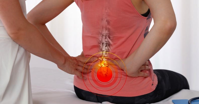
    Problems appear to only seriously arise when the inflammatory trigger is non-infectious inflammation.

    In such cases, excessive tissue fibrosis creates local impairments in biomechanical function.

    This impairment in local biomechanical function affects performance, can generate pain, and accelerate degenerative changes. These impairments can adversely affect the patient for years or even decades.

    Fortunately, abnormal tissue fibrosis can be minimized with early, persistent, controlled motion. Once established, abnormal tissue fibrosis can be improved with the use of a variety of motion applications.

    Cyriax’s text (1) states the following:

    “The excessive reaction of tissues to an injury is conditioned by the overriding needs of a process designed to limit bacterial invasion.

    If there is to be only one pattern of response, it must be suited to the graver of the two possible traumas. However, elaborate preparation for preventing the spread of bacteria is not only pointless after an aseptic injury, but is so excessive as to prove harmful in itself. The principle on which the treatment of post-traumatic inflammation is based is that the reaction of the body to an injury unaccompanied by infection is always too great.” (Cyriax, p.14)

    Cyriax finds support in the sports trauma text authored by physicians Steven Roy and Richard Irvin (4), who state:

    “It is important to realize that the body’s initial reaction to an injury is similar to its reaction to an infection. The reaction is termed inflammation and may manifest macroscopically (such as after an acute injury) or at a microscopic level, with the latter occurring particularly in chronic overuse conditions.” (Roy, p. 125)

    Additional support for these concepts from Cyriax and Roy/Irvin are the writings of physician I. Kelman Cohen and associates (5). In their 1992 text Wound Healing, these authors note:

    “There are two important consequences of being a warm-blooded animal. One is that body fluids make optimal culture media for bacteria. It is to the animal’s advantage, therefore, to heal wounds with alacrity in order to reduce chances of infection.”

    “The prompt development of granulation tissue forecasts the repair of the interrupted dermal tissue to produce a scar.” In addition to providing tensile strength, scars are believed to be a barrier to infectious migration.

    The chronic nature of this scar tissue or fibrosis is expressed in the 1998 article by Thomas Melham and associates (6).

    These authors note that post-traumatic scar tissue can cause pain with activity, pain on palpation, decreased range of motion, and loss of function, and that these problems are resistant to surgery and to conventional physical.

    Excessive scar tissue contributes to chronic soft tissue dysfunction that cause significant disabilities and time lost from work or training activities, and these problems are often difficult to successfully treat. The authors extensively elaborate on the mechanical and neurological adverseness caused by connective tissue fibrosis, noting:

    “Many athletes develop excessive connective tissue fibrosis (scar tissue) or poorly organized scar tissue in and around muscles, tendons, ligaments, joints, and myofascial planes as a result of acute trauma, recurrent microtrauma, immobilization, or as a complication of surgical intervention.”

    “This can lead to soft tissue adhesions, tendonitis, tendonosis, fascial restrictions, and chronic inflammation or dysfunction which in many cases responds poorly to conventional treatments.”

    These authors present an argument that carefully and precisely applied external forces “appear to stimulate connective tissue remodeling through resorption of fibrosis, along with inducing repair and regeneration of collagen secondary to fibroblast recruitment.”

    Pain, Healing, and MOTION…

    As noted above, abnormal tissue fibrosis can be minimized with early, persistent, controlled motion.

    Once established, abnormal tissue fibrosis can be improved with the use of a variety of motion applications.

    Support for the value in using motion to treat soft-tissue injuries has been throughout the literature for decades.

    As an example, Beverly Hills neurosurgeon Emil Seletz, associated with the medical school at the University of California, Los Angeles (UCLA), noted in the Journal of the American Medical Association in 1958, the following, with respects to the management of whiplash soft-tissue injuries (7):

    “During injury, hemorrhage within the capsular ligaments gives rise to swelling of the nerves and eventually adhesions between the dural sleeve and the nerve root; these factors give rise to symptoms that may be prolonged for months or even years after the injury.”

    “In reviewing the types of treatment with a number of specialists in this field, it is found that, while therapy naturally varies to suit the individual need, it consists primarily of local heat in the form of hot wet packs and cervical traction, followed by very gentile massage and manual rotations.”

    “The importance of a carefully planned scheme of treatment must be emphasized to the patient, and treatments must be religiously carried out daily during the first two or three weeks (and then about three times weekly), depending, of course, on the individual case.”

     “Delay or faulty treatment leads to adhesions about the facets and scarring about the capsular ligaments, persistent spasm, congestive lymph edema, and fibrosis of muscles, swelling, and eventual adhesions of nerves within the nerve root canals.”

    “The resultant faulty posture in neglected cases enhances the degeneration of the intervertebral disks, as well as spur formation in the lateral co-vertebral articulations, which on the roentgenogram has come to be known as traumatic arthritis.”

    “I cannot too strongly emphasize the urgency of early and persistent therapy, always by a specialist in this field.”

    “Occasionally, a patient is seen with persistent complaints of head, neck, and shoulder pain, who has had on surgical exposure persistent swelling and adhesions of several nerve roots within the dural sleeve of exit. It is most likely that early, persistent, and adequate therapy by those expertly trained in physical medicine will prevent most patients from developing a surgical condition.”

    On this very same topic, Cyriax’s comments include a review of the 1940 primary research by ML Stearns (8), stating:

    “Her (Stearns) main conclusion on the mechanics of the formation of scar tissue was that external mechanical factors, were responsible for the development of the fibrillary network into orderly layers.

    Within four hours of applying a stimulus, an extensive network of fibrils was already visible around the fibroblasts; during the course of 48 hours this became dense enough to hide the cells almost completely: and in 12 days a heavy layer of fibrils had appeared.

    At first the fibrils developed at random, but later they acquired a definite arrangement, apparently as a direct result of the mechanical factors.

    Of these factors, movement is obviously the most important and equally obvious it is most effective and least likely to cause pain before the fibrils have developed an abnormal firm attachment to neighboring structures.

    When free mobility was encouraged from the onset, the fibers in the scar were arranged lengthwise as in a normal ligament.

    Gentle passive movements do not detach fibrils from their proper formation at the healing breach but prevent their continued adherence at normal sites.

    The fact that the fibrils rapidly spread in all directions provides sufficient reason for beginning movements at the earliest possible moment; otherwise they develop into strong fibrous scars (adhesions) that so often cause prolonged disability after a sprain.” (Cyriax, p. 15)

    Cyriax notes further:

    “When pain is due to bacterial inflammation, Hilton’s advocacy of rest remains unchallenged and is today one of the main principles of medical treatment.

    When, however somatic pain is caused by inflammation due to trauma, his ideas require modification.

    When non-bacterial inflammation attacks the soft tissues that move, treatment by rest has been found to result in chronic disability, later, although the symptoms may temporarily diminish.  

    Hence, during the present century, treatment by rest has given way to therapeutic movement in many soft tissue lesions.

    Movement may be applied in various ways: the three main categories are:
    (a) active and resistive exercises:
    (b) passive, especially forced movement: and
    (c) deep massage.” (Cyriax, p.14)

    “Tension within the granulation tissue lines the cells up along the direction of stress.

    Hence, during the healing of mobile tissues, excessive immobilization is harmful.

    It prevents the formation of a scar strong in the important direction by avoiding the strains leading to due orientation of fibrous tissue and also allows the scar to become unduly adherent, e.g. to bone.” (Cyriax, p.15

    In 1983, sports physicians Steven Roy and Richard Irvin also note (4):

    “The trauma, or initial lesion, leads to an increase of the friction that occurs between moving tissues as well as to a release of chemical mediators, both of which may start the inflammatory process.

    This process may present macroscopically with a number of signs, particularly (a) pain (b) swelling, and (c) redness and warmth. However, microtrauma may not present with any of these signs, particularly during the early stages, even though the inflammation is proceeding at the microscopic level.” (p. 125)

    “The injured tissues next undergo remodeling, which can take up to one year to complete in the case of major tissue disruption.

    The remodeling stage blends with the later part of the regeneration stage, which means that motion of the injured tissues will influence their structure when they are healed.

    This is one reason why it is necessary to consider using controlled motion during the recovery stage.

    If a limb is completely immobilized during the recovery process, the tissues may emerge fully healed but poorly adapted functionally, with little chance for change, particularly if the immobilization has been prolonged.

    Another reason for encouraging controlled motion is that any adhesions that develop will be flexible and will thus allow the tissues to move easily on each other.

    Caution should be observed during the first two weeks, as mentioned previously, as the tensile strength of the tissues may be markedly reduced.” (p.127)

    In 1986, physician John Kellett notes (9):

    Acute inflammation is beneficial when one has acute infection. However, the “acute inflammatory phase of the body’s response to trauma is apparently of no benefit.”

    “The micropathology of acute soft tissue trauma has been investigated. Healing of ligaments and soft tissue injuries in general has been shown to occur by fibrous repair (scar tissue) and not by regeneration of the damaged tissue.”

    “Early mobilization, guided by the pain response, promotes a more rapid return to full activity.”

     “Early mobilization, guided by the pain response, promotes a more rapid return to full functional recovery.”

    “The collagen is remodeled to increase the functional capabilities of the tendon or ligament to withstand the stresses imposed upon it.”

    “It appears that the tensile strength of the collagen is quite specific to the forces imposed on it during the remodeling phase: i.e. the maximum strength will be in the direction of the forces imposed on the ligament.”

    Dr. Kellett summarizes the benefits of early mobilization following soft tissue injury as follows:

    1)   Improvement of bone and ligament strength, reducing recurrence of injury.

    2)   The strength of repaired ligaments is proportional to the mobility of the ligament, resulting in larger diameter collagen fiber bundles and more total collagen.

    3)   “Collagen fiber growth and realignment can be stimulated by early tensile loading of muscle, tendon, and ligament.”

    4)   Collagen formation is not confined to the healing ligaments, but adheres to surrounding tissues. The formation of these adhesions between repairing tissues and adjacent structures is minimized by early movement.

    5)   With motion, “joint proprioception is maintained or develops earlier after injury, and this may be of importance in preventing recurrences of injuries and in hastening full recovery to competitive fitness.”

    6)   The nutrition to the cartilage is better maintained with early mobilization.

    7)   Following this acute inflammatory phase and largely guided by the pain response of the    patient, early mobilization is commenced, based upon the premise that the stress of movement on repairing collagen is largely responsible for the orientation and tensile strength of the tendons and ligaments.

    Dr. Cohen (5) and associates also comment even further on the value of range of motion exercises in the management of soft tissue injury, by stating:

    “During the phase of wound contraction, the active cellular process is locked into position by increasing amounts of rigid collagenous scar. Frequent, gentle exercise can be used to put an extremity joint through a full range of motion and keep the newly developing scar tissue stretched and remodeled. Frequent use of the range of motion exercises is important to keep the developing and contracting scar tissue from becoming a rigid, fixed scar contracture. Range of motion exercises concentrate on remodeling the newly laid collagen before it develops into a rigid scar contracture.” (p. 110)

    In 1994, Halldor Jonsson and associates (10) performed surgical evaluations of 50 patients with chronic whiplash symptoms, showing a “high incidence of discoligamentous injuries in whiplash-type distortions.” The authors noted:

    “The injured spinal segments had become increasingly stiffer over 5 years, which may reflect healing of unrecognized soft tissue injuries.”

    “The most likely source of radicular symptoms is perineural scarring. Therefore, patients with neck distortions after traffic accidents should be mobilized early within the limits of pain to prevent scar transformation of hidden injuries.”

    In 1996, orthopedic surgeon Joseph Buckwalter, MD, from the University of Iowa, adds to the concepts with the following points from an article published in the journal Hand Clinics (11):

    1)   Treatment of tissue injuries with prolonged rest delays recovery and can cause irreversible changes in tissue strength and function.

    2)   Early motion of tissue injuries maintains the structure and composition of normal bone, tendon, ligament, articular cartilage and muscle.

    3)   Immobilization of dense fibrous tissues (tendon, ligament, and joint capsule) causes the tissues to be weaker and stiffer.

    4)   Complete restoration of normal ligament insertion structure and mechanical properties require up to one year of activity, which can mean some patients may require a year of management following these injuries.

    5)   Ageing decreases the adaptive response to repetitive loading, indicating that older patients do not respond as well to the same treatment delivered to younger patients, and that older patients may require more treatment and have a worse prognosis for complete recovery.

    6)   Early motion during the repair and remodeling phases of healing can decrease or prevent adhesions.

    In 1997, US President Bill Clinton tore the tendon of his quadriceps at the attachment to the patella. After surgical repair, President Clinton was put into a passive range of motion device to improve the timing and quality of healing of his injury.

    The device used to treat Mr. Clinton was researched by Canadian orthopedic surgeon Robert Salter. Dr. Salter has published many primary research studies on the physiological effects of passive motion.

    Much of this research is summarized in his 1993 book Continuous passive Motion, A Biological Concept for the Healing and Regeneration of Articular Cartilage, Ligaments, and Tendons; From Origination to Research to Clinical Applications (12).

    It can be reasonably assumed that President Clinton received the best treatment in the world for his injuries. All indications reflect that he enjoyed a speedy and complete recovery.

    Lastly, as presented here, an excellent review on The Concept Of Motion was published in the journal The Physician and Sports Medicine in 2000 by Pekka Kannus, MD, Ph.D. (13).

    Dr. Kannus is chief physician and head of the Accident and Trauma Research Center and sports medicine specialist at the Tampere Research Center of Sports Medicine at the UKK Institute in Tampere, Finland. His article titled “Immobilization or Early Mobilization After an Acute Soft-Tissue Injury?” notes:

    “Experimental and clinical studies demonstrate that early, controlled mobilization is superior to immobilization for primary treatment of acute musculoskeletal soft-tissue injuries and postoperative management.”

    Prolonged inflammation may lead to excessive scarring. Therefore, early, effective treatment seeks to prevent prolonged inflammation and excessive scarring.

    “The current literature on experimental acute soft-tissue injury speaks strongly for the use of early, controlled mobilization rather than immobilization for optimal heating.”

    Experimentally induced ligament tears in animals heal much better with early, controlled mobilization than with immobilization.

    “The superiority of early controlled mobilization has been especially clear in terms of quicker recovery and return to full activity without jeopardizing the subjective or objective long-term outcome.”

     “Controlled experimental and clinical trials have yielded convincing evidence that early, controlled mobilization is superior to immobilization for musculoskeletal soft-tissue injuries. This holds true not only in primary treatment of acute injuries, but also in their postoperative management. The superiority of early controlled mobilization is especially apparent in terms of producing quicker recovery and return to full activity, without jeopardizing the long-term rehabilitative outcome. Therefore, the technique can be recommended as the method of choice for acute soft-tissue injury.”

    Two Additional Supportive Studies…

    Suportive Study #1
    Early Mobilization of Acute Whiplash Injuries (14)

    British Medical Journal
    March 1986

    In this study, 61 whiplash-injured patients were randomized to treatment with either “a period of immobility using a soft collar and simple analgesia before gradual mobilization” (standard treatment), or a alternative treatment involving “daily neck exercises and mobilization.” The authors concluded that:

    “Results showed that eight weeks after the accident the degree of improvement seen in the actively treated group compared with the group given standard treatment was significantly greater for both cervical movement and intensity of pain.”

    “Our results confirmed expectations that initial immobility after whiplash injuries gives rise to prolonged symptoms whereas a more rapid improvement can be achieved by early active management without any consequent increase in discomfort.”

    Supportive Study #2
    Early Intervention in Whiplash-Associated Disorders
    A Comparison of Two Treatment Protocols (15)

    Spine
    July 15, 2000

    This study was designed as a prospective randomized trial in 97 patients with a whiplash injury caused by a motor vehicle collision. Patients were randomly assigned to initial cervical collar or to early active mobilization. The authors concluded:

    “In patients with whiplash-associated disorders caused by a motor vehicle collision, treatment with frequently repeated, active submaximal movements combined with mechanical diagnosis and therapy is more effective in reducing pain than a standard program of initial rest, recommended use of a soft collar, and gradual self-mobilization.”

    “The main finding in this study was that active treatment of whiplash associated disorder resulted in a significantly greater pain reduction than standard [initial immobilization] treatment.”

    “In patients with WAD caused by a motor vehicle collision, early treatment with frequently repeated active submaximal movements combined with mechanical diagnosis and therapy is more effective in reducing pain than treatment with initial rest, recommendation of a soft collar, and a gradual introduction of home exercises.”

    Conclusions

    The discussion and references above support the concept that adverse pathogens cause tissue destruction and subsequent inflammation. The body appears to respond in a manner to wall-off the area of inflammation by over healing the region with a fibrous response.

    The fibrous response appears to be a physical barrier, reducing the ability of the pathogens to spread to other regions of the body, thereby improving the host’s chances for survival.

    However, when inflammation is caused by non-infectious mechanisms, the same fibrotic tissue response occurs. In such cases, without infectious pathogens, the fibrotic tissue response is excessive, resulting in mechanical harm to the host.

    This harmful tissue fibrosis is worsened with early immobilization of the affected tissues.

    This tissue fibrosis is minimized with early persistent controlled mobilization.

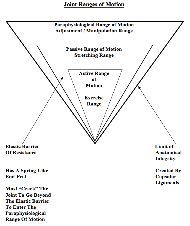
    Established harmful tissue fibrosis is best managed with specific controlled motion for purpose of adhesion rupture and remodeling. The motion to treat established harmful fibrotic tissue should be individualized to the needs of the patient.

    Different fibrotic tissues respond optimally to different categories of controlled motion application:

    1)   Periarticular tissue fibrosis responds optimally to joint adjustments / specific line-of-drive            manipulation.

    2)   Muscle fibrosis responds well to active resistive exercise.

    3)   Non-contractile tissue (tendon, fascia, ligament, etc.) fibrosis responds best to manually applied tissue friction.

    Most patients have a combination of tissues that are adversely affected, depending on the mechanism of injury or stress.

    Consequently, a combination of these applications of controlled motion, by someone who is expertly trained, is often required to achieve timely, efficient, and long-lasting clinical improvements.

    REFERENCES

    1)   Cyriax, James; Textbook of Orthopaedic Medicine, Diagnosis of Soft Tissue Lesions, Bailliere            Tindall, Volume 1, eighth edition, 1982.

    2)   Boyd, William, Pathology, Lea and Febiger, 1952.

    3)   Guyton, Arthur, Textbook of Medical Physiology, Saunders, 1986.

    4)   Roy, Steven; Irvin, Richard; Sports Medicine: Prevention, Evaluation, Management, and        Rehabilitation, Prentice-Hall, 1983.

    5)   Cohen, I. Kelman; Diegelmann, Robert F; Lindbald, William J; Wound Healing, Biochemical &           Clinical Aspects, WB Saunders, 1992.

    6)   Melham TJ, Sevier TL, Malnofski MJ, Wilson JK, Helfst RK, Chronic ankle pain and fibrosis             successfully treated with a new noninvasive augmented soft tissue mobilization technique   (ASTM); Medicine Science Sports Exercise, June 1998; 30(3): 801-4.

    7)   Seletz E, Whiplash Injuries, Neurophysiological Basis for Pain and Methods Used for           Rehabilitation; Journal of the American Medical Association, November 29, 1958, pp. 1750–1755.

    8)   Stearns, ML, Studies on development of connective tissue in transparent chambers in rabbit’s ear;      American Journal of Anatomy, vol. 67, 1940, p. 55.

    9)   Kellett J, Acute soft tissue injuries–a review of the literature; Medicine and Science in Sports and Exercise. Oct. 1986;18(5):489-500.

    10)  Jonsson H, Cesarini K, Sahlstedt B, Rauschning W, Findings and Outcome in Whiplash-Type Neck Distortions; Spine, Vol. 19, No. 24, December 15, 1994, pp 2733-2743.

    11) Buckwalter J, Effects of Early Motion on Healing of Musculoskeletal Tissues, Hand Clinics, Volume 12, Number 1, February 1996.

    12) Salter R, Continuous Passive Motion, A Biological Concept for the Healing and Regeneration of        Articular Cartilage, Ligaments, and Tendons; From Origination to Research to Clinical      Applications, Williams and Wilkins, 1993.

    13) Kannus P, Immobilization or Early Mobilization After an Acute Soft-Tissue Injury?; The Physician And Sports Medicine; March, 2000; Vol. 26 No 3, pp. 55-63.

    14) Mealy K, Brennan H, Fenelon GCC; Early Mobilization of Acute Whiplash Injuries; British Medical Journal, March 8, 1986, 292(6521): 656-657.

    15) Rosenfeld M, Gunnarsson R, Borenstein P, Early Intervention in Whiplash-Associated Disorders, A Comparison of Two Treatment Protocols; Spine, 2000;25:1782-1787.