Category: Advanced

  • Chiropractic Care and the Opioid Crisis

    Chiropractic Care and the Opioid Crisis

    More Evidence that Chiropractic Care Significantly Reduces the Use of Opioid Drugs

    THE OPIATE/OPIOID PROBLEM

    The government of the state of Oregon notes (1):

    Opium is made from the poppy plant.

    Opiates are compounds that can be purified directly from opium without modification. This includes morphine, codeine, methadone, and heroine.

    Few people understand that dextromethorphan is an opiate. It is available in the United States without prescription in products such as NyQuil, Robitussin, TheraFlu, and Vicks. Dextromethorphan is primarily used as a cough suppressant.

    Opioids are a synthetic form of opium that are made in a lab. Drug companies have created more than 500 different opioid molecules, including:

    • OxyContin (oxycodone)
    • Percocet (oxycodone)
    • Vicodin (hydrocodone)
    • Dilaudid    (hydromorphone)
    • Demerol
    • Imodium
    • Darvon
    • Darvocet
    • Opana
    • Fentanyl

    Fentanyl is legally prescribed as Ultiva, Sublimaze, and the Duragesic patch.

    Both opiates and opioids are drugs known as “narcotics.” Narcotic means sleep-inducing or pain suppressing.

    The Centers for Disease Control and Prevention of the United States Government note (2):

    • 75% of the 92,000 drug overdose deaths in 2020 involved narcotics (opiates/opioids).
    • Over 82% of these deaths involved synthetic opioids, primarily fentanyl.

    Side effects of opiates/opioids include insomnia, constipation, “jittery nerves,” and nausea (2). They cause life-threatening side effects such as shallow breathing and slowed heart rate, leading to loss of consciousness and death.

    Opiates/opioids can cause addiction. They can make your brain and body believe the drug is necessary for survival. As you learn to tolerate the dose you’ve been prescribed, you may find that you need even more medication to relieve the pain. “More than 2 million Americans misuse opioids, according to the National Institute on Drug Abuse.”

    According to the American Society of Anesthesiologists, heroin is an illegal and highly addictive form of opiate with no sanctioned medical use (3).

    The Department of Justice of the United States government notes (4):

    • In 2021, 107,622 lives were lost in the United States due to a drug overdose. This is an average of nearly 295 people per day.
    • Drug overdoses are the leading cause of death for Americans ages 18-45; 66% of these overdose deaths were attributable to opioids, primarily by illicit fentanyl.
    • Fentanyl “is the most dangerous drug threat facing our nation.”
    • Fentanyl “is a deadly synthetic opioid that is being mixed into heroin, cocaine, and other street drugs.”

    Cocaine is NOT an opiate. It is a powerfully addictive stimulant drug made from the leaves of the coca plant native to South America.

    Physician Paul Offit, MD, in his 2017 book Pandora’s Lab: Seven Stories of Science Gone Wrong, notes (5):

    • The dangers, addiction, and suffering attributed to the opium poppy have been recognized for 6000 years.
    • Opium was used by the ancient Greeks, including Hippocrates and Galen. The ancient Romans also used opium. The Carthaginian general Hannibal used it to kill himself in 183 B.C.
    • Arab merchants brought opium to China in the seventh century A.D. After Chinese citizens began to smoke opium, up to 90% of the population became addicted to it.
    • Britain and China fought two Opium Wars between 1839 and 1860. China tried to stop the British importation of the drug that was enslaving their country.
    • Americans consumed liquid opium, laudanum, including George Washington, Mary Todd Lincoln, and the wife of lawman Wyatt Earp.
    • The German drug company Bayer began distributing heroin in 1900. It was marketed as being safe for children and pregnant women.
    • In 1986, respected physician and pain specialist, Russell Portenoy, MD, opened the door for doctors to prescribe long-term, high-dose narcotics. He published his paper in the journal Pain (6). His article reported the stories of 38 people who were on high-dose narcotics, including OxyContin, for pain control. He assured physicians that there was little associated addiction and/or death. Dr. Portenoy reasoned that, “opioid maintenance therapy can be safe.” He encouraged physicians to get over their fear of painkillers, what he called “opiophobia.”
    • In late 1995, the FDA approved Purdue Pharma’s timed-released version of OxyContin (time-released oxycodone). OxyContin became one of the most addictive narcotics ever sold, and the leading cause of accidental deaths in the United States.

    Also, in 2017, the United States’ problem with opiates/opioids was quantified in the journal Annals of Internal Medicine in a study titled (7):

    Prescription Opioid Use, Misuse, and Use Disorders in U.S. Adults

    This survey used 51,200 adult subjects. The authors found:

    • 8 million (37.8%) U.S. civilian, non-institutionalized adults used prescription opioids.
    • 5 million adults misused opiate drugs (12.5%).
    • 9 million US adults officially have an opiate use disorder.
    • “More than one third of U.S. civilian, non-institutionalized adults reported prescription opioid use in 2015, with substantial numbers reporting misuse and use disorders.”

    The authors note that the numbers they present are undoubtedly under representative of the opioid problem because they did not include an assessment of groups that are likely to take and to abuse these drugs, including:

    • They did not survey homeless persons who were not living in shelters.
    • They did not survey active-duty military personnel.
    • They did not survey anyone in jail or other institutions.

    An important 2018 study on the topic of opiates/opioids was published in the Journal of the American Medical Association, titled (8):

    Effect of Opioid vs Non-opioid Medications on Pain-Related Function in Patients with Chronic Back Pain or Hip or Knee Osteoarthritis Pain:

    This study was a Randomized Clinical Trial that involved 234 subjects. Eligible patients had moderate to severe chronic back pain or hip or knee osteoarthritis pain despite analgesic use. Chronic Pain was defined as pain nearly every day for 6 months or more. The authors state:

    “Rising rates of opioid overdose deaths have raised questions about prescribing opioids for chronic pain management.”

    “Because of the risk for serious harm without sufficient evidence for benefits, current guidelines discourage opioid prescribing for chronic pain.”

    “Studies have found that treatment with long-term opioid therapy is associated with poor pain outcomes, greater functional impairment, and lower return to work rates.”

    “Treatment with opioids was not superior to treatment with non-opioid medications for improving pain-related function over 12 months. Results do not support initiation of opioid therapy for moderate to severe chronic back pain or hip or knee osteoarthritis pain.”

    This study was reviewed by the Back Letter in an article titled (9):

    Landmark Trial Punctures the Myth That Opioids Provide Powerful Relief of Chronic Pain

    The Back Letter makes the following points:

    “The deadly opioid overtreatment epidemic picked up steam in the late 1980s and early 1990s with the misguided notion that opioids are painkillers that can be used safely and effectively in the long-term treatment of chronic back pain—or other forms of noncancer chronic pain.”

    “The intervening years—and as many as 300,000 deaths in the related opioid overdose epidemic—have rebutted the idea that opioids can be used safely on a mass basis.”

    “Opioids are perceived as strong pain relievers, but our data showed no benefits of opioid therapy over non-opioid medication therapy for pain.”

    “The data do not support opioids’ reputation as ‘powerful painkillers.’”

    “This is an impressive study…. It is the first clinical trial comparing opioid and non-opioid medications with long-term follow-up. It provides strong evidence that opioids should not be the first line of treatment for chronic musculoskeletal pain.”

    “Opioids are not achieving the benefits for which they are marketed. And everyone is now well aware of the adverse effects of opioids.”

    Chiropractic v. Opiates/Opioids

    In 2007, a study was published in the journal Clinical Therapy, titled (10):

    Narcotic Drug Use Among Patients with Lower Back Pain in Employer Health Plans

    The study sample included 13,760 patients with low back pain (LBP) due to mechanical causes. Almost half of the patients with LBP (45%) used narcotic drugs. Pertaining to chiropractic, the authors noted:

    “Patients with LBP who received chiropractic services were less likely to use narcotic drugs. Chiropractic care appears to be a substitute treatment to pain medication and other health care services in patients with LBP due to the different sequence of services for pain treatment.”

    In 2014, a study was published in the journal Neurology, titled (11):

    Opioids for Chronic Non-cancer Pain:
    A Position Paper of the American Academy of Neurology

    The author, Gary Franklin, MD, notes:

    • There is no evidence from clinical trials that opioids could be safely and effectively used in patients with chronic non-cancer pain.
    • Studies show that long-term use of opioid pain drugs causes them to lose their analgesic effect because of drug tolerance or opioid-induced hyperalgesia.
    • The use of opioid drugs on low back-injured workers does not cause meaningful improvement in pain and function.
    • When workers are given opioids for low back injuries, there is a doubling of the development of long-term disability.
    • The author recommends “against use of opioids for mild to moderate pain conditions, such as chronic musculoskeletal conditions, headache, and fibromyalgia.”
    • “In the long run, the use of opioids chronically for most routine conditions, such as chronic low back pain, chronic headaches, or fibromyalgia, will not prove to be worth the risk.”
    • “Cognitive–behavioral therapy, structured exercise, spinal manipulation, and interdisciplinary rehabilitation, although proven to be moderately effective in treating subacute and chronic low back pain, are often either not available or not adequately funded.”
    • “The risks for chronic opioid therapy for some chronic conditions such as headache, fibromyalgia, and chronic low back pain likely outweigh the benefits.”

    In 2018, a study was published in The Journal of Alternative and Complementary Medicine, titled (12)

    Association Between Utilization of Chiropractic Services for Treatment of Low-Back Pain and Use of Prescription Opioids

    The authors used a retrospective cohort design to analyze health insurance claims of 6,868 low back pain subjects from New Hampshire. In 2015, New Hampshire had the second-highest age adjusted rate of drug overdose deaths in the United States, a 31% increase from the previous year and more than double the national rate. The subjects were aged 18–99 years. The authors note:

    “More aggressive pain management efforts that began in the 1990s have led to an epidemic of prescriptions for opioid pain medications in the U.S.”

    “More than 650,000 opioid prescriptions are dispensed per day in the United States.”

    “One out of five patients with non-cancer pain or pain-related diagnoses is prescribed opioids in office-based settings.”

    “There is little evidence that opioids improve chronic pain, function, or quality of life.”

    “Among U.S. adults prescribed opioids, 59% reported having back pain.”

    “Among New Hampshire adults with office visits for non-cancer low-back pain, the adjusted likelihood of filling a prescription for an opioid analgesic was 55% lower for recipients of services provided by doctors of chiropractic compared with non-recipients.” 

    “Pain management services provided by doctors of chiropractic may allow patients to use lower or less frequent doses of opioids, leading to lower costs and reduced risk of adverse effects.”  

    “[Chiropractic care] could exert a positive impact on patients with low-back pain by reducing unnecessary care, lowering costs, and improving safety.”

    “Pain relief resulting from services delivered by doctors of chiropractic may allow patients to use lower or less frequent doses of opioids, leading to reduced risk of adverse effects.”

    Also, in 2018, the journal Pain Medicine published a study titled (13):

    Opioid Use Among Veterans of Recent Wars Receiving Veterans Affairs [VA] Chiropractic Care

    The authors are from Yale School of Medicine, School of Medicine Boston University, and University of Massachusetts Medical School. The VA began providing chiropractic services on-site in 2004 and has expanded implementation each year thereafter. In the VA, chiropractic patients are seen overwhelmingly for low back and/or neck musculoskeletal pain conditions. In private sector populations, increases in chiropractic care is correlated with reduced opioid use.

    A 2016 VA Health Services Research conference “recommended broader uptake of a group of evidence-based nonpharmacological therapies,” including spinal manipulation, massage, acupuncture, exercise, and patient education, which “are the core components of multimodal chiropractic care in the VA.” The authors state:

    “As reduction in opioid use remains a national priority, a better understanding of the relationship between opioid use and chiropractic services is needed to inform research and policy efforts aimed to assess and/or optimize the delivery of chiropractic care in the VA.”

    “Apart from the potential to reduce pain and improve function in patients with musculoskeletal conditions, chiropractic care may have an impact on opioid use in such patients.”

    “Chiropractic care is more likely to be a replacement for, rather than an addition to, opioid therapy for chronic musculoskeletal pain conditions in the VA.”

    “Our results, along with the previous literature, suggest that expanding access to chiropractic care should be a key policy consideration for the VA, congruent with national initiatives aimed to increase the use of evidence-based nonpharmacological treatments for chronic musculoskeletal pain.”

    In 2019, a study was published in the journal BMJ Open titled (14):

    Observational Retrospective Study of the Initial Healthcare Provider for New-onset Low Back Pain with Early and Long-term Opioid Use

    The authors examined the association of initial conservative therapy provider treatment (chiropractors, acupuncturists, physical therapists) on opioid use in a national sample (216,504) of individuals with a new-onset low back pain (LBP). The most frequent initial conservative provider seen was a chiropractor. The authors note:

    “One of the most common conditions for which opioids are prescribed is low back pain (LBP).”

    “Comparisons of the treatment patterns of primary care physicians and conservative therapists (defined as chiropractors, physical therapists, acupuncturists) suggest that the use of conservative therapies for LBP may decrease the likelihood of opioid use.” 

    “For early opioid use, patients initially visiting chiropractors had 90% decreased odds.”

    “Patients who received initial treatment from chiropractors or physical therapists had decreased odds of short-term and long-term opioid use compared with those who received initial treatment from primary care physicians.”

    • Chiropractic: 90% reduction
    • Physical Therapy: 85% reduction

    “Initial visits to chiropractors or physical therapists are associated with substantially decreased early and long-term use of opioids.”

    In 2020, a study was published in the journal Pain Medicine, titled (15):

    Association Between Chiropractic Use and Opioid Receipt Among Patients with Spinal Pain

    The authors are from Yale School of Medicine. This meta-analysis used 6 studies, 5 that focused on back pain and 1 on neck pain: “All six studies (62,624 patients) provided sufficient data and were judged similar enough to be pooled for meta-analysis.” Studies that evaluated spinal manipulation delivered by providers other than chiropractors were excluded. The authors note:

    “Chiropractors predominantly manage spinal conditions, with back conditions being the most common reason to seek chiropractic care.”

    “Chiropractors provide many of the non-pharmacological treatments recommended by clinical practice guidelines for spinal pain, including spinal manipulation, patient education, exercise, acupuncture, and massage.”

    “Ideally, non-pharmacological therapies including multimodal chiropractic care are utilized as frontline treatments to ultimately avoid the prescription of opioids, which are associated with poor outcomes for low back pain and chronic pain.”  

    “The main finding of the review was that all included studies demonstrated a negative association between use of chiropractic care and opioid prescription receipt.”

    “The current study adds to the small but increasing body of evidence demonstrating that access to and utilization of chiropractic services are negatively associated with opioid use.”

    “This review demonstrated an inverse association between chiropractic use and opioid receipt among patients with spinal pain.”  

    “Chiropractic users had 64% lower odds of receiving an opioid prescription than nonusers.”

    Also, in 2020, a study was published in the journal Pain Medicine, titled (16):

    Impact of Chiropractic Care on Use of Prescription Opioids in Patients with Spinal Pain

    The objective of this study was to evaluate the impact of chiropractic utilization upon use of prescription opioids among patients with spinal pain. It involved 101,221 subjects, aged 18-84 years, from Connecticut, Massachusetts, and New Hampshire. Subjects were followed for up to six years. The authors note:

    “Among patients with spinal pain disorders, for recipients of chiropractic care, the risk of filling a prescription for an opioid analgesic over a six-year period was reduced by half, as compared with non-recipients.”

    “Among those who saw a chiropractor within 30 days of being diagnosed with a spinal pain disorder, the reduction in risk was greater as compared with those who visited a chiropractor after the acute phase had passed.”

    “Multiple opportunities for patient–doctor interaction may allow the chiropractor to review clinical progress, advise on home exercise, ergonomics, and other self-management strategies, and provide reassurance, all of which may help improve outcomes and reduce the need for medication.”

    “[There is] accumulating evidence for increased utilization of chiropractic services as an upstream strategy for reducing dependence upon prescription opioid medications.”

    In 2022, a study was published in the Journal of Chiropractic Medicine, titled (17):

    Associations Between Early Chiropractic Care and Physical Therapy on Subsequent Opioid Use Among Persons with Low Back Pain

    The objective of this study was to estimate the association between early use of physical therapy (PT) or chiropractic care and opioid use in individuals with low back pain (LBP). It assessed 40,929 patients with LBP. The authors note:

    “Nearly 80% of opioid users take their pain medication long-term.”

    “Chiropractors have historically focused on manipulation to improve function of the spine to alleviate back pain.”

    “The use of chiropractic care within 30 days of LBP diagnosis was associated with diminished use of opioids in the short term and, in particular, the long term, in which the risk of long-term opioid use was almost cut in half.”

    “Chiropractic care was associated with substantial reduction in likelihood of any opioid use and long-term opioid use [by 44%].”

    Also, in 2022, a study was published in the journal Chiropractic & Manual Therapies, titled (18):

    Association Between Chiropractic Care and Use of Prescription Opioids Among Older Medicare Beneficiaries with Spinal Pain

    This retrospective observational study examined 55,949 Medicare beneficiaries diagnosed with spinal pain, of whom 9,356 were recipients of chiropractic care and 46,593 were non-recipients. The authors note:

    “Opioid analgesics continue to be widely prescribed for spinal pain despite current evidence-based clinical guidelines that identify non-pharmacological therapies as the preferred first-line approach.”

    “Chiropractic care is an alternative to opioid analgesia for spinal pain.”

    “The adjusted risk of filling an opioid prescription within 365 days of first office visit was 56% lower among recipients as compared to nonrecipients.”

    Among early recipients of chiropractic care, the reduction of filling an opioid prescription was 62% lower as compared to non-recipients.

    “Our results suggest that—in addition to lower cost and more efficient utilization of clinical resources—early chiropractic care for spinal pain is also associated with improved patient safety as compared to conventional medical care, at least with regard to use of opioids.”

    “Among older Medicare beneficiaries with spinal pain, use of chiropractic care is associated with significantly lower risk of filling an opioid prescription.”

    SUMMARY

    A bold but logical conclusion from these presented studies is that all patients of all age groups with spine pain syndromes should try chiropractic care prior to using opiate/opioid pain drugs.

    REFERENCES

    1. https://www.oregon.gov/adpc/pages/opiate-opioid.aspx; accessed February 23, 2023.
    2. https://www.cdc.gov/opioids/basics/index.html; accessed February 23, 2023.
    3. https://www.asahq.org/madeforthismoment/pain-management/opioid-treatment/what-are-opioids/; accessed February 23, 2023.
    4. https://www.justice.gov/opioidawareness/opioid-facts; accessed February 23, 2023.
    5. Offit PA; Pandora’s Lab: Seven Stories of Science Gone Wrong; National Geographic; 2017.
    6. Portenoy RK, Foley KM; Chronic use of opioid analgesics in non-malignant pain: report of 38 cases; Pain; May 1986; Vol. 25; No. 2; pp. 171-186.
    7. Han B, Wilson M. Compton WM, Blanco C, Crane E, Lee J, Jones CM; Prescription Opioid Use, Misuse, and Use Disorders in U.S. Adults: 2015 National Survey on Drug Use and Health; Annals of Internal Medicine; September 2017; Vol. 167; No. 5; pp. 293-301.
    8. Krebs EE, Gravely A, Nugent S, Jensen AC, DeRonne B, Goldsmith ES, Kroenke K, Bair MJ, Noorbaloochi S; Effect of Opioid vs Non-opioid Medications on Pain-Related Function in Patients With Chronic Back Pain or Hip or Knee Osteoarthritis Pain: The SPACE Randomized Clinical Trial; Journal of the American Medical Association; March 6, 2018; Vol. 319; No. 9; pp. 872-882.
    9. Back Letter; Landmark Trial Punctures the Myth That Opioids Provide Powerful Relief of Chronic Pain; 32; No. 7; July 2017.
    10. Rhee Y, Taitel MS, Walker DR, Lau DT; Narcotic drug use among patients with lower back pain in employer health plans: A retrospective analysis of risk factors and health care services; Clinical Therapy; 2007; Vol. 29; Supplemental; pp. 2603–2612.
    11. Franklin GM; Opioids for Chronic Non-cancer Pain: A Position Paper of the American Academy of Neurology; Neurology; September 30, 2014; Vol. 83; pp. 1277-1284.
    12. Whedon JM, Toler AWJ, Goehl JM, Kazal LA; Association Between Utilization of Chiropractic Services for Treatment of Low-Back Pain and Use of Prescription Opioids; The Journal of Alternative and Complementary Medicine; June 2018; Vol. 24; No. 4; pp. 552-556.
    13. Lisi AJ, Corcoran KL, DeRycke EC, Bastian LA, Becker WC and 11 more; Opioid Use Among Veterans of Recent Wars Receiving Veterans Affairs Chiropractic Care; Pain Medicine; September 1, 2018; Vol. 19; Supplemental; pp. S54–S60.
    14. Kazis LE, Ameli O, Rothendler J, Garrity B, Cabral H, McDonough C, Carey K, Stein M, Sanghavi D, Elton D, Fritz J, Saper R; Observational Retrospective Study of the Initial Healthcare Provider for New-onset Low Back Pain with Early and Long-term Opioid Use; BMJ Open; September 2019; Vol. 9; No. 9; e028633.
    15. Corcoran KL, Bastian LA, Gunderson CG, Steffens C, Brackett A, Lisi AJ; Association Between Chiropractic Use and Opioid Receipt Among Patients with Spinal Pain: A Systematic Review and Meta-analysis; Pain Medicine; February 1, 2020; Vol. 21; No. 2; pp. e139-e145.
    16. Whedon JM, Toler AWJ, Kazal LA, Bezdjian S, Goehl JM, Greenstein J; Impact of Chiropractic Care on Use of Prescription Opioids in Patients with Spinal Pain; Pain Medicine; December 25, 2020; Vol. 21; No. 12; pp. 3567-3573.
    17. Acharya M, Chopram D, Smith AM, Fritz JM, Martin BC; Associations Between Early Chiropractic Care and Physical Therapy on Subsequent Opioid Use Among Persons with Low Back Pain in Arkansas; Journal of Chiropractic Medicine; June 2022; Vol. 21; pp. 67-76.
    18. Whedon JM, Uptmor S, Toler AJW, Bezdjian S, MacKenzie TA, Kazal LA; Association Between Chiropractic Care and Use of Prescription Opioids Among Older Medicare Beneficiaries with Spinal Pain: A Retrospective Observational Study; Chiropractic & Manual Therapies; January 31, 2022; Vol. 30; No. 1.
  • Lumbar Disc Herniation with Radiculopathy and Chiropractic Manipulation Effectiveness, Safety, Prevention

    Lumbar Disc Herniation with Radiculopathy and Chiropractic Manipulation Effectiveness, Safety, Prevention

    The bulging disc and the protruded disc are the categories most often seen in chiropractic clinical practice. The category and magnitude of disc herniations can only be determined by advanced imaging, especially MRI and/or CT.

    Effectiveness

    There is a long and rich history showing the effectiveness of spinal manipulation for the management of low back and leg pain (radiculopathy), briefly reviewed below:

    ••••

    In 1954, RH Ramsey, MD, published a study titled (1):

    Conservative Treatment of Intervertebral Disk Lesions

    This study appeared in the Instructional Course Lectures of the American Academy of Orthopedic Surgeons. Dr. Ramsey states:

    “The conservative management of lumbar disk lesions should be given careful consideration because no patient should be considered for surgical treatment without first having failed to respond to an adequate program of conservative treatment.”

    “From what is known about the pathology of lumbar disk lesions, it would seem that the ideal form of conservative treatment would theoretically be a manipulative closed reduction of the displaced disk material.”

    “Many forms of manipulation are carried out by orthopaedic surgeons and by cultists and this form of treatment will probably always be a controversial one.”

    “The patient lies on his side on the edge of the table facing the surgeon and the leg that is up is allowed to drop over the side of the table, tending to swing the up-side of the pelvis forward. The arm that is up is allowed to drop back behind the patient, tending to pull the shoulder back. The surgeon then places one hand on the patient’s shoulder and his opposite forearm on the patient’s iliac crest. Simultaneously, the shoulder is thrust suddenly back, rotating the torso in one direction while the iliac crest is thrust down and forward, rotating the pelvis in the opposite direction. This gives the lumbar spine a twist that frequently causes an audible and palpable crunch. This procedure is then repeated with the patient on his other side. The patient is then turned on his back and his hips and knees are hyperflexed sufficiently to forcibly flex the lumbar spine which tends to open up the disk spaces posteriorly.”

    ••••

    In 1969, physicians JA Mathews and DAH Yates from the Department of Physical Medicine, St. Thomas’ Hospital, London, published a study titled (2):

    Reduction of Lumbar Disc Prolapse by Manipulation

    The authors evaluated a number of patients that presented with an acute onset of low back pain and radiculopathy that did not respond to rest. Diagnostic epidurography showed clinically relevant small disc protrusions with antalgia and positive lumbar spine nerve stretch tests. These patients were then treated with long-lever rotation manipulations of the lumbar spine. The manipulations were repeated until abnormal symptoms and signs had disappeared. Following the manipulations there was resolution of signs, symptoms, antalgia, and reduction in the size of the protrusions. Drs. Mathews and Yates state:

    “The frequent accompaniment of acute onset low back pain by spinal deformity suggests a mechanical factor, and the accompanying abnormality of straight-leg raise or femoral stretch test suggests that the lesion impinges on the spinal dura matter of the dural nerve sheaths.”

    “Rotation manipulations apply torsion stress throughout the lumbar spine. If the posterior longitudinal ligament and the annulus fibrosus are intact, some of this torsion force would tend to exert a centripetal force, reducing prolapsed or bulging disc material.” 

    “The results of this study suggest that small disc protrusions were present in patients presenting with lumbago and that the protrusions were diminished in size when their symptoms had been relieved by manipulations.”

    These authors conclude: “it seems likely that the reduction effect [of the disc protrusion] is due to the manipulating thrust used.”

    ••••

    In another study from 1969, BC Edwards compared the effectiveness of heat/massage/exercise to spinal manipulation in the treatment of 184 patients that were grouped according to the presentation of back and leg pain. The study was published in the Australian Journal of Physiotherapy (3). This study was reviewed by Augustus White, MD, and Manohar Panjabi, PhD, in their 1990 book, Clinical Biomechanics of the Spine (4). Drs. White and Panjabi make the following points pertaining to the Edwards article:

    “A well-designed, well executed, and well-analyzed study.”

    In the group with pain radiating into the buttock, “the results were slightly better with manipulation, and again they were achieved with about half as many treatments.”

    In the groups with pain radiation to the knee and/or to the foot, “the manipulation therapy was statistically significantly better,” and in the group with pain radiating to the foot, “the manipulative therapy is significantly better.”

    “This study certainly supports the efficacy of spinal manipulative therapy in comparison with heat, massage, and exercise. The results (80 – 95% satisfactory) are impressive in comparison with any form of therapy.”

    ••••

    In 1977, the third edition of Orthopaedics, Principles and Their Applications was published (5). It contains a section titled:

    Treatment of Intervertebral Disc Herniation With Manipulation

    “Manipulation. Some orthopaedic surgeons practice manipulation in an effort at repositioning the disc. This treatment is regarded as controversial and a form of quackery by many men.  However, the author has attempted the maneuver in patients who did not respond to bed rest and were regarded as candidates for surgery. Occasionally, the results were dramatic.

    Technique. The patient lies on his side on the edge of the table facing the surgeon, and the uppermost leg is allowed to drop forward over the edge of the table, carrying forward that side of the pelvis.  The uppermost arm is placed backward behind the patient, pulling the shoulder back.  The surgeon places one hand on the shoulder and the other on the iliac crest and twists the torso by pushing the shoulder backward and the iliac crest forward.  The maneuver is sudden and forceful and frequently is associated with an audible and palpable crunching sound in the lower back.  When this is felt, the relief of pain is usually immediate.  The maneuver is repeated with the patient on the opposite side.”

    ••••

    In 1987, physicians Paul Pang-Fu Kuo and Zhen-Chao Loh published a study pertaining to lumbar disc protrusions and spinal manipulation, titled (6):

    Treatment of Lumbar Intervertebral Disc Protrusions by Manipulation

    The authors performed a series of eight manipulations on 517 patients with protruded lumbar discs. Their outcomes were quite good, with 84% achieving a successful outcome and only 9% not responding. Only 14% suffered a reoccurrence of symptoms at intervals ranging from two months to twelve years. These authors state:

    “Manipulation of the spine can be effective treatment for lumbar disc protrusions.”

    “Most protruded discs may be manipulated. When the diagnosis is in doubt, gentle force should be used at first as a trial in order to gain the confidence of the patient.”

    “During manipulation a snap may accompany rotation. Subjectively it has dramatic influence on both patient and operator and is thought to be a sign of relief.”

    “Gapping of the disc on bending and rotation may create a condition favorable for the possible reentry of the protruded disc into the intervertebral cavity, or the rotary manipulation may cause the protruded disc to shift away from pressing on the nerve root.”

    ••••

    In 1989, the Journal of Manipulative and Physiological Therapeutics published a case study of a patient with an “enormous central herniation lumbar disc” who underwent a course of side posture manipulation (7). The patient improved considerably with only 2 weeks of treatment. The authors state:

    “It is emphasized that manipulation has been shown to be an effective treatment for some patients with lumbar disc herniation.”

    ••••

    In 1995, chiropractors PJ Stern, Peter Côté, and David Cassidy published a study titled (8):

    A Series of Consecutive Cases of Low Back Pain with Radiating Leg Pain Treated by Chiropractors

    The authors retrospectively reviewed the outcomes of 59 consecutive patients complaining of low back and radiating leg pain, and were clinically diagnosed as having a lumbar spine disk herniation. Ninety percent of these patients reported improvement of their complaint after chiropractic manipulation. They concluded:

    “Based on our results, we postulate that a course of non-operative treatment including manipulation may be effective and safe for the treatment of back and radiating leg pain.”

    ••••

    In 2006, physicians Valter Santilli, MD, Ettore Beghi, MD, and Stefano Finucci, MD, published an article in The Spine Journal titled (9):

    Chiropractic Manipulation in the Treatment of Acute Back Pain and Sciatica with Disc Protrusion

    The purpose of this study was to assess the short- and long-term effects of spinal manipulations on acute back pain and sciatica with disc protrusion. It is a randomized double-blind trial comparing active and simulated manipulations for these patients. The study used 102 patients. The manipulations or simulated manipulations were done 5 days per week by experienced chiropractors for up to a maximum of 20 patient visits, “using a rapid thrust technique.” Re-evaluations were done at 15, 30, 45, 90, and 180 days. The authors found:

    “Active manipulations have more effect than simulated manipulations on pain relief for acute back pain and sciatica with disc protrusion.”

    “At the end of follow-up, a significant difference was present between active and simulated manipulations in the percentage of cases becoming pain-free (local pain 28% vs. 6%; radiating pain 55% vs. 20%).”

    “Patients receiving active manipulations enjoyed significantly greater relief of local and radiating acute LBP, spent fewer days with moderate-to-severe pain, and consumed fewer drugs for the control of pain.”

    ••••

    In 2014, an interdisciplinary group of physicians, chiropractors, and researchers published a study in the Annals of Internal Medicine, titled (10):

    Spinal Manipulation and Home Exercise with Advice for Subacute and Chronic Back-Related Leg Pain

    This study included 192 patients who were suffering from back-related leg pain for at least 4 weeks. Treatment lasted 12 weeks. The authors concluded:

    “For leg pain, spinal manipulative therapy plus home exercise and advice had a clinically important advantage over home exercise and advice (difference, 10 percentage points) at 12 weeks.”

    “Spinal manipulative therapy with home exercise and advice improved self-reported pain and function outcomes more than exercise and advice alone at 12 weeks.”

    “For patients with subacute and chronic back-related leg pain, spinal manipulative therapy in addition to home exercise and advice is a safe and effective conservative treatment approach, resulting in better short-term outcomes than home exercise and advice alone.”

    ••••

    In another 2014 study, a group of multidisciplinary researchers and chiropractic clinicians from Switzerland presented a prospective study involving 148 patients with low back and leg pain. The study was published in the Journal of Manipulative and Physiological Therapeutics and titled (11):

    Outcomes of Acute and Chronic Patients with Magnetic Resonance Imaging–Confirmed Symptomatic Lumbar Disc Herniations Receiving High-Velocity, Low-Amplitude, Spinal Manipulative Therapy:

    The purpose of this study was to document outcomes of patients with confirmed, symptomatic lumbar disc herniations and sciatica that were treated with chiropractic side posture high-velocity, low-amplitude, spinal manipulation to the level of the disc herniation. The authors concluded:

    “The proportion of patients reporting clinically relevant improvement in this current study is surprisingly good, with nearly 70% of patients improved as early as 2 weeks after the start of treatment. By 3 months, this figure was up to 90.5% and then stabilized at 6 months and 1 year.”

    “A large percentage of acute and importantly chronic lumbar disc herniation patients treated with chiropractic spinal manipulation reported clinically relevant improvement.”

    “Even the chronic patients in this study, with the mean duration of their symptoms being over 450 days, reported significant improvement, although this takes slightly longer.”

    “A large percentage of acute and importantly chronic lumbar disc herniation patients treated with high-velocity, low-amplitude side posture spinal manipulative therapy reported clinically relevant ‘improvement’ with no serious adverse events.”

    “Spinal Manipulative therapy is a very safe and cost-effective option for treating symptomatic lumbar disc herniation.”

    ••••

    In 2016, Richard Deyo, MD, MPH, and Sohail Mirza, MD, MPH published a review in The New England Journal of Medicine, titled (12):

    Herniated Lumbar Intervertebral Disk

    Drs. Deyo and Mirza advocate 6 weeks of non-pharmacological conservative care for patients with a herniated lumbar disc, as long as there are no neurological red-flags. They note that some patients will benefit with 12 weeks of conservative care. They clearly indicate that chiropractic spinal manipulation is both safe and usually effective in the management of patients suffering with discogenic radiculopathy, stating:

    A randomized trial of chiropractic manipulation for subacute or chronic back related leg pain “showed that manipulation was more effective than home exercise with respect to pain relief at 12 weeks.”

    “A randomized trial involving patients who had acute sciatica with MRI-confirmed disk protrusion showed that at 6 months, significantly more patients who underwent chiropractic manipulation had an absence of pain than did those who underwent sham manipulations (55% vs. 20%).”

    Safety

    In 1981, a study was published in the journal Spine, titled (13):

    The Relevance of Torsion to the Mechanical Derangement of the Lumbar Spine

    The authors applied rotational stresses to the lumbar spine discs of cadavers. They noted that the limit of lumbar spinal segmental rotation was not created by the disc, but rather by the facet joint. During rotational stress, the compression facet is the first structure to yield at the limit of torsion, and this occurs after about 1-2° of rotation. The authors state:

    “Much greater angles are required to damage the intervertebral disc, so torsion seems unimportant in the etiology of disc degeneration and prolapse.”

    “Because of the protection offered by the compression facet, the intervertebral disc is subjected to relatively small stresses and strains in the physiologic range of torsion. By the time the facets are damaged, the disc is rotated only about one-third to one-tenth of its maximum angle and is bearing a small fraction of the torque required to rupture it.” 

    “Except in cases of extreme trauma and as a sequel to crushing of the apophyseal joints, axial rotation can play no major part in the mechanical derangement of the intervertebral disc in life.”

    ••••

    In 1983, the same group (as #13) published an updated cadaver study in journal Spine, titled (14):

    The Mechanical Function of the Lumbar Apophyseal Joints

    Based upon their experiments, the authors concluded that the facet joints “prevent excessive movement from damaging the discs: the posterior annulus is protected in torsion by the facet surfaces and in flexion by the capsular ligaments.” They note that the facets only allow at most 2° of rotation, and also note that the disc will completely recover from all rotational stresses that are less then 3°. The authors state:

    “In flexion, as in torsion, the apophyseal joints protect the intervertebral disc.”

    “The function of the lumbar apophyseal joints is to allow limited movement between vertebrae and to protect the discs from shear forces, excessive flexion, and axial rotation.”

    ••••

    In 1995, a third updated article was published by this group, appearing in the journal Clinical Biomechanics, titled (15):

    Recent Advances in Lumbar Spinal Mechanics and their Clinical Significance

    Once again, these authors note that rotational loading of the lumbar spinal motor unit will always damage the facet joints “long before the disc.” Despite the supposition that lumbar spinal manipulation, and especially primary rotational manipulation, may injure the intervertebral disc, these cadaver biomechanical studies indicate that such injuries are not biomechanically possible.

    ••••

    In 1993, chiropractor J. David Cassidy, chiropractor Haymo Thiel, and physician (orthopedic surgeon) William Kirkaldy-Willis published a “Review of the Literature” article, titled (16):

    Side Posture Manipulation for Lumbar Intervertebral Disk Herniation

    These authors cite the above studies (13, 14, 15) on human cadavers that show the annulus of the disc is quite resistant to rotational stresses. Specifically, a normal disc did not show failure until 22.6° of rotational stress, and a degenerated disc could withstand an average of 14.3° of rotational stress. They conclude “torsional failure of the lumbar disk first requires fracture of the posterior joints” before there is any annular tearing. These authors state:

    “The treatment of lumbar disk herniation by side posture manipulation is not new and has been advocated by both chiropractors and medical manipulators.”

    “The treatment of lumbar intervertebral disk herniation by side posture manipulation is both safe and effective.”

    ••••

    In 2018, a team of Canadian researchers from multiple universities and health care facilities published a study in the European Spine Journal, titled (17):

    Chiropractic Care and Risk for Acute Lumbar Disc Herniation:
    A Population-based Self-controlled Case Series Study

    The objective was to investigate the association between chiropractic care and acute lumbar disc herniation and contrast this with the association between primary care physician care and acute lumbar disc herniation. This is the first population-based epidemiologic investigation of the association between chiropractic care and acute lumbar disc herniation. The study subjects included the entire population in Ontario’s (CAN) provincial healthcare system over an 11-year period, representing over 100 million person-years of observation. The authors were able to identify all surgically managed cases of acute lumbar disc herniation, visits to chiropractors, and to primary care providers. These authors state:

    “If chiropractic treatment occurs before a lumbar disc herniation progresses to radiculopathy or neurologic deficit and is thus diagnosed, then the [chiropractic] treatment itself can be erroneously blamed for causing the lumbar disc herniation.” 

    “This systematic error—known as protopathic bias—is a type of reverse-causality bias due to processes that occur before a diagnosed or measured outcome event.”

    “Given that deteriorating outcome can initially present as low back pain, it is possible that these patients seek chiropractic care in the prodromal phase of deteriorating outcome, implying that an observed association between chiropractic care and acute deteriorating outcome may not be causal.”

    “Since patients also commonly see primary care physicians for back pain and this healthcare encounter is unlikely to cause disc herniation, an observed association between PCP visits and acute deteriorating outcome could be attributed to care seeking for the initial symptoms of deteriorating outcome (protopathic bias).”

    “The risk for acute lumbar disc herniation with early surgery associated with chiropractic visits was no higher than the risk associated with primary care physician visits.”

    “Our analysis suggests that patients with prodromal back pain from a developing disc herniation likely seek healthcare from both chiropractors and primary care physicians before full clinical expression of acute lumbar disc herniation.”

    “We found no evidence of excess risk for acute lumbar disc herniation with early surgery associated with chiropractic compared with primary medical care.” 

    The analysis “suggested a positive safety profile for chiropractic care relative to the baseline risk represented by primary care physician care.”

    Prevention

    In 2013, a study on more than 3,000 occupational low-back injured patients from the state of Washington was published in the journal Spine (18). These patients were followed prospectively for 3 years. The authors found that, after adjusting for severity of injury and other variables, that having chiropractic care significantly reduced the odds of having a future back surgery. The authors state:

    “In Washington State worker’s compensation, injured workers may choose their medical provider. Even after controlling for injury severity and other measures, workers with an initial visit for the injury to a surgeon had almost nine times the odds of receiving lumbar spine surgery compared to those seeing primary care providers, whereas workers whose first visit was to a chiropractor had significantly lower odds of surgery [by 78%].”

    “Approximately 43% of workers who saw a surgeon had surgery within 3 years, in contrast to only 1.5% of those who saw a chiropractor.”

    ••••

    Very recently (December 2022), a study was published evaluating the odds of patients suffering from lumbar disc herniation and radiculopathy over a period of 1-2 years. The study was published in the BMJ Open, titled (19):

    Association Between Chiropractic Spinal Manipulation and Lumbar Discectomy in Adults with Lumbar Disc Herniation and Radiculopathy

    The study was developed by a multidisciplinary research team. The authors assessed two matched cohorts of 5,785 patients with a mean 37 years. They note that it is common for patients with lumbar disc herniations and radiculopathy to receive chiropractic care or undergo surgery to remove herniated disc material, a procedure called discectomy. Prior studies have found that patients who initiate care for low back pain with a chiropractor have significantly reduced odds of having discectomy.

    In this study, the relative odds for discectomy were significantly reduced in the chiropractic cohort compared with the cohort receiving other care over 1-year (by 69%) and 2-year follow-up (by 77%). This study shows that patients initially receiving chiropractic care for lumbar disc herniation with radiculopathy have reduced odds of discectomy over 1-year and 2-year follow-up.

    ••••

    The discussion and studies presented here indicate:

    • Chiropractic spinal manipulation is very effective for the management of discogenic low back pain and discogenic radiculopathy.
    • Chiropractic spinal manipulation is very safe in the management of discogenic low back pain and discogenic radiculopathy.
    • There is evidence that chiropractic care can reduce the need for discectomy back surgery.

    REFERENCES

    1. Ramsey RH; Conservative Treatment of Intervertebral Disk Lesions; American Academy of Orthopedic Surgeons, Instructional Course Lectures; 1954; Vol. 11; pp. 118-120.
    2. Mathews JA and Yates DAH; Reduction of Lumbar Disc Prolapse by Manipulation; British Medical Journal; September 20, 1969; No. 3; pp. 696-697.
    3. Edwards BC; Low Back Pain and Pain Resulting from Lumbar Spine Conditions: A Comparison of Treatment Results; Australian Journal of Physiotherapy; 1969; Vol. 15; No. 3; pp. 104-110.
    4. White AA, Panjabi MM; Clinical Biomechanics of the Spine; Second edition, JB Lippincott Company; 1990.
    5. Turek S; Orthopaedics, Principles and Their Applications; JB Lippincott Company; 1977; page 1335.
    6. Kuo PP and Loh ZC; Treatment of Lumbar Intervertebral Disc Protrusions by Manipulation; Clinical Orthopedics and Related Research; February 1987; No. 215; pp. 47-55.
    7. Quon JA, Cassidy JD, O’Connor SM, Kirkaldy-Willis WH; Lumbar Intervertebral Disc Herniation: Treatment by Rotational Manipulation; Journal of Manipulative and Physiological Therapeutics; June 1989; Vol. 12; No. 3; pp. 220-227.
    8. Stern PJ, Côté P, Cassidy JD; A Series of Consecutive Cases of Low Back Pain with Radiating Leg Pain Treated by Chiropractors; Journal of Manipulative and Physiological Therapeutics; Jul-Aug 1995; Vol. 18; No. 6; pp. 335-342.
    9. Santilli V, Beghi E, Finucci S; Chiropractic Manipulation in the Treatment of Acute Back Pain and Sciatica with Disc Protrusion: A Randomized Double-blind Clinical Trial of Active and Simulated Spinal Manipulations; The Spine Journal; March-April 2006; Vol. 6; No. 2; pp. 131–137.
    10. Bronfort G, Hondras M, Schulz CA, Evans RL, Long CR, PhD; Grimm R; Spinal Manipulation and Home Exercise with Advice for Subacute and Chronic Back-Related Leg Pain: A Trial with Adaptive Allocation; Annals of Internal Medicine; September 16, 2014; Vol. 161; No. 6; pp. 381-391.
    11. Leemann S, Peterson CK, Schmid C, Anklin B, Humphreys BK; Outcomes of Acute and Chronic Patients with Magnetic Resonance Imaging–Confirmed Symptomatic Lumbar Disc Herniations Receiving High-Velocity, Low Amplitude, Spinal Manipulative Therapy: A Prospective Observational Cohort Study with One-Year Follow-Up; Journal of Manipulative and Physiological Therapeutics; March/April 2014; Vol. 37; No. 3; pp. 155-163.
    12. Deyo R, Mirza S; Herniated Lumbar Intervertebral Disk; New England Journal of Medicine; May 5, 2016; Vol. 374; No. 18; pp. 1763-1772.
    13. Adams MA, Hutton WC; The Relevance of Torsion to the Mechanical Derangement of the Lumbar Spine; Spine; May/June 1981; Vol. 6, No. 3; pp. 241-248.
    14. Adams MA, Hutton WC; The Mechanical Function of the Lumbar Apophyseal Joints; Spine; April 1983; Vol. 8; No. 3; pp. 327-330.
    15. Adams MA, Dolan P; Recent advances in lumbar spinal mechanics and their clinical significance; Clinical Biomechanics; January 1995; Vol. 10; No. 1; pp. 3-19.
    16. Cassidy JD, Thiel HW, Kirkaldy-Willis WH; Side Posture Manipulation for Lumbar Intervertebral Disk Herniation; Journal of Manipulative and Physiological Therapeutics; February 1993; Vol. 16; No. 2; pp. 96-103.
    17. Hincapié CA, Tomlinson GA, Côté P, Rampersaud YR, Jadad AJ, Cassidy JD; Chiropractic Care and Risk for Acute Lumbar Disc Herniation: A Population-based Self-controlled Case Series Study; European Spine Journal; July 2018; Vol. 27; No. 7; pp. 1526–153.
    18. Keeney BJ, Fulton-Kehoe D, Turner JA, Wickizer TM, Chan KCG, Franklin GM; Early Predictors of Lumbar Spine Surgery after Occupational Back Injury: Results from a Prospective Study of Workers in Washington State; Spine; May 15, 2013; Vol. 38; No. 11; pp. 953-964.
    19. Trager RJ, Daniels CJ, Perez JA, Casselberry RM, Dusek JA: Association Between Chiropractic Spinal Manipulation and Lumbar Discectomy in Adults with Lumbar Disc Herniation and Radiculopathy: Retrospective Cohort Study Using United States’ Data; BMJ Open; December 16, 2022; Vol. 12; No. 12; Article e068262.
  • Spine Pain Syndromes: Chiropractic Care vs. Pain Drugs

    Spine Pain Syndromes: Chiropractic Care vs. Pain Drugs

    The “gold standard” for health care research is the randomized controlled trial, or RCT. A lay explanation for a typical RCT study is:

    • Researchers gather a group of individuals that have the same problem.
    • The problem is quantified, often by using standardized measurement outcomes.
    • The group is randomized to be treated differently. The quantification of the problem should be quite close between groups during this randomization.
    • At the end of a pre-determined treatment period, study subjects are reassessed using the standardized measurement outcomes. This allows the researchers to determine the benefit (or lack of benefit) of the various treatment interventions.

    ••••

    In 1990, a study was published in the British Medical Journal, titled (1):

    Low Back Pain of Mechanical Origin:
    Randomized Comparison of Chiropractic
    and Hospital Outpatient Treatment

    This study was a randomized comparison of chiropractic and hospital outpatient treatment in the management of low back pain. It is noteworthy that chiropractic care does not involve pharmacology (pain drugs). This randomized trial involved 741 patients. The patients were followed for a period between 1–3 years. The authors concluded:

    “Chiropractic treatment was more effective than hospital outpatient management, mainly for patients with chronic or severe back pain.”

    “There is, therefore, economic support for use of chiropractic in low back pain, though the obvious clinical improvement in pain and disability attributable to chiropractic treatment is in itself an adequate reason for considering the use of chiropractic.” 

    “Chiropractic was particularly effective in those with fairly intractable pain-that is, those with a history of severe pain.”

    “Patients treated by chiropractors were not only no worse off than those treated in hospital but almost certainly fared considerably better and that they maintained their improvement for at least two years.”

    “The results leave little doubt that chiropractic is more effective than conventional hospital outpatient treatment.”

    As noted above, this study was published in the British Medical Journal. The month after this study was published (June 1990), the medical journal Lancet published an editorial pertaining to the study, stating the following (2):

    “Chiropractors and Low Back Pain”

    [The study (1)] “showed a strong and clear advantage for patients with chiropractic.”

    The advantage for chiropractic over conventional hospital treatment was “not a trivial amount” and “reflects the difference between having mild pain, the ability to lift heavy weights without extra pain, and the ability to sit for more than one hour, compared with moderate pain, the ability to lift heavy weights only if they are conveniently positioned, and being unable to sit for more than 30 minutes.”

    “This highly significant difference occurred not only at 6 weeks, but also for 1, 2, and even (in 113 patients followed so far) 3 years after treatment.”

    “Surprisingly, the difference was seen most strongly in patients with chronic symptoms.”

    “Chiropractic treatment should be taken seriously by conventional medicine, which means both doctors and physiotherapists.”

    The authors of the British Medical Journal study (1) note that if all back-pain patients without manipulation contraindications were referred for chiropractic instead of hospital treatment, there would be significant annual treatment cost reductions, a significant reduction in sickness days, and a significant savings in social security payments.

    The authors of this study (1) wanted a longer-term follow-up on their patient population. This later study was completed and published in the British Medical Journal in 1995, and titled (3):

    Randomised Comparison of Chiropractic
    and Hospital Outpatient
    Management for Low Back Pain:
    Results from Extended Follow Up

    The same authors, from the Medical Research Council Epidemiology and Medical Care Unit, Wolfson Institute of Preventive Medicine, Medical College of St. Bartholomew’s Hospital, London, reminded the readers that their original study involved 741 men and women aged 18-64 years with low back pain, and that the primary measurement outcome used was the change in the total Oswestry Low Back Questionnaire score. Like the original 1990 study, this study is also a randomized comparison design.

    The objective of this study was to compare the effectiveness over three years of chiropractic and hospital outpatient management for low back pain. As noted above, the authors remind the readers that their 1990 study “reported greater improvement in patientswith low back pain treated by chiropractic compared with those receiving hospital outpatient management.” In this 3-year follow-up study, the authors asked the study subjects whether they thought their allocated treatment had helped their back pain. The authors found:

    “According to total Oswestry scores improvement in all patients at three years was about 29% more in those treated by chiropractors than in those treated by the hospitals.”

    “The beneficial effect of chiropractic on pain was particularly clear.”

    “[More subjects] rated chiropractic helpful at three years than hospitalmanagement.”

    “[Improvements] were all significantly greater in those treated by chiropractic, including the changes early on-that is, at six weeks and six months.”

    “At three years the results confirm the findings of an earlier report that when chiropractic or hospital therapists treat patients with low back pain as they would in day to day practice those treated by chiropractic derive more benefit and longterm satisfaction than those treated by hospitals.”

    “The substantial benefit of chiropractic on intensity of pain is evident early on and then persists.”

    “The results of our trial show that chiropractic has a valuable part to play in the management of low back pain.”

    “Chiropractic seems to be more effective than hospital management, possibly because more treatments are spread over longer time periods.”

    The Oswestry scores in this follow-up study found more than pain improvement with chiropractic care. Also significantly improved were:

    • The ability to sit for more than a shorttime
    • Sleeping ability
    • Personal care
    • Lifting
    • Walking
    • Standing
    • Sex life
    • Social life
    • Travelling

    An important finding from the 1990 study that was confirmed in this 1995 study is that the longer and more serious the low back pain, the more likely the patient was to benefit from chiropractic care.

    ••••

    In 2002, a study was published in the Annals of Internal Medicine, titled (4):

    Manual Therapy, Physical Therapy,
    or Continued Care by a General Practitioner
    for Patients with Neck Pain:
    A Randomized Controlled Trial

    The authors defined “manipulation” as:

    “Orthopedic manipulative (manual) therapy is a specialization within physical therapy and provides comprehensive conservative management for pain and other symptoms of neuro-musculo-articular dysfunction in the spine and extremities.”

    The authors compared the effectiveness of manual therapy, physical therapy, and pharmacology care provided by a general practitioner physician for the treatment of neck pain. This study is a randomized controlled trial (RCT) design, involving 183 patients. The authors concluded:

    “Manual therapy scored consistently better than the other two interventions on most outcome measures.”

    “In daily practice, manual therapy is a favorable treatment option for patients with neck pain compared with physical therapy or continued [drug] care by a general practitioner.”

    “The success rates for manual therapy were statistically significantly higher than those for physical therapy.”

    “Manual therapy scored better than physical therapy on all outcome measures…”      

    “Range of motion improved more markedly for those who received manual therapy or physical therapy than for those who received continued [physician drug] care.”

    “The postulated objective of manual therapy in the restoration of normal joint motion, was achieved, as indicated by the relatively large increase in the range of motion of the cervical spine.”

    “Patients receiving manual therapy had fewer absences from work than patients receiving physical therapy or continued [physician drug] care.”

    “Manual therapy seems to be a favorable treatment option for patients with neck pain.”

    “Primary care physicians should consider manual therapy when treating patients with neck pain.”

    In this study, the primary authors are physical therapists, and they found that manual manipulative therapy was superior to traditional physical therapy in the management of neck pain.

    ••••

    In 2003, a study published in the journal Spine, titled (5):

    Chronic Spinal Pain:
    A Randomized Clinical Trial Comparing
    Medication, Acupuncture, and Spinal Manipulation

    Important for this discussion is acknowledging that this study is a randomized controlled clinical trial design. The initial phase of the study was of nine weeks duration. The second phase was a reassessment at a one-year follow-up.

    The authors randomized 115 patients suffering from chronic neck/back pain to three groups:

    • Daily prescription Nonsteroidal Anti-inflammatory Drugs (NSAIDs) prescribed by a medical doctor.
    • Needle Acupuncture by a certified needle acupuncturist, two patient visits per week.
    • Spinal manipulation by a licensed chiropractor. The manipulations were high-velocity, low-amplitude thrust spinal manipulation to a joint with reduced mobility. Two treatments per week were given.

    The measurement outcomes used included the Oswestry Back Pain Disability Index (Oswestry), the Neck Disability Index (NDI), the Short-Form-36 Health Survey questionnaire (SF-36), visual analog scales (VAS) of pain intensity, and ranges of spinal movement. Subjects were assessed initially and then again at 2, 5, and 9 weeks after the beginning of treatment.

    The authors note that a pathologic cause cannot be identified for most episodes of spinal pain, and clinicians often have great difficulty establishing the underlying cause. They state that only about 15% of patients receive a definitive diagnosis for their spinal pain; it is often impossible to reach a specific diagnosis.

    The authors discuss that the proportion of primary care patients with uncomplicated spinal pain who have poor outcomes appears to be higher than generally recognized by either patients and physicians. Yet, physicians are highly likely to recommend the use of Nonsteroidal Anti-inflammatory Drugs (NSAIDs). The authors state:

    “Adverse reactions to nonsteroidal anti-inflammatory (NSAID) medication have been well documented.”

    “Gastrointestinal toxicity induced by NSAIDs is one of the most common serious adverse drug events in the industrialized world.”

    Chiropractic care was included in this study because “numerous studies have shown that patients with low back pain exhibit abnormal spinal motion.” The authors suspected that chiropractic manipulation to joints with a reduced range of motion would be superior to acupuncture and drug care.

    In the randomization of the subjects in this study, the average duration of spinal pain symptoms was 8.3 years for the chiropractic spinal manipulation group, 6.4 years for the medication group, and 4.5 years for the acupuncture group. Clearly, the chiropractic patient group was statistically much more chronic than the other treatment groups.

    The outcomes for this study showed that the highest proportion of early (asymptomatic status) recovery was found for chiropractic spinal manipulation (27.3%), followed by acupuncture (9.4%), and lastly by medication (5%). The chiropractic spinal manipulation achieved the best overall results, with improvements of 50% on the Oswestry scale, 38% on the NDI, 47% on the SF-36, and 50% on the VAS for back pain, 38% for lumbar standing flexion, 20% for lumbar sitting flexion, 25% for cervical sitting flexion, and 18% for cervical sitting extension.

    The authors note:

    “The consistency of the results provides evidence that in patients with chronic spinal pain, [chiropractic spinal] manipulation, if not contraindicated, results in greater short-      term improvement than acupuncture or medication.”

    “The highest proportion of asymptomatic patients before or at the week 9 assessment was found in the [chiropractic spinal] manipulation group (27.3%) followed by the acupuncture group (9.4%) and the medication group (5%).”

    “Manipulation yielded the best results over all the main outcome measures.”

    “The results of this efficacy study suggest that [chiropractic] spinal manipulation, if not contraindicated, may be superior to needle acupuncture or medication for the successful treatment of patients with chronic spinal pain syndrome.”

    “In summary, the significance of the study is that for chronic spinal pain syndromes, it appears that [chiropractic] spinal manipulation provided the best overall short-term results, despite the fact that the spinal manipulation group had experienced the longest pretreatment duration of pain.”

    The authors noted that the NSAIDs used in this study did not achieve a marked improvement (only 5%) in chronic spinal pain, and they caused adverse reactions in 6.1% of the patients. More subjects suffered an adverse reaction to the drugs (6.1%) than were benefited from the drugs (5%). The authors state that there is insufficient evidence for the use of NSAIDs to manage chronic low back pain.

    Even though the chiropractic treatment group was the most chronic (8.3 years), 27.3% recovered with 18 spinal adjustments over a period of 9 weeks, or less. This means that better than every fourth patient became asymptomatic with 9 weeks or less of chiropractic spinal manipulation, even though they had been chronic for more than 8 years.

    The chiropractic spinal manipulation group showed significantly greater improvement in subjective complaints, functional abilities, objective range of spinal motion, and in general health status than acupuncture and medication groups. The chiropractic patients recorded no adverse events to treatment.

    In the treatment of chronic spinal pain, chiropractic manipulation is superior to acupuncture and medication.

    As noted, this study was a nine week-duration clinical trial. The authors wanted to reassess patient clinical status, using the same measurement outcomes, more than a year later. The results were published in 2005 in the Journal of Manipulative and Physiological Therapeutics, titled (6):

    Long-Term Follow-up
    of a Randomized Clinical Trial
    Assessing the Efficacy of Medication,
    Acupuncture, and Spinal Manipulation
    for Chronic Mechanical Spinal Pain Syndromes

    The authors restate that chronic spinal pain is often triggered by injury or disease. Chronic spinal pain has an immense impact on public health, poses an enormous financial strain on the health systems in developed countries, and affects the economy by lost working time through illness.

    They note that chronic spinal pain is recurrent in nature for many patients, is a main cause of absence from work, and it has a high incidence in society. The patients in this study had chronic spinal pain (average duration of more than 2 years) and had long histories of having sought pain relief. The authors note:

    “The overall results of this extended follow-up efficacy study appear to favor the application of manipulation [which successfully achieves long-term benefits in chronic spinal pain syndrome patients].”

    “Statistical testing revealed that only in the manipulation group, 5 of the 7 observed improvements were statistically significant which compares with only 1 item in each of the acupuncture and the medication groups, respectively.”

    “In patients with chronic spinal pain syndromes, [chiropractic] spinal manipulation, may be the only treatment modality of the assessed regimens that provides broad and significant long-term benefit.”

    “Spinal manipulation appeared to provide the highest satisfaction.”

     “Patients who have chronic mechanical spinal pain syndromes and received spinal manipulation gained significant broad-based beneficial short-term and long-term outcomes.”

    The authors were particularly negative on the results offered by medications for the management of chronic spinal pain, noting that “medication did not achieve an improvement in chronic spinal pain.”

    ••••

    “Dogma” is a belief or set of beliefs that is widely accepted by the members of a group without being questioned or doubted. Dogma is often associated with religions. However, dogma is also pervasive in science. In his 2022 book Transformer, professor of evolutionary biochemistry at University College London, Nick Lane, notes that dogmas can hold scientific progress back decades.

    An approach to a medical problem that for decades had been established and accepted, without question or doubt as the absolute best management to a medical problem, may be turned on its head with advancing investigations and evidence. Such an affront to established medical dogma will initially be met with great resistance, and acceptance may take a long time.

    Biochemically, it is known that there is a link between inflammation and pain. Inflammation alters the threshold of the pain nerves, bringing more pain signals to the brain. Hence, anti-inflammatory efforts will help reduce pain. When it was learned that the chemistry of pain could be reduced by giving the patient nonsteroidal or steroid anti-inflammatory drugs, the pharmacological management of pain quickly became patented, and then marketed to doctors, medical associations, insurance companies, government guidelines, government insurance programs, and to the public. In fact, nonsteroidal anti-inflammatory drugs (NSAIDs) for pain were awarded the 1982 Nobel Prize in Medicine (8).

    The routine uses of anti-inflammatory drugs to treat spinal pain makes biochemical sense, and they have been proven to be helpful for patients. However, there are problems that are under emphasized or completely ignored:

    • Anti-inflammatory drugs do not work well as compared to nonpharmacological alternatives such as chiropractic, acupuncture, exercise, etc. (9).
    • Anti-inflammatory drugs are associated with numerous and often serious side effects (5, 6).
    • Using anti-inflammatory drugs for acute low back pain may predispose the patient to becoming a chronic low back pain sufferer. The irony of this statement and its challenge to orthodoxy has the potential to forever shake up the management of acute low back pain. The science behind this is reviewed below.

    In May 2022, a unique and important article was published pertaining to the use of anti-inflammatory drugs (NSAIDs or steroids) and the future incidence of chronic low back pain. It was published in the journal Science Translational Medicine, titled (10): 

    Acute Inflammatory Response Via Neutrophil Activation
    Protects Against the Development of Chronic Pain

    The authors note that chronic pain inflicts huge societal costs in terms of management, loss of work productivity, and effects on quality of life. They also note that chronic low back pain is the most frequently reported chronic pain condition, stating:

    “LBP ranks the highest of all chronic conditions in terms of years lived with disability, with its prevalence and burden increasing with age.”

    The authors note that the transition from acute to chronic pain is critically important but poorly understood. Therefore, they investigated the pathophysiological mechanisms underlying the transition from acute to chronic low back pain (LBP). The study involved an assessment of 98 LBP subjects. Clinicians followed the standardized protocol for treating patients with acuteLBP with NSAIDs or systemic steroidal drugs to reduce the acute inflammatory response.

    Statistically, the results were shocking. Taking anti-inflammatory drugs significantly increased the odds for transitioning from acute to chronic low back pain. The authors explain their findings by noting that anti-inflammatory drugs inhibit white blood immune system cells (neutrophils) from working properly in the long-term, resulting in chronic low back pain. They state:

    “Current treatments for LBP often target the immune system and include nonsteroidal anti-inflammatory drugs (NSAIDs), acetaminophen, and corticosteroids, although all of these drug classes are minimally effective at best.”

    “Early treatment with a steroid or nonsteroidal anti-  inflammatory drug (NSAID) led to prolonged pain despite being analgesic in the short term.”

    “The management of acute inflammation may be counterproductive for long-term outcomes of LBP sufferers.”

    “Drugs that inhibit inflammation might interfere with the natural recovery process, thus increasing the odds for chronic pain.”

    “Results indicate the importance of the up-regulation of the inflammatory response at the acute stage of musculoskeletal pain as a protective mechanism against the development of chronic pain.”

    “The beginning of the inflammatory process programs its resolution, and it is thus the failure to initiate an appropriate inflammatory response that may lead to chronic pain.”

    The acute treatment of inflammation with either a steroid or a NSAID, “although both effectively reducing pain behavior during their administration—greatly prolonged the resolution of neuropathic, myofascial, and especially inflammatory pain states.”

    “Individuals with acute back pain were at greater risk [76%] of developing chronic back pain if they reported NSAID usage than if they were not taking NSAIDs, adjusting for age, sex, [and] ethnicity.”

    “Our conclusions may have a substantial impact on medical treatment of the most common presenting complaint to healthcare professionals.”

    SUMMARY AND CONCLUSIONS

    Pharmacology for pain management has many concerns. They are actually not very effective. They are associated with multiple serious side-effects in a linear fashion (the more one takes the greater the risks of suffering side-effects). The scariest recent discovery is that taking anti-inflammatory drugs for acute back pain may significantly increase the risks of becoming a chronic back pain sufferer.

    In contrast, chiropractic care for back pain is highly effective, has high levels of patient satisfaction, routinely outperforms pain drugs in randomized clinical trials, and carries none of the risks associated with pain drugs.

    REFERENCES

    1. Meade TW, Dyer S, Browne W, Townsend J, Frank OA; Low Back Pain of Mechanical Origin: Randomized Comparison of Chiropractic and Hospital Outpatient Treatment; British Medical Journal; June 2, 1990; Vol. 300; pp. 1431-1437.
    2. Editorial; Chiropractors and Low Back Pain; Lancet; July 28, 1990; p. 220.
    3. Meade TW, Dyer s, Browne W, Frank AO;Randomised Comparison of Chiropractic and Hospital OutpatientManagement for Low Back Pain: Results from Extended Follow Up; British Medical Journal;August 5, 1995; Vol. 311; pp. 349-51.
    4. Hoving JC, Koes BW, de Vet HCW, van der Windt DAWM, Assendelft WJJ, Mameren H, Devillé WLJM; Manual Therapy, Physical Therapy, or Continued Care by a General Practitioner for Patients with Neck Pain; A Randomized Controlled Trial; Annals of Internal Medicine; May 21, 2002; Vol. 136; No. 10; pp. 713-722.
    5. Giles LGF; Muller R; Chronic Spinal Pain: A Randomized Clinical Trial Comparing Medication, Acupuncture, and Spinal Manipulation; Spine; July 15, 2003; Vol. 28; No. 14; pp. 1490-1502.
    6. Muller R, Giles LGF; Long-Term Follow-up of a Randomized Clinical Trial Assessing the Efficacy of Medication, Acupuncture, and Spinal Manipulation for Chronic Mechanical Spinal Pain Syndromes; Journal of Manipulative and Physiological Therapeutics; January 2005; Vol. 28; No. 1; pp. 3-11.
    7. Lane N; Transformer: The Deep Chemistry of Life and Death; WW Norton & Company; 2022.
    8. https://www.nobelprize.org/prizes/medicine/1982/summary/ (accessed January 5, 2023).
    9. Foreman J; A Nation in Pain, Healing Our Biggest Health Problem; Oxford University Press; 2014.
    10. Parisien M, Lima LV, Dagostino C, El-Hachem N, and 16 more; Acute Inflammatory Response Via Neutrophil Activation Protects Against the Development of Chronic Pain;Science Translational Medicine; May 11, 2022; Vol. 14; Article eabj9954.
  • Osteoarthritis and Chiropractic Care

    Osteoarthritis and Chiropractic Care

    Evolving Models

     A large recent review of the chiropractic profession confirms that chiropractors specialize in the management of back and neck pain (1). The study, from the National Health Interview Survey, which is the principal and reliable source of comprehensive health care information in the United States, found that 63% of chiropractic patients presented with low back pain and 30% presented with neck pain. Overall, 93% of chiropractic patients presented with spinal pain complaints (1).

    When assessing spinal pain complaints, it is commonplace for chiropractors to take x-rays. Spinal x-rays serve many purposes. The primary purposes for taking spinal x-rays were summarized in 2020 in the journal Quantitative Imaging in Medicine and Surgery. The study was titled (2):

    The Role of Radiography in the Study of Spinal Disorders

    The authors state:

    “Radiography plays an important role in many conditions affecting the spine.”

    The authors advocate spinal x-rays for the assessment of these potential problems:

    • Traumatic Fracture
    • Congenital Abnormalities
    • Osteoporotic Vertebral Fracture
    • Degenerative Spinal Pathology
    • Spinal Instability
    • Spondylolisthesis
    • Alterations in Spinal Curvature
    • Inflammatory Pathology
    • Infectious Pathology
    • Neoplastic Pathology

    The authors note that the most frequent origin of spinal pain is classified as “non-specific” (approximately 70%) because the specific nociceptive source cannot be proven. The second most prevalent cause of spinal pain is mechanical. Mechanical spinal pain includes degenerative disorders of the spine, alignment abnormalities, and vertebral fractures. X-rays are very valuable in diagnosing all of these mechanical causes of back pain.

    Spinal degenerative pathology is also known as:

    • Arthritis
    • Osteoarthritis
    • Degenerative Arthritis
    • Degenerative Joint Disease (DJD)
    • Spondylosis, primarily applied to the spinal facet joints
    • Spondyloarthritis, again primarily applied to the spinal facet joints
    • Degenerative Disc Disease (DDD)

    Degenerative spinal pathology can affect any and all of the spinal joints. Degenerative spinal pathology is extremely common in adults, and its incidence increases with age (3).

    Spinal x-rays are one of the most frequently employed modalities used in the study of spinal pain. Publications advocate taking x-rays on patients for these reasons (2):

    • When the patient presents with a history of trauma that may result in fracture or instability.
    • When there is suspicion of vertebral compression fracture secondary to osteoporosis or from a history of steroid use.
    • When there is a history of congenital pathologies, including hyperkyphosis, hyperlordosis, scoliosis, kyphoscoliosis, hemi- vertebrae, block vertebrae, Klippel-Feil syndrome, sacralization, lumbarization, facetal tropism, etc.
    • When there is a history or suspicion of spondylolisthesis, Scheuermann’s disease, and/or Schmorl nodes.

    History records that the chiropractic profession pioneered the use of x-rays on the spine in 1910 (4, 5). At the time, spinal x-rays were referred to as “spinographs.”

    Today, spinal x-rays are at the center of much of chiropractic education and clinical practice. Chiropractic educational curriculum has between 6-10 courses that are entirely dedicated to the taking and interpreting of x-rays. Additionally, x-rays are woven into the majority of clinical science and adjustive technique courses.

    Leading scientific/medical journals have confirmed the competency of chiropractors in reading/interpreting spinal x-rays (6, 7, 8).

    A study published in 1997 in the Journal of Manipulative and Physiological Therapeutics detailed the use of x-rays by chiropractors (9):

    • 74% of chiropractors have x-ray facilities in their offices.
    • 71% use x-rays to screen for contraindications to chiropractic care.
    • 63% use x-rays to assess existence of pathological conditions.
    • 51% use x-rays to observe/measure altered biomechanics and posture.
    • 84% of the chiropractors refer to medical radiologists and/or to chiropractic radiologists for a formal interpretation of their radiographs.

    A valid concern pertaining to x-ray exposure is the ionization issue and the cost to patients and to the health-care system. These concerns were addressed in 2017 in the journal Mayo Clinic Proceedings: Innovations, Quality & Outcomes (10). The authors note that chiropractors frequently use spinal x-rays. They also speculate that the increased use of x-ray imaging may be responsible for the reduction of the use of advanced imaging, such as MRI and CT, resulting in a net benefit for both the patient and the reimbursing parties, saving the system money, time, and personnel resources.

    In chiropractic clinical practice, ablative pathology (infection, malignancy, tumor, etc.) is quite rare. In contrast, degenerative arthritic changes are very common. An important article pertaining to degenerative arthritis was published in 2006 by Susan Garstag, MD, and Todd Stitik, MD (11).

    These authors note that osteoarthritis is the most prevalent form of arthritis and a major cause of disability in people aged 65 and older. Osteoarthritis affects the majority of adults over age 55, and that 58% of those older than 70 years have symptomatic osteoarthritis.

    These authors categorize joint degenerative changes to local factors and systemic factors:

    Local Factors include:

      • Altered joint biomechanics
      • Malalignment
      • Muscle weakness
      • Impaired proprioception neurology

    Systemic Factors include:

      • Age
      • Genetic Factors
      • Nutritional Factors, especially inflammatory profile and free radical burden (generators of oxidative stress)

    Drs. Garstag’s and Stitik’s comments linking osteoarthritis to malalignment, muscle weakness, impaired proprioception, and altered joint biomechanics are particularly interesting to chiropractors, as these are all components of what chiropractors call the subluxation.

    Traditional chiropractic teaches that untreated joint subluxations lead to osteoarthritic changes.

    There is no doubt that spinal degenerative changes can cause spinal pain syndromes. Yet, there is also no doubt that spinal degenerative changes can exist in subjects that have no pain whatsoever (3, 11, 12, 13, 14, 15, 16).

    Less controversial is the contention that asymptomatic spinal degenerative changes are a weakness that increases the vulnerability of the afflicted articulations to injury and pain when subjected to increased stress, prolonged stress, or injury:

    • In 1964, whiplash injury expert and pioneer, Ruth Jackson, MD, published an article titled “The Positive Findings in Neck Injuries” in the American Journal of Orthopedics. Dr. Jackson’s conclusions in this article were based on her evaluation of 5,000 injured patients. She notes (17): 

    “[Pre-existing pathological conditions of the cervical spine, when injured], “result in more damage than would be anticipated in a so-called ‘normal’ cervical spine.”

    • In 1977, Samuel Turek, MD, clinical professor from the Department of Orthopedics and Rehabilitation at the University of Miami School of Medicine, and author of the reference text, Orthopaedic Principles and their Applications, states (18):

    “The injury may be compounded by the presence of degenerative disease of the spine.”

     “With advancing age, especially in the presence of degenerative disease, the tissues become inelastic and are easily torn.”

    • In 1981, Rene Cailliet, MD, professor and rehabilitation specialist from the University of Southern California, and author of the book Neck and Arm Pain, states (19):

    “The pre-existence of degeneration may have been quiescent in that no symptoms were noted, but now minor trauma may ‘decompensate’ the safety margin and symptoms occur.”

    • In 1983, Norris and Watt followed 61 whiplash-injured patients for a minimum of six months in order to establish factors that were prognostic for recovery. They published their findings in the British Journal of Bone and Joint Surgery, titled “The Prognosis of Neck Injuries Resulting from Rear-End Vehicle Collisions.” Their conclusions include (20):

    “Factors which adversely affect prognosis include the presence of objective neurological signs, stiffness of the neck, [loss of cervical lordosis], and pre-existing degenerative spondylosis.”

    “Pre-existing degenerative changes in the cervical spine, no matter how slight, do appear to affect the prognosis adversely.”

    • In 1985, Webb, in his article titled “Mechanisms and Patterns of Tissue Injury,” notes (21):

    “Degenerative joint disease is recognized as a major influence on subsequent tissue damage both in severity and pattern.”

    “In any individual where changes consistent with degenerative joint disease are present, one can expect the injury to be more severe or a very minor injury to produce severe symptoms requiring prolonged treatment.”

    • In 1986, Arthur Ameis, MD, from the Faculty of Medicine at the University of Toronto, notes (22):

    “For the elderly, neck injury can be very serious.  The degenerative spine is biomechanically ‘stiffer’, behaving more like a single long bone than like a set of articulating structures.  Deforming forces are less evenly dissipated, and more damage is done.”

    • In 1987, physicians Edward Dunn and Steven Blazar authored “Soft-Tissue Injuries of the Lower Cervical Spine” for the American Academy of Orthopedic Surgeons. In this publication they note (23):

    “If present, degenerative changes should be duly noted as they may affect the prognosis.” 

    “…pre-existing degenerative changes adversely affected the outcome.”

    • In 1988, Mairmaris and colleagues published a study titled “Whiplash Injuries of the Neck.” They reviewed 102 whiplash-injured patients 2 years after injury. They concluded (24):

    “The analysis of the radiological results showed that pre-existing degenerative changes in the cervical spine are strongly indicative of a poor prognosis.”

    • In 1988, physician Hirsch and colleagues published a paper titled “Whiplash Syndrome, Fact or Fiction?” in Orthopedic Clinics of North America. These authors note (25):

    “[Pre-existing structural changes and degenerative changes are] frequently associated with a more difficult, more prolonged, and less complete recovery.”

    “These changes may include the presence of osteophytes, foraminal encroachment on the oblique projections, and the presence of intervertebral disc space narrowing.” 

    “When hyperextension injury occurs in the presence of pre-existing osteophyte formation, there is further narrowing of the spinal canal, which increases the potential for injury to the nerve roots or cord.”

    • In their 1988 reference text on whiplash injuries titled Whiplash Injuries, The Acceleration/Deceleration Syndrome, Steve Foreman and Arthur Croft note (26):

    “…the presence of preexisting degenerative changes, no matter how slight, appears to alter the prognosis adversely.”

    • In 1989, physician Porter published an article in the British Medical Journal titled “Neck Sprains After Car Accidents.” He noted (27):

    “Pre-existing degenerative changes may worsen the prognosis.”

    • In 1991, Watkinson, along with Gargan and Bannister, radiographically reviewed 35 whiplash-injured patients 10.8 years after injury. In this study, 87% of patients with spondylosis on initial radiographs reported continued symptoms, compared with only 20% of patients with normal initial radiographs. They concluded (28):

    “Patients with degenerative changes initially have more symptoms after 2 years than those with normal radiographs at the time of injury.”

    “Degenerative changes occurred significantly more frequently in patients who had sustained soft tissue injuries than in a control population.”

    • In 1995, physicians Jerome Schofferman and S. Wasserman published an article in Spine titled “Successful treatment of low back pain and neck pain after a motor vehicle accident despite litigation.” The authors evaluated 39 consecutive patients with low back pain or neck pain that resulted from a motor vehicle accident. These authors noted (29):

    “Pre-existing degenerative changes on initial x-rays, no matter how slight, had a worse prognosis.”

    • In 1996, Squires, Gargan and Gordon Bannister, published a 15.5-year follow-up evaluation of 40 patients who had been injured in a motor vehicle collision in the British Journal of Bone and Joint Surgery, titled “Soft-tissue Injuries of the Cervical Spine. 15-year Follow-up.” In this article, these authors note (30): 

    “80% of the patients who had deteriorated in the last five years had degenerative changes.” 

    “100% of patients with severe ongoing problems had cervical degeneration at 11 years after injury.”

    • In 1999, the reference text Whiplash and Related Headaches, by neurologist Bernard Swerdlow, MD, makes the following point (31):

    “[Risk factors that may lead to chronicity include] pre-existing degenerative osteoarthritic changes.”

    “Other conditions that may pre-exist the accident that may contribute to a chronic state following the accident are osteoarthritis, degeneration of vertebral body joints, disc degeneration and inflammatory processes.” 

    “Studies indicate that pre-existing osteoarthritic changes contributed to alter the prognosis adversely.” 

    “As we get older there is a degeneration of the intervertebral disc. This degeneration affects the height of the disc. When there is loss of disc height, then this may cause a decrease in motion of the posterior facets and lead to restriction of motion at that level. Therefore, the biomechanical function of these vertebrae are affected.”

    • In 2002, in their reference text titled Whiplash, Gerard Malanga, MD and Scott Nadler, DO, state (32):

    “Several researchers have associated poor clinical outcomes with spondylosis, reporting a higher prevalence of spondylosis in patients with continued symptoms.”

    “It is certainly theoretically possible that symptoms from a previously asymptomatic cervical spondylosis are precipitated by trauma and are responsible for the continuing pain.”

    “It is generally accepted, for example, that a previously asymptomatic hip or knee with long-standing radiographic degenerative changes can become painful after an apparently minor injury.”

    “It seems reasonable to presume that a similar outcome can occur with so-called soft tissue strains to the cervical spine.”

    • In 2005, physician Schenardi published a study titled “Whiplash injury, TOS and double crush syndrome, Forensic medical aspects.” The author addresses the issue of pre-injury cervical spine degeneration, stating (33):

    “[A substantial percentage of people will have whiplash symptoms for more than a few months], especially the elderly or those with pre-existing neck problems who may develop chronic long-term problems which may never resolve.”

    ••••

    The benefits of spinal adjusting for patients suffering from spinal degenerative arthritis is attributed primarily to two mechanisms:

    A) Mechanical dispersion of inflammatory chemicals (34):

    Spinal joints with degenerative arthritis tend to also have stiffness, a reduction in the range of motion. Spinal adjusting improves spinal motion, and improved spinal motion disperses the accumulation of inflammatory chemicals that initiate pain.

    B) Closure of the pain gate (35):

    The pain electrical signal in the brain can be “blocked” by the closing of the “pain gate.” Spinal stiffness allows the pain gate to be open to pain electrical signals. Spinal adjusting improves mechanical motion and closes the pain gate to these electrical signals. This explanation was championed by Canadian orthopedic surgeon William H. Kirkaldy-Willis in 1985. Dr. Kirkaldy-Willis stated (35):

    “[Melzack and Wall proposed the Gate Theory of Pain in 1965, and this theory has] withstood rigorous scientific scrutiny.”

    “The central transmission of pain can be blocked by increased proprioceptive input.” “[Pain is facilitated by] lack of proprioceptive input.” “[This is why it is important for] early mobilization to control pain after musculoskeletal injury.”

    The facet capsules are densely populated with mechanoreceptors: “Increased proprioceptive input in the form of spinal mobility tends to decrease the central transmission of pain from adjacent spinal structures by closing the gate. Any therapy which induces motion into articular structures will help inhibit pain transmission by this means.”

    This closure of the pain gate model of the mechanism of pain suppression by spinal adjusting has continued to gain support (36, 37). The model indicates that the adjustment activates regions in the brain and brainstem that inhibit pain. This model is continuing to gain support and advancement. The most recent published advancement appeared in the journal Current Rheumatology Reports June 2022, titled (38):

    Autonomic Nervous System Dysregulation and Osteoarthritis Pain:
    Mechanisms, Measurement, and Future Outlook

    This review discusses potential overlap among autonomic dysregulation, osteoarthritis (OA) progression, and chronic pain.”

    The authors are from the University of Florida, and their study was funded by the National Institutes of Health. As with the pain gate, the autonomic nervous system resides in the brain and the brainstem. Consequently, it is plausible that when spinal adjusting closes the pain gate that it also influences the autonomic nervous system.

    The authors note that the autonomic nervous system changes in chronic pain states and that it plays a role in osteoarthritis progression. There are functional overlaps between the autonomic nervous system and pain processing centers in the brain.

    The authors note that medications fail to fully resolve pain and have serious long-term side effects. Consequently, there is a need for a paradigm shift to create new solutions for osteoarthritis treatment, especially for nonpharmacological therapeutics.

    Specifically, the authors note that the pathophysiologic progression of osteoarthritis is enhanced by increased activity of the sympathetic nervous system and inhibited by increased activity of the parasympathetic nervous system. The sympathetic nervous system primarily uses the neurochemical norepinephrine. The parasympathetic nervous system primarily uses the neurochemical acetylcholine. The authors make these points:

    • Increased sympathetic tone and release of norepinephrine activates pain-sensing nerve fibers.
    • Increased parasympathetic tone and release of acetylcholine attenuates pain-sensing nerve fibers.
    • Increased parasympathetic tone increases signals to the gut, which “regulates the production and absorption of nutrients and the diversity of gut microbiota.” This explains the physiologic link between osteoarthritis and gut dysbiosis.

    The authors note that non-pharmacological interventions that target the autonomic nervous system are becoming increasingly supported.  They also note the link between the autonomic nervous system and osteoarthritis.

    Although chiropractors use spinal adjusting (specific line-of-drive manipulation) for the management of spine pain syndromes, the mechanisms for the proven benefits continue to be updated. A few of these supportive studies are briefly presented here:

    The Neurochemically Diverse Intermedius Nucleus of the Medulla as a Source of Excitatory and Inhibitory Synaptic Input to the Nucleus Tractus Solitarii (39)

    The Intermedius Nucleus of the Medulla: A Potential Site for the Integration of Cervical Information and the Generation of Autonomic Responses (40)

    Cerebral Metabolic Changes in Men After Chiropractic Spinal Manipulation for Neck Pain (41)

    Neck Muscle Afferents Influence Oromotor and Cardiorespiratory Brainstem Neural Circuits (42)

    Measurable Changes in the Neuro-endocrinal Mechanism Following Spinal Manipulation (43) 

    Glucose Metabolic Changes in the Brain and Muscles of Patients with Nonspecific Neck Pain Treated by Spinal Manipulation Therapy (44)

    SUMMARY and CONCLUSIONS

    As the evolving models for the influence of chiropractic spinal manipulation in the management of spinal osteoarthritic changes and related spine pain syndrome, the presented studies support these concepts:

    • Spinal osteoarthritic changes are very common and nearly universal in humans older than age 50.
    • Spinal osteoarthritic changes may be asymptomatic. Yet, asymptomatic spinal osteoarthritic changes are a biomechanical weakness, reducing the ability of the arthritic joint to optimally disperse the forces imparted to the joint during stress, load, and injury. Hence, asymptomatic spinal osteoarthritic joints are likely to become symptomatic when exposed to trivial or prolonged biomechanical stress.
    • Despite the model or models of explanation for the pain associated with osteoarthritic changes, chiropractic manipulative management is both very safe and effective.

    REFERENCES

    1. Adams J, Peng W, Cramer H, Sundberg T, Moore C; The Prevalence, Patterns, and Predictors of Chiropractic Use Among US Adults: Results From the 2012 National Health Interview Survey; Spine; December 1, 2017; Vol. 42; No. 23; pp. 1810–1816.
    2. Santiago FR, Ramos-Bossini AJL, Wáng YXJ, Zúñiga DL; The Role of Radiography in the Study of Spinal Disorders; Quantitative Imaging in Medicine and Surgery; 2020; Vol. 1; No. 12; pp. 2322-2355.
    3. Brinjikji W, Luetmer PH, Comstock B, Bresnahan BW, Chen LE, Deyo RA, Halabi S, Turner JA, Avins AL, James K, Wald JT, Kallmes DF, Jarvik JG; Systematic Literature Review of Imaging Features of Spinal Degeneration in Asymptomatic Populations; American Journal of Neuroradiology (AJNR); April 2015; Vol. 36; No. 4; pp. 811–816.
    4. Young KJ; Evaluation of Publicly Available Documents to Trace Chiropractic Technique Systems That Advocate Radiography for Subluxation Analysis: A Proposed Genealogy; Journal of Chiropractic Humanities; December 2014; Vol. 21; No. 1; pp. 1–24.
    5. Jenkins HJ, Downie AS, Moore CS, French SD; Current evidence for spinal X-ray use in the chiropractic profession: A narrative review; Chiropractic & Manual Therapies; November 21, 2018; Vol. 26; No. 48.
    6. Taylor JA; Clopton P; Bosch E; Miller KA; Marcelis S; Interpretation of abnormal lumbosacral spine radiographs. A test comparing students, clinicians, radiology residents, and radiologists in medicine and chiropractic; Spine; May 15, 1995; Vol. 20; No. 5; pp. 1147-1153.
    7. Assendelft WJ, Bouter LM, Knipschild PG, Wilmink JT; Reliability of lumbar spine radiograph reading by chiropractors; Spine; June 1, 1997; Vol. 22; No. 11; pp. 1235-1241.
    8. de Zoete A, Assendelft WJ, Algra PR, Oberman WR, Vanderschueren GM, Bezemer PD; Reliability and validity of lumbosacral spine radiograph reading by chiropractors, chiropractic radiologists, and medical radiologists; Spine; September 1, 2002; Vol. 27; No. 17; pp. 1926-1933.
    9. Harger BL, Taylor JA, Haas M; Nyiendo J; Chiropractic radiologists: A survey of chiropractors’ attitudes and patterns of use; Journal of Manipulative and Physiological Therapeutics; June 1997; Vol. 20; No. 5; pp. 311-314.
    10. Horn ME, George SZ, Fritz JM; Influence of Initial Provider on Health Care Utilization in Patients Seeking Care for Neck Pain; Mayo Clinic Proceedings: Innovations, Quality & Outcomes; October 19, 2017; Vol. 1; No. 3; pp. 226-233.
    11. Garstang SV, Stitik TP; Osteoarthritis: Epidemiology, Risk Factors, and Pathophysiology; American Journal of Physical Medicine and Rehabilitation;November 2006; Vol. 85; No. 11; pp. S2-S11.
    12. Boden SD, Davis DO, Dina TS, Patronas NJ, Wiesel SW; Abnormal magnetic-resonance scans of the lumbar spine in asymptomatic subjects. A prospective investigation; Journal of Bone and Joint Surgery; March 1990; Vol. 72; No. 3; pp. 403-408.
    13. Greenberg JO, Schnell; Magnetic resonance imaging of the lumbar spine in asymptomatic adults. Cooperative study–American Society of Neuroimaging; Journal of Neuroimaging; February 1991; Vol. 1; No. 1; pp. 2-7.
    14. Jensen MC, Brant-Zawadzki MN, Obuchowski N, Modic MT, Malkasian G, Ross JS; Magnetic resonance imaging of the lumbar spine in people without back pain; New England Journal of Medicine; July 14, 1994; Vol. 331; Vol. 2; pp. 69-67.
    15. Kanayama M, Togawa D, Takahashi C, Terai T, Hashimoto T; Cross-sectional magnetic resonance imaging study of lumbar disc degeneration in 200 healthy individuals; Journal of Neurosurgery Spine; October 2009; Vol. 11; No. 4; pp. 501-507.
    16. Kalichman L, Kim DH, Li L, Guermazi A, Hunter DJ; Computed tomography-evaluated features of spinal degeneration: prevalence, intercorrelation, and association with self-reported low back pain; Spine Journal; March 2010; Vol. 10; No. 3; pp. 200-208.
    17. Jackson R; The Positive Findings in Neck Injuries; American Journal of Orthopedics; August-September, 1964; pp. 178-187.
    18. Turek S; Orthopaedics Principles and their Applications; Lippincott; 1977; p. 740.
    19. Cailliet R; Neck and Arm Pain; F. A. Davis Company; 1981; p. 103.
    20. Norris SH, Watt I; The Prognosis of Neck Injuries Resulting From Rear-end Vehicle Collisions; The Journal Of Bone And Joint Surgery (British); November 1983; Vol. 65-B.
    21. Webb; Whiplash: Mechanisms and Patterns of Tissue Injury; Journal of the Australian Chiropractors’ Association; June 1985.
    22. Ameis A; Cervical Whiplash: Considerations in the Rehabilitation of Cervical Myofascial Injury; Canadian Family Physician; September 1986.
    23. Dunn EJ, Blazer S; Soft-tissue injuries of the lower cervical spine; Instructional course lectures; 1987; Vol. 36; 499-512.
    24. Miamaris C, Barnes MR, Allen MJ; Whiplash Injuries of the Neck: A Retrospective Study; Injury; November 1988; Vol. 19; No. 6; pp. 393-396.
    25. Hirsch SA, Hirsch; PJ; Hiramoto H, Weiss A; Whiplash Syndrome. Fact or Fiction?; Orthopedic Clinics of North America; October 1988; Vol. 19; No. 4; pp. 791-795.
    26. Foreman S, Croft A; Whiplash Injuries, The Acceleration/Deceleration Syndrome; Williams & Wilkins; 1988; p. 389 and p. 395.
    27. Porter KM; Neck Sprains after Car Accidents; British Medical Journal; April 15, 1989; Vol. 298; pp. 973-974.
    28. Watkinson A, Gargan M, Bannister G; Prognostic Factors in Soft Tissue Injuries of the Cervical Spine; Injury, the British Journal of Accident Surgery; July 1991; pp. 307-309.
    29. Schofferman J, Wasserman S; Successful Treatment of Low Back Pain and Neck Pain After a Motor Vehicle Accident Despite Litigation; Spine; May 1, 1994; Vol. 19; No. 9; pp. 1007-1010.
    30. Squires B, Gargan M, Bannister G; Soft-tissue Injuries of the Cervical Spine, 15-year Follow-up; Journal of Bone and Joint Surgery (British); November 1996; Vol. 78-B; No. 6; pp. 955-957.
    31. Swerdlow B; Whiplash and Related Headaches; CRC press; 1999; p. 1040.
    32. Malanga G, Nadler S; Whiplash; Hanley & Belfus; 2002; p. 91.
    33. Schenardi C; Whiplash injury, TOS and Double Crush Syndrome, Forensic Medical Aspects; Acta Neurochirurgica; supplement; Vol. 92; 2005; pp. 25-27.
    34. Mooney V; Where Is the Pain Coming From?; Spine; October 1987; Vol. 12; No. 8; pp. 754-759.
    35. Kirkaldy-Willis WH, Cassidy JD; Spinal Manipulation in the Treatment of Low back Pain; Canadian Family Physician; March 1985; Vol. 31; pp. 535-540.
    36. Vicenzino B, Collins D, Wright A; The Initial Effects of a Cervical Spine Manipulative Physiotherapy Treatment on the Pain and Dysfunction of Lateral Epicondylalgia; Pain; November 1996; Vol. 68; No. 1; pp. 69-74.
    37. Savva C, Giakas G, Efstathiou M; The Role of the Descending Inhibitory Pain Mechanism in Musculoskeletal Pain Following High-Velocity, Low Amplitude Thrust Manipulation: A Review of the Literature; Journal of Back and Musculoskeletal Rehabilitation; 2014; Vol. 27; No. 4; pp. 377–382.
    38. Yeater TD, Cruz CJ, Cruz‑Almeida Y, Allen KD; Autonomic Nervous System Dysregulation and Osteoarthritis Pain: Mechanisms, Measurement, and Future Outlook; Current Rheumatology Reports; June 2022; Vol. 24; No. 6; pp. 175-183.
    39. Edwards IJ, Dallas ML, Poole SL, Milligan CJ, Yanagawa Y, Szabo G, Erdelyi F, Deuchars SA, Deuchars J; The Neurochemically Diverse Intermedius Nucleus of the Medulla as a Source of Excitatory and Inhibitory Synaptic Input to the Nucleus Tractus Solitarii; The Journal of Neuroscience; August 1, 2007; Vol. 27; No. 31; pp. 8324-8333.
    40. Edwards IJ, Deuchars SA, Deuchars J; The Intermedius Nucleus of the Medulla: A Potential Site for the Integration of Cervical Information and the Generation of Autonomic Responses; Journal of Chemical Neuroanatomy; November 2009; Vol. 38; pp. 166–175.
    41. Ogura T, Tashiro M, Masud M, Watanuki S, Shibuya K, Yamaguchi K, Itoh M, Fukuda H, Yanai K; Cerebral Metabolic Changes in Men After Chiropractic Spinal Manipulation for Neck Pain; Alternative Therapies Health Medicine; Nov-Dec 2011; Vol. 17; No. 6; pp. 12-17.
    42. Edwards IJ, Lall VK, Paton JF, Yanagawa Y, Szabo G, Deuchars SA, Deuchars J; Neck Muscle afferents Influence Oromotor and Cardiorespiratory Brainstem Neural Circuit; Brain Structure & Function; 2015; Vol. 220; No. 3; pp. 1421-1436.
    43. Sampath KK, Mani R, Cotter JD, Tumilty S; Measurable Changes in the Neuro-endocrinal Mechanism Following Spinal Manipulation; Medical Hypotheses; December 2015; Vol. 85; No. 6; pp. 819-824.
    44. Inami A, Ogura T, Watanuki S, Masud M, Shibuya K, Miyake M, Matsuda R, Hiraoka K, Itoh M, Fuhr AW, Yanai K, Tashiro M; Glucose Metabolic Changes in the Brain and Muscles of Patients with Nonspecific Neck Pain Treated by Spinal Manipulation Therapy: A [18F]FDG PET Study; Evidence-Based Complementary and Alternative Medicine; 2017; Article 4345703.

    “Authored by Dan Murphy, D.C.. Published by ChiroTrust® – This publication is not meant to offer treatment advice or protocols. Cited material is not necessarily the opinion of the author or publisher.”

  • Spine and Extremity Pain

    Spine and Extremity Pain

    The Relationship Between Spine Pain and Different Mechanisms of Extremity Pain

    Three Simple Categories of Patients

    Chiropractors have a public and health care (scientific) perception as the “go to” provider for low back pain and neck pain. This was confirmed in a large recent review of the chiropractic profession. It was published in the journal Spine in December 2017, and titled (1):

    The Prevalence, Patterns, and Predictors of Chiropractic Use Among US Adults
    Results From the 2012 National Health Interview Survey

    The data for this study was from the National Health Interview Survey, which is the principal and reliable source of comprehensive health care information in the United States. The authors note that there are more than 70,000 practicing chiropractors in the United States. Chiropractors use manual therapy to treat musculoskeletal and neurological disorders. The authors state:

    “The most common complaints encountered by a chiropractor are back pain and neck pain and is in line with systematic reviews identifying emerging evidence on the efficacy of chiropractic for back pain and neck pain.”

    “Back pain (63.0%) and neck pain (30.2%) were the most prevalent health problems for chiropractic consultations and the majority of users reported chiropractic helping a great deal with their health problem and improving overall health or well-being.”

    “Our analyses show that, among the US adult population, spinal pain and problems – specifically for back pain and neck pain – have positive associations with the use of chiropractic.”

    “Chiropractic services are an important component of the healthcare profession for patients affected by musculoskeletal disorders (especially for back pain and neck pain) and/or for maintaining their overall well-being.”

    In contrast to common lay perception, chiropractic adjustments (specific line-of-drive manipulations) only rarely “unpinch” pinched nerves (2). Compressive neuropathology is the rarest type of spine pain seen in chiropractic clinical practice (2).

    PAIN NEUROLOGY

    Pain is an electrical signal interpreted by the brain (3, 4). The electrical signal of pain is brought to the brain by nerves (5). The pain electrical signal most often begins in response to tissue inflammation (6, 7).

    EMBRYOLOGY

    The nervous system develops in segments called neuromeres.

    The low back neuromere is shared with that of the pelvis and legs.

    The neck neuromere is shared with the shoulders and arms.

    Pain in the Brain

    Pain in the Brain
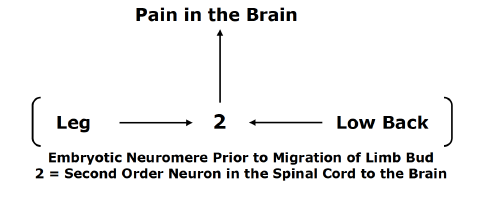
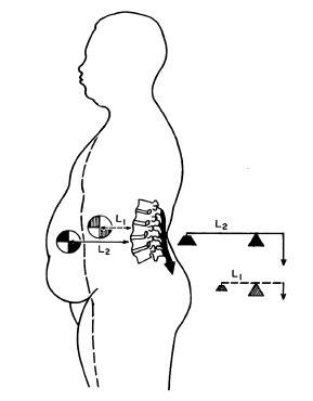
The nerves of the legs and low back arise from the same embryologic neuromere. The nerves of the legs and low back use the same second order neuron in the spinal cord to send the pain electrical signal to the brain. With the growth of the fetus, the limb bud migrates, but it maintains its embryotic neuromere.

    This organization of the nervous system can confuse the brain as to the site of origin of the pain signal. The brain may perceive low back inflammation and/or irritation and hence pain as arising in the leg, and vice versa: a leg problem may be interpreted as arising from the low back. This phenomenon is called referred pain. Identifying and treating the actual site of the pain signal is quite important in achieving a good clinical outcome.

    The same referred pain phenomenon exists between the arms and neck, as well as in other parts of the body.

    Neuroanatomy

    Spinal Cord

    A simple but accurate way to categorize the nervous system is into two separate but integrated systems (above):

    1) The MOTOR nerve system:

    • The motor nerve system is shown on the left side of the drawing.
    • The motor nerve system begins in the brain, travels down the spinal cord, exits the spinal column, and innervates the muscles.
    • The motor nerves are also called efferent nerves because they originate in the central nervous system, leave the spinal column, and innervate muscles.
    • The motor nerve system controls movement and strength.

    2)  The SENSORY nerve system:

    • The sensory nerve system is shown on the right side of the drawing.
    • The sensory nerve system brings electrical signals from the peripheral body into the spinal cord, up the spinal cord, and into the brain.
    • The sensory nerves are also called afferent nerves because they originate in the peripheral body, travel to the spinal column, and terminate in the brain where the electrical signal is perceived and interpreted.
    • Pain and temperature are sensory nerve signals (there are others).

    Category 1 Patients
    Inflammatory Pain

    In 1976, spine care pioneer Alf Nachemson, MD, noted that only tissues that have a sensory nerve supply are capable of sending the pain signal to the brain (8). Almost all tissues of the spinal column have a sensory nerve supply and are therefore capable of initiating pain perception. In 1991, Stephen Kuslich, MD, and colleagues, using invasive irritations on 700 conscious subjects, documented the spinal tissues that are capable of initiating low back pain. These include (9):

    • Skin
    • Superficial Muscles
    • Deep Muscles
    • Intervertebral Disc
    • Facet Joint Capsules
    • Periosteum of the Vertebral Bone
    • Nerve roots

    Chronic low back pain was primarily attributed to the intervertebral disc (9). The perspective that chronic low back pain is primarily discogenic is supported by others (10, 11, 12).

    In the neck, similar to the low back, the same tissues are capable of initiating pain. In contrast to the low back, persistent neck pain is primarily attributed to the facet joint capsules (13, 14).

    Successful management of Category 1 Patients (Inflammatory Pain) involves a variety of anti-inflammatory interventions. Some, such as the commonly used non-steroidal anti-inflammatory drugs (NSAIDs), are helpful but are associated with horrible and unacceptable side effects (15, 16). Ice is often helpful.

    Chiropractic care is proven to be very successful in the treatment of chronic low back and neck pain. Often, chiropractic care is superior to alternative types of management for both, including prescription nonsteroidal anti-inflammatory drugs, physical therapy, exercise, and needle acupuncture (2, 15, 17, 18, 19, 20, 21, 22, 23). Undoubtedly, this is the basis for clinical guidelines to routinely advocate chiropractic care and spinal manipulation for the treatment of spinal pain (24, 25, 26, 27, 28, 29).

    The benefits of spinal adjusting in these patients are attributed primarily to two mechanisms:

    A) Mechanical dispersion of inflammatory chemicals (10):

    Spinal adjusting improves spinal motion, and improved spinal motion disperses the accumulation of inflammatory chemicals. This is especially true with respect to discogenic pain, as the intervertebral disc is avascular. Only improved motion can disperse the nociceptive chemicals.

    When Vert Mooney, MD, was the president of the International Society for the Study of the Lumbar Spine, his Presidential Address emphasized this concept, stating (10):

    “Mechanical events can be translated into chemical events related to pain.”

    “The fluid content of the disk can be changed by mechanical activity.”

    “Mechanical activity has a great deal to do with the exchange of water and oxygen concentration [in the disc].”  

     “Research substantiates the view that unchanging posture, as a result of constant pressure such as standing, sitting or lying, leads to an interruption of pressure-dependent transfer of liquid. Actually, the human intervertebral disk lives because of movement.”

    “In summary, what is the answer to the question of where is the pain coming from in the chronic low-back pain patient? I believe its source, ultimately, is in the disk. Basic studies and clinical experience suggest that mechanical therapy is the most rational approach to relief of this painful condition.”

    “Prolonged rest and passive physical therapy modalities no longer have a place in the treatment of the chronic problem.”

    B) Closure of the pain gate (2, 30, 31):

    The pain electrical signal can be “blocked” by closing the “pain gate.” Spinal adjusting can initiate a mechanical electrical signal that does this. This concept was first proposed by pain researchers Ronald Melzack and Patrick Wall in 1965 (30). Their theory is known as the Gate Control Theory of Pain.

    In 2002, the British Journal of Anaesthesia published a study reaffirming the validity of the Gate Theory of Pain in an article titled (31):

    Gate Control Theory of Pain Stands the Test of Time

    Theoretically, spinal stiffness would allow the pain gate to be open to nociceptive electrical signals; spinal adjusting would improve mechanical motion and close the pain gate to nociceptive signals. This explanation was first advanced by Canadian orthopedic surgeon Kirkaldy-Willis in 1985. Dr. Kirkaldy-Willis stated (2):

    Melzack and Wall proposed the Gate Theory of Pain in 1965, and this theory has “withstood rigorous scientific scrutiny.”

    “The central transmission of pain can be blocked by increased proprioceptive input.” Pain is facilitated by “lack of proprioceptive input.” This is why it is important for “early mobilization to control pain after musculoskeletal injury.”

    The facet capsules are densely populated with mechanoreceptors. “Increased proprioceptive input in the form of spinal mobility tends to decrease the central transmission of pain from adjacent spinal structures by closing the gate. Any therapy which induces motion into articular structures will help inhibit pain transmission by this means.”

    Other publications have supported this model (32, 33).

    Category 2 Patients
    Sclerogenic Referral Pain

    Many patients who suffer with low back pain also have pain in their leg(s). Many patients with neck pain will also have pain in their arm(s). Although such a clinical presentation leads to concerns of spinal nerve compression (Category 3 Patients), it is usually something else. It is referred pain, as described above in the section on EMBRYOLOGY. Technically, it is also known as Sclerogenic pain, Sclerotomic pain, or Sclerotogenous pain.

    Sclerogenic leg pain occurs as a consequence of an irritation of spinal tissues below the deep fascia (deep spinal muscles, spinal ligaments, intervertebral disc, etc.). It does not involve irritation, inflammation or compression of the spinal nerve roots.

    The existence and patterns of sclerogenic pain has been documented for nearly a century (34, 35, 36, 37, 38, 39, 40, 41, 42).

    Sclerogenic pain often subjectively presents as a deep, diffuse dull ache that is not in a dermatomal pattern. It is difficult for the patient to precisely locate the pain on the skin; the discomfort is perceived deep to the skin.

    Typically, sclerogenic pain presents with these examination findings:

    • Spinal compression tests (Spurling’s, Kemp’s, etc.) are usually negative.
    • Valsalva is usually negative.
    • Straight leg raising and the brachial plexus tension tests are usually negative.
    • Myotomal strength tests are usually normal.
    • Deep tendon reflexes are usually normal and symmetrical. In general, sclerogenic pain is not complicated or dangerous. Chiropractic spinal adjustments primarily affect the deep spinal tissues that are responsible for the sclerogenic pain referral. Consequently, chiropractic spinal adjusting is very effective in improving and/or resolving both back pain and referred sclerogenic leg pain.

    Category 3 Patients
    Nerve Root Involvement
    Irritation, Inflammation, Compression

    Between every spinal segmental level there is a spinal nerve (nerve root). This arrangement exists in all spinal regions: neck, mid back, and low back. Spinal pathology has the potential to irritate, inflame, and/or compress the nerve root. The technical term for these nerve root problems is radiculopathy.

    IVF / Spinal nerve

    In the neck, the spinal nerve root extends down the shoulder and into the arm(s). In the low back, the spinal root extends down the pelvis and into the leg(s). Consequently, when a nerve is irritated, inflamed, or compressed, it generates symptoms (pain, numbness, tingling, hypersensitivity, burning, achiness, etc.) and/or functional disturbances (weakness, atrophy, etc.) in the arm(s) and/or leg(s).

    Nerve root irritation

    The most common cause of nerve root irritation, inflammation, or compression is herniation of the intervertebral disc. Other causes include arthritic changes (degenerative joint disease, degenerative disc disease, spondylosis) causing narrowing of the intervertebral foramen.

    Radiculopathy is more complex and concerning than local pain or sclerogenic pain syndromes. As a rule, nerve root irritation and inflammation resolve with chiropractic spinal adjusting. However, they often require longer treatment duration and more frequent chiropractic visits. To rule out serious pathology that may be causing the irritation and/or inflammation, chiropractors may use diagnostic imaging, primarily x-rays. On occasion, the chiropractor may determine that advanced diagnostic imaging (MRI, CT, etc.) is warranted.

    Compressive radiculopathy is the most concerning clinical syndrome seen in chiropractic clinical practice. This is because excessive or prolonged compression may lead to death of some of the nerve fibers resulting in permanent functional impairments.

    Typical clinical findings in patients with compressive radiculopathy include:

    • Positive spinal compression tests (Spurling’s (neck), Kemp’s (low back), etc.).
    • Positive Valsalva test.
    • Positive straight leg raising (low back) and/or the brachial plexus tension (neck) tests.
    • Myotomal (muscle) weakness.
    • Reduced deep tendon reflexes.
    • Altered superficial sensation in a dermatomal pattern.

    Red flag findings that warrant referral for additional investigations include:

    • Muscle group atrophy.
    • Loss of normal function of bladder, bowels, or sexual function (difficulty starting, difficulty ending, dripping, loss of sensation, etc.).
    • Saddle anesthesia (loss of sensation in the area of the buttocks that would contact a saddle when sitting).
    • Loss of bowel, bladder, and/or sexual function.

    In addition to diagnostic imaging (x-rays, MRI, and/or CT scans), compressive radiculopathy may warrant the use of neuro-diagnostic testing (electromyography, nerve conduction studies, etc.).

    Statistically, compressive neuropathology is very rare, constituting only 1-2% of chiropractic clinical practice. For seven decades, studies have shown that spinal adjusting is appropriate and usually successful in the management of compressive radiculopathy (43, 44, 45 46, 47 48, 49, 50, 51, 52, 53, 54, 55).

    Rarely, patients suffering from compressive radiculopathy will require a surgical decompression. Chiropractors are trained to monitor patient progress for any symptoms or signs that might benefit or require a surgical consultation. 

    SUMMARY

    Chiropractors primarily treat and manage spinal pain syndromes. Ninety-three percent of initial chiropractic care is for the management of low back and/or neck pain complaints (1). On these patients, a number of mechanisms may be responsible for improvement in the patients’ signs and symptoms. The explanations most commonly used are (A) the dispersion of inflammatory chemicals and (B) the initiation of a neurological sequence of events that closes the pain gate.

    When a patient presents with spine pain and extremity (arm or leg), complaints (pain, tingling, numbness, etc.) clinical thinking changes. Initially, the chiropractor will determine if the extremity complaint is sclerogenic referred or if it is compressive radiculopathy. In the case of sclerogenic referral, successful management of the deep spinal tissue irritations will resolve the extremity symptomatology, and this is effectively accomplished with spinal adjusting. Typically, the spine complaints and the extremity symptoms will resolve together.

    In the case of suspected compressive radiculopathy, chiropractors will do a more detailed evaluation, which may include imaging and/or neuro-diagnostics. The chiropractor may decide to treat the condition, refer the patient to another provider, or co-treat the patient with another provider.

    REFERENCES

    1. Adams J, Peng W, Cramer H, Sundberg T, Moore C; The Prevalence, Patterns, and Predictors of Chiropractic Use Among US Adults: Results From the 2012 National Health Interview Survey; Spine; December 1, 2017; Vol. 42; No. 23; pp. 1810–1816.
    2. Kirkaldy-Willis WH, Cassidy JD; Spinal Manipulation in the Treatment of Low back Pain; Canadian Family Physician; March 1985; Vol. 31; pp. 535-540.
    3. Kandel E, Schwartz J, Jessell T; Principles of Neural Science; McGraw-Hill; 2000.
    4. Ambron R; The Brain and Pain: Breakthroughs in Neuroscience; Columbia University Press; 2022.
    5. Gunn CC; The Gunn Approach to the Treatment of Chronic Pain: Intramuscular Stimulation for Myofascial Pain of Radiculopathic Origin; Churchill Livingston; 1996.
    6. Boswell M, Cole BE, Editors; Weiner’s Pain Management: A Practical Guide for Clinicians; American Academy of Pain Management; Seventh Edition; 2006.
    7. Omoigui S; The Biochemical Origin of Pain: The Origin of All Pain is Inflammation and the Inflammatory Response: Inflammatory Profile of Pain Syndromes; Medical Hypothesis; 2007; Vol. 69; pp. 1169 – 1178.
    8. Nachemson A; The Lumbar Spine, An Orthopedic Challenge; Spine; March 1976; Vol. 1; No 1; pp. 59-71.
    9. Kuslich S, Ulstrom C, Michael C; The Tissue Origin of Low Back Pain and Sciatica: A Report of Pain Response to Tissue Stimulation During Operations on the Lumbar Spine Using Local Anesthesia; Orthopedic Clinics of North America; April 1991; Vol. 22; No. 2; pp. 181-187.
    10. Mooney V; Where Is the Pain Coming From?; Spine; October 1987; Vol. 12; No. 8; pp. 754-759.
    11. Izzo R, Popolizio T, D’Aprile P, Muto M; Spine Pain; European Journal of Radiology; May 2015; Vol. 84; pp. 746–756.
    12. Bogduk N; Functional Anatomy of the Spine; Handbook of Clinical Neurology; Vol. 136; pp. 675-688.
    13. Bogduk N, Aprill C; On the Nature of Neck Pain, Discography and Cervical Zygapophysial Joint Blocks; Pain; August 1993; Vol. 54; No. 2; pp. 213-217.
    14. Bogduk N; On Cervical Zygapophysial Joint Pain After Whiplash; Spine; December 1, 2011; Vol. 36; No. 25S; pp. S194–S199.
    15. Giles LGF; Muller R; Chronic Spinal Pain: A Randomized Clinical Trial Comparing Medication, Acupuncture, and Spinal Manipulation; Spine; July 15, 2003; Vol. 28; No. 14; pp. 1490-1502.
    16. Maroon JC, Bost JW; Omega-3 Fatty Acids (Fish Oil) as an Anti-Inflammatory: An Alternative to Nonsteroidal Anti-inflammatory Drugs for Discogenic Pain; Surgical Neurology; April 2006; Vol. 65; 326– 331.
    17. Meade TW, Dyer S, Browne W, Townsend J, Frank OA; Low back pain of mechanical origin: Randomized comparison of chiropractic and hospital outpatient treatment; British Medical Journal; June 2, 1990; Vol. 300; pp. 1431-1437.
    18. The Lancet; Chiropractors and Low Back Pain; July 28, 1990, p. 220.
    19. Muller R, Giles LGF; Long-Term Follow-up of a Randomized Clinical Trial Assessing the Efficacy of Medication, Acupuncture, and Spinal Manipulation for Chronic Mechanical Spinal Pain Syndromes; Journal of Manipulative and Physiological Therapeutics; January 2005; Vol. 28; No. 1; pp. 3-11.
    20. Cifuentes M, Willetts J, Wasiak R; Health Maintenance Care in Work-Related Low Back Pain and Its Association with Disability Recurrence; Journal of Occupational and Environmental Medicine; April 14, 2011; Vol. 53; No. 4; pp. 396-404.
    21. Senna MK, Machaly SA; Does Maintained Spinal Manipulation Therapy for Chronic Nonspecific Low Back Pain Result in Better Long-Term Outcome? Randomized Trial; Spine; August 15, 2011; Vol. 36; No. 18; pp. 1427–1437.
    22. Woodward MN, Cook JCH, Gargan MF, Bannister GC; Chiropractic Treatment of Chronic ‘Whiplash’ Injuries; November 1996; Injury; Vol. 27; No. 9; pp. 643-645.
    23. Khan S, Cook J, Gargan M, Bannister G; A symptomatic classification of Whiplash Injury and the Implications for Treatment; The Journal of Orthopaedic Medicine; 1999; Vol. 21; No. 1; pp. 22-25.
    24. Chou R, Qaseem A, Snow V, Casey D, Cross JT, Shekell, Owens DK; Diagnosis and Treatment of Low Back Pain; Annals of Internal Medicine; Vol. 147; No. 7; October 2007; pp. 478-491.
    25. Chou R, Huffman LH; Non-pharmacologic Therapies for Acute and Chronic Low Back Pain; Annals of Internal Medicine; October 2007; Vol. 147; No. 7; pp. 492-504.
    26. Globe G, Farabaugh RJ, Hawk C, Morris CE, Baker G, DC, Whalen WM, Walters S, Kaeser M, Dehen M, DC, Augat T; Clinical Practice Guideline: Chiropractic Care for Low Back Pain; Journal of Manipulative and Physiological Therapeutics; January 2016; Vol. 39; No. 1; pp. 1-22.
    27. Wong JJ, Cote P, Sutton DA, Randhawa K, Yu H, Varatharajan S, Goldgrub R, Nordin M, Gross DP, Shearer HM, Carroll LJ, Stern PJ, Ameis A, Southerst D, Mior S, Stupar M, Varatharajan T, Taylor-Vaisey A; Clinical practice guidelines for the noninvasive management of low back pain: A systematic review by the Ontario Protocol for Traffic Injury Management (OPTIMa) Collaboration; European Journal of Pain; Vol. 21; No. 2; February 2017; pp. 201-216.
    28. Qaseem A, Wilt TJ, McLean RM, Forciea MA; Noninvasive Treatments for Acute, Subacute, and Chronic Low Back Pain: A Clinical Practice Guideline from the American College of Physicians; For the Clinical Guidelines Committee of the American College of Physicians; Annals of Internal Medicine; April 4, 2017; Vol. 166; No. 7; pp. 514-530.
    29. Oliveira CB, Maher CG, Pinto RZ, et al.; Clinical Practice Guidelines for the Management of Non-Specific Low Back Pain in Primary Care: An Updated Overview; European Spine Journal; November 2018; Vol. 27; No. 11; pp. 2791-2803.
    30. Melzack R, Wall P; Pain Mechanisms: A New Theory; Science; November 19, 1965; Vol. 150; No. 3699; pp. 971-979.
    31. Dickenson AH; Gate Control Theory of Pain Stands the Test of Time; British Journal of Anaesthesia; June 2002; Vol. 88; No. 6; pp. 755-757.
    32. Vicenzino B, Collins D, Wright A; The Initial Effects of a Cervical Spine Manipulative Physiotherapy Treatment on the Pain and Dysfunction of Lateral Epicondylalgia; Pain; November 1996; Vol. 68; No. 1; pp. 69-74.
    33. Savva C, Giakas G, Efstathiou M; The Role of the Descending Inhibitory Pain Mechanism in Musculoskeletal Pain Following High-Velocity, Low Amplitude Thrust Manipulation: A Review of the Literature; Journal of Back and Musculoskeletal Rehabilitation; 2014; Vol. 27; No. 4; pp. 377–382.
    34. Kellgren JH; A Preliminary Account of Referred Pain Arising from Muscle; The British Medical Journal; February 12, 1938; pp. 325-327.
    35. Kellgren JH; On the distribution of pain arising from deep somatic structures with charts of segmental pain areas; Clinical Science; 1939; Vol. 4; pp. 35-46.
    36. Inman VT, Saunders JB; Anatomicophysiological Aspects of Injuries to the Intervertebral Disc; Journal of Bone and Joint Surgery; April 1947; Vol. 29; No. 2; pp. 461–468.
    37. Sinclair DC, Feindel WH, Weddell G, Falconer MA; The Intervertebral Ligaments as a source of Segmental Pain; The Journal of Bone and Joint Surgery, British; August 1, 1948; Vol. 30-B; No. 3; pp. 515-521
    38. Feinstein B, Langion JNK, Jameson RM, Schiller F; Experiments on Pain Referred from Deep Somatic Tissues; Journal of Bone and Joint Surgery, American; October 1954; Vol. 36-A; No. 5; pp. 981-997.
    39. Hockaday JM, Whitty CW; Patterns of Referred Pain in the Normal Subject; Brain; September 1967; Vol. 90; No. 3; pp. 481-496.
    40. Travell J, Simons D; Myofascial pain and dysfunction, the trigger point manual; New York; Williams & Wilkins; 1983.
    41. Travell J, Simons D; Myofascial pain and dysfunction, the trigger point manual: THE LOWER EXTREMITIES; New York; Williams & Wilkins; 1992.
    42. Simons D, Travell J; Travell & Simons’, Myofascial pain and dysfunction, the trigger point manual: Volume 1, Upper Half of Body; Baltimore; Williams & Wilkins; 1999.
    43. Ramsey RH; Conservative Treatment of Intervertebral Disk Lesions; American Academy of Orthopedic Surgeons; Instructional Course Lectures; Vol. 11; 1954; pp. 118-120.
    44. Mathews JA, Yates DAH; Reduction of Lumbar Disc Prolapse by Manipulation; British Medical Journal; September 20, 1969; No. 3; pp. 696-697.
    45. Edwards BC; Low back pain and pain resulting from lumbar spine conditions: a comparison of treatment results; Australian Journal of Physiotherapy; September 1969; Vol. 15; No. 3; pp. 104-110.
    46. Turek S; Orthopaedics, Principles and Their Applications; JB Lippincott Company; 1977; page 1335.
    47. Kuo PP, Loh ZC; Treatment of Lumbar Intervertebral Disc Protrusions by Manipulation; Clinical Orthopedics and Related Research; February 1987; No. 215; pp. 47-55.
    48. Quon JA, Cassidy JD, O’Connor SM, Kirkaldy-Willis WH; Lumbar intervertebral disc herniation: treatment by rotational manipulation; Journal of Manipulative and Physiological Therapeutics; June 1989; Vol. 12; No. 3; pp. 220-227.
    49. Cassidy JD, Thiel HW, Kirkaldy-Willis WH; Side posture manipulation for lumbar intervertebral disk herniation; Journal of Manipulative and Physiological Therapeutics; February 1993; Vol. 16; No. 2; pp. 96-103.
    50. Stern PJ, Côté P, Cassidy JD; A series of consecutive cases of low back pain with radiating leg pain treated by chiropractors; Journal of Manipulative and Physiological Therapeutics; Jul-Aug 1995; Vol. 18; No. 6; pp. 335-342.
    51. Santilli V, Beghi E, Finucci S; Chiropractic manipulation in the treatment of acute back pain and sciatica with disc protrusion: A randomized double-blind clinical trial of active and simulated spinal manipulations; The Spine Journal; March-April 2006; Vol. 6; No. 2; pp. 131–137.
    52. McMorland G, Suter E, Casha S, du Plessis SJ, Hurlbert RJ; Manipulation or Microdiskectomy for Sciatica? A Prospective Randomized Clinical Study; Journal of Manipulative and Physiological Therapeutics; October 2010; Vol. 33; No. 8; pp. 576-584.
    53. Bronfort G, Hondras M, Schulz CA, Evans RL, Long CR, Grimm R; Spinal Manipulation and Home Exercise with Advice for Subacute and Chronic Back-Related Leg Pain: A Trial With Adaptive Allocation; Annals of Internal Medicine; September 16, 2014; Vol. 161; No. 6; pp. 381-391.
    54. Leemann S, Peterson CK, Schmid C, Anklin B, Humphreys BK; Outcomes of Acute and Chronic Patients with Magnetic Resonance Imaging–Confirmed Symptomatic Lumbar Disc Herniations Receiving High-Velocity, Low Amplitude, Spinal Manipulative Therapy: A Prospective Observational Cohort Study with One-Year Follow-Up; Journal of Manipulative and Physiological Therapeutics; March/April 2014; Vol. 37; No. 3; pp. 155-163.
    55. Fritz JM, Lane E, McFadden M, Brennan G, Magel JS, Thackeray A, Minick K, Meier W, Greene T; Physical Therapy Referral from Primary Care for Acute Back Pain with Sciatica: A Randomized Controlled Trial; Annals of Internal Medicine; January 2021; Vol. 174; No. 1; pp. 8-17.
    56. Ghasabmahaleh SH, Rezasoltani Z, Dadarkhah A, Hamidipanah S, Mofrad RK, Sharif Najafi S; Spinal Manipulation for Subacute and Chronic Lumbar Radiculopathy: A Randomized Controlled Trial; The American Journal of Medicine; January 2021; Vol. 134; No. 1; pp. 135−141.
  • Neck Injury and the Brain

    Neck Injury and the Brain

    Background Information

    Mild traumatic brain injuries are also known as concussions. It is estimated that these injuries have a prevalence of 3.8 million per year in the United States (1). Despite this high incidence, mild traumatic brain injuries and concussions are one of the least understood injuries facing the sports healthcare and the neuroscience communities today (2).

     Introduction

    Sir Isaac Newton (1643-1727) explained the three universal laws of motion in his 1687 book Mathematical Principles of Natural Philosophy. Newton’s first law of motion states that every object will remain at rest or in uniform motion in a straight line unless compelled to change its state by the action of an external force. This tendency to resist changes in a state of motion is termed inertia.

    The inertial tendency of an object is proportional to the object’s weight. The greater the weight, the greater the inertia. As an analogy, it is more difficult to change the resting state of a 100-pound rock as compared to that of a 10-pound rock. It is also more difficult to stop the uniform motion of a 100-pound rock as compared to that of a 10-pound rock.

    Newton’s Law of Inertia applies to the human body. Different parts of the human body have different inertias between them. Of particular importance is the inertial differences between the human head and the person’s trunk.

    Neck Hyperextension As Trunk Is Pushed Under Head

    A classic example of these inertial concepts is the rear-end motor vehicle collision. In a rear-end motor vehicle collision, the struck vehicle, its seat, and trunk of the occupant are quickly propelled forward, while the head, having its own inertial mass, will remain at rest. Thus, the head remains still while the body is moved forward under the head. This gives the appearance that the head is extending upon the trunk, the so-called “hyperextension” phase of a rear-end motor vehicle collision.

    Other inertial differences within the human body, pertinent to this discussion, are between the skull and the brain, and between the brain and the spinal cord.

    Two Types of Brain Injury

    (please forgive the crassness of the analogy)

    One

    The stationary head can be injured by a direct blow. The head might be struck by a hammer, bat, or a falling object. The skull will hopefully protect the brain from injury by dispersing the forces of the blow.

    Wearing a hard-hat or a helmet adds to the protection of the brain by enhancing the dispersion of the forces from the blow. Hence, the ubiquitous advocacy of helmet wearing throughout society, from sports to family bike riding, etc.

    Direct blow brain injury can occur without direct injury to the neck. It can also occur without any inertial injury to the neck, to the brain, and/or to the spinal cord.

    Direct blow brain injury is only rarely encountered in chiropractic clinical practice.

    Two

    The brain can sustain an inertial injury. Inertial brain injury can occur without any blow to the head/skull. The injury occurs as a consequence of the brain smashing into the inside of the skull.

    A societally understood but horrific example of inertial brain injury is shaken baby syndrome. The violent shaking of a baby causes serious inertial injuries (including death) to the baby’s brain, spinal cord, and neck. Yet, often there is no direct blow to the baby’s head, neck, or body.

    Brain inertial injuries are the rule in motor vehicle collisions and in most of sport’s head trauma.

    Elite athletes can run 100 yards in 10 seconds, traveling about 20 miles per hour. The athlete’s body, neck, spinal cord, brain, and skull are all moving together at 20 miles per hour.  Should the athlete collide with a wall, goal post, another player, etc., and his/her body fully stop in a fraction of a second, the brain in the skull will continue to propel forward at the pre-collision 20 miles per hour because of the law of inertia. The brain will collide with the inside of the skull and sustain an inertial injury.

    Importantly, this inertial brain injury will occur whether the athlete is wearing a helmet or not. Ironically, wearing a helmet adds to the weight of the head, increasing its inertia. Increasing the weight of the head with a helmet increases the inertial loads and injuries to the neck. Helmets primarily protect against direct blow head/skull injuries. Helmets increase neck inertial injuries. As noted below, helmets increase brain inertial injuries:

    Give an elite football player (American) the best helmet available. Ask the player to run as fast as he is comfortable and crash his helmet, head first, into a brick wall. How fast will he run?

    Remove the helmet from the player and again ask him to run as fast as he is comfortable and crash his head, without a helmet, into a brick wall. How fast will he run?

    Important Points:

    When it comes to crashing one’s head into a brick wall, the player with the helmet will run faster towards the wall.

    • Using the helmet will reduce direct blow brain injury. This is good.
    • Using the helmet, because of the faster running speed, will increase inertial brain injury. This is not

    In vivo, the brain is exceptionally soft, delicate, and vulnerable. The outside of the skull is mostly smooth. However, the inside of the skull is not smooth. Inside the skull there are numerous sharp and jagged bony contours and ridges. Inertial stress between the skull and the brain rakes the soft brain over the bony ridges, resulting in appreciable damage.

    Key Points:

    One can sustain a direct blow brain injury without sustaining an inertial injury to the neck, brain, and/or spinal cord.

    Essentially all inertial brain injuries will also cause an inertial injury to the neck and its structures, including the joints, discs, muscles, nerves, and potentially the spinal cord.

    Clinical Diagnosing of Inertial Brain Injury

    The most widely accepted criteria for traumatic brain injury are from the American Congress of Rehabilitation Medicine, which include (3):

    • Any period of loss of consciousness.
    • Any loss of memory for events immediately before or after the accident (posttraumatic amnesia).
    • Any alteration in mental state at the time of the accident, including feeling dazed, disoriented, or confused.
    • Any focal neurological deficits that may or may not be transient but that do not exceed: loss of consciousness of 30 minutes; an initial Glasgow Coma Scale (GCS) score of 13-15 (explained below); posttraumatic amnesia of 24 hours.

    This definition includes these mechanisms of injury:

    • The head being struck.
    • The head striking an object.
    • The brain undergoing an acceleration/deceleration movement (i.e.,whiplash) without direct external trauma to the head.

    ••••

    The Glasgow Coma Scale (GCS) is a neurological scale used to give a reliable and objective assessment of the conscious state of a person, adult or child, primarily following head injury. It was published in 1974 by professors of neurosurgery at the University of Glasgow’s Institute of Neurological Sciences (4):

    GCS  less than 8–9         Severe Brain Injury

    GCS 9–12           Moderate Brain Injury

    GCS 13-15          Minor Brain Injury

    • The scale is composed of three tests: eye, verbal, and motor
    • A score of 15 is essentially normal, a fully awake person.
    • A score of 3 indicates a deep coma.

    A copy of the Glasgow Coma Scale is included at the end of this paper.

    ••••

    Recovery from traumatic brain injury is measured using two outcomes:

     Functional Recovery

    Subjective (symptom) Recovery

     ••••

    Functional recovery is assessed using the Glasgow Outcome Scale-Extended (GOS-E) score (5). This tool for functional recovery is the most widely used outcome measure in traumatic brain injury and is widely recommended by international bodies, including the National Institutes of Health:

    • full functional recovery = GOS-E score of 8
    • incomplete recovery = GOS-E score less than 8: 1 indicates death; 4 indicates severe disability; 5 indicates moderate disability; 7 indicates unable to return to preinjury functioning; 8 indicates full recovery or return to baseline function

    A copy of the GOS-E is included at the end of this paper.

    ••••

    Subjective (symptom) Recovery is assessed using the Rivermead Post Concussion Symptoms Questionnaire (RPQ) (6). The RPQ asks about 16 new or worsened symptoms since the injury; scores range from 0 (best) to 64 (worst).

    A copy of the Rivermead Post Concussion Symptoms Questionnaire is included at the end of this paper.

    •••••••••

    A recent study (August 2022) looked at the long-term (6 month) recovery rates in individuals who had sustained a mild traumatic brain injury. The study was published in the Journal of the American Medical Association Network Open and titled (7):

    Outcomes in Patients with Mild Traumatic Brain Injury

    Without Acute Intracranial Traumatic Injury

    The objective of this study was to describe the 2-week and 6-month recovery outcomes in a cohort of 991 patients with mild traumatic brain injury, a Glasgow Coma Scale score of 15, and a negative head CT scan that had been taken within 24 hours of injury. Subjects’ mean age was 39 years, 64% male and 36% female. As noted above, patient assessments were done using:

    Glasgow Coma Scale for severity of injury.

    Glasgow Outcome Scale-Extended for functional recovery.

     Rivermead Post Concussion Symptoms Questionnaire for subjective (symptom) recovery.

    The authors note that most of the more than 3 million new cases of traumatic brain injury in the United States each year are classified as mild.

    Medical care practitioners often assume that patients with mild traumatic brain injury will improve over time with the only intervention being rest, and there will be no long-term sequelae.

    Despite such optimism, 21% of patients with mild traumatic brain injury “will experience mental health problems, including posttraumatic stress disorder and depression; cognitive and behavioral impairment; and changes in memory, attention, and motivation, which are associated with loss of work days and unemployment.”

    The outcomes presented in this prospective study of nearly 1,000 subjects is summarized in the chart below (7):

    GOS-E Score at 2 Weeks

    Increased RPQ Symptoms at 2 Weeks

    Increased RPQ Symptoms at 6 Months

    1-5

    48%

    30%

    6

    33%

    17%

    7

    19%

    9%

    8

    6%

    0.0%


    GOS-E Score at 6 Months

    Increased RPQ Symptoms at 6 Months

    1-5

    55%

    6

    42%

    7

    17%

    8

    0.0%


    Two weeks after the injury, 27% had functional recovery and 73% had incomplete recovery.

    Six months after the injury, 44% had functional recovery and 56% had incomplete recovery. More than half of the patients who sustained a mind traumatic brain injury with a negative CT scan had not functionally recovered 6 months after injury.

    Only 12% of the patients noted meaningful subjective recovery between 2 weeks and 6 months, indicating that a “wait and see” management is certainly inadequate for these patients.

    In this cohort of mild brain injured subjects, the majority were neither functionally or subjectively recovered at 6 months after injury. The authors state:

    “This study found that most [mild traumatic brain injury] participants with a GCS score of 15 and negative head CT scan reported incomplete recovery at 2 weeks and 6 months after their injury.”

    Traumatic Brain Injury and the Neck

    In 2015, an important study on this topic was published in the journal The Physician and Sports Medicine, and titled (8):

    The Role of the Cervical Spine in Post-concussion Syndrome

    The authors note that there is considerable overlap of the signs and symptoms of mild traumatic brain injury and of whiplash neck injury. This overlap of presentation may cause confusion as to the source of symptomatology. A “wait and see” approach to management of an assumed brain injury may be completely inappropriate if in fact the symptoms are attributed to the neck.

    This paper reviews the existing literature surrounding the numerous proposed theories of post-concussive syndrome and introduces another potential, and very treatable, cause of this chronic condition: cervical spine dysfunction due to concomitant whiplash-type injury.

    The authors provide a comparative chart contrasting mild traumatic brain injury with whiplash neck injury:

    Signs and Symptoms of Mild Traumatic Brain Injury

    Signs and Symptoms of Whiplash Injuries

    Headache

    Pressure in Head

    Headache

    Neck pain

    Neck/shoulder pain

    Reduced/painful neck movements

    Nausea/vomiting

    Nausea/vomiting

    Dizziness

    Balance problems

    Dizziness

    Unsteadiness

    Blurred Vision

    Sensitivity to Light

    Vision problems

    Difficulty remembering

    Confusion

    Feeling Like “In a Fog”

    Difficulty Concentrating

    Memory problems

    Problems Concentrating

    Sensitivity to Noise

    Ringing in Ears

    Feeling Slowed Down

    “Don’t Feel Right”

    Nervous / Anxious / Irritable

    Sadness / More Emotional

    Fatigue / Low Energy /Drowsiness

    Trouble Falling Asleep

    Reduced/painful Jaw Movements

    Numbness, Tingling or Pain in Arm or Hand

    Numbness, Tingling or Pain in Leg or Foot

    Difficulty Swallowing


    The symptoms of headache and dizziness that are so prevalent in concussion-type injuries may actually be the result of neck injury mechanisms. Numerous brain stem structures receive synaptic inputs from the second cervical dorsal root ganglion afferents, including (9):

    • Lateral cervical nucleus
    • Central cervical nucleus
    • Caudal projections to C5 level
    • Cuneate nucleus, lateral cuneate nucleus
    • Nucleus tractus solitarius
    • Intercalatus nucleus
    • Nucleus X of the vestibular system
    • Trigemino-cervical nucleus

    In this article, the authors discuss the cases of 5 patients with diagnosed post-concussive syndrome, who experienced very favorable outcomes following various treatment and rehabilitative techniques aimed at restoring cervical spine function. The treatment included chiropractic spinal manipulation.

    These authors propose that a cervical injury, suffered concurrently at the time of the mild traumatic brain injury, acts as a “major symptomatic culprit in many post-concussive syndrome patients.”

    These authors state:

    “Any significant blunt impact and/or acceleration/deceleration of the head will also result in some degree of inertial loading of the neck potentially resulting in strain injuries to the soft tissues and joints of the cervical spine.”

     “Acceleration/deceleration of the head–neck complex of sufficient magnitude to cause mild traumatic brain injury is also likely to cause concurrent injury to the joints and soft tissues of the cervical spine.”

    “[It is] well established that injury and/or dysfunction of the cervical spine can result in numerous signs and symptoms synonymous with concussion, including headaches, dizziness, as well as cognitive and visual dysfunction, making diagnosis difficult.”

    It has been known since 2006 that brain-injured athletes concurrently injure their cervical spines (10). Injury or dysfunction of the cervical spine has been shown to cause headaches, dizziness and loss of balance, nausea, visual and auditory disturbances, reduced cognitive function, and many other signs and symptoms considered synonymous with concussion.

    In this study (8), the authors present five cases of patients diagnosed with post-concussive syndrome who were treated successfully in a chiropractic clinic. Their improvement was rapid and documented using standard measurement outcomes. The improved clinical outcome results were long-lasting.

    The treatment included:

    • Active Release Therapy
    • Localized vibration therapy over the affected muscles
    • Spinal manipulative therapy of the restricted joints
    • Low-velocity mobilizations (on 1 patient)

    The authors concluded:

    “Management of persistent post concussive symptoms through ongoing brain rest is outdated and demonstrates limited evidence of effectiveness in these patients.”

    “[Instead, there is evidence that] skilled, manual therapy- related assessment and rehabilitation of cervical spine dysfunction should be considered for chronic symptoms following concussion injuries.”

    This study highlights the lack of understanding by athletes, the public, and healthcare providers that it is essentially impossible to sustain a traumatic brain injury without also injuring the soft tissues of the cervical spine. It is anatomically and biologically probable that these cervical spine injuries cause many, if not most, of the symptoms of the post-concussion syndrome.

    It is gratifying to see a published study showing that traditional chiropractic management of post-concussive syndrome patients resulted in rapid and sustained improvement in post-concussive signs and symptoms, allowing the athlete to return to full competition.

    It is recommended that all patients who are likely suffering from a mild traumatic brain injury be referred to a chiropractor for cervical spine evaluation and treatment. This is especially important in light of the poor functional and subjective outcomes associated with the “wait and see” approach to management (7).

    It is also recommended that all patients suffering from the post-concussive syndrome (long-term sequelae to mild traumatic brain injury) should be referred to a chiropractor for cervical spine evaluation and treatment.

    REFERENCES:

    1. Langlois JA, Rutland-Brown W, Wald MM; The Epidemiology and Impact of Traumatic Brain Injury: A Brief Overview; Journal of Head Trauma Rehabilitation; September-October 2006; Vol. 21; No. 5; pp. 375–378.
    2. Thompson J, Sebastianelli W, Slobounov S; EEG and Postural Correlates of Mild Traumatic Brain Injury in Athletes; Neuroscience Letters; April 4, 2005; Vol. 377; No. 5; pp. 158–163.
    3. Kay T, Harrington DE, Adams R, et. al; Definition of Mild Traumatic Brain Injury; Journal of Head Trauma and Rehabilitation; 1993; Vol. 8; No. 3; pp. 86-87.
    4. Teasdale G, Jennett B; Assessment of Coma and Impaired Consciousness: A Practical Scale; Lancet; July 13, 1974; Vol. 304; No. 7872; pp. 81-84.
    5. Wilson JT, Pettigrew LE, Teasdale G; Structured Interviews for the Glasgow Outcome Scale and the Extended Glasgow Outcome Scale: Guidelines for Their Use; Journal of Neurotrauma; August 1998; Vol. 15; No. 8; pp. 573-585.
    6. King NS, Crawford S, Wenden FJ, Moss NE, Wade DT; The Rivermead Post-concussion Symptoms Questionnaire: A Measure of Symptoms Commonly Experienced After Head Injury and its Reliability; Journal of Neurology; September 1995; Vol. 242; No. 9; pp. 587-592.
    7. Madhok DY, Rodriguez RM, Barber J, Temkin NR, Markowitz AJ, Kreitzer N, Manley GT; Outcomes in Patients with Mild Traumatic Brain Injury Without Acute Intracranial Traumatic Injury; JAMA Network Open; August 1, 2022; Vol. 5; No. 8; Article e2223245.
    8. Marshall CM, Vernon H, Leddy JJ, Baldwin BA; The Role of the Cervical Spine in Post-concussion Syndrome; July 2015; Vol. 43; No. 3; pp. 274-284.
    9. Bogduk N; Anatomy and Physiology of Headache; Biomedicine and Pharmacotherapy; 1995; Vol. 49; No. 10; pp. 435-445.
    10. Hynes LM, Dickey JP; Is there a Relationship Between Whiplash-Associated Disorders and Concussion in Hockey?; Brain Injury; February 2006; Vol. 20; No. 2; pp. 179-188.


    “Authored by Dan Murphy, D.C.. Published by ChiroTrust® – This publication is not meant to offer treatment advice or protocols. Cited material is not necessarily the opinion of the author or publisher.”


    GLASGOW COMA SCALE: the highest possible score is 15:


    Eye Opening 

    1. Never (no eye opening)
    2. To Pain Stimulus (squeezing the lunula area of the patient’s fingernail)
    3. To Sound, specifically to speech
    4. Spontaneous

    Verbal Responses 

    1. None
    2. Incomprehensible Sounds (moaning but no words)
    3. Inappropriate Words (random or exclamatory speech, but no conversational exchange; speaks words but no sentences)
    4. Confused Conversation (The patient responds to questions coherently but there is some disorientation and confusion)
    5. Oriented (Patient responds coherently and appropriately to questions such as the patient’s name and age, where they are and why, the year, month, etc.)

    Motor Responses

    1. None, no motor response
    2. Extension Response – Decerebrate posturing (adduction of arm, internal rotation of shoulder, pronation of forearm and extension at elbow, flexion of wrist and
    3. fingers, leg extension, plantar-flexion of foot)
    4. Abnormal Flexion Response – Decorticate posturing (internal rotation of shoulder, flexion of forearm and wrist with clenched fist, leg extension, plantar-flexion of foot)
    5. Normal Flexion (withdrawal) – Withdrawal from pain (absence of abnormal posturing; unable to lift hand past chin with supraorbital pressure pain applied;
    6. but does pull away when nailbed is pinched)
    7. Localizes Pain – Localizes to pain (purposeful movements towards painful stimuli; e.g., brings hand up beyond chin when supraorbital pressure applied)
    8. Obeys commands (the patient does simple things as asked)


    The Glasgow Outcome Scale-Extended (GOS-E)


    1. Dead


    1. Vegetative – Condition of unawareness with only reflex responses but with periods of spontaneous eye opening.


    1. Lower Severe Disability – Patient fully dependent for all activities of daily living; Requires assistance to be available constantly; Unable to be left alone at night.


    1. Upper Severe Disability – Can be left alone at home for up to 8 hours but remains dependent. Unable to use public transport or shop by themselves.


    1. Lower Moderate Disability – Able to return to work in sheltered workshop or non-competitive job. Rarely participates in social and leisure activities. Ongoing daily psychological problems (quick temper, anxiety, mood swings, depression).


    1. Upper Moderate Disability – Able to return to work. Participates in social and leisure activities less than half as often. Weekly psychological problems.


    1. Lower Good Recovery – Returned to work. Participates in social and leisure activities a little less and has occasional psychological problems.


    1. Upper Good Recovery – Full recovery with no current problems relating to injury.


    The Rivermead Post-Concussion Symptoms Questionnaire


    Please compare yourself now (in the last 24 hours) with before the accident:

    • 0 = Not experienced at all
    • 1 = Not more of a problem
    • 2 = A mild problem
    • 3 = A moderate problem
    • 4 = A severe problem

    Headaches………………………………………….. 0 1 2 3 4


    Feelings of Dizziness ………………………………0 1 2 3 4


    Nausea and/or Vomiting …………………………. 0 1 2 3 4


    Noise Sensitivity, easily upset by loud noise ….0 1 2 3 4


    Sleep Disturbance ………………………………….0 1 2 3 4


    Fatigue, tiring more easily ………………………..0 1 2 3 4


    Being Irritable, easily angered …………………..0 1 2 3 4


    Feeling Depressed or Tearful …………………….0 1 2 3 4


    Feeling Frustrated or Impatient …………………0 1 2 3 4


    Forgetfulness, poor memory …………………….0 1 2 3 4


    Poor Concentration ………………………………. 0 1 2 3 4


    Taking Longer to Think ………………………….. 0 1 2 3 4


    Blurred Vision …………………………………….. 0 1 2 3 4


    Light Sensitivity, easily upset by bright light …0 1 2 3 4


    Double Vision ………………………………………0 1 2 3 4


    Restlessness ……………………………………….0 1 2 3 4


    Are you experiencing any other difficulties?


    1. __________________________________ 0 1 2 3 4


    1. __________________________________ 0 1 2 3 4


  • The Importance of Sagittal Posture

    The Importance of Sagittal Posture

    The historic seminal book on orthopedics was written by Nicholas Andry in 1741 (1). The primary discussion of his book addressed the treatment of spinal distortions, beginning in childhood. Andry was a professor of medicine at the University of Paris.

    The word orthopedic is a composite of two Greek words:

    Ortho, meaning straight.

    Pedic, meaning child.

    Andry’s book emphasized the fact that children who did not grow straight had many health problems and a difficult life.

    •••••••••

    The sagittal plane divides the body into two equal halves.

     The sagittal plane divides the body into two equal halves.

    Postural distortions that move the spine and head backwards along the sagittal plane are called posterior distortions. Postural distortions that move the spine and head forwards along the sagittal plane are called anterior distortions. The emphasis of this discussion is anterior postural distortions along the sagittal plane.

    The primary emphasis of the chiropractic community, by far, is the treatment of spinal pain syndromes. This was detailed in the most comprehensive review of the chiropractic profession in the modern era (2). Ninety-three percent of patients who initially chose to see chiropractors did so because of a spinal pain complaint. There is no doubt that chronically altered posture is related to spinal musculoskeletal pain syndromes (3, 4).

    Biomechanics

    Upright posture is a first-class lever mechanical system, such as a teeter-totter or seesaw (5, 6). In the first-class mechanical system, the fulcrum is where the forces are the greatest. For upright human posture, the fulcrum is the vertebral column and its joints.

    When a patient has anterior postural distortions along the sagittal plane, their head is projected forward of the fulcrum. Yet, the patient does not fall onto their face. Their muscles, on the opposite side of the fulcrum, will contract to balance the patient’s stance, but at a high price. The joints of the vertebral column (the fulcrum) are subjected to very high loads, accelerating joint breakdown, degeneration, irritations, inflammation, and pain. Simultaneously, the constant contraction of the posture balancing muscles causes muscle problems, including inflammation, fibrosis, and pain (7).

    When a patient has anterior postural distortions along the sagittal plane, their head is projected forward of the fulcrum

    An example is presented by Rene Cailliet, MD (7). Dr. Cailliet notes that if one’s 10-pound head is anteriorly displaced along the sagittal plane by 3 inches, the required counter balancing muscle contraction on the opposite side of the fulcrum (the vertebrae) would be 30 lbs. (10 lbs. X 3 inches):

     An example is presented by Rene Cailliet, MD (7). Dr. Cailliet notes that if one’s 10-pound head is anteriorly displaced along the sagittal plane by 3 inches, the required counter balancing muscle contraction on the opposite side of the fulcrum (the vertebrae) would be 30 lbs. (10 lbs. X 3 inches):

    The constant muscle contraction required to balance anterior sagittal postural distortions creates muscle fatigue, inflammation, fibrosis, and eventually leads to chronic musculoskeletal pain syndromes (7). Collectively, these muscle syndromes are known as myofascial pain syndromes (8, 9, 10).

    The constant muscle contraction required to balance anterior sagittal postural distortions creates muscle fatigue, inflammation, fibrosis, and eventually leads to chronic musculoskeletal pain syndromes (7). Collectively, these muscle syndromes are known as myofascial pain syndromes (8, 9, 10).

    Modern Problems

     There is a modern epidemic of poor posture, neck pain, shoulder pain, arm/hand neurological symptoms, and accelerated spinal degenerative arthritis. This modern trend is attributed to the excessive use of hand-held communication devices with the head/neck bent forward, or anterior postural distortions into the sagittal plane. Both lay media and professional publications have labeled this postural distortion as “Text Neck” or “Tech Neck.”

    In 2017, Reuters Health published an article titled (11):

    Leaning Forward During Phone Use May Cause ‘Text Neck’

    This article makes the following points:

    • People often look down when using their smartphones, particularly when texting.
    • Studies have found that people hold their necks at around 45 degrees forward when using their smartphones.
    • When in a neutral position looking forward, the head weighs about 10 to 12 pounds. At a 15-degree forward flexion position, it functions as if it weighs 27 pounds.
    • At 60 degrees of forward flexion, the stress on the spine increases to about 60 pounds.
    • These prolonged abnormal stresses on a growing spinal column may lead to abnormal spinal development with dire long-term spinal health consequences in adulthood.
    • Spine surgeons are noticing an increase in patients with neck and upper back pain, likely related to poor posture during prolonged smartphone use.
    • Young patients who should not have back and neck issues are reporting disk herniations and spinal alignment problems.
    • “In an X-ray, the neck typically curves backward, and what we’re seeing is that the curve is being reversed as people look down at their phones for hours each day.”
    • Simple lifestyle changes are suggested to relieve the stress from the “text neck” posture, including holding cell phones in front of the face while texting, and using two hands and two thumbs to create a more symmetrical and comfortable position for the spine.
    • Also, people who work at computers or on tablets should use an elevated monitor stand so it sits at a natural horizontal eye level.
    • Take frequent rest breaks and/or engage in some physical exercise that can strengthen the neck and shoulder muscles. Ex 1: Lie on a bed and hang one’s head backward over the edge, extending the neck to restore the normal arc in the neck. Ex 2: While sitting/standing, attempt to align the neck with the ears over the shoulders and the shoulders over the hips.

     In 2018, The New York Post published an article titled (12):

     Tech is Turning Millennials into a Generation of Hunchbacks

    This article profiles a young man who began suffering from upper-back pain and neck soreness while in his late teens, subsequent to a habit of hunching over his cellular phone. As his symptoms progressed he developed constant pain, he hunched his shoulders, and the pain caused him to wake up numerous times throughout every night, causing constant fatigue. His upper back and neck would become incredibly tight; his neck was always bent forward.

    After a decade of suffering, the young man’s chiropractor diagnosed him with “tech neck.”  The loss or reversal of the normal cervical curve into the anterior sagittal plane is easily diagnosed with postural x-rays. The author notes:

    “Undoing the damage is a process that includes breaking bad habits, taking standing breaks and doing exercises such as yoga, foam rolling and stretches that promote good carriage and strengthen core and upper body muscles. Experts also advise patients to hold mobile devices with their elbows at 180 degrees so the screen is in front of their faces.”

    Treatment options include chiropractic, restorative postural traction, postural exercises, and core exercises.

    In 2014, Kenneth K. Hansraj, MD, published an article in the journal Surgical Technology International, titled (13):

    Assessment of Stresses in the Cervical Spine Caused by Posture and Position of the Head

    Dr. Hansraj is Chief of Spine Surgery at the New York Spine Surgery & Rehabilitation Medicine facility. He notes that billions of people are using cell phone devices on the planet, essentially in poor posture. Consequently, the purpose of this study was to assess the forces incrementally seen by the neck (cervical spine) as the head is tilted forward into worsening forward head posture.

    Dr. Hansraj indicates that an average person spends 2-4 hours a day with their heads tilted forward reading and texting on their smart phones / devices, amassing 700-1400 hours of excess, abnormal cervical spine stress per year. A high school student may spend an extra 5,000 hours in poor posture per year.

    Dr. Hansraj created a cervical spine model to calculate the forces experienced by the cervical spine when in incremental flexion (forward head position). His mathematical analysis used a head weight of 13.2 pounds. His calculations are as follows:

    Soft tissues (ligaments, cartilage, tendons, muscles, etc.) undergo viscoelastic creep when prolonged loads are applied. The tissues mechanically distort and eventually adapt to the applied loads. This includes the tissue distortions caused by head-down posture and the forward bending of the cervical spine while viewing or working on hand-held cellular devices (13).

    Dr. Hansraj states:

     “Poor posture invariably occurs with the head in a tilted forward position and the shoulders drooping forward in a rounded position.”

    “The weight seen by the spine dramatically increases when flexing the head forward at varying degrees.”

    “Loss of the natural curve of the cervical spine leads to incrementally increased stresses about the cervical spine. These stresses may lead to early wear, tear, degeneration, and possibly surgeries.”

    Creepy Things

    Biological soft tissues (ligaments, cartilage, tendons, muscles, etc.) adapt to prolonged postural distortions. Soft tissues literally change in response to these distortions. Over time they become longer or shorter in response to the positions they are habitually exposed to (14). The technical term for this premise is called viscoelastic creep (6).

    Biological systems adapt to prolonged mechanical loads. Tissues literally change in response to these loads; they adapt to become longer or shorter in response to the loads (11). This includes the biomechanical loads caused by the prolonged use of cellular devices. The technical term for this premise is called viscoelastic creep (12).

    Once soft tissues undergo viscoelastic creep, the correction of the postural distortion is no longer a simple thing. Neurological postural distortions are routinely improved quickly with chiropractic adjusting. However, the reversal of viscoelastic creep postural distortions are more complex. Creep distortions often respond best to both chiropractic adjusting and to rehabilitation programs that involve heel lifts, traction, correctional fulcrum blocks, tissue work, exercise, and more.

    Associated biomechanical problems to having one’s head bent forward in the sagittal plane is a compensatory rigid upper thoracic spine hyperkyphosis and an overall forward leaning of the entire spinal column into the anterior sagittal plane. The relevance of both of these problems is detailed below.

    A Review of Pertinent Studies

    In 2004, the Journal of the American Geriatrics Society published a study titled (15):

     Hyperkyphotic Posture Predicts Mortality in Older Community-Dwelling Men and Women: A Prospective Study

    This was a prospective study that included 1,353 participants. Kyphotic posture was measured as the number of 1.7-cm blocks needed to be placed under the participant’s head to achieve a neutral head position when lying supine on a rigid radiology table. Individuals with hyperkyphosis cannot lie flat with their heads touching a flat surface unless they hyperextend their necks. Study participants were followed for an average of 4.2 years. Hyperkyphotic posture was specifically associated with an increased rate of death due to atherosclerosis. These authors note:

    “Hyperkyphosis is associated with restrictive pulmonary disease and poor physical function, suggesting that hyperkyphosis might be associated with other adverse health outcomes.”

    “With increasing kyphotic posture, there was a trend towards greater mortality.”

    “For deaths due to atherosclerosis, participants with hyperkyphotic posture had a significant 2.4 times greater rate of death.”

    “It is possible that hyperkyphotic posture reflects an increased rate of physiological aging.”

     ••••

    In 2005, the journal Spine published a study titled (16):

    The Impact of Positive Sagittal Balance in Adult Spinal Deformity

    The authors measured the pain, systemic health, and disability status of 298 individuals and compared such measurements to a radiographic measurement of sagittal plane postural balance. A full-spine (36 inch) lateral x-ray was exposed. A plum line was dropped from the body of the C7 vertebrae and measured with respects to the articulating surface of L5 with the sacral base. All measures of health status showed significantly poorer scores as C7 plumb line deviation increased in the forward direction (anterior to the sacral base). The authors note:

    “Patients with relative kyphosis in the lumbar region had significantly more disability than patients with normal or lordotic lumbar sagittal measures.”

    “This study shows that although even mildly positive sagittal balance is somewhat detrimental, severity of symptoms increases in a linear fashion with progressive sagittal imbalance.”

    “There was clear evidence of increased pain and decreased function as the magnitude of positive [forward] sagittal balance increased.”

    “This study shows that although even mildly positive [forward] sagittal balance is somewhat detrimental, severity of symptoms increases in a linear fashion with progressive [forward] sagittal imbalance.”

    spine sagittal view

    ••••

    In 2013, The Journals of Gerontology: Series A: Biological Sciences published a study titled (17):

    Spinal Posture in the Sagittal Plane is Associated with Future Dependence in Activities of Daily Living

    The authors measured spinal postures in 804 older adults (aged 65–94 years) to determine if anterior sagittal postures were associated with the need for future assistance in Activities of Daily Living (ADL). They found that anterior sagittal posture that pitched the body and head forward was significantly associated with the need for future assistance in the person’s Activities of Daily Living. The authors state:

    “Accumulated evidence shows how important spinal posture is for aged populations in maintaining independence in everyday life.”

    “Spinal posture changes with age, but accumulated evidence shows that continued good spinal posture is important in allowing the aged to maintain independent lives.”

    “Declines in balance and gait skills caused by inclination lead to falls and fractures, and that these negative outcomes in turn lead to dependence in ADL among elderly people.”

    “Even mildly positive sagittal balance is somewhat detrimental, the decline in health status increases in a linear fashion with progressive sagittal imbalance.”

    “[The] results indicate that attention needs to be paid to inclination in spinal posture to identify elderly people at high risk of becoming dependent in ADL.”

     ••••

    In 2022, the journal BMJ Open published a study titled (18):

    Association of Kyphotic Posture with Loss of Independence and Mortality in a Community-Based Prospective Cohort Study

    The objective of this study was to investigate the association between anterior sagittal kyphotic posture and future loss of independence and mortality in community-dwelling older adults. It is a prospective study that assessed 1,621 community-dwelling adults aged ≥65 years at the time of their baseline health check-up. Subjects were prospectively followed for a medium of 5.8 years. The authors note:

    “There has been a growing concern regarding the association between kyphotic posture and serious health-related outcomes, such as loss of independence and mortality.”

    “Kyphotic posture was associated with loss of independence and mortality in community-dwelling older adults.”

    The authors note that the deleterious effects of kyphotic posture on health include:

    • A decline in physical function
    • Impairment in pulmonary function
    • Increased pain
    • Gastro-esophageal reflux disease
    • Poor quality of life
    • Increase in falls

    ••••

    Also, in 2022, the journal Scientific Reports published an article titled (19):

    Detection of Cognitive Decline by Spinal Posture Assessment in Health Exams of the General Older Population

    The authors analyzed cognitive function test scores as determined by Montreal Cognitive Assessment and Mini-Mental State Examination tests in 411 subjects and compared the scores to their anterior sagittal spinal balance. The authors noted:

    “Spinal balance anteriorization can be regarded as an easily visible indicator of latent cognitive decline in community-dwelling older people.”

    “Visible clues as to the anteriorization of spinal balance can help to more easily monitor for signs of impending cognitive impairment, which may lead to dementia and frailty.”

    “Our results showed that the anteriorization of spinal balance existed at the onset of cognitive impairment.”

    “When visible appearance changes in posture occur, appropriate diagnostic measures are advised in consideration of the possibility of cognitive impairment to help prevent frailty, dementia, and bedridden status.”

     The authors suggest that improvements in anterior sagittal plane postural distortions may reduce the incidence and progression of cognitive decline.

    ••••

    Chiropractors have always understood the adverseness of postural distortions and especially a forward shift in sagittal spinal alignment. A number of chiropractic techniques are primarily concerned with assessing, preventing, and changing these and other postural distortions. The chiropractic interventions used often involve combinations of certain spinal adjustments (specific manipulations), ergonomic advice, spinal exercises, and extension (mirror image reversal) traction. These techniques are taught at both the chiropractic university/college level as well as in post-graduate classes. A handful of the published outcomes on these chiropractic approaches for the management of anterior sagittal postural distortions are referenced here (20, 21, 22, 23, 24, 25, 26, 27, 28).

    REFERENCES

    1. Biedermann H; Manual Therapy in Children; Churchill Livingstone; 2004.
    2. Adams J, Peng W, Cramer H, Sundberg T, Moore C; The Prevalence, Patterns, and Predictors of Chiropractic Use Among US Adults: Results From the 2012 National Health Interview Survey; Spine; December 1, 2017; Vol. 42; No. 23; pp. 1810–1816.
    3. Kendall HO, Kendall FP, Boynton DA; Posture and Pain; Williams and Wilkins; 1985.
    4. Mennell JM; “The Forward Head Syndrome” in The Musculoskeletal System, Differential Diagnosis from Symptoms and Physical Signs; Aspen; 1992.
    5. Cailliet R; Low Back Pain Syndrome; 4th edition; FA Davis Company; 1981.
    6. White AA, Panjabi MM; Clinical Biomechanics of the Spine; Second Edition; Lippincott; 1990.
    7. Cailliet R; Soft Tissue Pain and Disability; 3rd Edition; FA Davis Company; 1996.
    8. Travell J, Simons D; Myofascial pain and dysfunction, the trigger point manual; New York: Williams & Wilkins; 1983.
    9. Travell J, Simons D; Myofascial pain and dysfunction, the trigger point manual: THE LOWER EXTREMITIES; New York: Williams & Wilkins; 1992.
    10. Simons D, Travell J; Travell & Simons’, Myofascial pain and dysfunction, the trigger point manual: Volume 1: Upper Half of Body; Baltimore; Williams & Wilkins; 1999.
    11. Carolyn Crist C; Leaning forward during phone use may cause ‘text neck’; Reuters Health; April 14, 2017.
    12. Fleming K; Tech is Turning Millennial into a Generation of Hunchbacks; The New York Post; March 5, 2018.
    13. Hansraj KK; Assessment of Stresses in the Cervical Spine Caused by Posture and Position of the Head; Surgical Technology International; November 2014; Vol. 25; pp. 277-279.
    14. Bowman K; Move Your DNA: Restore Your Health Through Natural Movement;
    15. Kado DM, Huang MH, Karlamangla AS, Barrett-Connor E, Greendale GA; Hyperkyphotic Posture Predicts Mortality in Older Community-Dwelling Men and Women: A Prospective Study; Journal of the American Geriatrics Society; October 2004; Vol. 52; No. 10; pp. 1662-1667.
    16. Glassman SD, Bridwell K, Dimar JR, Horton W, Berven S, Schwab F; The Impact of Positive Sagittal Balance in Adult Spinal Deformity; Spine; September 15, 2005; Vol. 30; No. 18; pp. 2024-2029.
    17. Kamitani K, Michikawa T, Iwasawa S, Eto N, Tanaka T, Takebayashi T, Nishiwaki T; Spinal Posture in the Sagittal Plane is Associated with Future Dependence in Activities of Daily Living: A Community-Based Cohort Study of Older Adults in Japan; The Journals of Gerontology: Series A: Biological Sciences; July 2013; Vol. 68; No. 7; pp. 869-875.
    18. Hijikata Y, Kamitani T, Sekiguchi M, Otani K, Fukuhara S, Konno S, Takegami M, Yamamoto Y; Association of Kyphotic Posture with Loss of Independence and Mortality in a Community-Based Prospective Cohort Study: The Locomotive Syndrome and Health Outcomes in Aizu Cohort Study; BMJ Open; March 31, 2022; Vol. 12; No. 3; e052421.
    19. Nishimura H, Ikegami S, Uehara M, Takahashi J, Tokida R, Kato H; Detection of Cognitive Decline by Spinal Posture Assessment in Health Exams of the General Older Population; Scientific Reports; May 19, 2022; Vol. 12; No. 1; pp. 8460.
    20. idealspine.com.
    21. Leach RA; An evaluation of the effect of chiropractic manipulative therapy on hypolordosis of the cervical spine; Journal of Manipulative and Physiological Therapeutics; March 1983; Vol. 6; No. 1; pp. 17-23.
    22. Harrison DD, Jackson BL, Troyanovich S, Robertson G, de George D, Barker WF; The Efficacy of Cervical Extension-Compression Traction Combined with Diversified Manipulation and Drop Table Adjustments in the Rehabilitation of Cervical Lordosis: A Pilot study; Journal of Manipulative and Physiological Therapeutics; September 1984; Vol. 17; No. 7; pp. 454-464.
    23. Troyanovich SJ, Harrison DE, Harrison DD; Structural rehabilitation of the spine and posture: Rationale for treatment beyond the resolution of symptoms; Journal of Manipulative and Physiological Therapeutics; January 1998; Vol. 21; No. 1; pp. 37-50.
    24. Harrison DE, Harrison, DD, Haas JW; CBP Structural Rehabilitation of the Cervical Spine; 2002.
    25. Harrison DE, Cailliet R, Harrison DD, Janik TJ, Holland B; A new 3-point bending traction method for restoring cervical lordosis and cervical manipulation: A nonrandomized clinical controlled trial; Archives of Physical Medicine and Rehabilitation; April 2002; Vol. 83; No. 4; pp. 447-453.
    26. Morningstar MW, Strauchman MN, Weeks DA; Spinal manipulation and anterior headweighting for the correction of forward head posture and cervical hypolordosis: A pilot study; Journal of Chiropractic Medicine; Spring 2003; Vol. 2; No. 2; pp. 51-54.
    27. Harrison DE, Harrison DD, Betz JJ, Janik TJ, Holland B, Colloca CJ, Haas JW; Increasing the cervical lordosis with chiropractic biophysics seated combined extension-compression and transverse load cervical traction with cervical manipulation: nonrandomized clinical control trial; Journal of Manipulative and Physiological Therapeutics; March-April 2003; Vol. 26; No. 3; pp. 139-2151.
    28. Ferrantelli JR, Harrison DE, Harrison DD, Stewart D; Conservative treatment of a patient with previously unresponsive whiplash-associated disorders using clinical biomechanics of posture rehabilitation methods; Journal of Manipulative and Physiological Therapeutics; March-April 2005; Vol. 28; No. 3; pp. e1-8.

    “Authored by Dan Murphy, D.C.. Published by ChiroTrust® – This publication is not meant to offer treatment advice or protocols. Cited material is not necessarily the opinion of the author or publisher.”

  • Whiplash Injury and Stress Induced Analgesia and the Connection with Chiropractic Spinal Adjusting

    Whiplash Injury and Stress Induced Analgesia and the Connection with Chiropractic Spinal Adjusting

    “Nothing in Biology Makes Sense Except in the Light of Evolution”
    – Theodosius Dobzhansky (1900-1975)
    Ukrainian-American Geneticist and Evolutionary Biologist

    Historical Background

    Most humans older than 60 years of age have a detailed memory of the Vietnam War. The Vietnam War extended 20 years, from 1955 to 1975.

    United States soldier-on-the-ground involvement in the war extended primarily from 1965 to 1975. The collective consciousness of America was largely fixated on the war. It had a profound influence on the American society and the world. For many, the influence is still profound (2022).

    The peak year for US soldier deaths in Vietnam was 1968 with 16,899 (1). That same year (1968), Stanford University biology professor Paul Ehrlich, PhD, wrote a book that may have received more publicity had it not been released at the height of the Vietnam War. In his book The Population Bomb, Dr. Ehrlich notes that planet earth could only produce enough food to feed about 4 billion people, which was the approximate population of our planet in 1968. Dr. Ehrlich (and his wife Anne Ehrlich, uncredited) predicted that humans were on the cusp of mass starvation and worldwide famine.

    When Dr. Ehrlich wrote his book, he did not know that there was already a person on the problem. Norman Borlaug, PhD, (d. 2009, age 95), was an American microbiologist from the University of Minnesota. In 1968 he was in Mexico, genetically manipulating wheat, drastically increasing per acre crop yields (3, 4, 5).

    This year, 2022, human death rates are often constant headline news stories. Infection, war, murder, suicide, overdoses, disasters, disease (heart disease, cancer, etc.), accidents and more. Yet, this year, the population of planet earth will surpass 8 billion people, twice as many people as Dr. Ehrlich predicted our planet could feed.

    Ironically, in 2016, the journal Lancet published a study looking at adult body-mass index in 200 countries from 1975 to 2014 (6). The study had 756 global collaborators and it involved more than 19 million participants. Shockingly, per capita, every country became heavier.

    Dr. Ehrlich is now 90 years of age. Dr. Borlaug (d. 2009) was awarded the 1970 Nobel Prize for his genetic work on wheat. It is often noted that Dr. Borlaug prevented more human starvation and suffering (literally billions of people) than any person in history.

    Biological Background
    (very simplified)

    Our genetic material passes parental traits to their offspring during conception (7, 8). Our genetic material exists in strands of 23 pairs of molecules called chromosomes. Any one strand is made up of molecules called deoxyribonucleic acid, or DNA. The information used to create proteins exists in DNA. This information is our genetic code.

    Plants, animals, most microbes, etc., share the “letters” of the genetic code. In essence, there are only 4 letters: A, Adenine; C, Cytosine; G, Guanine; t, Thymine.

    Each letter is known as a nucleotide base. In total, the human genome is comprised of about 3 billion nucleotide bases (3,117,275,501 is the exact number, reference #9).

    Proteins are made from 20 different molecules called amino acids.

    Three adjacent nucleotide bases are called a codon. A codon is associated with one amino acid.

    The assembled sequence of amino acids creates a protein. Some proteins are made from a smaller number of amino acids: insulin is 51 amino acids long. Other proteins are much longer: collagen is about 1,000 amino acids long (the structural protein in skin, tendons, ligaments, cartilage, etc.). [see graphic on following page]

    The section of DNA containing the nucleotide bases that code for a single protein is called a gene. Human DNA contains about 20,000 genes.


    Feeding the World Background
    (very simplified)

    In the mid-1800s (about 1840-1874), an unexpected global powerhouse was the country of Peru (and to a lesser extent, contested, Chile). It had been understood that the rate-limiting ingredient in growing food was the availability of nitrogen. Nitrogen is the primary ingredient in guano (poop from birds, bats, etc.). Islands off the west coast of Peru, owned by Peru (contested, especially with Chile), had been accumulating guano for millennia, often reaching heights of hundreds of feet. Ships from around the globe would travel to these islands, often waiting for months, to load their hulls with guano to grow food in their nations.

    By about 1874, guano reserves had been depleted. Suddenly, humans, who had transitioned largely from hunter-gathers to farmers for their food supplies, were in trouble. By the turn of the century (1900), experts estimated that global food production could only feed about 1.5 billion persons, and the approximate human population was about 1.5 billion persons.

    A German chemist, Fritz Haber, PhD, went to work to solve and prevent what could become a catastrophic problem: wide-spread global human starvation. Haber was aware that about 70% of air is nitrogen, but accessing that nitrogen seemed impossible because of the nitrogen-nitrogen triple bond. Haber overcame that obstacle with extreme pressure (compression) and high heat in the presence of hydrogen. The results were an ammonia-based nitrogen fertilizer that was used to grow crops (10). Suddenly, earth could feed 4 billion people.

    Dr. Haber’s discovery was credited in 1909, and he was awarded the 1918 Nobel Prize in chemistry. Yet, his contributions are tainted because Haber’s work had a darker side:

    • Haber discovered that his ammonia-based nitrogen fertilizers were highly explosive. His technology and facilities were used to make the bombs of World War I (1914-1918) and beyond through today, killing millions.
    • Haber also created the horrific toxic gasses used to kill solders in the trenches of World War I. In their first application, about 6,000 soldiers died. Overall, about 100,000 soldiers were killed, and about a million were permanently disabled.
    • Production of ammonia-based nitrogen fertilizers emits more global-warming carbon dioxide than any other chemical-making process (10).
    • Water pollution from ammonia-based nitrogen fertilizers creates oxygen-depleted dead zones which further significantly contribute to global warming. (10).

    Connecting Dots

    Learning and understanding evolutionary biology is a fascinating scientific endeavor (11). Species, including humans, adapt to environmental changes, or they become extinct. Adaptations occur slowly over many (thousands) generations and thousands of years. Environmental changes that occur too quickly can have huge negative impacts on human health.

    Without the rapid innovations of Drs. Haber and Borlaug, planet earth would be a much different place. Dr. Haber’s rapid environmental change was based on chemistry. Dr. Borlaug’s accomplishments were based upon genetic manipulation. Today, rapid environmental changes are often a combination of both chemistry and genetics. For example, crops (corn, soy, wheat, etc.) are genetically altered to withstand the toxic effects of the chemical herbicide glyphosate (12).

    Initially, the innovations of Drs. Haber and Borlaug were viewed only as positive. Yet, history is suggesting that such rapid changes may have had negative consequences (12, 13).

    The point of this is to briefly discuss evolutionary biology. One such way in which humans have adapted evolutionarily is their perception of pain, especially of post-traumatic whiplash injury pain.

    •••••••••

    Overwhelmingly (93%) of the public seeks chiropractic care for the management of spinal pain syndromes (14). Sixty-three percent of patients go to chiropractors for the management of back pain and 30% for neck pain.

    Whiplash injury primarily causes neck pain (15, 16, 17, 18). Chiropractic care is very successful at treating neck pain (19, 20, 21) and specifically whiplash injury neck pain (22, 23, 24).

    Whiplash injuries are not only a health problem. Whiplash injuries often involve the carelessness or negligence of another party/person. This means that whiplash injuries are also legal problems.

    An expert is a person who has obtained a high level of knowledge on a specific topic by virtue of their education, training, and/or experience. The chiropractor treating whiplash-injured patients is considered by all parties (patients, attorneys, insurance providers, etc.) to be an expert. All parties to the whiplash injury rely on the opinions of the treating chiropractor to explain the particulars of a specific patient’s condition. This includes topics such as injury mechanism, causation, clinical symptoms, examination and imaging findings, treatment recommendations, prognosis, work/leisure restrictions, long-term and/or permanent impairments, complicating factors unique to an individual and/or injurious event, and much more. Treating chiropractors who may not have a full understanding of expert issues may harm the legal component of a case.

    A common expert obstacle pertaining to the acute whiplash injury is the delay of symptomology. The delay of symptomology for those involved in motor vehicle collisions is more the rule than the exception. The parties involved in the crash often have no symptoms at the accident scene. Individuals initially believe that they are uninjured and so inform other parties at the accident scene. If police officers arrive and make a police report, the parties involved often report to the officers that they are not injured and the report will record that sentiment.

    Although subjectively asymptomatic for pain, crash scene subjects are often slightly dazed and confused. Many report that the loudness of the crash startled them. They are exchanging personal and insurance information. Their hearts are racing and their blood pressure is elevated. They are confused and angered. They are concerned about being blamed for the collision or assigning blame to others. Their minds are preoccupied with concern over the welfare of others involved who may be injured, including passengers in their own vehicle, which often included family members or perhaps children. Everything becomes a worry, including work, school, shopping, doctors, fixing the vehicle, etc.

    All of these events have triggered the classic flight-or-fight response. The flight-or-fight response was first described by Harvard Medical School physician and physiologist Walter Cannon, MD, in 1915 (25) (d. 1945, age 73). As with everything in evolutionary biology, the flight-or-fight response is a genetic neuro-hormonal response that improves the survival of the individual and hence the survival of the human species. Individuals with a robust flight-or-fight response survive frightening and/or dangerous situations, and they pass those genetic characteristics to their offspring. Individuals with a lesser flight-or-fight response do not survive. This means that humans today genetically retain a robust flight-or-fight response.

    An interesting and important aspect of the flight-or-fight response is that once it is triggered, the individual will ignore or be unaware of injury and/or pain. Through the genetics of evolutionary biology, the ability to ignore injury and pain would give an individual a selective advantage for survival. In the Paleolithic era, if injured or wounded in a fight or hunt or during an escape, suppression of pain would enhance survival. The technical terminology for this phenomenon is Stress-Induced Analgesia.

    Often, whiplash-injured patients will not complain of pain until the day following injury, and sometimes pain will be delayed for several days or longer. This delay in symptom expression is often used to argue a variety of points:

    • The patient was not injured.
    • Patient injuries were, at most, minor, or the patient would have expressed immediate symptoms.
    • The patient has become aware that they may be eligible for some sort of financial compensation. The motive for obtaining this financial compensation is referred to as secondary gain. Quite often, such financial compensation requires health care provider documentation for symptoms. An argument is made that the patient now seeks health care provider assessment in order to secure secondary gain.
    • The patient is faking or embellishing symptoms (malingering) for other motives, such as sympathy, time off, justification of reduced work-loads or family obligations, etc.

    A very common explanation for delayed symptoms following whiplash injury uses these points:

    • Pain occurs as a consequence of an accumulation of inflammatory chemicals.
    • Injury produces and releases pain producing inflammatory chemicals.
    • The pain producing inflammatory chemicals must reach a set quantity in order to cause the pain neuron action potential.
    • The accumulation of these pain producing inflammatory chemicals increases over hours or days until excitation threshold is achieved, accounting for symptom delay.

    Although this explanation may have some validity, the stress-induced analgesia model is increasingly being accepted (26, 27). In 2009, the   journal Progress in Neurobiology published a study titled (26):

    Stress-Induced Analgesia

    The authors, from the National University of Ireland, produced the most detailed review of the literature on the topic, citing 350 references. The authors review the neuroanatomy, neurophysiology, and both the animal and human experimental basis for stress-induced analgesia.

    The authors note that pain is an evolutionarily defense response to an aversive or noxious stimulus. They define stress-induced analgesia as a pain suppression response that occurs during or following exposure to a stressful or fearful stimulus. The authors make these important points:

    “For over 30 years, scientists have been investigating the phenomenon of pain suppression upon exposure to stressful stimuli, commonly known as stress-induced analgesia.”

    “Stress-induced analgesia is influenced by age, gender, and prior experience to stressful, painful, or other environmental stimuli.”

    “From an evolutionary perspective, stress-induced analgesia may be thought of as a component of the fight or flight response.”

    “Tending to a painful injury would not be conducive to the survival of an organism if further injury or death were threatened.”

    “Once the organism is no longer in danger, however, elevated nociception, expressed upon extinction of the aversive response, could be beneficial as normal behaviors may aggravate the injury.”

    “Predator–prey interactions most likely played a major role in the evolutionary development of stress-induced analgesia.”

    “Predator preference for injured prey, based on maximizing energy expenditure to consumption efficiency, may have led to a selective pressure for the evolution of animals which express stress-induced analgesia in threatening situations.”

    The authors conclude that from an evolutionary perspective, the induction of fear, concern, worry, confusion, noise, stress, etc., decreases the sensitivity to pain. As the neuro-endocrine basis for these emotions decrease, pain is perceived.

    Stress-induced analgesia is discussed in detail in the 2022 book titled (27):

    The Brain and Pain:
    Breakthroughs in Neuroscience

    The author, Richard Ambron, PhD, is emeritus professor of pathology, anatomy, and cell biology at Columbia University. Dr. Ambron makes these comments:

    “… you are walking in the woods and twist your ankle. You sit down on a log in considerable pain, but if a bear were to suddenly appear, you would get up and run as if you weren’t in pain. … you are experiencing what is known as stress-induced analgesia, i.e., a painful injury that would be incapacitating under normal circumstances can be ignored in order to escape death.”

    “Stress-induced analgesia was a well-documented phenomenon during World War I, when soldiers with grievous wounds disregarded their pain in order to escape danger. … it was clear from the experiences of the soldiers that pain is not an automatic response to an injury.”

    “…stress-induced analgesia was predicated on the idea that the body has an internal mechanism for dealing with pain.”

    Dr. Ambron’s explanation for the mechanism behind stress-induced analgesia is that stress neurologically activates the descending pain inhibitory control system. Importantly, there is compelling evidence that chiropractic spinal adjustments are effective in reducing pain because manipulation also activates the descending pain inhibitory control system (28, 29).

    Stress-induced analgesia is a neurological phenomenon. Its existence is independent of the degree of actual tissue injury. Additional support for whiplash injury pain (or pain suppression) as being primarily a neurological phenomenon appeared in the journal Frontiers in Neurology, in 2022. The study was titled (30):

    The Whiplash Disease Reconsidered

    The authors are from the Department of Neurology, University of Copenhagen, Denmark. The authors emphasize the common delay in injury symptomology following motor vehicle collisions and argue that this delay is best explained neurologically rather than by tissue injury. These authors suggest an evolving neurological mismatch disorder is an explanation for whiplash injury pain. They state:

    “It is assumed that a whiplash-type trauma is causing an acute tissue injury such as a distortion or sprain in the neck followed by neck pain and headache, which then tends to become a chronic pain condition.”

    “This tissue injury is supposed to be the cause of the acute symptoms, which then may be followed by secondary neuromodulation of the central nociceptive perception, causing a chronic condition of central hypersensitization resulting in lowered pain threshold and hyperalgesia.”

    These authors suggest that whiplash injury initiates a dysfunction in the central nervous system. They present an argument and evidence that the causative pathology of chronic whiplash disorder is “a central nervous system disorder,” and not an ongoing musculoskeletal injury. They suggest that effective treatment for chronic whiplash is through interventions that alter nociceptive neurology as opposed to interventions that primarily target injured soft tissues.

    The Bottom Line

    Stress-induced analgesia is the genetic consequence of evolutionary biology. It represents an adaptation that increases an individual’s survivability in dire situations. Modern humans continue to exhibit this genetic trait of neurological pain suppression. Modern examples include battlefield injuries and whiplash injury, and countless other events.

    Stress-induced analgesia is neurologically driven by the descending pain inhibitory control system. The descending pain inhibitory control system is also activated by chiropractic spinal adjustments (28, 29).

    A model evolves that suggests that chronic whiplash pain persists because of a neurological mismatch in the operation of the descending pain inhibitory control system. Chiropractors assert that spinal adjusting helps correct this neurological mismatch, explaining why chiropractic care is often successful in treating chronic spinal pain complaints.

    Additionally, chiropractic spinal adjusting has the unique ability to not only improve the descending pain inhibitory control system mismatch, but to also improve the timing and quality of the original soft tissue injury. This hypothesis is supported by Yale Medical School’s leading biomechanical expert Manohar Panjabi, PhD (31).

    Conversational expertise on the topics, especially on the issue of delayed pain following whiplash injury, is very valuable in both patient education and for any med-legal inquiries.

    REFERENCES


    1. “Vietnam War U.S. Military Fatal Casualty Statistics, Electronic Records Reference Report”. U.S. National Archives. 30 April 2019. DCAS Vietnam Conflict Extract File record counts by CASUALTY CATEGORY.
    2. Ehrlich PR; The Population Bomb; Ballatine Books; 1968.
    3. Fuhrman J; Fast Food Genocide: How Processed Food is Killing us and What we Can do About It; HarperOne; 2017.
    4. Offit PA; Pandora’s Lab; Seven Stories of Science Gone Wrong; National Geographic; 2017.
    5. Mann CC; The Wizard and the Prophet; Two Remarkable Scientists and Their Dueling Visions to Shape Tomorrow’s World; Knopf; 2018.
    6. NCD Risk Factor Collaboration; Trends in Adult Body-mass Index in 200 Countries from 1975 to 2014: A Pooled Analysis of 1698 Population-based Measurement Studies with 19.2 Million Participants; Lancet; April 2, 2016; Vol. 387; pp. 1377-1396.
    7. Farkas DH; DNA Simplified II: The Illustrated Hitchhiker’s Guide to DNA; AACC Press; 1999.
    8. Farkas DH; DNA from A to Z; AACC Press; 2004.
    9. Moskowitz C; Graphic Science; Scientific American; August 2022; p. 92.
    10. Elkin E, Gebre S, Boesler M; Making Do With Less Fertilizer; Bloomberg Businessweek; April 16, 2002; pp. 10-11.
    11. Heying H, Weinstein B; A Hunter-Gatherer’s Guide to the 21st Century; Evolution and the Challenges of Modern Life; Portfolio/Penguin; 2021.
    12. Seneff S; Toxic Legacy: How the Weedkiller GLYPHOSATE is Destroying Our Health and the Environment; Chelsea Green Publishing; 2021.
    13. Garrett L; The Coming Plague: Newly Emerging Diseases in a World Out of Balance; Farrar, Straus and Giroux; 1994.
    14. Adams J, Peng W, Cramer H, Sundberg T, Moore C; The Prevalence, Patterns, and Predictors of Chiropractic Use Among US Adults: Results From the 2012 National Health Interview Survey; Spine; December 1, 2017; Vol. 42; No. 23; pp. 1810–1816.
    15. Jackson R; The Cervical Syndrome; fourth edition; Charles Thomas; 1978.
    16. Foreman S, Croft A; Whiplash Injuries, The Acceleration/Deceleration Syndrome; Williams & Wilkins; 1988.
    17. Spitzer WO, Skovron ML, Salmi LR, Cassidy JD, Duranceau J, Suissa S, et al; Scientific monograph of the Quebec Task Force on Whiplash-Associated Disorders: Redefining Whiplash and its Management; Spine; April 15, 1995; Vol. 20; No. 8 supplemental; pp. 1S–73S.
    18. Nordhoff L; Motor Vehicle Collision Injuries, Biomechanics, Diagnosis, and Management; Second Edition; Jones and Bartlett; 2005.
    19. Hoving JL, Koes BW, de Vet HCW, van der Windt DAWM, Assendelft WJJ, van Mameren H, Devillé WLJM, Pool JJM, Scholten RJPM, Bouter LM; Manual Therapy, Physical Therapy, or Continued Care by a General Practitioner for Patients with Neck Pain: A Randomized, Controlled Trial; Annals of Internal Medicine; May 21, 2002; Vol. 136; No. 10; pp. 713-722.
    20. Giles LGF, Muller R; Chronic Spinal Pain: A Randomized Clinical Trial Comparing Medication, Acupuncture, and Spinal Manipulation; Spine July 15, 2003; Vol. 28; No. 14; pp. 1490-1502.
    21. Muller R, Giles LGF; Long-Term Follow-up of a Randomized Clinical Trial Assessing the Efficacy of Medication, Acupuncture, and Spinal Manipulation for Chronic Mechanical Spinal Pain Syndromes; Journal of Manipulative and Physiological Therapeutics; January 2005; Vol. 28; No. 1; pp. 3-11.
    22. Woodward MN, Cook JCH, Gargan MF, Bannister GC; Chiropractic Treatment of Chronic ‘Whiplash’ Injuries; November 1996; Injury; Vol. 27; No. 9; pp. 643-645.
    23. Khan S, Cook J, Gargan M, Bannister G; A symptomatic classification of Whiplash Injury and the Implications for Treatment; The Journal of Orthopaedic Medicine; 1999; Vol. 21; No. 1; pp. 22-25.
    24. Fernández-de-las-Peñas C, Fernández-Carnero J, Palomeque del Cerro L; Miangolarra-Page JC; Manipulative Treatment vs. Conventional Physiotherapy Treatment in Whiplash Injury: A Randomized Controlled Trial; Journal of Whiplash & Related Disorders; 2004; Vol. 3; No. 2.
    25. Cannon WB; Wisdom of the Body; W.W. Norton & Company; 1932.
    26. Butler RK, Finn DP; Stress-Induced Analgesia; Progress in Neurobiology; 2009; Vol. 88; Vol. 3; pp. 184–202.
    27. Ambron R; The Brain and Pain: Breakthroughs in Neuroscience; Columbia University Press;
    28. Vicenzino B, Collins D, Wright A; The Initial Effects of a Cervical Spine Manipulative Physiotherapy Treatment on the Pain and Dysfunction of Lateral Epicondylalgia; Pain; November 1996; Vol. 68; No. 1; pp. 69-74.
    29. Savva C, Giakas G, Efstathiou M; The role of the descending inhibitory pain mechanism in musculoskeletal pain following high-velocity, low amplitude thrust manipulation: a review of the literature; Journal of Back Musculoskeletal Rehabilitation; 2014; Vol. 27; No. 4; pp. 377-382.
    30. Astrup J, Gyntelberg F; The Whiplash Disease Reconsidered; Frontiers in Neurology; March 10, 2022; Vol. 13; Article 821097.
    31. Panjabi MM; A Hypothesis of Chronic Back Pain: Ligament Subfailure Injuries Lead to Muscle Control Dysfunction; European Spine Journal; May 2006; Vol. 15; No. 5; pp. 668-676.
  • The Intersecting of Two Lives: They Forever Improved Our Understanding of Back Pain

    The Intersecting of Two Lives: They Forever Improved Our Understanding of Back Pain

    Architectural Mechanical History

    For thousands of years, pyramids have been known around the globe: the Middle East, Asia, India, China, the Americas, etc. Also known for millennia are the architectural wonders of Greece, Rome, and the churches/cathedrals throughout Europe and Asia. When viewing these wonders, one questions, how was any of this possible? These marvels were constructed without the benefits of electricity, power tools, light bulbs, gasoline, cranes, calculators, computers, efficient supply chains, etc. The laborers did not benefit from the modern understanding of treatment for disease and/or injury, sanitation, reliable clean water, access to optimum nutrition, etc. Yet, their work product remains for all to see.

    The basic understanding of architectural mechanics dates back thousands of years. Yet, on occasion, an architectural mechanical mishap will occur.

    Pisa, Italy, has its notorious “Leaning Tower.” Groundbreaking for the Leaning Tower of Pisa was in the year 1173, and it was completed in 1372. The angle of the lean was 4 degrees and has increased over the centuries, putting the tower at risk of falling over.

    The Millennium Tower is located in downtown San Francisco, California. It is a 58-story skyscraper. It was opened in 2009. In 2016, residences were informed that the building was progressively tilting. Cracking noted in 2018 alarmed everyone that a serious mechanical problem existed; the lean distance from the roof was 18 inches. By 2022, the lean had increased to 28 inches. Stabilization solutions are not easy, inexpensive, or guaranteed.

    The message is simple yet critical: the most important mechanical concept in construction is to have a solid base, a solid foundation.

    Buildings are solely mechanical. When a building leans too far, it risks falling over. In contrast, upright human posture is not solely mechanical. When humans tilt in any direction during the activities of life (posture, ergonomics, work-leisure activities, etc.), falling over rarely occurs. Human upright posture is biomechanical.

    Levers

    Upright human posture is a first-class lever mechanical system (1, 2). When humans lean (tilt), in any direction, muscles contract in a manner to counter-balance the lean, thus keeping the human upright (1, 2).

    The first-class lever mechanical system has three components:

    • Weight
    • Fulcrum
    • Effort

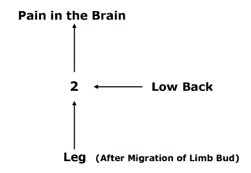
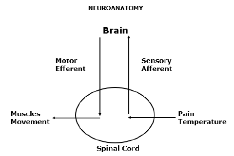
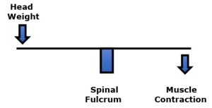
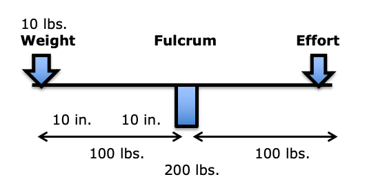
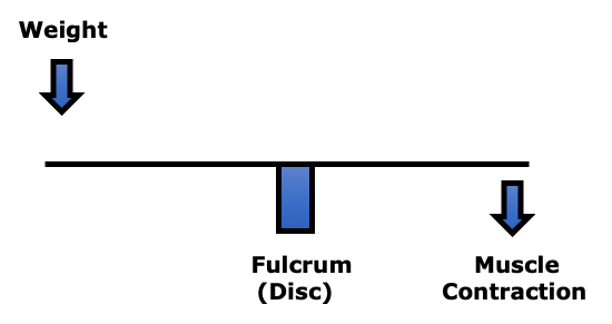
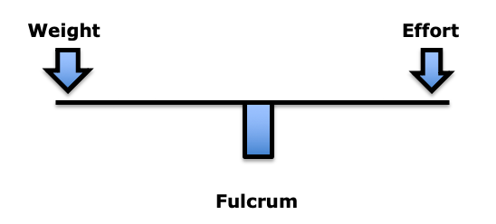
    In the first-class lever mechanical system, the fulcrum is between the weight and the effort. The fulcrum is the site of greatest mechanical stress.

    In this model, weight is important. But, more important than weight is the distance the weight is from the fulcrum. This is load (3). To maintain upright posture, the load must be counterbalanced by effort on the opposite side of the fulcrum. This effort is supplied by muscle contraction. For example:

    If the weight is 10 lbs., and the distance from the fulcrum was 10 inches (the lever arm), the load on the fulcrum would be 100 lbs. (10 X 10). In order to remain balanced, the effort (muscle contraction) on the opposite side of the fulcrum would have to also be 100 lbs. The effective total load applied to the fulcrum would be 200 lbs. Thus, an actual weight of 10 lbs. would have an effective load on the fulcrum of 200 lbs.

    In summary, the load experienced at the fulcrum of a first-class lever system is dependent upon three factors:

    • The magnitude of the weight
    • The distance the weight is away from the fulcrum (lever arm)
    • The addition of the counterbalancing muscle effort required to remain balanced

    In the low back, the fulcrum of the first-class lever of upright posture is primarily the intervertebral disc. In the low back, the facet joints bear very little weight (4). In the low back, postural distortions primarily affect the lumbar spine intervertebral discs.

    In their 1990 book Clinical Biomechanics of the Spine (1), Augustus White, MD, and Manohar Panjabi, PhD, state:

    “The load on the discs is a combined result of the object weight, the upper body weight, the back-muscle forces, and their respective lever arms to the disc center.”

    In her book Move Your DNA: Restore Your Health Through Natural Movement, biomechanist Katy Bowman clearly explains the difference between weight and load, emphasizing that the real problem is the load, which is the weight multiplied by a lever distance from a fulcrum, and of course adding the counter-balancing efforts of muscles. She states (3):

    “The loads created by gravity depend upon our physical position relative to the gravitational force.”

    The load created by gravity differs depending on alignment with the “perpendicular force of gravity.”

    “Every unique joint configuration, and the way that joint configuration is positioned relative to gravity, and every motion created, and the way that motion was initiated, creates a unique load that in turn creates a very specific pattern of strain in the body.” This is called “load profile.”

    “It’s not the weight that breaks you down, it’s the load created by the way you carry it.”

    “Loads are often oversimplified to ‘weight’ because it makes them easier to understand, but there is much more going on with your sore knee (or foot, or back, or pelvic floor) than your weight.”

    “Weight is not the be-all and end-all of loads. When you want to improve your health, it’s much more important to consider how you carry your weight than to spend hours contemplating the lone data point that is Your Weight.”

    The Intervertebral Disc
    (the fulcrum)

    The primary reason people seek chiropractic care is for chronic low back pain (5). Studies indicate that chronic low back pain is primarily discogenic (6, 7, 8, 9, 10). It is also understood that the intervertebral disc is particularly intolerant to rotational stress (4).

    The fibers of the annulus (outer portion) of the intervertebral disc are arranged in layers, and each layer is crossed in opposite directions. During chronic rotational stress on the disc, half of the annular fibers become tense, and the other half become lax. Since rotational stress applied to the annulus is resisted by only half of the annular fibers, the disc is operating at only half strength. This increases the vulnerability of the disc to injury and degenerative disease.

    Crossed Annular Fibers of the Intervertebral Disc

    A common cause of chronic rotational stress on the L5-S1 intervertebral disc is postural pelvic unleveling. This was established in 1983 in a study where standing radiographs of the pelvis and lumbar spine were exposed in 288 consecutive patients with chronic low back pain and in 366 asymptomatic controls (11). Findings showed that 73% of the subjects assessed had meaningful inequality of a lower limb (>5 mm shortness), resulting in pelvic unleveling. The incidence of pelvic unleveling in LBP patients was significantly higher than in asymptomatic controls (more than twice as much).

    The counter-rotational stresses on the L5-S1 intervertebral disc:

    Axial View From Above

    • The L5 spinous process will rotate to the right of midline, towards the side of the long leg and/or higher hemi-pelvis. This causes a counterclockwise rotation of the L5-S1 intervertebral disc.
    • The pubic symphysis and pelvis will also rotate to the right of midline, also towards the side of the long leg and/or higher hemi-pelvis. Because the pubic symphysis is in the anterior, this causes a clockwise rotation of the pelvis and sacrum, and a clockwise rotation of the L5-S1 intervertebral disc.
    • This results in “significant” counter-rotational stresses, primarily at the L5-S1 intervertebral disc. The consequences of these counter- rotational stresses at the L5 disc are accelerated disc degeneration and degradation, back pain, and sciatica. Such aberrant biomechanics, especially when chronic and even when asymptomatic, predispose the intervertebral disc to injury and pain.

    John Fitzgerald Kennedy

    John Fitzgerald Kennedy was born in 1917. Growing up he suffered from a number of health problems, including scarlet fever, whooping cough, measles, chicken pox, an appendectomy, colitis, and a chronically bad back. His earliest low back pain diagnosis was attributed to a herniated disc injury sustained while playing football (12).

    Prior to the United States involvement in World War II (December 7, 1941), Kennedy was medically disqualified from Officer Candidate School because of his bad back. Through perseverance, Kennedy eventually earned the rank of Lieutenant and was assigned as a commander of a patrol torpedo boat (PT-109) in the South Pacific.

    While on patrol in the South Pacific, August 2, 1943, Kennedy’s PT-109 was struck and split in half by a much larger, fast-moving Japanese destroyer. Two of Kennedy’s crew were killed outright, and Kennedy’s already weak back was significantly reinjured (probably a second disc herniation).

    In the aftermath of the destruction of his PT-109, Kennedy’s heroism to save his crew became legendary. His behavior became the topic of articles, books, movies, and political speeches. Kennedy was awarded a Purple Heart as well as the Navy and Marine Corps Medal for his leadership and courage.

    The Secretary of the Navy stated:

    “For extremely heroic conduct as Commanding Officer of Motor Torpedo Boat 109 following the collision and sinking of that vessel in the Pacific War area on August 1–2, 1943. Unmindful of personal danger, Lieutenant Kennedy unhesitatingly braved the difficulties and hazards of darkness to direct rescue operations, swimming many hours to secure aid and food after he had succeeded in getting his crew ashore. His outstanding courage, endurance and leadership contributed to the saving of several lives and were in keeping with the highest traditions of the United States Naval Service.”

    When Kennedy and his crew were eventually rescued on August 8, 1943, Kennedy was non-ambulatory. He was given a cane to help him up. That cane, along with a picture of Kennedy using it, can be seen in the Smithsonian Museum in Washington, D.C. (13).

    Despite his back injury, Kennedy only took a month off before returning to command another PT boat. As a consequence of his back injuries, Kennedy was relieved of command on November 18, 1941. He spent months at a military hospital recovering from his back injuries. Because of his low back physical disability, Kennedy was honorably discharged on March 1, 1945.

    In 1946, Kennedy was elected to Congress. In 1952, Kennedy was elected to the Senate. During these years, Kennedy’s back problems continued to worsen (14).

    In 1954, Senator Kennedy underwent a spinal fusion operation (14):

    This fusion operation resulted in chronic infection and the surgical wound was not healing. In February 1955, in yet another operation, the fusion hardware was surgically removed. Kennedy remained non-ambulatory through May of 1955. In desperation, Kennedy was referred to an internal medicine cardiologist from Cornell University, Janet Travell, MD.

    Janet Travell

    Janet Travell was born in New York in 1901. She died in 1997 at the age of 96. Both of her parents were physicians. In 1926, she received her medical degree from Cornell University Medical College in New York City, where she graduated at the head of her class. She was the first female to graduate from Cornell. In the first three decades of her professional career, she practiced cardiology while teaching pharmacology at Cornell. Her interest in musculoskeletal pain came about as a consequence of the neck, trunk, shoulder, and arm pains her cardiac patients suffered (15).

    When Dr. Travell first saw Senator Kennedy in 1955, she had already developed a reputation for excellence in managing chronic musculoskeletal disorders (16, 17, 18, 19). Her experience and expertise led her to look to mechanical causes and/or contributors to Kennedy’s chronic disabling back problems.

    Dr. Travell “discovered that one of [Senator] Kennedy’s legs was shorter than the other and made heel lifts for all of his left shoes to counter that additional source of stress on his back…. Dr. Travell had a workbench in her office and made lifts for both patients and family members. ‘One of the first things I did for him [Kennedy] was to institute a heel lift—a correction for the difference in leg length.’” (20)

    At Dr. Travell’s direction, Senator Kennedy spent his 38th birthday in the hospital. Neurosurgeons T. Glenn Pait and Justin T. Dowdy note (14):

    “In 1955, Kennedy was introduced to Dr. Janet Travell, a Cornell University pharmacologist and internal medicine specialist.”

    “Dr. Travell promptly admitted Senator Kennedy to the New York Hospital-Cornell Medical Center to jump-start his rehabilitation program.”

    “[Travell] introduced Kennedy to what would, in a few short years, become a symbol of his presidency—the rocking chair.”

     “More importantly, it marks the end of his major back surgeries and a shift in focus toward muscular and environmental factors contributing to his back pain.”

    “The emphasis regarding treatment of his back would move in a more cautious direction going forward, and notable functional restoration would be seen over the next few months.”

    Importantly, Drs. Pait and Dowdy also note (14):

    “In addition to the rocking chair, Travell made other changes, including providing Kennedy with a heel lift— raising the possibility of pelvic obliquity and leg-length discrepancy as a contributing factor in Kennedy’s low back pain.”

     In 2003, Dr. Travell’s daughter wrote (15):

    “Senator Kennedy received so much relief of pain from my mother’s medical treatments that he had ‘new hope for a life free from crutches if not from backache’.”

    In 2003, James Bagg wrote this, pertaining to Dr. Travell (20):

    “Jack Kennedy saw a great many physicians over the course of his short life, but one of them, according to his brother Bobby, enabled Jack to become President of the United States.”

    Kennedy considered Travell to be a medical genius (15). When Kennedy was elected to the presidency of the United States, he asked Dr. Travell to be his personal White House Physician, becoming the first woman and one of the few civilians (non-military) to hold that post. After President Kennedy’s assassination in 1963, his successor, President Lyndon B. Johnson, asked Dr. Travell to stay on in the White House as his physician. A year and a half later, she resigned to return to private practice and to the position of Associate Clinical Professor of Medicine at George Washington University. She became Emeritus Clinical Professor of Medicine from 1970–1988 and Honorary Clinical Professor of Medicine from 1988 until her death in 1997. Dr. Travell remained professionally active until the end of her life, writing articles, giving lectures, and attending conferences.

    In her writings and books, Dr. Travell recognized that a common yet overlooked “perpetuating factor” for chronic discogenic back pain and counterbalancing muscular stress was a structural difference in the length of the lower limbs (21, 22, 23). It has been documented since 1946 that about 75% of people have legs of unequal lengths, and that about a third of people have leg length differences that contribute to chronic back pain. As a rule, the sacrum is lower on the side of the short leg (see drawing below). The spinal column initially tilts towards the short leg, then compensates back to the midline as a consequence of chronic contraction of the quadratus lumborum muscle (24).

    Posterior to Anterior View From Behind

    • The sacrum is lower on the side of the short leg (left in this drawing).
    • The spinal column initially tilts towards the short leg, then compensates back to the midline as a consequence of contraction of the quadratus lumborum
    • The lumbar spinous processes (posterior) rotate towards the long leg. The pubic symphysis (anterior) also rotates towards the long leg. The consequent counter-rotational forces abnormally stress the L5 intervertebral disc.

    Dr. Travell noted that the solution was a proper heal lift, inserted into the shoe with the goal of leveling the pelvis and removing the counter-rotational stress of the L5 intervertebral disc. This intervention was included in her management of Senator Kennedy.

    SUMMARY

    The war heroism of Lieutenant Kennedy, his political importance as Congressman, Senator, and President of the United States, and the fact that the best of contemporary medical care failed to help his chronic back problems and disability, led to a profound change in the management of back pain. Janet Travell, a cardiologist with an interest in musculoskeletal pain syndromes, looked at then Senator Kennedy’s back from a mechanical perspective. Her care included the use of a rocking chair and the insertion of a heel lift to level his pelvis and remove chronic rotational stress to his L5 intervertebral disc. This approach would also relieve the chronic contraction of the counter-balancing muscles, markedly reducing fulcrum (disc) stress.

    Travell’s approach worked, at least better than any other intervention, especially considering the tremendous damage and infections that had already been done by failed surgeries.

    These and other mechanical interventions are common place, the rule, in chiropractic clinical practice. Over the decades, numerous additional studies continued to document the relationship between the anatomical short leg syndrome, pelvic unleveling, and chronic back pain (25, 26, 27, 28, 29, 30, 31, 32, 33, 34).

    REFERENCES

    1. White AA, Panjabi MM; Clinical Biomechanics of the Spine; Second Edition; Lippincott; 1990.
    2. Cailliet R; Soft Tissue Pain and Disability; 3rd Edition; FA Davis Company; 1996.
    3. Bowman K; Move Your DNA: Restore Your Health Through Natural Movement; Propriometrics Press; 2017.
    4. Kapandji IA; The Physiology of the Joints; Volume 3; The Trunk and the Vertebral Column; Churchill Livingstone; 1974.
    5. Adams J, Peng W, Cramer H, Sundberg T, Moore C; The Prevalence, Patterns, and Predictors of Chiropractic Use Among US Adults; Results From the 2012 National Health Interview Survey; Spine; December 1, 2017; Vol. 42; No. 23; pp. 1810–1816.
    6. Nachemson AL; The Lumbar Spine, An Orthopedic Challenge; Spine; Vol. 1; No. 1; March 1976; pp. 59-71.
    7. Mooney V; Where Is the Pain Coming From?; Spine; Vol. 12; No. 8; 1987; pp. 754-759.
    8. Kuslich S, Ulstrom C, Michael C; The Tissue Origin of Low Back Pain and Sciatica: A Report of Pain Response to Tissue Stimulation During Operations on the Lumbar Spine Using Local Anesthesia; Orthopedic Clinics of North America; Vol. 22; No. 2; April 1991; pp. 181-7.
    9. Izzo R, Popolizio T, D’Aprile P, Muto M; Spine Pain; European Journal of Radiology; May 2015; Vol. 84; pp. 746–756.
    10. Groh AMR, Fournier DE, Battie MC, Seguin CA; Innervation of the Human Intervertebral Disc: A Scoping Review; Pain Medicine; June 4, 2021; Vol. 22; No. 6; pp. 1281–1304.
    11. Friberg O; Clinical Symptoms and Biomechanics of Lumbar Spine and Hip Joint in Leg Length Inequality; Spine; September 1983; Vol. 8; No. 6; pp. 643-651.
    12. https://www.jfklibrary.org/learn/about-jfk/life-of-john-f-kennedy
    13. https://www.smithsonianmag.com/history/remembering-pt-109-63427209/
    14. Pait TG, Dowdy JT; John F. Kennedy’s Back: Chronic Pain, Failed Surgeries, and the Story of its Effects on His Life and Death; Journal of Neurosurgery Spine; September 2017; Vol. 27; No. 3; pp. 247-255.
    15. Wilson V; Janet G. Travell, MD: A Daughter’s Recollection; Texas Heart Institute Journal; 2003; Vol. 30; No. 1; pp. 8–12.
    16. pain-education.com/dr-travell.html‎; A Tribute to Dr. Janet Travell; accessed December 11, 2013.
    17. Lacayo R; How Sick Was J.F.K.?; TIME; November 24, 2002.
    18. Altman LK, Purdum TS; In J.F.K. File, Hidden Illness, Pain and Pills; The New York Times; November 17, 2002.
    19. Dallek R; The Medical Ordeals of JFK; The Atlantic; December 2002.
    20. Bagg JE; The President’s Physician; Texas Heart Institute Journal; 2003; 30; No. 1; pp. 1–2.
    21. Travell J, Simons D; Myofascial Pain and Dysfunction, the Trigger Point Manual; New York; Williams & Wilkins; 1983.
    22. Travell J, Simons D; Myofascial Pain and Dysfunction, the Trigger Point Manual: THE LOWER EXTREMITIES; New York; Williams & Wilkins; 1992.
    23. Simons D, Travell J; Travell & Simons’, Myofascial Pain and Dysfunction, the Trigger Point Manual: Volume 1, Upper Half of Body; Baltimore; Williams & Wilkins; 1999.
    24. Rush WA, Steiner HA; A Study of Lower Extremity Length Inequality; American Journal of Roentgenology and Radium Therapy; November 1946; Vol. 51; No. 5; pp. 616-623.
    25. Sicuranza B, Richards J, Tisdall L; The Short Leg Syndrome in Obstetrics and Gynecology; American Journal of Obstetrics and Gynecology; May 15, 1970; Vol. 107; No. 2; pp. 217-219.
    26. Giles LG, Taylor JR; Low-back Pain Associated with Leg Length Inequality; Spine; 1981 Sep-Oct; Vol. 6; No. 5; pp. 510-251.
    27. Friberg O; Clinical Symptoms and Biomechanics of Lumbar Spine and Hip Joint in Leg Length Inequality; Spine; September 1983; Vol. 8; No. 6; pp. 643-651.
    28. Gofton JP; Persistent Low Back Pain and Leg Length Disparity; Journal of Rheumatology; August 1985; Vol. 12; No. 4; pp. 747-750.
    29. Helliwell M; Leg Length Inequality and Low Back Pain; The Practitioner; May 1985; Vol. 229; pp. 483-485.
    30. Defrin R, Benyamin SB, Dov Aldubi R, Pick CG; Conservative Correction of Leg-Length Discrepancies of 10 mm or Less for the Relief of Chronic Low Back Pain; Archives of Physical Medicine and Rehabilitation; November 2005; Vol. 86; No. 11; pp. 2075-2080.
    31. Golightly YM, Tate JJ, Burns CB, Gross MT; Changes in Pain and Disability Secondary to Shoe Lift Intervention in Subjects With Limb Length Inequality and Chronic Low Back Pain; Journal of Orthopaedic & Sports Physical Therapy; Vol. 37; No. 7; July 2007; pp. 380-388.
    32. Balik SM, Kanat A, Erkut A, Ozdemir B, Batcik OE; Inequality in Leg Length is Important for the Understanding of the Pathophysiology of Lumbar Disc Herniation; Journal of Craniovertebral Junction; Spine; April-June 2016; Vol. 7; No. 2; pp. 87-90.
    33. Cambron JA, Dexheimer JM, Duarte M, Freels S; Shoe Orthotics for the Treatment of Chronic Low Back Pain: A Randomized Controlled Trial; Archives of Physical Medicine and Rehabilitation; September, 2017; Vol. 98; No. 9; pp. 1752-1762.
    34. Sheha ED; Steinhaus ME; Kim HJ; Cunningham ME; Fragomen AT; Rozbruch SR; Leg-Length Discrepancy, Functional Scoliosis, and Low Back Pain; Journal of Bone and Joint Surgery Reviews; August 8, 2018; Vol. 6; No. 8; pp. e6.
  • The Uniqueness of Chiropractic Thinking, Approach, and Treatment of Spinal Syndromes

    The Uniqueness of Chiropractic Thinking, Approach, and Treatment of Spinal Syndromes

    In his 1985 book, The Dental Physician (1), dentist Alfred Fonder, DDS, presents a series of case studies showing the integrated posture between the feet, legs, pelvis, lumbar spine, thoracic spine, cervical spine and temporomandibular joint.

    His message is simple: a mechanical problem in any one part of the human body will affect and cause mechanical problems in the entire kinetic chain of alignment and motion.

    The entire body is mechanically integrated.

    ••••

    In 1895, German physicist Wilhelm Conrad Roentgen discovered x-rays and radiographs. For this discovery, in 1901, he was awarded the first Nobel Prize in Physics (2). With the addition of spinal x-rays, the understanding of spinal and whole-body biomechanics drastically changed.

    ••••

    In 1916, Harvard Orthopedic Surgeon Robert W. Lovett, MD, published the third edition of his book Lateral Curvature of the Spine and Round Shoulders (3). This text has many examples of spinal radiographs, showing a biomechanical relationship between leg length, pelvic leveling, spinal scoliosis, and back pain.

    ••••

    In 1927, American surgeon Dudley Joy Morton, MD, described a common foot problem that caused not only chronic foot pain and disability, but also affected the ankle, knee, pelvis and spine (4). Dr. Morton named the problem “Morton’s Toe.” Morton’s Toe is an anatomically short first metatarsal, giving the foot the appearance of an abnormally long second toe. The syndrome would cause abnormal stress at the first metatarsal-phalangeal joint, and the patient would compensate with altered foot and kinetic ambulatory function which could influence whole body mechanical functions, symptoms, and signs.

    ••••

    The work of Morton became instrumental in the treatment of senator John F. Kennedy’s chronic low back pain by physician Janet Travell, MD, in 1955 (5). Then Lieutenant Kennedy’s notorious back problems were triggered during the legendary sinking of his boat PT-109 in the Pacific during WWII.

    Kennedy never fully recovered from his back injuries. In 1954, Kennedy underwent a second attempted spinal fusion operation, and it did not go well. He nearly died, and his recovery took 8 months. The following year, Kennedy came under the care of myofascial pain expert Janet Travell, MD (6). After studying the work of Dr. Morton, Dr. Travell realized that all mechanical problems could cause compensatory contraction in the musculature system, leading to treatable findings called trigger points and a diagnosis of myofascial pain syndrome.

    Dr. Travell’s treatment of Senator Kennedy in 1955 was a resounding success, and it was headline news. When Kennedy was elected president of the United States (taking office in 1961), he chose Dr. Travell to be his personal White House Physician. Dr. Travell was the first female and civilian physician to hold this prestigious office (7, 8, 9).

    When Dr. Travell first began treating Senator Kennedy’s mechanical problems and associated trigger points, he was non-ambulatory. His improvement was so impressive that Dr. Travell’s daughter wrote (6):

    “Senator Kennedy received so much relief of pain from my mother’s medical treatments that he had ‘new hope for a life free from crutches if not from backache.’”

    In 2003, James Bagg wrote [pertaining to Dr. Travell] (5):

    “Jack Kennedy saw a great many physicians over the course of his short life, but one of them, according to his brother Bobby, enabled Jack to become President of the United States.”

    The major revelation of Dr. Travell was that a toe problem would cause a foot problem which would cause a leg problem which would cause a spine problem. The resolution of the spinal complaints would require first rectifying the toe problem. Dr. Travell realized that both a toe problem and an anatomical short leg could both, independently, cause a spine problem. Dr. Travell realized that Senator Kennedy’s left leg was three-quarters of an inch shorter than that of his right leg.

    ••••

    In 1946, Lieutenant Colonel Weaver A. Rush and Captain Howard A. Steiner of the X-Ray Department of the Regional Station Hospital of Fort Leonard Wood, Missouri, exposed upright lumbosacral x-rays on 1,000 soldiers (10). All study subjects suffered from low back pain. These authors noted:

    • 23% of the soldiers had legs of equal length.
    • 77% of the soldiers had unequal length of their legs.

    These authors noted that the short leg was associated with a tilt of the pelvis and a scoliosis. They noted:

    “[Whenever there is a pelvic tilt], there exists coincidentally a scoliosis of the lumbar spine.”

    “Because this scoliosis, in all instances, compensates for the tilt of the pelvis, it is referred to by us as compensatory scoliosis.”

    “The existence of this compensatory scoliosis in the presence of a tilted pelvis due to shortening of one or the other lower extremity is believed by us to have clinical significance.”

    “It was a general consistent observation that the degree of scoliosis was proportionate to the degree of pelvic tilt. An individual who has a shortened leg will have to compensate completely if he intends to hold the upper portion of his body erect or in the midsagittal plane.”

    “A consistent observation which has been made is that in those cases with a shortened leg there is a corresponding tilt of the pelvis and a compensatory scoliosis of the lumbar spine.”

    Lieutenant Colonel Rush and Captain Steiner observed that leg length differences exceeding 5 mm were associated with the greatest low back pain or disability, and therefore 5 mm is labeled as being a “marked difference.” The authors stated:

    “For this reason, it is our opinion that the existence of such a condition [a short leg exceeding 5 mm] is significant from the standpoint of symptomatology and disability.”

    Dr. Travell measured Senator Kennedy’s left short leg at about three quarters of an inch, or about 18 mm.

    Senator Keneddy's left short leg

    Long Right Leg        Short Left Leg


    ••••

    In 1980, physician, neurologist, and chiropractor Scott Haldeman wrote (11):

    “If one leg is only a quarter of an inch shorter, the entire body can be tilted enough to cause pain throughout the skeletal system. So slight a difference can distort the entire skeleton, causing a ‘seesaw’ condition that pulls one shoulder down a full inch. This may never be recognized until the person is hurt in a fall or an accident.”

    Dr. Haldeman is from the Department of Neurology, University of California, Irvine, California. The shoulder tilt magnification compensation to a pelvic unleveling may result in symptoms including neck pain and headaches. Once again, it is noted that the different regions of the spine are mechanically interconnected.

    ••••

    In 1982, Richard Rothman, MD, PhD, and Frederick Simeone, MD, published the second edition of their book, The Spine (12). Dr. Rothman was a Professor of Orthopaedic Surgery at the University of Pennsylvania School of Medicine, and Chief of Orthopedic Surgery at the Pennsylvania Hospital in Philadelphia (d. 2018). Dr. Simeone was a Professor of Neurosurgery at the University of Pennsylvania School of Medicine, Chief of Neurosurgery at the Pennsylvania Hospital, and Director of Neurosurgery at the Elliott Neurological Center of Pennsylvania Hospital.

    Chapter 2 of Drs. Rothman’s and Simeone’s book is titled:

    “Applied Anatomy of the Spine”

    This chapter is written by Wesley Parke, PhD. In 1982, Dr. Parke was a Professor and Chairman of the Department of Anatomy at the University of South Dakota School of Medicine (d. 2005). In this chapter, Dr. Parke writes:

    “Although the 23 or 24 individual motor segments must be considered in relation to spinal column as a whole, no congenital or acquired disorder of a single major component of a unit can exist without affecting first the functions of the other components of the same unit and then the functions of other levels of the spine.”

    Dr. Parke is also noting that the entire spinal column is an integrated functioning unit.

    ••••

    In 1987, an important addition to the understanding of modern integrative biomechanics was published by Finish physician Ora Friberg, MD (14). Dr. Friberg exposed standing radiographs of the pelvis and lumbar spine in 288 consecutive patients with chronic low back pain and in 366 asymptomatic controls. His findings showed that 73% of the subjects assessed had meaningful inequality of a lower limb (>5 mm shortness). The incidence of leg length inequality in low back pain patients was significantly higher than in asymptomatic controls (more than twice as much).

    Importantly, Dr. Friberg emphasized the existence of counter-rotational stresses on the L5-S1 intervertebral disc in the presence of pelvic unleveling. The lumbar intervertebral discs are intolerant of chronic rotational stress because the nature of the crisscross annulus orientation essentially reduces mechanical integrity by half (15, 16). This concept would have a particular relevance to the chronic back problems and failed surgeries of former US President John Kennedy.

     Crisscross Annular Fibers of the Intervertebral Disc

    Axial View From Above

    Midline

    Short Left Leg  Long Right Leg

    The L5 spinous process will rotate to the right of midline, towards the side of the long leg (counterclockwise rotation).

    The pubic symphysis and pelvis will also rotate to the right of midline, towards the side of the long leg (clockwise rotation).

    This results in clinically meaningful counter-rotational stresses, primarily at the L5-S1 intervertebral disc. The consequences of these counter-rotational stresses at the L5 disc are accelerated disc degeneration and degradation, back pain, and sciatica.

    ••••

    The concept of the entire spine functioning as a single integrated unit was nicely noted in the reference books (1987, #17; 1994, #18) written by rheumatologist John Bland, MD (d. 2008). Dr. Bland was a Professor of Medicine at the University of Vermont College of Medicine. His books were titled (17, 18):

    Disorders of the Cervical Spine

    In this book, Dr. Bland states:

    “We tend to divide the examination of the spine into regions: cervical, thoracic, and lumbar spine clinical studies.

    This is a mistake.

    The three units are closely interrelated structurally and functionally – a whole person with a whole spine.

    The cervical spine may be symptomatic because of a thoracic or lumbar spine abnormality, and vice versa!

    Sometimes treating a lumbar spine will relieve a cervical spine syndrome, or proper management of the cervical spine will relieve low backache.”

    ••••

    In 1989, podiatrist Steven Subotnick, DPM, published his book titled Sports Medicine of the Lower Extremity (19). Dr. Subotnick was a Clinical Professor in the Departments of Biomechanics and Surgery at the California College of Podiatric Medicine, San Francisco, California. In his book, Dr. Subotnick notes that a pronation of the foot would result in a functionally short leg and a compensatory scoliosis. The scoliosis would affect the thoracic spine, the scapula, and result in a “compensation on the cervical area.”

    Dr. Subotnick is another provider noting the integrated function between the foot, leg, pelvis, and entire spinal column.

    ••••

    Recently, three reference texts have been published emphasizing the integrative nature of whole-body biomechanics:

    • Energy Medicine, The Scientific Basis, by James Oschman, PhD, 2000 (20).
    • Anatomy Trains: Myofascial Meridians for Manual and Movement Therapists; by Thomas Myers, 2001 (21).
    • Move Your DNA: Restore Your Health Through Natural Movement, Katy Bowman, 2017 (22).

    ••••

    In his 2000 book Energy Medicine, The Scientific Basis, James Oschman, PhD, notes that the entire body is mechanically interconnected via a connective tissue cytoskeletal matrix called the “tensegrity matrix.” He notes that mechanical providers of care, including chiropractors, solve health problems by attending to the quality of the tensegrity matrix.

    Dr. Oschman notes that gravity is the most potent physical influence in any human life. He notes that tensegrity accounts for the fact that inflexibility or shortening in one tissue influences structure and movement in other tissues. He notes that the entire body is mechanically integrated and interconnected. An imbalance in one part of the body will affect the whole body. He states:

    “The basic principle of gravitational biology is known to any child who plays with blocks. The center of gravity of each block must be vertically above the center of gravity of the one below, to have a stable, balanced arrangement. If the center of gravity of one block lies outside of the gravity line, stability is compromised.”

    “There is only one stable, strain-free arrangement of the parts of the human body. Any variation from this orientation will require corresponding compensations in other parts of the support system. Misalignment of any part will affect the whole system.”

    ••••

    In her 2017 book Move Your DNA: Restore Your Health Through Natural Movement, biomechanist Katy Bowman agrees with Dr. Oschman, noting, “gravity is one force your body responds to constantly.” Ms. Bowman discusses the importance of understanding that humans experience loads 100% percent of the time while existing and functioning in a gravity environment.

    Each tissue type responds differently to a load, yet “they are all connected, which means that a load you perceive as only happening in one part of your body is actually affecting all other parts of you, and affecting each part uniquely.” Once again, this is an understanding and statement that the whole body is mechanically integrated and interconnected. Ms. Bowman notes:

    “Tissues that spend most of their time in a fixed position will adapt to that position by making alterations that are fairly permanent.”

    “An under-moved area of the body will experience increases in the connective tissues.”

    Ms. Bowman calls these “extra- connected” areas of the body “sticky spots.”

    “On the cellular level, a sticky spot interferes with the transmission of forces throughout your tissues—mechanical signals that give cells context about loads placed upon them as well as position.”

    When a joint has a sticky spot, “you compensate by moving other joints,” which may “come with a heavy dose of damage.” Areas just outside of the sticky spot “experience unnaturally high loads.”

    “We need a tool to measure the loads, both on the whole body and on every body part. The tool I use is alignment.”

    The “sticky spots” discussed by Ms. Bowman are an orthopedic component of the joint dysfunction that chiropractors describe as the subluxation. Common terminology for “sticky spots” within chiropractic education and clinical practice is “the fibrosis of repair.” Chiropractors are well aware that mechanical care given to a “sticky spot” will mechanically influence the entire system. Chiropractors are also aware that compensatory sticky spots also require mechanical care to enhance optimum and speedy resolution of clinical symptoms and signs.

    ••••

    In 2004, clinicians from Rey Juan Carlos University, Spain, published a randomized control trial of mechanical based care for the management of whiplash injury (23). The aim of this clinical trial was to compare the results obtained with a manipulative protocol from the results obtained with a conventional physiotherapy treatment in patients suffering from whiplash injury. The authors used 380 acute whiplash injury (less than 3 months duration) subjects. All subjects were Quebec Task Force grades II and III:

    GRADE II = neck complaint and musculoskeletal signs

    GRADE III = neck complaint, musculoskeletal signs, and neurologic signs

    The authors note:

    “The goal of joint manipulation is to restore maximal, pain-free movement of the musculoskeletal system.”

    “Our clinical experience with these patients [whiplash-injured] has demonstrated that manipulative treatment gives better results than conventional physiotherapy treatment.”

    Manipulation is “effective in the management of whiplash injury.”

    “Manipulative treatment is more effective in the management of whiplash injury than conventional physiotherapy treatment.”

    An interesting observation by these authors is that optimal management of the neck and head complaints required that manipulation also had to be applied to mechanical findings in the lumbar spine and pelvis. Once again, this concept supports the concept that the spine is a single functioning unit: a whole person with a whole spine.

    ••••

    In 2005, physician Steven Glassman, MD, and colleagues, published a study of 752 patients by looking at the effect of sagittal spinal balance on pain and health profiles (24). Using a plumb line, the authors assessed the magnitude of a forward head/neck complex and how it affected low back pain and disability. The authors noted:

    “There was clear evidence of increased pain and decreased function as the magnitude of positive sagittal balance increased.”

    Once again, it is clear that neck alignment has a significant influence on low back pain.

    ••••

    In 2015, a group of physicians from New York, Chicago, Virginia, Oregon, Texas, California, Colorado, and the International Spine Study Group, published a study documenting the influence of the neck on the low back. Similar to the Glassman study above, the authors measured sagittal spinal balance using a plumb line.

    The authors note that when the head is sagittally forward of the sacrum, the patient will have an increase in low back pain and disability. They also note that any improvement in cervical spinal sagittal alignment will proportionally improve low back pain and disability.

    Putting It All Together

    Patients present to chiropractors with a variety of musculoskeletal complaints, primarily back and neck pain (26). The information presented here shows that the entire body is mechanically integrated. It is standard for a chiropractor to evaluate the entire body mechanically, regardless as to the actual location of the patient’s primary complaint. Examining and treating the entire spine, including regions that may be asymptomatic, will as a rule enhance optimal clinical improvement. Such care will logically expand to include the feet, knees, and hips.


    REFERENCES

    1. Fonder A; The Dental Physician; Medical-Dental Arts; 1985.
    2. https://www.nobelprize.org/prizes/physics/1901/rontgen/facts/; accessed May 27, 2022.
    3. Lovett RW; Lateral Curvature of the Spine and Round Shoulders; third edition; P. Blakiston’s Son & Co.; Philadelphia; 1916.
    4. Morton DJ; METATARSUS ATAVICUS: The Identification of a Distinctive Type of Foot Disorder; Journal of Bone and Joint Surgery, American; July 1, 1927; 9; No. 3; pp. 531-544.
    5. Bagg JE; The President’s Physician; Texas Heart Institute Journal; 2003; 30; No. 1; pp. 1–2.
    6. Wilson V; Janet G. Travell, MD: A Daughter’s Recollection; Texas Heart Institute Journal; 2003; Vol. 30; No. 1; pp. 8–12.
    7. Lacayo R; How Sick Was J.F.K.?; TIME; November 24, 2002.
    8. Altman LK, Purdum TS; In J.F.K. File, Hidden Illness, Pain and Pills; The New York Times; November 17, 2002.
    9. Dallek R; The Medical Ordeals of JFK; The Atlantic; December 2002.
    10. Rush WA, Steiner HA; A Study of Lower Extremity Length Inequality; American Journal of Roentgenology and Radium Therapy; November 1946; Vol. 56; No. 5; pp. 616-623.
    11. Haldeman S; Modern Developments in the Principles and Practice of Chiropractic; Appleton-Century-Crofts; New York; 1980.
    12. Rothman RH, Simeone FA; The Spine; second edition; WB Saunders Company; 1982.
    13. Parke WW; “Applied Anatomy of the Spine” chapter 2 in Rothman and Simeone; The Spine; second edition; WB Saunders Company; 1982.
    14. Friberg O; The statics of postural pelvic tilt scoliosis: A radiographic study on 288 consecutive chronic LBP patients; Clinical Biomechanics; November 1987; Vol. 2; No. 4; pp. 211-219.
    15. Kapandji IA; The Physiology of the Joints; Volume 3; The Trunk and
    16. the Vertebral Column; Churchill Livingstone; 1974.
    17. White AA, Panjabi MM; Clinical Biomechanics of the Spine; Second Edition; JB Lippincott Company; 1990.
    18. Bland J; Disorders of the Cervical Spine; WB Saunders Company; 1987; p. 84.
    19. Bland J; Disorders of the Cervical Spine; WB Saunders Company; 1994; p. 119.
    20. Subotnick SI; Sports Medicine of the Lower Extremity; Churchill Livingstone; 1989.
    21. Oschman J; Energy Medicine, The Scientific Basis; Churchill Livingstone; 2000.
    22. Meyers TW; Anatomy Trains: Myofascial Meridians for Manual and Movement Therapists; Churchill Livingstone; 2001.
    23. Bowman K; Move Your DNA: Restore Your Health Through Natural Movement; Propriometrics Press; 2017.
    24. Fernández-de-las-Peñas C, Fernández-Carnero J, Palomeque del Cerro L; Miangolarra-Page JC; Manipulative Treatment vs. Conventional Physiotherapy Treatment in Whiplash Injury: A Randomized Controlled Trial; Journal of Whiplash & Related Disorders; 2004; Vol. 3; No. 2.
    25. Glassman SD, Bridwell K, Dimar JR, Horton W, Berven S, Schwab F; The Impact of Positive Sagittal Balance in Adult Spinal Deformity; Spine; September 15, 2005; Vol. 30; No. 18; pp. 2024-2029.
    26. Protopsaltis TS, Scheer JK, BS; Terran JS, and 12 others; How the Neck Affects the Back: Changes in Regional Cervical Sagittal Alignment Correlate to HRQOL Improvement in Adult Thoracolumbar Deformity Patients at 2-year Follow-up [HRQOL = health-related quality of life]; Journal of Neurosurgery Spine; August 2015; Vol. 23; No. 2; pp. 153–158.
    27. Adams J, Peng W, Cramer H, Sundberg T, Moore C; The Prevalence, Patterns, and Predictors of Chiropractic Use Among US Adults: Results From the 2012 National Health Interview Survey; Spine; December 1, 2017; Vol. 42; No. 23; pp. 1810–1816.