Category: Advanced

  • Cost-Efficiency and Effectiveness of Chiropractic for Musculoskeletal Complaints

    Cost-Efficiency and Effectiveness of Chiropractic for Musculoskeletal Complaints

    LEGITIMIZING BACKGROUND

    The United States National Library of Medicine (NLM) is operated by the United States federal government, and is the world’s largest medical library. It is located in Bethesda, Maryland. The NLM is part of the National Institutes of Health (NIH), U.S. Department of Health and Human Services. NLM started in 1836 as a small collection of medical books and journals in the office of the U.S. Army Surgeon General (1).

    As the world’s largest medical library, a search engine for the content of the NLM was developed and is called PubMed (2). PubMed was developed and is maintained by the National Center for Biotechnology Information (NCBI) at the U.S. National Library of Medicine (NLM), located at the National Institutes of Health (NIH). PubMed is a globally free resource supporting the search and retrieval of peer-reviewed biomedical and life sciences literature with the aim of improving health–both globally and personally. Presently (2020), the PubMed database contains more than 30 million citations and abstracts of peer-reviewed biomedical literature. Citations in PubMed primarily stem from the biomedicine and health fields, and related disciplines such as life sciences, behavioral sciences, chemical sciences, and bioengineering.

    The PubMed system was offered free to the public starting in June 1997. “If your journal was in MEDLINE/PubMed, it had gone through an exhaustive evaluation, and had earned a badge of legitimacy.” (3)

    The Journal of Chiropractic Humanities is PubMed indexed. It is also currently indexed in Cumulative Index to Nursing and Allied Health Literature (CINAHL), Manual Alternative and Natural Therapy Index System (MANTIS), and the Index to Chiropractic Literature (ICL). (4) The webpage for the journal states (4):

    The Journal of Chiropractic Humanities is a peer-reviewed journal devoted to providing a forum for the chiropractic profession to disseminate information dedicated to chiropractic humanities. The primary purpose of the Journal of Chiropractic Humanities is to foster scholarly debate and interaction within the chiropractic profession regarding the humanities, which includes history, philosophy, linguistics, literature, jurisprudence, ethics, theory, sociology, comparative religions, and aspects of social sciences that address historical or philosophical approaches. The journal’s objective is to fulfill this purpose through careful editorial review and publication of expert work, by creating legitimate dialogue in a field where a diversity of opinion exists, and by providing a professional forum for interaction of these views.

    ••••••••••

    By a significant percentage, the primary reason patients go to chiropractors is for the management of spinal (low back and neck) pain (5). The actual number is 93% (63% for low back pain, 30% for neck pain). The effectiveness of chiropractic care for spinal pain is unquestioned and hence chiropractic care (spinal manipulation) is routinely included in spine pain clinical guidelines (6, 7, 8, 9, 10).

    Increasingly larger percentages of healthcare for Americans is through various government programs (Medicare, Medicaid (Medical), Affordable Health Care (Obamacare), etc.). Costs are always a concern in these programs. An honest and thorough assessment of the costs for chiropractic services for the management of musculoskeletal spine pain is long overdue.

    Such an appraisal was published in the PubMed indexed Journal of Chiropractic Humanities and titled (11):

    Cost-Efficiency and Effectiveness of Including
    Doctors of Chiropractic to Offer Treatment Under Medicaid:

    A Critical Appraisal of Missouri Inclusion
    of Chiropractic Under Missouri Medicaid

    As noted in the title, the article is a critical appraisal of the cost effectiveness for the inclusion of chiropractic services under Missouri Medicaid. Yet, the author’s presentation includes data from published studies throughout the United States’ healthcare delivery system pertaining to the value of chiropractic. Overall, 79 studies are cited. The lead author John R. McGowan, PhD, is an accounting professor at Saint Louis University, St. Louis, Missouri. Undoubtedly as a consequence of his accounting expertise, Dr. McGowan’s study includes extensive mathematical analysis, displayed in many graphs and tables.

    All chiropractors and/or public healthcare policy decision makers who are engaging in efforts to expand the use and inclusion of chiropractic for the complaints of neck and/or back pain should make use of this vital article.

    The authors begin by noting that the vetting of the benefits of chiropractic for the complaints of neck and/or back pain has been flawed, resulting in flawed conclusions that are both harmful to the chiropractic profession as well as harmful to the public good. They state:

    “Policymakers may unintentionally rely on flawed assumptions and methodologies such as static scoring, which we propose results in flawed conclusions.”

    The objectives of this study were to critically evaluate the methodology and conclusions of the fiscal notes prepared by the state of Missouri for including Doctors of Chiropractic (DCs) under Missouri Medicaid and to calculate the savings if Chiropractors were allowed to offer treatment under Missouri Medicaid.

    These authors suggest that when the Missouri Health Division initially assessed the value of Chiropractic, for the Missouri Medicaid program, for the management of neck and low back pain, that their scoring approach was (“unintentionally”) flawed, and as such they undervalued the fiscal benefits of chiropractic. Hence, these authors re-evaluated the value of chiropractic for the Missouri Medicaid program using improved assumptions.

    These authors assessed 3 cost saving values attributed to chiropractic care:

    • Cost savings from chiropractic care v. traditional MD/DO care
    • Cost savings from reductions in spinal surgery
    • Cost savings from reduced use and abuse of opioid prescription drugs

    This article proposes that chiropractors deliver care that is more effective and less costly than the current model.

    Reimbursement for chiropractic care is provided by Medicare in all 50 states, and by Medicaid in most states. The decision to provide coverage in each state is based on policymakers’ perceptions of both benefits and cost. If policymakers’ perceptions are based upon flawed assumptions, everyone loses.

    The distribution of providers for spine care in the United States shows that chiropractic care is second, accounting for more than a quarter of all cases:

    • 61% Medical Doctor and/or Doctor of Osteopathy (MD/DO)
    • 28% Doctor of Chiropractic (DC)
    • 11% from a combination of providers

    The study cites support indicating that orthopedic conditions contribute to more medical expenses than any other condition, and that back and neck pain account for a greater percentage of orthopedic expenses than all other orthopedic conditions. Spine pain is among the top 10 most costly of all medical conditions treated in the United States. They estimate that the 2018 annual expenditures on low back and neck pain is about $120 billion, and that these expenditures do not include those from the decrease in employee productivity. The authors make these points:

    • “Spine pain is a pervasive health care problem in the United States.”
    • “There is an 80% chance that people will have back pain at some point in their lives.”
    • “In the US, neck pain is one of the top musculoskeletal disorders in the adult population.”
    • The fourth most pervasive reason for disability globally is neck pain.
    • “Between 1990 and 2010, the US had a 29% increase in neck pain.”
    • “More US adults have chronic pain (headache, neck or back pain, arthritis, or joint pain) than heart disease, diabetes, and cancer combined.”
    • “Chiropractic-delivered services for back and neck pain are significantly more cost-effective when compared with all alternative approaches.”
    • “The benefits of care provided by doctors of chiropractic (DCs) have been demonstrated by research throughout the US health care system.”
    • “Although there are potentially numerous beneficial clinical outcomes from DC treatment, perhaps the greatest clinical outcome is the reduction of neck and low back pain.”

    The authors cite these studies to support their arguments:

    A 4-year study compared health care expenditures of 700,000 health plan members who had a chiropractic benefit with 1 million members of the same plan without the same benefit and concluded that systematic access to managed chiropractic care lowers overall health care costs and results in improved clinical outcomes. (12)

    A joint study between the American Pain Society and the American College of Physicians considered non-pharmacologic therapies available for acute and chronic low back pain, showing (7):

    • Spinal manipulation was recognized by the panel as an effective treatment for both acute and chronic low back pain.
    • The panel noted that over 90% of spinal manipulations are performed by chiropractors.

    In 2009, Arnold Milstein of Mercer Health and Benefits and Niteesh Choudhry of Harvard Medical School compared chiropractic-delivered care to that provided by medical physicians (13). The authors note:

    • The annual cost for treatment of neck pain by chiropractors was lower than treatment from medical physicians.
    • “When considering effectiveness and cost together, chiropractic physician care for low back pain and neck pain is highly cost-effective and represents a good value in comparison to medical physician care.”
    • “Our findings in combination with existing US studies published in peer-reviewed scientific journals suggest that chiropractic-delivered care for the treatment of low back and neck pain is likely to achieve equal or better health outcomes at a cost that compares very favorably to most therapies that are routinely covered in US health benefit plans.”
    • “Both value and clinical outcomes show marked improvement with the addition of chiropractic coverage for the treatment of low back and neck pain in US employer-sponsored health benefit plans.”

    Over a 2-year period, a BlueCross/BlueShield of Tennessee study examined a fully insured population of 85,000 subscribers (14). Their findings include:

    • “Patients who experience low back pain and seek treatment first from a chiropractic rather than an MD/DO save 40% on average of health care-related costs.”
    • “Episodes of care started with a chiropractor were 20% less expensive than episodes started with an MD/DO.”
    • For BlueCross/BlueShield of Tennessee, chiropractic-initiated episodes of care could lead to an annual cost savings of approximately $2.3 million.
    • “Insurance companies may be inadvertently paying more for care with restricted access to chiropractic-initiated care than if such restrictions were removed.”

    The authors note that seeing a chiropractor does not increase overall healthcare spending. They report:

    • “Adults in the United States who chose chiropractic care, the most common type of complementary and alternative medicine (CAM) used by people with spine problems, has increased by 57% from 1997 to 2006.” (15)
    • “Treatment by CAM providers lowers cost when compared with treatment from traditional medical providers.” (16)
    • Chiropractors provided approximately 75% of all CAM services (17).
    • Overall medical spending is not increased from seeking care from any CAM provider, including a chiropractor.
    • “Hospitalization expenditures were significantly lower for those who used complementary and alternative providers, including chiropractic.”

    A Canadian Study showed that chiropractic care was more cost-effective than usual care (18).

    • Chiropractor-delivered spinal manipulations were very effective for low back pain compared to usual care administered by primary care MDs.
    • Chiropractic-delivered manipulation is less costly than physiotherapy or GP care (19).
    • “Manual therapy techniques are more cost-effective for low back and shoulder pain and disability than care delivered by general practitioners (GPs) alone or with exercise, spinal stabilization, GP advice, advice to remain active, or brief pain management.”
    • Chiropractic-delivered manipulations are less costly and more effective for neck pain than the alternatives.
    • “Evidence showed manual therapy to be economically advantageous compared with other treatments of musculoskeletal conditions.”
    • “Chiropractic-delivered therapy is equally effective and more cost-efficient that usual medical care.”

    A 2015 study from the journal Spine showed (20):

    • Of patients receiving chiropractic manipulation, 94% had a 30% reduction in low back pain after 4 weeks, while only 56% of medical care recipients had the same reduction.
    • “This suggests a 38% increase in effectiveness by seeing a DC first.”

    ••••••••••

    An important but often under appreciated (and unassessed) benefit of chiropractic care is its potential to reduce the costly and dangerous incidence of spinal surgery. The authors make these points:

    • “Research shows that chiropractors are the safest and most effective alternative to surgery for most cases of back pain.”
    • “Published research has shown that spine surgeries can be avoided through chiropractic-managed care.” (21)
    • The odds of surgery for those who first saw a chiropractor were only 1.4%, while the odds for those who first saw a surgeon were 42.7%.
    • “Back pain is over-treated with spine surgeries in the United States.” (22)
    • “Back surgeries are among the most expensive operations performed.” (23)
    • “Per numerous sources, total costs related to spine surgery are projected to be well more than $100,000.”
    • “60% of patients with sciatica benefited from spinal manipulation after attempting medical management and failing to experience any improvement.” (24)
    • Chiropractic treatment “leads to 60% reduction in spinal surgery.”
    • “In light of the research showing that chiropractic-managed care can reduce the incidence of spinal surgery, it is reasonable to suppose that a number of surgeries could be avoided through routine chiropractic management.”
    • “One fact is sure, patients in the Missouri Medicaid system would benefit from the avoidance of spinal surgeries, and this would lead to significant cost savings for the state.”

    ••••••••••

    Another important and also under-appreciated (and unassessed) benefit of chiropractic care is its potential to reduce the costly and dangerous use of narcotic drugs, the opioid epidemic. Opioids, especially with long-term use, increase the risks of addiction, overdose and death. According to the Centers for Disease Control, opioid use disorder and overdose have been a growing problem throughout the United States. The quantity of opioids prescribed has been dramatically increasing since the 1990s, and the number of overdoses and deaths from prescription opioids has also increased. A study in 2018, authored by physicians from Harvard, Johns Hopkins, and Louisiana State University stated (25):

    “Tragically, opioids claimed over 64,000 lives just last year.”

    Pertaining to the opioid epidemic, these authors (McGowan et al), make these points:

    • “Prescription opioid use is steadily increasing across the US and in Missouri.” (26)
    • “Opioid abuse affects all branches of society.”
    • “More than 130 people die each day in the US from opioid overdose.”
    • “The misuse of prescription pain relievers, heroin, and synthetic opioids such as fentanyl and other opioids constitute a serious national crisis.”
    • Opioid use is lower for patients who receive treatment from chiropractors compared to non-recipients (27).
    • “The probability of getting a prescription for opioids was 55% lower in the chiropractic recipient cohort.”
    • “Opioid prescription refills were 78% lower for patients who received treatment from chiropractors.” (28)
    • There is a “growing body of research that is promoting DC-delivered care as an alternative to the use and abuse of opioid prescription drugs.”
    • The over prescription of opioids, “38% of the US population, or 92 million adults, took doctor-prescribed Oxycontin or Percocet.” (29)
    • “The fact that chiropractors do not prescribe opioids” is important. “Chiropractors treat low back and neck pain without resorting to the prescription of opioids or any other type of drugs.”
    • “Missouri Policymakers Recognize Chiropractic Alleviates Opioid Cost” (30)
    • “The Missouri Oversight Committee recognized the savings that would accrue to the state if individuals sought DC care rather than traditional MD/DO care for neck and low back pain.”
    • “Chiropractic care would lead to reductions in costs from the abuse of opioid prescription drugs.”

    •••••

    This study by McGowan et al did not review these supportive investigations:

    • A study of 6,868 low back pain subjects from New Hampshire, published in The Journal of Alternative and Complementary Medicine, titled (31):

    Association Between Utilization of Chiropractic Services
    for Treatment of Low-Back Pain and Use of Prescription Opioids

    The study showed:

    “Among patients with low-back pain, recipients of services delivered by Doctors of Chiropractic have a lower likelihood of using prescription opioids [by 55%], compared with non-recipients.”

    Chiropractic care “could exert a positive impact on patients with low-back pain by reducing unnecessary care, lowering costs, and improving safety.”

    “Pain relief resulting from services delivered by Doctors of Chiropractic may allow patients to use lower or less frequent doses of opioids, leading to reduced risk of adverse effects.”

    “Conclusions: Among New Hampshire adults with office visits for non-cancer low-back pain, the likelihood of filling a prescription for an opioid analgesic was significantly lower for recipients of services delivered by Doctors of Chiropractic compared with non-recipients.”

    • A study was published in 2020 from Yale School of Medicine that assessed 62,624 subjects, in the journal Pain Medicine, titled (32):

    Association Between Chiropractic Use and Opioid Receipt
    Among Patients with Spinal Pain:
    A Systematic Review and Meta-analysis

    This study noted:

    “Utilization of chiropractic services are negatively associated with opioid use, and thus may warrant further investigation to determine if chiropractic care may be an effective component of opioid prescription reduction strategies.”

    “This review demonstrated an inverse association between chiropractic use and opioid receipt among patients with spinal pain.”

    “Chiropractic users had 64% lower odds of receiving an opioid prescription than nonusers.”

    “Developing a better understanding of the relationship between chiropractic care and opioid use has the potential to impact clinical decisions and health care policy, especially concerning access to chiropractic services within health care systems and sequencing with other therapies.”

    SUMMARY FROM McGOWAN et al

    The Missouri Health Division had used increasingly flawed assumptions and methodologies over the past years in their cost estimates pertaining to chiropractic care, resulting in flawed conclusions pertaining to chiropractic care.

    Current research on the cost-effectiveness of chiropractic-delivered care compared to usual medical care reveals:

    • “People with insurance coverage that includes chiropractic had lower annual health care costs.”
    • “Those who seek chiropractic-guided treatment first saved an average of 40% on annual health care costs.”
    • “Seeking treatment from a chiropractor did not add to overall medical spending.”
    • “Chiropractic-directed treatment led to a 60% decrease in spinal surgeries.”
    • “Chiropractic-directed care was 38% less costly than usual medical care.”
    • “Chiropractic-delivered care lowered costs and increased positive outcomes.”
    • “Too often, people who seek relief from lower back pain through usual medical care end up taking painkillers, which are addictive and lead to negative outcomes.”
    • “Patients who visit DCs not only find relief from neck and lower back pain at lower annual cost, but also they report their outcomes to be more effective.”
    • “Chiropractic care leads to cost savings from reduced use and abuse of opioid prescription drugs.”
    • “There are now a dozen states that have embraced alternative therapies such as DC care to combat the cost of the opioid abuse epidemic.”
    • “This study supports the proposition that treatment by DCs for neck and lower back pain may reduce the use and abuse of opioid prescription drugs.”
    • “Chiropractic treatment and care leads to a reduction in cost of spinal surgery.”

    REFERENCES

    1. https://www.nih.gov/about-nih/what-we-do/nih-almanac/national-library-medicine-nlm
    2. https://www.nlm.nih.gov/bsd/pubmed.html
    3. https://scholarlykitchen.sspnet.org/2017/09/07/confusion-journals-pubmed-now/
    4. https://www.journals.elsevier.com/journal-of-chiropractic-humanities
    5. Adams J, Peng W, Cramer H, Sundberg T, Moore C; The Prevalence, Patterns, and Predictors of Chiropractic Use Among US Adults; Results From the 2012 National Health Interview Survey; Spine; December 1, 2017; Vol. 42; No. 23; pp. 1810–1816.
    6. Chou R; Qaseem A, Snow V, Casey D, Cross JT, Shekelle P, Owens DK; Diagnosis and Treatment of Low Back Pain; Annals of Internal Medicine; Vol. 147; No. 7; October 2007; pp. 478-491.
    7. Chou R, Huffman LH, MS; Non-pharmacologic Therapies for Acute and Chronic Low Back Pain; Annals of Internal Medicine; October 2007; Vol. 147; No. 7; pp. 492-504.
    8. Globe G, Farabaugh RJ, Hawk C, Morris CE, Baker G, DC, Whalen WM, Walters S, Kaeser M, Dehen M, DC, Augat T; Clinical Practice Guideline: Chiropractic Care for Low Back Pain; Journal of Manipulative and Physiological Therapeutics; January 2016; Vol. 39; No. 1; pp. 1-22.
    9. Wong JJ, Cote P, Sutton DA, Randhawa K, Yu H, Varatharajan S, Goldgrub R, Nordin M, Gross DP, Shearer HM, Carroll LJ, Stern PJ, Ameis A, Southerst D, Mior S, Stupar M, Varatharajan T, Taylor-Vaisey A; Clinical practice guidelines for the noninvasive management of low back pain: A systematic review by the Ontario Protocol for Traffic Injury Management (OPTIMa) Collaboration; European Journal of Pain; Vol. 21; No. 2 (February); 2017; pp. 201-216.
    10. Qaseem A, Wilt TJ, McLean RM, Forciea MA; Noninvasive Treatments for Acute, Subacute, and Chronic Low Back Pain: A Clinical Practice Guideline from the American College of Physicians; For the Clinical Guidelines Committee of the American College of Physicians; Annals of Internal Medicine; April 4, 2017; Vol. 166; No. 7; pp. 514-530.
    11. McGowan JR, Suiter L; Cost-Efficiency and Effectiveness of Including Doctors of Chiropractic to Offer Treatment Under Medicaid: A Critical Appraisal of Missouri Inclusion of Chiropractic Under Missouri Medicaid; Journal of Chiropractic Humanities; December 2019; Vol. 10; No. 26; pp. 31-52.
    12. Legorreta A, Metz D, Nelson S, Ray H, Chernicoff O, DiNubile N; Comparative analysis of individuals with and without chiropractic: characteristics, utilization, and costs; Archives of Internal Medicine; October 11, 2004; Vol. 164; No. 18; pp. 1985-1992.
    13. Choudhry N, Milstein A; Do chiropractic physician services for treatment of low-back and neck pain improve the value of health benefit plans? An evidence-based assessment of incremental impact on population health and total health care spending; Mercer Health and Benefits; 2009.
    14. Liliedahl R, Finch M, Axene D, Goertz CM; Cost of care for common back pain conditions initiated with chiropractic doctor vs medical doctor/doctor of osteopathy as first physician: experience of one Tennessee-based general health insurer; Journal of Manipulative and Physiological Therapeutics; Nov-Dec 2010; Vol. 33; No. 9; pp. 640–643.
    15. Davis M, Sirovich B, Weeks W; Utilization and expenditures on chiropractic care in the United States from 1997 to 2006; Health Services Research; June 2010; Vol. 45; No. 3; pp. 748–761.
    16. Metz R, Nelson C, LaBrot T, Pelletier K; Chiropractic care: is it substitution care or add-on care in corporate medical plans? Journal of Occupational Environmental Medicine; August 2004; Vol. 46; No. 8; pp. 847–855.
    17. Ndetan H, Bae S, Evans M, Rupert R, Singh K; Characterization of health status and modifiable risk behavior among United States adults using chiropractic care as compared with general medical care; Journal of Manipulative and Physiological Therapeutics; Jul-Aug 2009; Vol. 32; No. 6; pp. 414–422.
    18. Bishop P, Quon J, Fisher C, Dvorak M; The chiropractic hospital-based interventions research outcomes (CHIRO) study: A randomized controlled trial on the effectiveness of clinical practice guidelines in the medical and chiropractic management of patients with acute mechanical low back pain; Spine Journal; December 2010; Vol. 10; No. 12; pp. 1055–1064.
    19. Tsertsvadze A, Clar C, Court R, Clarke A, Mistry H, Sutcliffe P; Cost-effectiveness of manual therapy for the management of musculoskeletal conditions: A systematic review and narrative synthesis of evidence from randomized controlled trials; Journal of Manipulative and Physiological Therapeutics; Jul-Aug 2014; Vol. 37; No. 6; pp. 343–362.
    20. Schneider M, Hass M, Glick R, Stevans J, Landsittel D; Comparison of spinal manipulation methods and usual medical care for acute and sub-acute low back pain: A randomized clinical trial; Spine; February 15, 2015; Vol. 40; No. 4; pp. 209–217.
    21. Keeney B, Fulton-Kehoe D, Turner J, Wickizer T, Chan K, Franklin G; Early predictors of lumbar surgery after occupational back injury: Results from a prospective study of workers in Washington State; Spine; May 15, 2013; Vol. 38; No. 11; pp. 953–964.
    22. Deyo R, Mirza S, Turner J, Martin B; Overtreating chronic back pain: Time to back off?; Journal of the American Board of Family Medicine; Jan-Feb 2009; Vol. 22; No. 1; pp. 62–68.
    23. Smith JC; Back surgery: Too many, too costly, and too ineffective; To Your Health; 2011; Vol. 5; No. 6.
    24. McMorland G, Suter E, Casha S, du Plessis S, Hurlbert R; Manipulation or microdiskectomy for sciatica? A prospective randomized clinical study; Journal of Manipulative and Physiological Therapeutics; October 2010; Vol. 33; No. 8; pp. 576–584.
    25. Jones MR, Viswanath O, Peck J, Kaye AD, Gill JS, Simopoulos TT; A Brief History of the Opioid Epidemic and Strategies for Pain Medicine; Pain Therapy; June 2018; Vol. 7; No. 1; pp. 13-21.
    26. Centers for Disease Control and Prevention/NCHS, National Vital Statistics System, Mortality, CDC WONDER. Atlanta, GA: US Department of Health & Human Services; 2018.
    27. Alexander GC, Lyapustina T; The prescription opioid addiction and abuse epidemic: how it happened and what we can do about it; Pharmacology Journal; 2015; Vol. 294; 7866.
    28. Dorr H, Townley C; Chronic pain management therapies in Medicaid: Policy considerations for non-pharmacological alternatives to opioids; National Academy for State Health Policy; August 2016.
    29. Hughes A, Williams M, Lipari R, Bose J, Copello E, Kroutil L; Prescription drug use and misuse in the United States: Results from the 2015 national survey on drug use and health; National Survey on Drug Use and Health Data Review; Substance Abuse and Mental Health Services Administration; September 2016.
    30. Taylor J; Missouri legislature passes measure to let Medicaid cover chiropractic care; MissouriNet; June 5, 2018.
    31. Whedon JM, Toler AWJ, Goehl JM, Kazal LA; Association Between Utilization of Chiropractic Services for Treatment of Low-Back Pain and Use of Prescription Opioids; The Journal of Alternative and Complementary Medicine; June 2018; Vol. 24; No. 6; pp. 552-556.
    32. Corcoran KL, Bastian LA, Gunderson CG, Steffens C, Brackett A, MA, Lisi AJ; Association Between Chiropractic Use and Opioid Receipt Among Patients with Spinal Pain: A Systematic Review and Meta-analysis; Pain Medicine; February 1, 2020; Vol. 21; No. 2; e139-e145.

    “Authored by Dan Murphy, D.C.. Published by ChiroTrust® – This publication is not meant to offer treatment advice or protocols. Cited material is not necessarily the opinion of the author or publisher.”


  • Whiplash Injury and the Uncinate Process

    Whiplash Injury and the Uncinate Process

    The Joints of Hubert von Luschka

    A Unique Chiropractic Perspective

    The spot where two bones come together is called a joint. Some joints do not allow for much motion, like the joints between the various bones of the skull. Other joints allow for smooth movement between the adjacent bones. These joints are called synovial joints.

    The spinal column is divided into three distinct regions:

    • Lumbar (low back)
    • Thoracic (middle back)
    • Cervical (neck)

    The spinal column displays great mobility. This allows humans to bend down and touch their toes and to rotate their head over their shoulder to check for traffic while driving. These magnitudes of motion are largely attributed to the spinal synovial joints.

    The three regions of the spinal column show large differences in their mobility (1, 2, 3). The least moveable region is the thoracic spine. This is because the ribs attach to the thoracic spine vertebrae, come around to the front of the body and attach to the sternum (breast bone).

    The mobility of the lumbar spine region is greater than that of the thoracic spine, but not as large as the cervical spine. The cervical spine has the greatest mobility, attributable to a special reason that is discussed below.

    A primary concern for chiropractors is the mobility between the synovial joints of the spinal column. Chiropractors assess spinal joint mobility using a variety of investigational tools (observation, imaging, palpation, etc.). Once the level and direction of the reduced motion is identified, chiropractors deliver a force to the joint to improve the direction and magnitude of its motion (4). Chiropractors call this specifically delivered force an adjustment (a specific line-of-drive manipulation).

    Improving synovial joint mobility has a number of physiological influences, the best understood of which is the closure of the pain gate (4, 5, 6). The bottom line is that this means that chiropractic spinal adjusting is quite effective in the management of spinal pain syndromes (4, 7, 8, 9, 10, 11, 12, 13, 14, 15, 16).

    Synovial joints have these defining characteristics:

    • Articular hyaline cartilage
    • An articular capsule that creates a joint cavity
    • The articular capsule is neurologically innervated with nociceptors (pain afferents), mechanoreceptors (for proprioception), and with sympathetic nerves
    • The joint cavity is filled with synovial fluid
    ucinate process

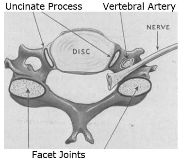
    The primary synovial joints of the spine are located in the posterior (back) half of the vertebrae, behind the exiting nerve root. These spinal synovial joints are known as the facet joints or the zygapophysial joints. Typically, each spinal vertebrae has four facet synovial joints:

    • Two on the top of the vertebrae (left and right)
    • Two on the bottom of the vertebrae (left and right)

    Uniquely, importantly, and the main topic of this paper, the cervical spine has four additional synovial joints per spinal vertebrae level (two on the top, left and right, and two on the bottom, left and right).

    In the graphic above, the view is axial, looking from above at the superior (top) margin of a cervical vertebrae. The facet joints, behind the nerve root, are clearly noted. Notice the structures labeled “Uncinate Process,” lateral to the intervertebral disc. The uncinate processes are a component of a synovial joint known as the Uncovertebral Joints.

    The uncinate processes and uncovertebral joints were first described in 1834, and later described in detail by German anatomist Hubert von Luschka in 1858. Hence, these joints are often referred to as the joints of von Luschka.

    Hubert von Luschka was a German anatomist. He was born is 1820. He was appointed a full professor of anatomy in 1855 at the University of Tubingen. He gained a noble title and began using von in his name in 1865.

    The joints of von Luschka are located in the cervical region of the spinal column between C3 through C7, in all specimens. In some individuals, these joints are also located in the upper thoracic spine region. These joints allow for cervical spinal flexion and extension while limiting cervical spinal lateral flexion.

    In 1958, the Journal of the American Medical Association published a study titled (17):

    Whiplash Injuries:
    Neurophysiological Basis for Pain
    and Methods Used for Rehabilitation

    This study appeared literally 100 years after the original descriptions of the joints of von Luschka by Herbert von Luschka. The author, Emil Seletz, MD, (b. 1907; d. 1999) was a neurosurgeon in Beverly Hills, California. His publications indicate that Dr. Seletz worked at the Los Angeles General Hospital, and he was faculty at the University of California, Los Angeles, Medical School. His is internet biography indicates he was chief of neurosurgery at Cedar’s Hospital (now called Cedars-Sinai Medical Hospital) in Los Angeles, and Professor of Neurological Surgery at the University of Southern California School of Medicine.

    In this article, Dr. Seletz profiles whiplash injuries, and it includes a discussion pertaining to the uncinate processes and the joints of von Luschka. The importance of this discussion lies in that these joints limit lateral flexion. Whiplash mechanisms that involve lateral flexion stress, or any mechanism where the patient’s head/neck are in rotation at the time of impact, have increased potential to cause injury to the uncinate processes and/or the joints of von Luschka.

    In this article, Dr. Seletz makes these points:

    • Cervical discs degenerate following whiplash trauma.
    • The cervical vertebrae also contain “special synovial joints,” the lateral body joints of von Luschka, first described in 1858.
    • In whiplash injury, the joints of von Luschka play a very significant clinical role.
    • “With degenerative thinning of the disk after trauma, the processes of the lateral joints [uncinate] are gradually forced outward and laterally, resulting in osteophyte formation.”
    • Lateral spurs from the joints of von Luschka compress the nerve root and can encroach on the vertebral artery and the vertebral nerve (sympathetic) within the foramen transversarium.
    • The joints of von Luschka are synovial joints with a cartilage cap.
    • “These spurs project even more, since the osteophytes are capped with cartilage and are considerably larger than is revealed by the roentgenogram.”

    The following year, 1959, Edward Compere, MD, and colleagues, published a study in the journal Orthopedics, titled (18):

    The Luschka Joints; Their Anatomy, Physiology and Pathology

    Dr. Compare was an orthopedic surgeon, and Professor of Bone and Joint Surgery at Northwestern University (Chicago). In this publication, the authors note that the joints of von Luschka demonstrated all of the elements of a true joint in microscopic sections. This adds support to the understanding that these joints are synovial.

    In 1978, Ruth Jackson, MD, published the fourth edition of her book, The Cervical Syndrome (19). Ruth Jackson, MD (1902-1994), was the world’s first female admitted into the American Academy of Orthopedic Surgeons (1937). She began her orthopedic private practice in Dallas in 1932. From 1936 to 1941, Dr. Jackson was Chief of Orthopaedics at Parkland Hospital in Fairmont, Texas. In 1945, she had her own private clinic built in Dallas. In 1956 she published the first edition of her acclaimed, authoritative book, The Cervical Syndrome. Dr. Jackson retired from clinical practice in 1989 at the age of 87 years.

    In her book, Dr. Jackson has a section pertaining to the joints of von Luschka, beginning with their injury vulnerability to lateral-flexion mechanical stress. She notes:

    • The joints of von Luschka “are covered with articular hyaline cartilage, as are all other articulating surfaces.”
    • “These articulating surfaces were described as true joints with capsules and synovial linings by von Luschka in 1858.”
    • “The adjacent surfaces at the sides of the bodies of the vertebrae are plated with articular cartilage,” and “capsular ligaments are present.”
    • “There is a definite space between the adjacent articular surfaces.”
    • “These articulations do satisfy the standard definition of synovial joints.”
    • “Osteophytic formations do occur at the margins of the articulations which is characteristic of synovial joints.”

    Most importantly, Dr. Jackson demonstrates that the injection of radiopaque dye into the joints of von Luschka shows that the material remains confines, as in being held by a capsule.

    In 2009, Jean-Michel Brismée, ScD, and colleagues from the School of Allied Health Sciences, Texas Tech University Health Sciences Center, published a study in the journal Spine titled (20):

    Immunohistochemical and Histological Study
    of Human Uncovertebral Joints

    The authors of this study claim that no research had investigated the presence of pain generating neurotransmitters within the uncovertebral cartilaginous and capsular tissue. Consequently, this study investigated the anatomy and innervation of the uncovertebral joints to determine if it is synovial in nature and if it was capable of generating pain.

    The study used two unembalmed fresh male human cadavers with a mean age of 83 years. Tissue from the uncovertebral capsule and cartilage was harvested from each uncovertebral surface from C2-C3 to C6-C7.

    The findings included the identification of chondrocytes and synoviocytes in the capsular tissue of each uncovertebral joints. This indicates that the uncinate joints have articular hyaline cartilage and a synovial capsule. This indicates that the uncovertebral joint is “synovial in nature.”

    The authors also assessed the harvested uncinate capsular tissue for neurofilaments using immunohistochemistry. The immunoreactivity findings indicated that the uncinate capsules also have the presence of both the somatic and autonomic nerve fibers. “These findings suggest that the uncovertebral joints are potential pain generators in the cervical spine.”

    The authors noted that these capsular and synovial tissue nerve fibers lead to pain perception and reflexive responses associated with synovial pathology. They make these comments:

    Biomechanically, the “uncovertebral joint is a major contributor to coupled motion at the lower cervical spinal segments and serves as a controlling factor of overall cervical segmental motion.”

    “A synovial or diarthrodial joint must exhibit a joint capsule, a synovial membrane, synovial fluid, and articular cartilage.” “Our present findings support the notion that the uncovertebral complex includes a synovial joint.”

    Disc degeneration always influences the uncinate joints, their biomechanics and their degenerative potential.

    Degenerative changes in the uncinate joints may translate into osteophytic projections that can compress adjacent cervical segmental nerves as they course through the intervertebral foramen or may cause cervical myelopathy.

    “Due to the close proximity of the uncinate process to the vertebral artery degenerative changes in the uncovertebral joint have been clinically related to vertebral artery compression, ultimately lending to the onset and severity of clinical vertebrobasilar insufficiency.”

    The authors conclude that there should be no controversy with regard to the anatomic and histological makeup of the uncovertebral joints. They are synovial joints that possess a joint cavity that is lined with a synovial membrane secretes synovial fluid. They imply that authors who claim otherwise are incorrect.

    In 2014, Jeffery Hartman, MD, from the College of Medicine, University of Saskatchewan, Canada, published a “Comprehensive Review” titled (21):

    Anatomy and Clinical Significance of
    the Uncinate Process and Uncovertebral Joint

    In this “comprehensive review,” Dr. Hartman cites 74 references of the literature, making this review the most extensive published review on this topic to date.

    Dr. Hartman notes that the uncinate process and its associated uncovertebral articulation are unique to the cervical spine. They are rudimentary at birth and develop and evolve with age. They are consistently found on the posterolateral aspect of the superior end plate of the third to seventh cervical vertebrae. However, in some individuals, they extend as low as to the T2 level.

    The uncinate processes guide the motions of the cervical spine. They also provide important lateral stability for the cervical spinal motions. The uncinate processes function to limit lateral flexion. Dr. Hartman states:

    “Although often overlooked due to their relatively small size the uncovertebral articulation has been shown to contribute to the stability of the cervical spine.”

    “The uncovertebral articulation contributes to the spinal motion segments mobility and stability as well as functions to protect the intervertebral foramen contents from herniated disc material.” [see Graphic 1]

    “The spinal segments’ mobility throughout flexion, extension, lateral flexion, and rotation is influenced by the structure of the uncinate processes and their articulation with the vertebra above.”

    Motion in the coronal plane that occurs during lateral flexion is coupled with axial rotation. The uncovertebral articulation is a significant contributor to this coupled motion.

    “In addition to influencing the mobility of the spinal segments the uncinate processes provide stability.”

    As noted above, the location of the uncinate processes makes them a barrier to lateral and posterior-lateral herniations of the cervical intervertebral disc. To support this, Dr. Hartman references a 2003 study from the journal Spine (22). It was a study of 200 patients with myelopathy or radiculopathy secondary to cervical intervertebral disc herniation. One hundred and ninety-eight (198) of the herniations were median or paramedian; only two were lateral herniations. Both of the two lateral herniations occurred at C7-T1, where the uncovertebral articulation is rudimentary or absent. The authors concluded:

    “It was concluded that the uncovertebral articulations prevent lateral herniations.”

    Lateral intervertebral disc herniations at C7-T1 are more common than other cervical segments because of the “lack of barrier provided by the uncinate processes.”

    The uncinate processes and joints are especially vulnerable to side-impact or rotation-type neck trauma. These articulations are commonly disrupted in head and neck injuries. Uncovertebral cleft hematomas are commonly seen in victims of traffic accidents. Following such trauma, the cervical intervertebral disc degenerates at an accelerated rate. This disc degeneration deflects more weight-bearing responsibility to the uncinate processes, accelerating spondylotic changes of these joints.

    The uncinate processes bear the load of the vertebra above. With age and/or injury, the intervertebral disc dehydrates and thins, leading to an increased load being supported by the uncinate processes. With this degeneration, the uncinate processes and uncovertebral articulations may compress the spinal nerve root and vertebral artery. Dr. Hartman makes these statements:

    “Dehydration and narrowing of the cervical intervertebral discs begin to occur in the fourth and fifth decades of life.”

    With age, the “uncinate process develops osteophytic lipping where the tip turns laterally.”

    “With narrowing of the intervertebral discs, the uncinate processes bear a greater load to support the weight of the head.”

    “The vertebra above comes to rest on the uncinate processes below which results in remodeling to the flattened and laterally directed configuration.”

    “The uncovertebral articulations are common sites for osteoarthritic changes.”

    “These changes are believed to be related to the dehydration and shrinkage of the intervertebral disc which leads to increased load and contact between the vertebra above and the uncinate processes below.”

    “The osteophytic spurring from the uncinate processes project laterally and thus can impinge on anatomical structures within the vicinity, including the spinal nerve root, vertebral artery, radicular (medullary) artery, cervical spinal cord, and cervical sympathetic trunk.”

    “The reduced blood flow through the radicular artery from external compression is a contributing factor to the development of cervical spondylotic myelopathy.”

    “Osteophytes that arise from the posterior aspect of the uncinate process project into the intervertebral foramen and encroach on its contents.”

    When the vertebral artery is compressed by osteophytes they are “more likely to be derived from the uncinate process than from the zygapophysial joints.”

    “In cervical spondylosis, the uncovertebral osteophytes are the most common cause of nerve root compression.”

    The degenerative changes described narrow the intervertebral foramen, increasing the incidence of nerve root irritation and/or compression:

    “The clinical picture resulting from cervical nerve root compression is one of pain, paraesthesia, diminished sensation to pinprick, diminished reflexes, muscle weakness, and, rarely, muscle wasting in the neck and ipsilateral upper extremity.”

    Dr. Hartman also notes that when there is an acute injury to the uncinate process, the synovial joint becomes inflamed and edematous. The resulting pain and spasm may precipitate an acute cervical torticollis.

    Summary and Concluding Remarks

    Ongoing evidence for 150 years continues to support the anatomy that the uncinate is a synovial joint with articular hyaline cartilage, a joint capsule, a synovial bag that produces synovial fluid, an is neurologically innervated and thus capable of producing pain. Like all other synovial joints, these joints of von Luschka may develop mechanical problems, open the pain gate, and cause neck pain.

    Many chiropractors contend that uncinate joint mechanical problems (chiropractic subluxation) can be improved with an appropriately delivered spinal adjustment. The uncinate joint subluxation is distinct from the facet joint subluxation. The adjustment of the uncinate joint subluxation is also distinct from the facet joint subluxation adjustment. Traditionally, the adjustment of facet joint subluxation is delivered at the back of the neck, often the contact is made with the fingertip or with an approved mechanical device. In contrast, the uncinate joint adjustment is often delivered to the front of the neck, and frequently the contact is made with the thumb or, again, with an approved mechanical device. The skills to successfully deliver these maneuvers is emphasized in chiropractic education.

    Lastly, with advancing degenerative changes to the uncinate processes, there may be irritation of the cervical nerve roots or possibly to the neurological controls of the vertebral artery. Ideally, carefully delivered chiropractic adjusting will improve biomechanical function and reduce the adverseness of such irritations.

    REFERENCES

    1. Cailliet R; Low Back Pain Syndrome; 4th edition; FA Davis Company; 1981.
    2. White AA, Panjabi MM; Clinical Biomechanics of the Spine; Second Edition; Lippincott; 1990.
    3. Kapandji AI; The Physiology of the Joints: The Spinal Column, Pelvic Girdle and Head; Seventh Edition; Handspring Publishing; 2019.
    4. Kirkaldy-Willis WH, Cassidy JD; Spinal Manipulation in the Treatment of Low Back Pain; Canadian Family Physician; March 1985; Vol. 31; pp. 535-540.
    5. Melzack R, Wall P; Pain mechanisms: a new theory; Science; November 19, 1965;150(3699); pp. 971-979.
    6. Dickenson AH; Gate Control Theory of Pain Stands the Test of Time; British Journal of Anaesthesia; June 2002; Vol. 88; No. 6; pp. 755-757.
    7. Giles LGF; Muller R; Chronic Spinal Pain: A Randomized Clinical Trial Comparing Medication, Acupuncture, and Spinal Manipulation; Spine July 15, 2003; Vol. 28; No. 14; pp. 1490-1502.
    8. Muller R, Giles LGF; Long-Term Follow-up of a Randomized Clinical Trial Assessing the Efficacy of Medication, Acupuncture, and Spinal Manipulation for Chronic Mechanical Spinal Pain Syndromes; Journal of Manipulative and Physiological Therapeutics; January 2005; Vol. 28; No. 1; pp. 3-11.
    9. Adams J, Peng W, Cramer H, Sundberg T, Moore C; The Prevalence, Patterns, and Predictors of Chiropractic Use Among US Adults; Results From the 2012 National Health Interview Survey; Spine; December 1, 2017; Vol. 42; No. 23; pp. 1810–1816.
    10. Meade TW, Dyer S, Browne W, Townsend J, Frank OA; Low back pain of mechanical origin: Randomized comparison of chiropractic and hospital outpatient treatment; British Medical Journal; Vol. 300; June 2, 1990; pp. 1431-7.
    11. Woodward MN, Cook JCH, Gargan MF, Bannister GC; Chiropractic treatment of chronic ‘whiplash’ injuries; Injury; November 1996; Vol. 27; No. 9; pp. 643-645.
    12. Khan S, Cook J, Gargan M, Bannister G; A symptomatic classification of whiplash injury and the implications for treatment; The Journal of Orthopaedic Medicine; Vol. 21; No. 1; 1999; pp. 22-25.
    13. Hoving JC, Koes BW, de Vet HCW, van der Windt DAWM, Assendelft WJJ, Mameren H, Devillé WLJM; Manual Therapy, Physical Therapy, or Continued Care by a General Practitioner for Patients with Neck Pain; A Randomized Controlled Trial; Annals of Internal Medicine; May 21, 2002; Vol. 136; No. 10; pp. 713-722.
    14. Fischgrund JS; Neck Pain, Monograph 27, American Academy of Orthopaedic Surgeons; 2004.
    15. César Fernández-de-las-Peñas; J. Fernández-Carnero; L. Palomeque del Cerro; Manipulative Treatment vs. Conventional Physiotherapy Treatment in Whiplash Injury: A Randomized Controlled Trial; Journal of Whiplash & Related Disorders; 2004; Vol. 3; No. 2.
    16. Langenfeld A, Humphreys K, Swanenburg J, Cynthia K. Peterson CK; Prognostic Factors for Recurrences in Neck Pain Patients Up to 1 Year After Chiropractic Care; Journal of Manipulative and Physiological Therapeutics; September 2015; Vol. 38; No. 7; pp. 458-464.
    17. Seletz E; Whiplash Injuries: Neurophysiological Basis for Pain and Methods Used for Rehabilitation; Journal of the American Medical Association; November 29, 1958; Vol. 168; No. 13; pp. 1750–1755.
    18. Compere EL; The Luschka Joints; Their Anatomy, Physiology and Pathology; Orthopedics; May 1959; Vol. 1; pp. 159-167.
    19. Jackson R; The Cervical Syndrome; fourth edition; Charles Thomas; 1978.
    20. Brismée JM, Sizer PS, Dedrick GS, Sawyer BG, Smith MP; Immunohistochemical and Histological Study of Human Uncovertebral Joints; Spine; May 20, 2009; Vol. 34; No. 12; pp 1257-1263.
    21. Hartman J; Anatomy and Clinical Significance of the Uncinate Process and Uncovertebral Joint: A Comprehensive Review; Clinical Anatomy; April 2014; Vol. 27; No. 3; pp. 431–440.
    22. Yamazaki S, Kokubun S, Ishii Y, Tanaka Y. 2003. Courses of cervical disc herniation causing myelopathy or radiculopathy: An analysis based on computed tomographic discograms; Spine; June 1, 2003; Vol. 28; No. 11; pp. 1171–1175.

    “Authored by Dan Murphy, D.C.. Published by ChiroTrust® – This publication is not meant to offer treatment advice or protocols. Cited material is not necessarily the opinion of the author or publisher.”
  • The Effects of Spinal Manipulation on the Musculoskeletal System

    The Effects of Spinal Manipulation on the Musculoskeletal System

    Spinal manipulation is one of the most ancient forms of treatment for musculoskeletal complaints, primarily for back pain and neck pain.

    In 1992, Robert Anderson, MD, PhD, DC, was the Director of Manual Medicine at the San Francisco Spine Institute, as well as Professor of Anthropology at Mills College in Oakland, California.

    At that time (1992), Scott Haldeman, DC, PhD, MD, was Associate Clinical Professor of Neurology at the University of California, Irvine, California.

    In 1992, the second edition of Dr. Haldeman’s book, Principles and Practice of Chiropractic, was published. Dr. Anderson wrote the first chapter of Dr. Haldeman’s book, titled (1):

    “Spinal Manipulation Before Chiropractic”

    Dr. Anderson notes that historically (for a least 2,500 years), spinal manipulation had been practiced in many parts of the world, primarily to treat musculoskeletal disorders. He presents the evidence of spinal manipulation being applied by a variety of providers in vast geographic regions, including: Bohemian (Czechoslovakia), China, Egypt, England, Finland, Greece, India, Japan, Latin America, Norway, Russia, and Wales.

    In many ways, the history of joint manipulation parallels the history of civilization. Another important review of manipulation through the ages was published in The Journal of Manual & Manipulative Therapy in 2007 and titled (2):

    A History of Manipulative Therapy

    This review makes these comments:

    “Manipulative therapy has known a parallel development throughout many parts of the world. The earliest historical reference to the practice of manipulative therapy in Europe dates back to 400 BCE.”

    “Historically, manipulation can trace its origins from parallel developments in many parts of the world where it was used to treat a variety of musculoskeletal conditions, including spinal disorders.”

    “It is acknowledged that spinal manipulation is and was widely practiced in many cultures and often in remote world communities such as by the Balinese of Indonesia, the Lomi-Lomi of Hawaii, in areas of Japan, China and India, by the shamans of Central Asia, by sabodors in Mexico, by bone setters of Nepal as well as by bone setters in Russia and Norway.”

    “Historical reference to Greece provides the first direct evidence of the practice of spinal manipulation.”

    “Hippocrates (460–385 BCE), who is often referred to as the father of medicine, was the first physician to describe spinal manipulative techniques.”

    “Claudius Galen (131–202 CE), a noted Roman surgeon, provided evidence of manipulation including the acts of standing or walking on the dysfunctional spinal region.”

    “Avicenna (also known as the doctor of doctors) from Baghdad (980–1037 CE) included descriptions of Hippocrates’ techniques in his medical text The Book of Healing.”

    “Nobody questions these early origins of manipulative therapy.”

    Prior to the modern era, for hundreds of years in developed and primitive societies throughout the world, practitioners of manipulation were known as bonesetters (2). All of this changed in 1874, and the global seat of change was in the United States of America.

    Andrew Taylor Still was a second-generation physician who became disillusioned with medicine following the death (from disease) of three of his children. Dr. Still conceived a theory whereby health could only be maintained and, therefore, disease defeated, by maintaining normal function of the musculoskeletal system (2). In 1892, Still established the American Osteopathic College in Kirksville, Missouri. By the time of his death in 1917, 3,000 Doctors of Osteopathy had graduated from his school.

    In 1895, “natural healer” Daniel David (DD) Palmer reasoned that when a vertebra was out of alignment, it caused pressure on nerves. In 1897, Palmer opened his first college, The Palmer College of Cure, now known as Palmer College of Chiropractic, Davenport, Iowa (2).

    In 1910, Palmer’s son Bartlett Joshua (BJ) Palmer, introduced the use of X-rays into Chiropractic. History records (2):

    “The G. I. Bill at the end of World War II enabled thousands of returning soldiers to bolster the ranks of the chiropractic profession.”

    “This influx seemed to provide an impetus that would propel the chiropractic profession to today’s status where it boasts 35 schools and colleges worldwide and, in the Western world at least, it is second only to the medical profession as a primary care healthcare provider.”

    How Does Spinal Manipulation Work?

    Pain Theory Background

    In 2017, a comprehensive and authoritative assessment of the chiropractic profession appeared in the orthopedic medical journal Spine, titled (3):

    The Prevalence, Patterns, and Predictors of
    Chiropractic Use Among US Adults:
    Results From the 2012 National Health Interview Survey

    The survey results indicated that 93% of patients go to chiropractors for the management of musculoskeletal pain syndromes. Specifically, 63% go to chiropractors for low back pain, and 30% for neck pain. Additionally, chiropractic spinal manipulation is very effective for the management of back and neck pain (3). The study showed:

    • 65% of the patients reported that chiropractic care helped their condition “a great deal.”
    • 26% of the patients reported that chiropractic care helped their condition “somewhat.”
    • Only 3% reported that the chiropractic care they received did not help them.

    In randomized clinical trials comparing chiropractic spinal manipulation to prescription pain medicines for chronic low back and neck pain, chiropractic manipulation was significantly more effective, registered no significant side effects, and displayed stable long-term clinical benefits (at the one year follow-up assessment) (4, 5).

    How does chiropractic spinal manipulation
    benefit people with spinal pain complaints?

    The Concept of Proprioception

    Proprioception is a sensory input from the peripheral musculoskeletal body (muscles, joints, tendons, ligaments, etc.) to the brain (central nervous system). Proprioceptive signals transmit mechanical information, especially mechanical factors such as position and movement. This concept is well-stated by attorney Chris Crowley and physician Henry Lodge, MD, in their book Younger Next Year, in a section they refer to as (6):

    “The Balancing Act”

    “Now it’s time to think about your brain and a concept called proprioception—the deceptively simple notion that you have to know where the different parts of your body are at all times.”

    “Your body is aware of exactly where each limb is in space every second, because each muscle, tendon, ligament and joint sends thousands of nerve fibers back to the brain through the spinal cord. Those fibers signal every nuance gradation of contraction, strength, muscular tone, orientation, position and movement at every moment of the day.”

    “Your brain keeps careful track of the location of every muscle and joint in you body every second, all day, every day, waiting for you to need the information.”

    Proprioception is both conscious and subconscious. In 1965, researchers discovered an incredibly important function for proprioception: pain control.

    In 1965, pain researchers became aware that the proprioceptive signals to the brain could block the pain signal to the brain. This concept was originally proposed by pain researchers Ronald Melzack and Patrick Wall (7). Their theory is known as the Gate Control Theory of Pain. Ronald Melzack, PhD, is a Canadian psychologist. Patrick Wall, MD (d. 2001), was a British neuroscientist and pain expert, as well as the first editor of the journal Pain.

    In 2002, the British Journal of Anaesthesia published a study reaffirming the validity of the Gate Theory of Pain in an article titled (8):

    Gate Control Theory of Pain Stands the Test of Time

    An oversimplified explanation of their Gate Control Theory of Pain is that the pain electrical signal to the brain can be blocked by non-painful electrical signals arising from other sensory afferents, especially from joint proprioceptors. Practically, this would mean that prolonged or static positions would reduce proprioception, allowing the pain signal to more readily enter the brain for pain perception. Similarity, if a person’s joints lost or reduced their normal movement arc, there would be a proportionate reduction of the proprioceptive sensory input to the brain. This would allow pain signals to enter the brain because the pain gate would be open.

    As noted above, chiropractic’s Daniel David Palmer believed that when a vertebra was out of alignment, it caused pressure on nerves (2). A specific directional manipulation (the chiropractic spinal adjustment) would improve alignment and reduce nerve pressure.

    Palmer’s “nerve pressure” theory is probably correct in only a minority of chiropractic patients. It was officially challenged by a vocally pro-chiropractic orthopedic surgeon from Saskatchewan, CAN. Dr. William H. Kirkaldy-Willis published his theory as to how/why chiropractic spinal manipulation helped those suffering from back pain in the journal Canadian Family Physician in 1985 (35 years prior to this writing), titled (9):

    Spinal Manipulation in the Treatment of Low Back Pain

    Dr. Kirkaldy-Willis (d. 2006) was a Professor Emeritus of Orthopedics and director of the Low-Back Pain Clinic at the University Hospital, Saskatoon, Canada. The abstract from his article makes these comments:

    “Spinal manipulation, one of the oldest forms of therapy for back pain, has mostly been practiced outside of the medical profession.”

    “Over the past decade, there has been an escalation of clinical and basic science research on manipulative therapy, which has shown that there is a scientific basis for the treatment of back pain by manipulation.”

    “Most family practitioners have neither the time nor inclination to master the art of manipulation and will wish to refer their patients to a skilled practitioner of this therapy.”

    “The physician who makes use of this resource will provide relief for many patients.”

    Dr. Kirkaldy-Willis notes that 80% of the population will experience low back pain sometime during their adult life. At any given time, 20-30% of the adult population is suffering from low back pain. Low back pain is the second most common cause of worker loss of productivity.

    Yet, in disagreement with chiropractic’s Daniel David Palmer, Dr. Kirkaldy-Willis argues that less than 10% of low back pain is due to pressure or entrapment of spinal nerves. He notes that it is unlikely that spinal manipulation replaces a vertebra that is out of alignment.

    In contrast, Dr. Kirkaldy-Willis applies Melzack and Wall’s Gate Theory of Pain to the chiropractic adjustment in his explanation as to how manipulation helps with pain.

    Dr. Kirkaldy-Willis begins his explanation by noting that with positional (alignment) problems there is a shortening of periarticular connective tissues and intra-articular adhesions may form. This orthopedic principle is supported by many others (10, 11, 12, 13, 14, 15, 16, 17, 17). This intra-articular and periarticular fibrosis would reduce joint motion. He proposes that a specific line-of-drive manipulation (chiropractic spinal adjustment) could break these adhesions and/or remodel fibrosis, also supported by others (11, 12, 13, 15, 16, 17, 18). He cites support for these concepts, including:

    • Following joint manipulation, there is consistently a measureable increase in the range of motion.
    • As a rule, there is an initial increase in symptoms after the first few manipulations probably as a result of breaking adhesions and tissue fibrosis. He states:

    “In almost all cases, however, this increase in pain is temporary and can be easily controlled by local application of ice.”

    “Patients undergoing manipulative treatment must therefore be reassured that the initial discomfort is only temporary.”

    “However, the gain in mobility must be maintained during this period to prevent further adhesion formation.”

    Dr. Kirkaldy-Willis then applies the improved articular motion to Melzack and Wall’s Gate Theory of Pain. He notes that this theory has “withstood rigorous scientific scrutiny,” stating:

    “The central transmission of pain can be blocked by increased proprioceptive input.”

    Pain is facilitated by “lack of proprioceptive input.”

    “Increased proprioceptive input in the form of spinal mobility tends to decrease the central transmission of pain from adjacent spinal structures by closing the gate. Any therapy which induces motion into articular structures will help inhibit pain transmission by this means.”

    Dr. Kirkaldy-Willis notes that at the end of the manipulation range of motion, “the limit of anatomical integrity is encountered. Movement beyond this limit results in damage to the capsular ligaments.” Consequently, joint manipulation “requires precise positioning of the joint at the end of the passive range of motion and the proper degree of force to overcome joint coaptation” (to overcome the resistance of the joint surfaces in contact). He concludes:

    “With experience, the manipulator can be very specific in selecting the spinal level to be manipulated.”

    “The physician who makes use of this resource will provide relief for many back pain patients.”

    Today (2020) this “Orthopedic Gate Theory” explanation for the mechanism of benefits from manipulation for pain control is widely popular. Yet, Dr. Kirkaldy-Willis also offers a concomitant reflex neurological model. He supports that during manipulation, the stretching of joint ligaments (capsules) will trigger a reflex that inhibits local musculature, also improving motion and closing the pain gate.

    ••••••••••

    An update to the effects of spinal manipulation was published last year (2019) in the journal Spine, titled (19):

    Neurophysiological Effects of High Velocity and Low Amplitude
    Spinal Manipulation in Symptomatic and Asymptomatic Humans:
    A Systematic Literature Review

    The objective of this study was to summarize the evidence of the neurophysiological effects of spinal manipulative therapy (SMT) with high velocity low amplitude thrust (HVLA-SMT). The authors searched the literature until July 2018 and only used controlled studies of at least moderate quality. They found 18 studies that met their inclusion criteria that involved 932 participants. Most studies on spinal manipulation focus on pain mechanisms and pain control.

    The authors note that spinal manipulative therapy with high-velocity low amplitude thrust (HVLA-SMT) is effective in reducing low back pain (LBP), and applies to both acute and chronic nonspecific LBP. They agree that spinal manipulation has both neurophysiological and biomechanical influences, and support that manipulation increases proprioception, stating:

    “The mechanical force of spinal manipulation primarily affects afferent neurons in the paraspinal tissue and triggers neurophysiological responses in the peripheral and central nervous system, eventually leading to pain inhibition.”

    The evidence presented in this study supports that spinal manipulation increases proprioception, which eventually leads to pain inhibition. This is consistent and supportive of Dr. Kirkaldy-Willis’s application of Melzack and Wall’s Gate Theory of Pain, above.

    ••••••••••

    Also in 2019 (April), a study was published in the Journal of Pain titled (20):

    Decreased Neurologic Pain Signature Activation Following
    Thoracic Spine Manipulation in Healthy Volunteers

    In this study, researchers from Stanford University employed functional magnetic resonance imaging (fMRI) on ten healthy volunteers to objectively measure pain intensity following spinal manipulation. This study was funded by the National Institute of Health, USA.

    The authors note that: “Spinal manipulation is commonly used when managing patients with neck and back pain.” Although they support the rationale that spinal manipulation elicits a mechanical effect on the spine that leads to therapeutic mechanical changes within the spine, they wanted to explore if central (brain) mechanisms were also being activated. The results of this study showed that spinal manipulation activates brain regions associated with pain inhibition.

    It has been observed for decades that spinal manipulation helps control pain. This study adds to the evidence, using the best available evidence (functional MRI), showing that spinal manipulation works in pain reduction, in part, by inhibiting pain-processing regions of the brain. Once again, the results of this study are consistent and supportive of Dr. Kirkaldy-Willis’s application of Melzack and Wall’s Gate Theory of Pain.

    ••••••••••

    A few months later in 2019 (October), this same group published a follow-up study titled (21):

    Evidence for Decreased Neurologic Pain Signature Activation
    Following Thoracic Spinal Manipulation
    in Healthy Volunteers and Participants with Neck Pain

    Once again, these authors used functional magnetic resonance imaging (fMRI) to investigate the effect of thoracic spinal manipulation on pain-related brain activity. They assessed 16 subjects with acute/subacute neck pain and compared them to 10 healthy volunteers. The spinal manipulation was a high-velocity low-amplitude end-range force directed to the T4-T5 motion segment. The brain fMRI images were performed on a 3T magnetic resonance scanner.

    The authors note that fMRIs can map pain processing in the central nervous system. They also note that the perception of pain is not encoded by a single brain area but distributed throughout the brain.

    Spinal manipulation is a mechanical thrust to spinal joints slightly beyond their passive range of motion. It is a common treatment for neck and back pain, theorized to mechanically affect the spine leading to therapeutic mechanical changes. Yet, spinal manipulation’s therapeutic action may also be mediated by inhibiting the central nervous system.

    This study confirmed that there is a centrally (brain) mediated therapeutic action of spinal manipulation for pain reduction. The authors note:

    “The findings provide evidence that spinal manipulation may alter the processing of pain-related brain activity within specific pain-related brain regions.”

    “A growing body of evidence is pointing towards neurophysiologic mechanisms of action underlying the pain modulating effects of spinal manipulation including both spinal and supraspinal mechanisms.”

    Consistent with the other studies above, and in support of the proprioception closure of the pain gate, these authors note:

    “Spinal manipulation is known to activate large diameter mechanoreceptors that in turn can inhibit the transmission of nociceptive signals at the spinal cord.”

    ••••••••••

    Earlier this year (2020), another study on this topic was published in the journal Cureus titled (22):

    The Effect of High Velocity Low Amplitude
    Cervical Manipulations on the Musculoskeletal System:
    Literature Review

    As the title indicates, the objective of the study was to describe how cervical manipulation could impact musculoskeletal disorders. A systematic search of the literature was carried out using PubMed; inclusion criteria was randomized controlled trial manuscripts published in peer-reviewed journals with individuals of all ages from 2005 to 2020. Subjects with skeletal muscle or health problems were evaluated as to the effects of cervical manipulation, comparing them with control subjects receiving placebo or another type of treatment.

    The authors note that the number of people suffering from skeletal muscle problems is constantly increasing. They attribute this to work activities that lead to prolonged wrong positions for a period of time.

    Pertaining to spinal manipulation, the authors note:

    “Spinal manipulation is a manual therapy technique used by chiropractors, osteopaths, physiotherapists and some doctors to treat skeletal muscle problems.”

    “In manual therapy, high velocity low amplitude (HVLA) cervical manipulation techniques are frequently used.”

    “The use of high velocity low amplitude (HVLA) techniques is growing as a therapeutic option.”

    “Cervical manipulations are effective in management of cervicalgia, epicondylalgia, temporomandibular joint disorders, and shoulder pain.”

    Both cervical manipulation and thoracic manipulation may lead to significant cervical pain improvement. The review of the literature presented indicates that spinal manipulation activates the supraspinal (brain and brainstem) neurological pain inhibitory control system. The authors note:

    “Manipulation involves segmental activation of inhibitory pathways that may lead to changes in pressure pain thresholds even at a distance [away from] of the manipulated area.”

    “HVLA techniques are able to produce more local hypoalgesia than other manual therapy techniques.”

    “The results of the research show that HVLA techniques, on subjects with musculoskeletal disorders, are able to influence pain modulation, mobility, and strength both in the treated area and at a distance.”

    For thousands of years and throughout the world there are observations that spinal manipulation helps people with musculoskeletal pain, especially spinal pain. For more than one hundred years, chiropractors, patients, governments, insurance companies, and scientists have realized that chiropractic spinal adjusting (specific manipulation) not only helps people with a variety of musculoskeletal pain syndromes, but that it probably works, at least in part, as a consequence of the activation of brain pain control circuitry.

    When spinal joints have reduced movement, the pain gate at that level is open. Chiropractic adjusting (specific manipulation) increases the firing of the proprioceptors, creating a neurological sequence of events that closes the pain gate.

    Proprioceptive integrity is a significant factor in the state of the pain gate. Improved proprioception closes the pain gate. Chiropractic adjusting improves proprioception.

    REFERENCES:

    1. Anderson R; “Spinal Manipulation Before Chiropractic”; in Haldeman S; Principles and Practice of Chiropractic; Second Edition; Appleton & Lang; 1992.
    2. Pettman E; A History of Manipulative Therapy; The Journal of Manual & Manipulative Therapy; Vol. 15; No. 3; (2007); pp. 165–174.
    3. Adams J, Peng W, Cramer H, Sundberg T, Moore C; The Prevalence, Patterns, and Predictors of Chiropractic Use Among US Adults; Results From the 2012 National Health Interview Survey; Spine; December 1, 2017; Vol. 42; No. 23; pp. 1810–1816.
    4. Giles LGF; Muller R; Chronic Spinal Pain: A Randomized Clinical Trial Comparing Medication, Acupuncture, and Spinal Manipulation; Spine July 15, 2003; Vol. 28; No. 14; pp. 1490-1502.
    5. Muller R, Giles LGF; Long-Term Follow-up of a Randomized Clinical Trial Assessing the Efficacy of Medication, Acupuncture, and Spinal Manipulation for Chronic Mechanical Spinal Pain Syndromes; Journal of Manipulative and Physiological Therapeutics; January 2005; Vol. 28; No. 1; pp. 3-11.
    6. Crowley C, Henry Lodge H; Younger Next Year: Live Strong, Fit, and Sexy—Until You’re 80 and Beyond; Workman Publishing; New York; 2007.
    7. Melzack R, Wall P; Pain mechanisms: a new theory; Science; November 19, 1965;150(3699); pp. 971-979.
    8. Dickenson AH; Gate Control Theory of Pain Stands the Test of Time; British Journal of Anaesthesia; June 2002; Vol. 88; No. 6; pp. 755-757.
    9. Kirkaldy-Willis WH, Cassidy JD; Spinal Manipulation in the Treatment of Low Back Pain; Canadian Family Physician; March 1985; Vol. 31; pp. 535-540.
    10. Viderman T; Experimental Models of Osteoarthritis: The Role of Immobilization; Clinical Biomechanics; November 1987; Vol. 2; No. 4; pp. 223-229.
    11. Stearns, ML; Studies on development of connective tissue in transparent chambers in rabbit’s ear; American Journal of Anatomy; Vol. 67; 1940; p. 55.
    12. Seletz E; Whiplash Injuries: Neurophysiological Basis for Pain and Methods Used for Rehabilitation; Journal of the American Medical Association; November 29, 1958; pp. 1750-1755.
    13. Cyriax J; Orthopaedic Medicine, Diagnosis of Soft Tissue Lesions; Bailliere Tindall; Vol. 1; 1982.
    14. Cohen IK, Diegelmann RF, Lindbald WJ; Wound Healing, Biochemical & Clinical Aspects; WB Saunders; 1992.
    15. Salter R; Continuous Passive Motion, A Biological Concept for the Healing and Regeneration of Articular Cartilage, Ligaments, and Tendons; From Origination to Research to Clinical Applications; Williams and Wilkins; 1993.
    16. Buckwalter J; Effects of Early Motion on Healing of Musculoskeletal Tissues; Hand Clinics; February 1996; Vol. 12; No. 1; pp. 13-24.
    17. Schleip R; Fascia; The Tensional Network of the Human Body; The Scientific and Clinical Applications in Manual and Movement Therapy; Churchill Livingstone; 2012.
    18. Bowman K; Move Your DNA: Restore Your Health Through Natural Movement; 2017.
    19. Wirth B, Gassner A, de Bruin ED, Axen I, Swanenburg J, Kim Humphreys BK, Schweinhardt P; Neurophysiological Effects of High Velocity and Low Amplitude Spinal Manipulation in Symptomatic and Asymptomatic Humans: A Systematic Literature Review; Spine; August 2019; Vol. 44; No. 15; pp. E914–E926.
    20. Weber K, Wager T, Elliott J, Mackey S, Liu W, Sparks C; Decreased Neurologic Pain Signature Activation Following Thoracic Spine Manipulation in Healthy Volunteers; The Journal of Pain; April 2019; Vol. 20; No. 4; Supplement; p. S69.
    21. Weber KA, Wager TD, Mackey S, Elliott JM; Evidence for Decreased Neurologic Pain Signature Activation Following Thoracic Spinal Manipulation in Healthy Volunteers and Participants with Neck Pain; NeuroImage: Clinical; October 18, 2019; Volume 24; 102042.
    22. Giacalone A, Febbi M, Magnifica F, Ruberti E; The Effect of High Velocity Low Amplitude Cervical Manipulations on the Musculoskeletal System: Literature Review; Cureus; April 15, 2020; Vol. 12; No. 4; pp. e7682.

    “Authored by Dan Murphy, D.C.. Published by ChiroTrust® – This publication is not meant to offer treatment advice or protocols. Cited material is not necessarily the opinion of the author or publisher.”

  • Low Back Pain and Neurological Balance

    Low Back Pain and Neurological Balance

    The Morning Shower

    The typical morning shower is neither 100% cold water nor 100% hot water, but rather a balance of the two. The perfect shower water temperature is a balance of hot and cold water.

    While enjoying the perfect shower, if suddenly the hot water is turned drastically higher, the brain perceives:

    Ouch! The water is too hot!

    In this situation, the cold water is where it is supposed to be, but the hot water is too high. Hot and cold are no longer in balance. The ouch suggests the experience is painful. [Remember, pain and temperature share the same neurological pathway to the brain]. The most common remedy is to quickly turn down the hot water to reestablish balance.

    Pain in General

    Most pain is an inflammatory event. Inflammatory chemicals irritate pain nerves. The understanding of the inflammatory chemical nature of pain was awarded the Nobel Prize in Physiology or Medicine in 1982 (1).

    In 2007, the journal Medical Hypothesis expands and elaborates on the relationship between pain and inflammation. The article is titled (2):

    The Biochemical Origin of Pain:
    The Origin of all Pain is Inflammation and the Inflammatory Response: Inflammatory Profile of Pain Syndromes

    The author states:

    “Every pain syndrome has an inflammatory profile consisting of the inflammatory mediators that are present in the pain syndrome.”

    “The key to treatment of Pain Syndromes is an understanding of their inflammatory profile.”

    “Our unifying theory or law of pain states: the origin of all pain is inflammation and the inflammatory response.”

    “Irrespective of the type of pain whether it is acute or chronic pain, peripheral or central pain, nociceptive or neuropathic pain, the underlying origin is inflammation and the inflammatory response.”

    “Activation of pain receptors, transmission and modulation of pain signals, neuro-plasticity and central sensitization are all one continuum of inflammation and the inflammatory response.”

    “Irrespective of the characteristic of the pain, whether it is sharp, dull, aching, burning, stabbing, numbing or tingling, all pain arises from inflammation and the inflammatory response.”

    Using our shower metaphor, inflammation is hot water. “Turning down the hot water” of pain perception is most commonly done by using nonsteroidal anti-inflammatory drugs (NSAIDs), or by using other anti-inflammatory drugs such as steroids. This approach seems logical, but there are problems:

    In 2003, the journal Spine states (3):

    “Adverse reactions to non-steroidal anti-inflammatory (NSAID) medication have been well documented.”

    “Gastrointestinal toxicity induced by NSAIDs is one of the most common serious adverse drug events in the industrialized world.”

    There is “insufficient evidence for the use of NSAIDs to manage chronic low back pain, although they may be somewhat effective for short-term symptomatic relief.”

    In 2006, the journal Surgical Neurology states (4):

    “More than 70 million NSAID prescriptions are written each year, and 30 billion over-the-counter NSAID tablets are sold annually.”

    “5% to 10% of the adult US population and approximately 14% of the elderly routinely use NSAIDs for pain control.”
    Almost all patients who take the long-term NSAIDs will have gastric hemorrhage, 50% will have dyspepsia, 8% to 20% will have gastric ulceration, 3% of patients develop serious gastrointestinal side effects, which results in more than 100,000 hospitalizations, an estimated 16,500 deaths, and an annual cost to treat the complications that exceeds 1.5 billion dollars.

    “NSAIDs are the most common cause of drug-related morbidity and mortality reported to the FDA and other regulatory agencies around the world.”

    One author referred to the “chronic systemic use of NSAIDs to ‘carpet-bombing,’ with attendant collateral end-stage damage to human organs.”

    In 2017 and 2019, cardiologist Steven Gundry, MD, explains how the consumption of NSAIDs disrupts the integrity of the gut protective mucus membrane, increasing adverse gut permeability (“leaky gut”), increasing one’s systemic inflammation and hence increasing pain. Dr. Gundry states (2017) (5):

    “Copious research published over the last half century reveals that gulping down apparently harmless NSAIDs is like swallowing a live grenade. These drugs blow gaping holes in the mucus-lined intestinal barrier.”

    “As a result, lectins, lipopolysaccharides, and living bacteria are able to deluge the breaks in your levee, flooding your body with foreign invaders.”

    “Inundated by these foreign proteins and other invaders, your immune system does what if does best, producing inflammation and pain. This pain in turn prompts you to down another NSAID, promoting a vicious cycle.” “… the more pain you have, the more NSAIDs you take.”

    “Increased intestinal permeability from … the regular use of NSAIDs and acid-reducing drugs, pro¬duces what is commonly called leaky gut syndrome.”

    “NSAIDs are both the number-one pharmaceutical seller and the number-one health menace.”

    In addition to these (and many other) side effect problems from the consumption of NSAIDs, there is another major concern. These anti-inflammatory drugs are not very effective, especially for chronic pain problems (17). If these drugs worked well, Americans would not need to fill 70 million prescriptions and consume 30 billion over-the-counter NSAID tablets yearly.

    Low Back Pain Paradigm Shift

    Drugs for the management of low back pain, especially for chronic low back pain, have sufficient down-sides. Consequently, in recent years, low back pain management and guidelines have advocated not using drugs but rather to use non-pharmacological approaches (7, 8, 9, 10, 11, 12, 13).

    Return to the Shower, A Different Model

    While enjoying the perfect shower, there is another method of creating the perception of too hot and/or pain: turn off the cold water. An interesting corollary is the understanding that the hot water is at the perfect level. The problem, the imbalance, is a lack of cold water. Yet, the brain perception of hot/pain is the same, ouch!

    The remedy is not to turn down (or off) the hot water because the hot water is where it is supposed to be. Rather, the remedy is to turn up the cold water.

    Why Do Patients Go to Chiropractors?

    By a significant percentage, the primary reason patients go to chiropractors is for the management of low back pain (14). The actual number is 63%. Many of these patients have already taken over-the-counter or prescription NSAIDs, with less than acceptable clinical improvement.

    Chiropractic spinal adjusting (specific spinal manipulation) is both safe and very successful for these patients (15, 16, 17, 18, 19).

    What Tissue is the Primary Source of Chronic Low Back Pain?

    The primary tissue responsible for chronic low back pain is the intervertebral disc (20, 21, 22, 23, 24, 25, 26, 27). It has been documented for decades that the intervertebral disc is innervated with nociceptors [hot water], and that inflammatory chemicals in the disc increases the depolarization of the disc nociceptors.

    What if, as is often the case, the patient has been taking NSAIDs, but the clinical improvement is unacceptable? Perhaps their pain persists because the cold water has been turned down or off. In such a case, the solution is to turn the cold water up, reestablishing balance.

    What Nerves Function as the Cold Water?

    In our model, the hot water represents the nociceptors (pain afferents). What represents the cold water? It is mechanical nerves that are also in the intervertebral disc.

    These mechanical nerves are referred to proprioceptors or mechanoreceptors. When spinal joints do not move appropriately, the mechanoreceptors [cold water] are turned down or perhaps turned off, leaving the nociceptors [hot water] unopposed. The result is pain, even though the nociceptors are actually functioning normally. There is an imbalance between the hot and cold water.

    What is the Gate Theory of Pain?

    The Gate Theory of Pain was proposed in 1965 by Ronald Melzack and Patrick Wall (28), and it has survived in depth scientific scrutiny and the test of time (29). It was reviewed and applied to chiropractic spinal adjusting by Canadian orthopedic surgeon William H. Kirkaldy-Willis, MD, in 1985 (30).

    Dr. Kirkaldy-Willis notes that Melzack and Wall’s Gate Theory of Pain, has “withstood rigorous scientific scrutiny.” He further states:

    “The central transmission of pain can be blocked by increased proprioceptive input.” Pain is facilitated by “lack of proprioceptive input.” This is why it is important for “early mobilization to control pain after musculoskeletal injury.”

    “Increased proprioceptive input in the form of spinal mobility tends to decrease the central transmission of pain from adjacent spinal structures by closing the gate. Any therapy which induces motion into articular structures will help inhibit pain transmission by this means.”

    Dr. Kirkaldy-Willis’ discussion of the Gate Theory of Pain involves the “closing” of the pain gate through the enhancement of proprioception (mechanoreception). Spinal manipulation improves the movement parameters of articulations. The improvement of joint motion results in a neurological sequence of events that “closes” the pain gate.

    In our model, spinal adjusting would turn up the cold water, reestablish balance with the hot water, and balance (eliminate) pain perception in the brain.

    Is There Evidence that Proprioceptors/Mechanoreceptors (cold water)
    Exist in the Intervertebral Disc?

    Melzack and Wall’s Gate Theory of Pain was published in 1965. Dr. Kirkaldy-Willis’ article on chiropractic spinal manipulation and the Gate Theory of Pain was published in 1985. Since 1985, a number of studies have investigated the anatomy and physiology of joint mechanoreceptors, often using human subjects. Several of these are presented here:

    In 1992, the journal Spine published a study titled (31):

    Neural Elements in Human Cervical Intervertebral Discs

    The authors note:

    • The intervertebral disc is innervated with mechanoreceptors, [cold water] perhaps as deep as to the nucleus pulposus.
    • These mechanoreceptors communicate to the central nervous system, providing basic proprioceptive function, including the sense of compression, deformation, and alignment. They state:

    “The presence of neural elements within the intervertebral disc indicates that the mechanical status of the disc is monitored by the central nervous system.”

    “The location of the mechanoreceptors may enable the intervertebral disc to sense peripheral compression or deformation as well as alignment.”

    In 1995, the journal Spine published a study titled (32):

    Mechanoreceptors in Intervertebral Discs:
    Morphology, Distribution, and Neuropeptides

    The authors documented the occurrence and morphology of
    mechanoreceptors in human intervertebral discs. They found that the lumbar intervertebral discs are innervated with mechanoreceptors, and that once again these mechanoreceptors provide basic proprioceptive function, including the maintenance of muscle tone and muscular reflexes. They state:

    Physiologically, these mechanoreceptors “provide the individual with sensation of posture and movement.”

    “In addition to providing proprioception, mechanoreceptors are thought to have roles in maintaining muscle tone and reflexes.”

    “Their presence in the intervertebral disc and longitudinal ligament can have physiologic and clinical implications.”

    In 2010, the Journal of Clinical Neuroscience published a study titled (33):

    An Immunohistochemical Study of Mechanoreceptors
    in Lumbar Spine Intervertebral Discs

    This study used twenty-five lumbar (L4–5 and L5–S1) fresh human
    intervertebral discs. Again, these lumbar intervertebral discs were
    innervated with mechanoreceptors.

    These mechanoreceptors are important in maintaining proper muscle
    tone and when dysfunctional can create intense muscle spasms. They also
    provide basic proprioceptive function, specifically the sense of compression, deformation, kinesthesia, and alignment. These authors state:

    “These receptors have a key role in the perception of joint position and adjustment of the muscle tone of the vertebral column.”

    “An important component of low back pain is an intense muscle spasm of the vertebral musculature, elicited through reflex arches mediated by specialized nerve endings.”

    “During axial loading of a motion segment, compressive stresses in the nucleus will generate tensile stresses in the peripheral annulus, which is rich in neural receptors.”

    “In conclusion, this study confirms the existence of an abundant network of encapsulated and non-encapsulated receptors in the intervertebral discs of the lower lumbar spine in normal human subjects. The principal role of encapsulated structures is assumed to be the continuous monitoring of position, velocity and acceleration (kinesthesia).”

    What is the Optimal Anatomical Balance Between Nociceptors (Hot Water) and Mechanoreceptors (Cold Water) in the Intervertebral Disc?

    The answer is unknown, but a study was published in 2012 suggesting that 24% of the neurofilaments in the intervertebral disc are nociceptors and 56% are mechanoreceptors. This would suggest, rounded, that there should be twice as much cold water (mechanoreceptors) as hot water (nociceptors) (34).

    What Can Cause an Anatomical Imbalance
    Between the Number of Nociceptors v. Mechanoreceptors?

    Why Is Disc Degenerative Disease So Often
    Associated with Chronic Low Back Pain?

    The intervertebral disc (IVD) is composed of three main anatomical regions:

    • The central nucleus pulposus (NP):In the normal IVD, the NP is both avascular and aneural (void of both blood supply and nerves).
    • The annulus fibrosus (AF): In the normal IVD, the NP is avascular, but the outer third or so of the AF is innervated with nerves, including nociceptors (hot water) and mechanoreceptors (cold water).
    • The cartilaginous endplate (CEP): In the normal IVD, the CEP is both vascular and has nerves. AF and NP nutrition (and hence health) is dependent upon movement through the porous CEP, which itself is dependent upon motor unit movement (23).

    Disc Degeneration has multiple causes:

    • Genetics
    • Age
    • Smoking
    • Glycating Diets (high carbohydrate consumption)
    • Injury (either macro or repetitive micro events)
    • Weight
    • Load
    • Reduced motion
    • Reduced blood supply to the vertebral body cartilaginous end plates
    • Inadequate nutrient supply
    • Infection
    • Autoimmune cascades
    • Combinations of multiple of these factors

    When the disc degenerates, more often than not it changes its anatomical innervation. This phenomenon is called:

    Hyperinnervation, Neoneuralisation, Receptive Field Enlargement

    Neoneuralisation was first described in 1997 and has been confirmed by many subsequent investigations (35, 36, 37, 38, 39, 40, 41). The nerves that are normally located only in the outer annulus fibrosis migrate into the inner annulus and even into the nucleus pulposus. As noted above, both the inner annulus and even the nucleus pulposus are normally without nerves.

    The titles alone of these publications tell a story:

    Nerve Ingrowth into Diseased Intervertebral Disc in Chronic Back Pain (35), 1997

    Innervation of “Painful” Lumbar Discs (36), 1997

    Nerve Growth Factor Expression and Innervation of the Painful Intervertebral Disc (37), 2002

    Increased Nerve and Blood Vessel Ingrowth Associated with Proteoglycan Depletion in an Ovine Annular Lesion Model of Experimental Disc Degeneration (38), 2002

    The Pathogenesis of Discogenic Low Back Pain (39), 2005

    Annulus Fissures Are Mechanically and Chemically Conducive to the Ingrowth of Nerves and Blood Vessels (40), 2012

    Nerves are More Abundant Than Blood Vessels in the Degenerate Human Intervertebral Disc (41), 2015

    Initially (23 years ago, 1997), the concept that nerves could migrate from the outer annulus of the IVD into the inner annulus and even nucleus pulposus was met with much skepticism. Today, there are many investigational studies that document this phenomenon. These studies are somewhat different investigational methods, are done by different groups in universities throughout the world and published in various respected journals. This constitutes convergent validity.

    Despite this documentation, most health care providers, including those that specialize in treating back pain, are unfamiliar with it, and often deny its existence.

    Histologically (looking at tissues through microscopes and often with the use of specific staining technology), different categories of nerves appear different from each other. Histological assessment can distinguish nociceptors (hot water) from mechanoreceptors (cold water).

    Interestingly and startling, the new nerves into the inner regions of the disc are classified as nociceptors. More nociceptors without more mechanoreceptors. Hence, beginning more than two decades ago, a unique explanation for back pain and chronicity began to emerge: more pain nerves grow into the disc, and without mechanoreceptors.

    The clinical relevance is that this neuroanatomical aspect of chronic low back pain perception complicates matters for both the patient and the doctor. Even minor mechanical dysfunction may result in pain perception. This may be the explanation for why some patients require mechanical care (chiropractic adjusting) at a greater frequency that others, and why home mechanical care (exercise, blocking, pulleys, etc.) is more necessary for some chronic patients than others.

    This explanation for chronic low back pain also underscores the concerns of long-term drugs for symptom control and would support the reason for long-term ongoing mechanical care.

    REFERENCES:

    1. http://www.nobelprize.org
    2. Omoigui S; The biochemical origin of pain: The origin of all pain is inflammation and the inflammatory response: Inflammatory profile of pain syndromes; Medical Hypothesis; 2007; Vol. 69; pp. 1169–1178.
    3. Giles LGF; Reinhold Muller R; Chronic Spinal Pain: A Randomized Clinical Trial Comparing Medication, Acupuncture, and Spinal Manipulation; Spine; July 15, 2003; Vol. 28; No. 14; pp. 1490-1502.
    4. Maroon JC, Bost JW; Omega-3 Fatty acids (fish oil) as an anti-inflammatory: An alternative to nonsteroidal anti-inflammatory drugs for discogenic pain; Surgical Neurology; April 2006; Vol. 65; pp. 326–331.
    5. Gundry S; The Plant Paradox: The Hidden Dangers in “Healthy” Foods that Cause Disease and Weight Gain; Harper Wave; 2017.
    6. Gundry S; The Longevity Paradox: How to Die Young at a Ripe Old Age; Harper Wave 2019.
    7. Ghildayal N, Johnson PJ, Evans RL, Kreitzer MJ; Complementary and Alternative Medicine Use in the US Adult Low Back Pain Population; Global Advances in Health and Medicine; January 2016; Vol. 5; No. 1; pp. 69-78.
    8. Roger Chou, MD; Amir Qaseem, MD, PhD, MHA; Vincenza Snow, MD; Donald Casey, MD, MPH, MBA; J. Thomas Cross Jr., MD, MPH; Paul Shekelle, MD, PhD; and Douglas K. Owens, MD, MS; Diagnosis and Treatment of Low Back Pain; Annals of Internal Medicine; Vol. 147; No. 7; October 2007; pp. 478-491.
    9. Roger Chou, MD, and Laurie Hoyt Huffman, MS; Non-pharmacologic Therapies for Acute and Chronic Low Back Pain; Annals of Internal Medicine; October 2007; Vol. 147; No. 7, pp. 492-504.
    10. Globe G, Farabaugh RJ, Hawk C, Morris CE, Baker G, DC, Whalen WM, Walters S, Kaeser M, Dehen M, DC, Augat T; Clinical Practice Guideline: Chiropractic Care for Low Back Pain; Journal of Manipulative and Physiological Therapeutics; January 2016; Vol. 39; No. 1; pp. 1-22.
    11. Wong JJ, Cote P, Sutton DA, Randhawa K, Yu H, Varatharajan S, Goldgrub R, Nordin M, Gross DP, Shearer HM, Carroll LJ, Stern PJ, Ameis A, Southerst D, Mior S, Stupar M, Varatharajan T, Taylor-Vaisey A; Clinical practice guidelines for the noninvasive management of low back pain: A systematic review by the Ontario Protocol for Traffic Injury Management (OPTIMa) Collaboration; European Journal of Pain; Vol. 21; No. 2 (February); 2017; pp. 201-216.
    12. Qaseem A, Wilt TJ, McLean RM, Forciea MA; Noninvasive Treatments for Acute, Subacute, and Chronic Low Back Pain: A Clinical Practice Guideline from the American College of Physicians; For the Clinical Guidelines Committee of the American College of Physicians; Annals of Internal Medicine; April 4, 2017; Vol. 166; No. 7; pp. 514-530.
    13. Abbasi J; Researching Nondrug Approaches to Pain Management; An Interview with Robert Kerns, PhD; Journal of the American Medical Association; March 28, 2018; E1.
    14. Adams J, Peng W, Cramer H, Sundberg T, Moore C; The Prevalence, Patterns, and Predictors of Chiropractic Use Among US Adults; Results From the 2012 National Health Interview Survey; Spine; December 1, 2017; Vol. 42; No. 23; pp. 1810–1816.
    15. Kirkaldy-Willis WH, Cassidy JD; Spinal Manipulation in the Treatment of Low back Pain; Canadian Family Physician; March 1985; Vol. 31; pp. 535-540.
    16. Meade TW, Dyer S, Browne W, Townsend J, Frank OA; Low back pain of mechanical origin: Randomized comparison of chiropractic and hospital outpatient treatment; British Medical Journal; June 2, 1990; Vol. 300; pp. 1431-1437.
    17. Giles LGF, Muller R; Chronic Spinal Pain: A Randomized Clinical Trial Comparing Medication, Acupuncture, and Spinal Manipulation; Spine; July 15, 2003; Vol. 28; No. 14; pp. 1490-1502.
    18. Muller R, Giles LGF; Long-Term Follow-up of a Randomized Clinical Trial Assessing the Efficacy of Medication, Acupuncture, and Spinal Manipulation for Chronic Mechanical Spinal Pain Syndromes; Journal of Manipulative and Physiological Therapeutics; January 2005; Vol. 28; No. 1
    19. Coulter ID, Crawford C, Hurwitz EL, Vernon H, Khorsan R, Booth MS, Herman PM; Manipulation and Mobilization for Treating Chronic Low Back Pain: A Systematic Review and Meta-analysis; The Spine Journal; May 2018; Vol. 18; No. 5; pp. 866-879.
    20. Smyth MJ, Wright V, Sciatica and the intervertebral disc. An experimental study; Journal of Bone and Joint Surgery [American]; Vol. 40; No. 1548; pp. 1401-1408.
    21. Bogduk N, Tynan W, Wilson A. S., The nerve supply to the human lumbar intervertebral discs; Journal of Anatomy; 1981; Vol. 132; No. 1; pp. 39-56.
    22. Bogduk N; The innervation of the lumbar spine; Spine; April 1983; Vol. 8; No. 3; pp. 286-293.
    23. Mooney V; Where Is the Pain Coming From?; October 1987; Spine; Vol. 12; No. 8; pp. 754-759.
    24. Kuslich S, Ulstrom C, Michael C; The Tissue Origin of Low Back Pain and Sciatica: A Report of Pain Response to Tissue Stimulation During Operations on the Lumbar Spine Using Local Anesthesia; Orthopedic Clinics of North America; Vol. 22; No. 2; April 1991; pp.181-187.
    25. Faustmann PM; Neuroanatomic basis for discogenic pain; Z Orthop Ihre Grenzgeb; Nov-Dec 2004; Vol. 142; No. 6; pp. 706-708. [Article in German]
    26. Ozawa T, Ohtori S, Inoue G, Aoki Y, Moriya H, Takahashi K; The Degenerated Lumbar Intervertebral Disc is Innervated Primarily by Peptide-Containing Sensory Nerve Fibers in Humans; Spine; October 1, 2006; Vol. 31; No. 21; pp. 2418-2422.
    27. Bogduk N, Aprill C, Derby R; Lumbar Discogenic Pain: State-of-the-Art Review; Pain Medicine; June 2013; Vol. 14; No. 6; pp. 813–836.
    28. Melzack R, Wall P; Pain mechanisms: a new theory; Science; November 19, 1965; Vol. 150; No. 3699; pp. 971-979.
    29. Dickenson AH; Gate Control Theory of Pain Stands the Test of Time; British Journal of Anaesthesia; June 2002; Vol. 88; No. 6; pp. 755-757.
    30. Kirkaldy-Willis WH, Cassidy JD; Spinal Manipulation in the Treatment of Low back Pain; Canadian Family Physician; March 1985; Vol. 31; pp. 535-540.
    31. Mendel T, Wink CS, Zimny ML; Neural elements in human cervical intervertebral discs; Spine; February 1992; Vol. 17; No. 2; pp. 132-135.
    32. Roberts S, Eisenstein SM, Menage J, Evans EH, Ashton IK; Mechanoreceptors in intervertebral discs: Morphology, distribution, and neuropeptides; Spine; December 15, 1995; Vol. 20; No. 24; pp. 2645-51.
    33. Dimitroulias A, Tsonidis C, Natsis K, Venizelos I, Djau SN, Tsitsopoulos P; An immunohistochemical study of mechanoreceptors in lumbar spine intervertebral discs; Journal of Clinical Neuroscience; June 2010; Vol. 17; No. 6; pp. 742-745.
    34. Fujimoto K, Miyagi M, Ishikawa T, Inoue G, and 16 more; Sensory and Autonomic Innervation of the Cervical Intervertebral Disc: The Pathomechanics of Chronic Discogenic Neck Pain; Spine; July 15, 2012; Vol. 37; No. 16; pp. 1357–1362.
    35. Freemont AJ, Peacock TE, Goupille P, Hoyland JA, O’Brien J, Jayson MI; Nerve ingrowth into diseased intervertebral disc in chronic back pain; Lancet; Jul 19, 1997; Vol. 350; No. 9072; pp. 178-81.
    36. Coppes MH, Marani E, Thomeer RT, Groen GJ; Innervation of “painful” lumbar discs; Spine; October 15, 1997; Vol. 22; No. 20; pp. 2342-2349.
    37. Freemont AJ, Watkins A, Le Maitre C, Baird P, Jeziorska M, Knight MT, Ross ER; O’Brien JP, Hoyland JA; Nerve growth factor expression and innervation of the painful intervertebral disc; Journal of Pathology; July 2002; Vol. 197; No. 3; pp. 286-92.
    38. Melrose J, Roberts S, Smith S, Menage J, Ghosh P; Increased nerve and blood vessel ingrowth associated with proteoglycan depletion in an ovine annular lesion model of experimental disc degeneration; Spine; June 15, 2002; Vol. 15; No. 12; pp. 15; pp. 1278-1285.
    39. Peng B, Wu W, Hou S, Zhang C, Li P, Yang Y; The pathogenesis of discogenic low back pain; Journal of Bone and Joint Surgery (Br); January 2005; Vol. 87-B; No. 1; 62-67.
    40. Stefanakis M, Al-Abbasi M, Harding I; Pollintine P, Dolan P, Tarlton J, Adams MA; Annulus Fissures Are Mechanically and Chemically Conducive to the Ingrowth of Nerves and Blood Vessels; Spine; October 15, 2012; Vol. 37; Number 22; pp. 1883–1891.
    41. Binch ALA, Cole AA, Breakwell LM, Michael ALR, Chiverton N, Creemers LB, Cross AK, Le Maitre CL; Nerves are More Abundant Than Blood Vessels in the Degenerate Human Intervertebral Disc; Arthritis Research & Therapy; December 2015; Vol. 17:370.

    “Authored by Dan Murphy, D.C.. Published by ChiroTrust® – This publication is not meant to offer treatment advice or protocols. Cited material is not necessarily the opinion of the author or publisher.”

  • Chiropractors and X-Rays

    Chiropractors and X-Rays

    Decision Making Standards of Care

    Considering Trauma, Disease, Degenerative Changes,
    Anomalies, and Biomechanics

    Weighing Risks Associated with Ionizing Radiation

    History has recorded chiropractic-like providers for millennia. In his 1992 book (1), Scott Haldeman, DC, PhD, MD (neurologist, University of California, Irvine), includes a chapter titled “Spinal Manipulation Before Chiropractic.” Written by anthropologist Robert Anderson, MD, PhD, DC, the chapter reviews evidence of the use of spinal manipulation for spinal pain throughout civilization spanning period of 5,000 years.

    The modern era of spinal manipulation providers began in 1895, in America, with Daniel David Palmer. Palmer coined the word “chiropractor.” “Chiro,” the Greek word for hand, and “practor” for practitioner. Essentially, for a provider who practices with his hands.

    Within a few decades of 1895, states began to license chiropractors.

    Today, chiropractors are licensed health care providers. They are licensed by the state in which they practice. To become licensed by the state, chiropractors must meet all educational requirements and pass all state approved or administered examinations. After licensure, chiropractors must continue to keep their license to practice updated, which again requires meeting continuing educational requirements established by the state and the state’s Board of Chiropractic Examiners (or other state board authority).

    With the increasing popularity and utilization of chiropractic, the US Department of Education began to formally scrutinize the education of chiropractors. In 1971, the US Department of Education formally recognized the Council on Chiropractic Education (CCE) to establish and accredit chiropractic education. Today (2020), all US chiropractic colleges must be accredited through the CCE.

    Although a chiropractic license in one state does not allow that chiropractor to practice in another state, all chiropractors are allowed certain federal benefits, like treating Medicare patients. As a consequence of CCE accreditation, chiropractic “standards of care” are similar throughout most US states.

    Licensing boards and other jurisdictions evaluate licensed health care professionals based upon that profession’s “standard of care.” Although the precise language varies somewhat state to state, the essence of the “standard of care” definition for chiropractic is for the chiropractor to behave in a manner consistent with the following:

    A chiropractor of ordinary learning, judgement, and skill would or would not do under the same or similar circumstances.

    The key word is ordinary. This excludes being judged by others that have advanced degrees, such as in chiropractic orthopedics, chiropractic neurology, or chiropractic radiology, etc.

    Chiropractic Standard of Care and X-Rays
    Pertinent History

    Historically, chiropractic and x-rays developed together. Both were launched in 1895: chiropractic by Daniel David Palmer and x-rays by Wilhelm Conrad Rontgen. Rontgen was awarded the very first Nobel Prize in Physics, in 1901, for his discovery. Some of the earliest applications of Rontgen’s x-rays were spinal x-rays (spinographs), pioneered by the son of Daniel David Palmer, Bartlett Joshua Palmer. The relationship between chiropractic and x-rays continues through today.

    Today’s chiropractor has extensive undergraduate education in radiology, and post-graduate certification in radiology is available. Most field practitioner chiropractors frequently attend continuing education classes in radiology. A number of states require continuing education in radiology to maintain an active license to practice. As such, determining if x-rays are necessary has become a component of chiropractic standards of care.

    Radiology is woven into the majority of clinical science and technique courses taught in chiropractic college. Leading scientific/medical journals have confirmed the competency of chiropractors in reading/interpreting spinal x-rays (3, 4, 5).

    An assessment of the use of x-rays by chiropractors found (6):

    • 74%  of the chiropractors had x-ray facilities in their offices.
    • 71%  used x-rays to screen for contraindications to chiropractic care.
    • 63% used x-rays to assess the possible existence of pathological conditions.
    • 51%  used x-rays to observe/measure altered biomechanics and posture.
    • 27%  used x-rays for medico-legal protection.
    • 84% of the chiropractors refer to medical radiologists and/or to chiropractic radiologists for a formal interpretation of their radiographs.

    The decision to take x-rays on a patient is a clinical call by the chiropractor. The decision is a combination of complaint, history, and examination findings. The chiropractor may decide to take x-rays prior to initiating treatment, or defer x-rays to observe a patient’s response to treatment, or decide that x-rays are not clinically required, at least at that time.

    Chiropractors take x-rays on patients for a variety of reasons, including:

    • Fracture — Especially if history is consistent with a force that may induce a broken bone.
    • Pathology — This includes infection, malignancy, and benign tumors.
    • Metabolic — This includes diagnosis such as rheumatoid arthritis, ankylosing spondylitis, Otto’s pelvis.
    • Developmental — Examples would include slipped capital femoral epiphysis, congenital hip dislocation, Legg-Calves-Perth’s disease, etc.
    • Degeneration — This would include disc disease, facet arthrosis, spondylosis, central canal stenosis, lateral recess stenosis, etc.
    • Anomalies — This would include block vertebrae, hemi vertebrae, demi vertebrae, Klippel-Feil syndrome, cervical ribs, os odontoideum, lumbosacral transitional segments (sacralization, lumbarization), facet tropism, dysplasia, agenesis, spina bifida, etc.
    • Biomechanics — This would include segmental malpositions, postural distortions, leg length inequality, scoliosis, ligamentous instabilities (stress radiography), etc. Many chiropractors base their adjustive line-of-drive on spinal biomechanical measurements.

    •••••••••

    Studies Supporting Chiropractic X-Rays

    In 1983, Stephen Kovach and Eldon Huslig published a study in the Journal of Manipulative and Physiological Therapeutics, titled (7):

    Prevalence of Diagnoses on the Basis of
    Radiographic Evaluation of Chiropractic Cases

    The authors showed the results of a review of all the radiographic examinations performed at the National College of Chiropractic Clinic during the 1982 calendar year. They show how these radiographs helped in the diagnosis of musculoskeletal, cardiopulmonary, and/or or abdominal syndromes. They state:

    “The use of plain film radiography has long been a staple of the chiropractic profession. Radiographic examinations are a valuable tool in the chiropractic diagnosis of a patient’s condition.”

    ••••••••••

    In 1984, Kovach and Huslig published another study in the Journal of Manipulative and Physiological Therapeutics, titled (8):

    Shoulder Pain and Pancoast tumor: A Diagnostic Dilemma

    In this study, the authors describe a case of shoulder pain radiating into the arm and ulnar side of the hand. Cervical spine radiographs were exposed. A careful evaluation showed a Pancoast tumor in the apical region of the lung. Pancoast tumors are malignant tumors. The authors note that radiography was necessary to acquire an accurate diagnosis and an appropriate referral.

    ••••••••••

    In 1992, Owens published a study in the Journal of Manipulative and Physiological Therapeutics, titled (9):

    Line Drawing Analyses of Static Cervical X-ray Used in Chiropractic

    Following an extensive review of the literature on this topic, the author states:

    “Reliability studies exist showing that inter- and intra-examiner reliability are sufficient to measure lateral and rotational displacements of C1 to within +/- 1 degree. This amount of error allows objective analysis of upper cervical x-rays to detect changes in the angular positional relationships of radiographic images on the order of those already seen clinically. Methods of cervical analysis that use relative angular measures of skeletal positioning are best able to control the effects of radiographic distortion.”

    This study supports the utilization of upper cervical spine radiographs to determine the measurable biomechanics of the atlas’ lateral and rotational displacements.

    ••••••••••

    The Chiropractic Biophysics, Inc., group prides itself on precision x-ray postural analysis of patients, both prior to and following a protocol of structural rehabilitation. Starting in the 1990s they performed a series of studies whose objective was to establish the reliability and validity of their postural radiological measurements. Their blinded radiological analysis was assessed with the help of the Department of Statistics, Temple University, Philadelphia, PA. They were able to prove both the reliability and validity of their x-ray measurement systems for postural analysis (10, 11, 12, 13, 14).

    ••••••••••

    In 2002, a study was published in the journal Spine, titled (15):

    Reliability and Validity of Lumbosacral Spine Radiograph Reading by Chiropractors, Chiropractic Radiologists, and Medical Radiologists

    The authors were from the Department of Radiology, Medical Center Alkmaar, Alkmaar, The Netherlands. The authors acknowledge that plain x-rays of the spine are an established part of chiropractic practice. Their study objective was to determine and compare the reliability and validity of contraindications to chiropractic treatment (infections, malignancies, inflammatory spondylitis, and spondylolysis/spondylolisthesis) detected by chiropractors, chiropractic radiologists, and medical radiologists on plain lumbosacral radiographs. Five chiropractors, three chiropractic radiologists and five medical radiologists read a set of 300 blinded lumbosacral radiographs, 50 of which showed an abnormality. The authors concluded:

    “All the professional groups could adequately detect contraindications to chiropractic treatment on radiographs. For this indication, there is no reason to restrict interpretation of radiographs to medical radiologists. Good professional relationships between the professions are recommended to facilitate interprofessional consultation in case of doubt by the chiropractors.”

    What Are the Risks/Benefits of Exposure
    from Diagnostic X-Ray Ionizing Radiation?

    Starting in the late 1920s, and especially after the use of atomic weapons at the end of WWII, it was assumed that any exposure to ionizing radiation was harmful (16). This concept is termed the linear no-threshold dose-response to ionizing radiation. However, this concept has recently been challenged from a number of sources.

    In 1979, a study was published in the journal Health Physics, titled (17):

    A Catalog of Risks

    The authors presented an analysis of loss of life expectancy attributed to a number of risk factors, as follows:

    These authors conclude that to increase life expectancy, the priorities should be:

    • Reduce the number of unmarried adults.
    • Control overweight problems.
    • Move to a better state, and do not move to a bad state. (Nevada, Mississippi, South Carolina, West Virginia)
    • Less attention should be paid to radiation hazards and catastrophes.

    ••••••••••

    Hormesis is a phenomenon of dose-response relationships in which something that produces harmful biological effects at moderate to high doses may produce beneficial effects at low doses.

    Hormesis was well explained in the July 2015 Scientific American by Mark Mattson, PhD (18). Dr. Mark Mattson has a PhD in biology. He is Chief of the Laboratory of Neurosciences at the National Institute on Aging, and Professor of Neuroscience at Johns Hopkins University. He is Editor-in-Chief of Ageing Research Reviews and NeuroMolecular Medicine, a Section Editor for Neurobiology of Aging, and an Associate Editor for Trends in Neurosciences.

    In this article, Dr. Mattson states:

    “Hormesis is a term used by toxicologists to refer to a biphasic dose response to an environmental agent characterized by a low dose stimulation or beneficial effect and a high dose inhibitory or toxic effect.”

    “Thus, a short working definition of hormesis is: a process in which exposure to a low dose of a chemical agent or environmental factor that is damaging at higher doses induces an adaptive beneficial effect on the cell or organism.”

    Dr. Mattson explains that hormesis is a fundamental concept in evolutionary biology. He notes, “thousands of published articles include data showing biphasic responses of cells or organisms to chemicals or changing environmental conditions.”

    Beginning several decades ago, credible studies authored by serious researchers in respected peer-reviewed journals began pointing out that exposure to low dose radiation, including from medical imaging, were hormetic. Such exposures would activate within the body a series of protective responses, including the activation of the endogenous anti-oxidant array. This up-regulation of endogenous protectors would neutralize damage caused by the radiation exposure (19, 20, 21, 22, 23, 24, 25, 26, 27).

    A literature search of the National Library of Medicine using PubMed with the terms “radiation hormesis” locates 428 articles (May 5, 2020).

    A central theme from these studies is that the authors are unyielding in their support for the understanding that high doses of radiation exposure are damaging and harmful. They are equally unyielding and insistent in the science that shows that low doses of radiation, including that from medical x-rays are not harmful, but rather hormetic.

    The disagreements in the peer reviewed literature pertaining to medical radiation exposure is so contentious that authors and publications have accused the initiators and perpetuators of the linear no-threshold dose-response to ionizing radiation perspective of “scientific misconduct.” (28)

    ••••••••••

    In 2017, an article was published in the Journal of Nuclear Medicine and titled (29):

    Subjecting Radiologic Imaging to
    the Linear No-Threshold Hypothesis:

    A Non Sequitur of Non-Trivial Proportion

    A non sequitur is Latin for “it does not follow.” It means “an invalid argument,” or a conclusion that is “fallacious.”

    The linear no-threshold hypothesis (LNTH) has been applied to low-dose ionizing radiation for more than 70 years but lacks a valid scientific foundation. Yet, “this hypothesis is the orthodox foundation of radiation protection science, in turn forming the basis of regulations and public policy.” The authors further state:

    “Radiologic imaging is claimed to carry an iatrogenic risk of cancer, based on an uninformed commitment to the 70-y-old linear no-threshold hypothesis (LNTH).”

    “Credible evidence of imaging-related low-dose carcinogenic risk is nonexistent; it is a hypothetical risk derived from the demonstrably false linear no-threshold hypothesis.”

    “The low-dose radiation of medical imaging has no documented pathway to harm.”

    The author’s primary criticism of the linear no-threshold hypothesis of low-dose radiation exposure is that its proponents purposefully ignore the theory’s “fatal flaw.” This flaw is that the proponents focus only on molecular damage while ignoring protective, organismal biologic responses. Earth’s life forms have developed adaptive, biologic repair and/or removal responses to radiation damage. Low doses of radiation stimulate these biological protective responses. Yet, high doses of radiation exposure overwhelm and inhibit such protection mechanisms. The authors state:

    “The primary linear no-threshold hypothesis fallacy is it excludes this evolutionary biology, ignoring the body’s differing responses to high versus low radiation doses.”

    Low-dose chronic radiation exposure is associated with two adaptive cellular responses: enhanced antioxidant defense and increased apoptotic response. The immune system generally keeps cancers in check, and cancers develop mainly when the immune system is suppressed. Low-dose radiation has been shown to stimulate the immune system, causing a reduction in cancer rates.

    The authors conclude that there is an unwarranted fear of low-dose radiation. The contemporary attitude towards health care x-ray exposure has resulted in unjustified “radiophobia,” a perspective that is misplaced, wrong, and non-scientific. The authors make a strong argument why such x-ray exposures should not be avoided based upon fear of radiation.

    ••••••••••

    In 2018, a study was published in the journal Dose-Response, and titled (30):

    X-Ray Imaging is Essential for Contemporary Chiropractic
     and Manual Therapy Spinal Rehabilitation:

    Radiography Increases Benefits and Reduces Risks

    The authors argue for the value of spinal x-rays for determining and measuring both postural and segmental mechanical abnormalities. They also argue that spinal x-rays improve diagnosis and reduce inappropriate treatment. Additionally, spinal x-rays can image both cautionary and absolute contraindications to manual therapy.

    The authors cite references to support that the radiation dose employed for plain spinal x-rays radiograph is very low, about 100 times below the documented threshold dose for harmful effects. Hence, medical, dental, and chiropractic x-rays must be considered to be safe.

    The authors point out the primary influence of x-ray exposure is the genesis of reactive oxygen species (ROS) that have the ability to exert a damaging influence on cellular DNA. However, the quantity of ROS produced by x-rays is minor compared to the very large quantity of ROS that is constantly produced by aerobic metabolism (breathing air). This makes x-ray generated ROS quantity negligible to human health.

    Also, all organisms, including humans, have evolved powerful protective mechanisms that prevent, repair, or remove damage in and to cells caused by ROS. Excessively damaged/cancerous cells may be destroyed by immune system mechanisms, preventing the growth and spread of cancerous cells.

    The levels of ROS produced by low-dose x-rays sends signals to upregulate many of the biological protection systems against aerobic ROS, other toxins, pathogens, and all damage events. This stimulation produces a range of beneficial effects, including a lower risk of cancer. The authors state:

    “Since low doses of radiation stimulate many protective systems, including the immune system, it is very unlikely that low-level radiation causes more damage than benefit.” [underline and italic added]

    “Rather than increasing risk, such exposures would likely stimulate the patient’s own protection systems and result in beneficial health effects.”

    “A radiograph may in fact stimulate our protective systems, which is a beneficial health effect.”  

    These authors argue that clinical practice guidelines should be updated and abandon unfounded bias against patient exposure to spinal x-rays.

    The authors of this article are from Chiropractic Biophysics, Inc. Chiropractic Biophysics is a nonprofit research organization that has published more than 200 peer-review scientific articles in a variety of chiropractic, medical, and scientific journals. Their emphasis is on analyzing biomechanical spinal problems and initiating a program of rehabilitation to improve the abnormal spinal findings towards a more normal anatomical pattern with a goal of improving neuromusculoskeletal physiology. The Chiropractic Biophysics group has been the most active and vocal group questioning decades-long standards pertaining to x-ray ionizing radiation exposure safety.

    This year (2020), the Chiropractic Biophysics group has published two additional extensive reviews of the literature studies pertaining to the topic of medical x-ray safety, specifically evaluating long-accepted premises (31, 32):

    • The linear no-threshold (LNTH) hypothesis
    • As Low As Reasonably Achievable (ALARA)

    These authors note:

    • ALARA is the acronym for “As Low As Reasonably Achievable.” It is a radiation protection concept borne from the linear no-threshold (LNT) hypothesis.
    • “There are no valid data today supporting the use of LNT in the low-dose range, so dose as a surrogate for risk in radiological imaging is not appropriate, and therefore, the use of the ALARA concept is obsolete.”
    • “Continued use of an outdated and erroneous principle unnecessarily constrains medical professionals attempting to deliver high-quality care to patients by leading to a reluctance by doctors to order images, and a resistance from patients/parents to receive images.”
    • ALARA and its continued endorsement of by regulatory bodies propagates “radiophobia.”
    • “The ALARA principle, as used as a radiation protection principle throughout medicine, is scientifically defunct and should be abandoned.”
    • “Ensuing fear-mongering media headlines of iatrogenic cancers from these essential medical diagnostic tools has led the public and medical professionals alike to display escalating radiophobia.”

    SUMMARY:

    All licensed health care providers, including chiropractors, are judged by the “standard of care.” Often, but not always, the standard of care for chiropractors requires taking (or referring out for) x-rays.

    There are many benefits for both the chiropractor and patient to having spinal x-rays to assist in the analysis, diagnosis, and treatment of spinal syndromes. It appears that in the clinical decision making to expose a patient to ionizing radiation, or not, the concepts of linear no-threshold and As Low As Reasonably Achievable are less concerning than once thought. The negative warnings surrounding x-rays appear to be overstated, and they may be nonexistent.

    REFERENCES:

    1. Anderson R; “Spinal Manipulation Before Chiropractic” in Principles and Practice of Chiropractic, Haldeman S; Appleton & Lang; 1992.
    2. Martin SC (1993); Chiropractic and the social context of medical technology; Technology and Culture; Vol. 34; No. 4; pp. 808–834.
    3. Taylor JA; Clopton P; Bosch E; Miller KA; Marcelis S; Interpretation of abnormal lumbosacral spine radiographs. A test comparing students, clinicians, radiology residents, and radiologists in medicine and chiropractic; Spine; May 15, 1995; Vol. 20; No. 5; pp. 1147-1153.
    4. Assendelft WJ, Bouter LM, Knipschild PG, Wilmink JT; Reliability of lumbar spine radiograph reading by chiropractors; Spine; June 1, 1997; Vol. 22; No. 11; pp. 1235-1241.
    5. de Zoete A, Assendelft WJ, Algra PR, Oberman WR, Vanderschueren GM, Bezemer PD; Reliability and validity of lumbosacral spine radiograph reading by chiropractors, chiropractic radiologists, and medical radiologists; Spine; September 1, 2002; Vol. 27; No. 17; pp. 1926-1933.
    6. Harger BL, Taylor JA, Haas M; Nyiendo J; Chiropractic radiologists: A survey of chiropractors’ attitudes and patterns of use; Journal of Manipulative and Physiological Therapeutics; June 1997; Vol. 20; No. 5; pp. 311-314.
    7. Kovach SG; Huslig EL; Prevalence of diagnoses on the basis of radiographic evaluation of chiropractic cases; Journal of Manipulative and Physiological Therapeutics; December 1983; Vol. 6; No. 4; pp. 197-201.
    8. Kovach SG; Huslig EL; Shoulder pain and Pancoast tumor: A diagnostic dilemma; Journal of Manipulative and Physiological Therapeutics; March 1984; Vol. 7; No. 1; pp. 25-31.
    9. Owens EF; Line drawing analyses of static cervical X ray used in chiropractic; Journal of Manipulative and Physiological Therapeutics; September 1992; Vol. 15; No. 7; pp. 442-449.
    10. Troyanovich SJ, Harrison DE, Harrison DD, Holland B, Janik TJ; Further analysis of the reliability of the posterior tangent lateral lumbar radiographic mensuration procedure: concurrent validity of computer-aided X-ray digitization; Journal of Manipulative and Physiological Therapeutics; September 1998; Vol. 21; No. 7; pp. 460-467.
    11. Troyanovich SJ, Harrison SO, Harrison DD, Harrison DE, Payne MR, Janik TJ, Holland B; Chiropractic biophysics digitized radiographic mensuration analysis of the anteroposterior lumbopelvic view: a reliability study; Journal of Manipulative and Physiological Therapeutics; June 1999; Vol. 22; No. 5; pp. 309-315.
    12. Troyanovich SJ, Harrison DE, Harrison DD, Holland B, Janik TJ; Chiropractic biophysics digitized radiographic mensuration analysis of the anteroposterior cervicothoracic view: a reliability study; Journal of Manipulative and Physiological Therapeutics; September 2000; Vol. 23; No. 7; pp. 476-482.
    13. Harrison DE, Holland B, Harrison DD, Janik TJ; Further reliability analysis of the Harrison radiographic line-drawing methods: crossed ICCs for lateral posterior tangents and modified Risser-Ferguson method on AP views; Journal of Manipulative and Physiological Therapeutics; February 2002; Vol. 25; No. 2; pp. 93-98.
    14. Harrison DE, Harrison DD, Colloca CJ, Betz J, Janik TJ, Holland B; Repeatability over time of posture, radiograph positioning, and radiograph line drawing: an analysis of six control groups; Journal of Manipulative and Physiological Therapeutics; February 2003; Vol. 26; No. 2; pp. 87-98.
    15. de Zoete A, Assendelft WJ, Algra PR, Oberman WR, Vanderschueren GM, Bezemer PD; Reliability and validity of lumbosacral spine radiograph reading by chiropractors, chiropractic radiologists, and medical radiologists; Spine; September 1, 2002; Vol. 27; No. 17; pp. 1926-1933.
    16. Calabrese E; Origin of the linearity no threshold (LNT) dose-response concept; Archives of Toxicology; September 2013; Vol. 87; No. 9; pp. 1621-1633.
    17. Cohen B, Lee IS; A Catalog of Risks; Health Physics; June 1979; Vol. 36; pp. 707-722.
    18. Mattson MP; Toxic Chemicals in Fruits and Vegetables Are What Give Them Their Health Benefits; Scientific American; July 2015; Vol. 313; No. 1.
    19. Pollycove M, Feinendegen LE; Molecular biology, epidemiology, and the demise of the linear no-threshold (LNT) hypothesis; C R Acad Sci III; Feb-Mar 1999; Vol. 32; No. 2-3; pp. 197-204.
    20. Calabrese EJ, Baldwin LA; Radiation hormesis: the demise of a legitimate hypothesis; Human Experimental Toxicology; January 2000; Vol. 1; No. 1; pp. 76-84.
    21. Feinendegen LE, Pollycove M; Biologic responses to low doses of ionizing radiation: detriment versus hormesis. Part 1. Dose responses of cells and tissues; Journal of Nuclear Medicine; July 2001; Vol. 42; No. 7; pp. 17N-27N.
    22. Pollycove M, Feinendegen LE; Biologic responses to low doses of ionizing radiation: Detriment versus hormesis. Part 2. Dose responses of organisms; Journal of Nuclear Medicine; September 2001; Vol. 42; No. 9; pp. 26N-32N.
    23. Pollycove M, Feinendegen LE; Radiation-induced versus endogenous DNA damage: possible effect of inducible protective responses in mitigating endogenous damage; Human Experimental Toxicology; June 2003; Vol. 22; No. 6; pp. 290-306.
    24. Feinendegen LE, Pollycove M, Sondhaus CA; Responses to low doses of ionizing radiation in biological systems; Nonlinearity Biol Toxicology Medicine; July 2004; Vol. 2; No. 3; pp. 143-171.
    25. Feinendegen LE, Pollycove M, Neumann RD; Whole-body responses to low-level radiation exposure: new concepts in mammalian radiobiology; Experimental Hematology; April 2007; Vol. 35; No. 4 (Suppl 1); pp. 37-46.
    26. Feinendegen LE, Pollycove M, Neumann RD; Low-dose cancer risk modeling must recognize up-regulation of protection; Dose Response; December 10, 2009; Vol. 8; No. 2; pp. 227-52.
    27. Calabrese EJ; Flaws in the LNT single-hit model for cancer risk: An historical assessment; Environ Research; October 2017; Vol. 158; pp. 773-788.
    28. Calabrese EJ; LNTgate: How scientific misconduct by the U.S. NAS led to governments adopting LNT for cancer risk assessment; Environmental Research; July 2016; Vol. 148; pp. 535-546.
    29. Siegel JA, Pennington CW, Sacks B; Subjecting Radiologic Imaging to the Linear No-Threshold Hypothesis: A Non Sequitur of Non-Trivial Proportion; Journal of Nuclear Medicine; January 2017; Vol. 58; No. 1; pp. 1–6.
    30. Paul A. Oakley PA, Jerry M. Cuttler JM, Deed E. Harrison DE; X-Ray Imaging is Essential for Contemporary Chiropractic and Manual Therapy Spinal Rehabilitation: Radiography Increases Benefits and Reduces Risks; Dose-Response: An International Journal; April-June 2018; pp. 1-7.
    31. Oakley PA, Harrison DE; Death of the ALARA [As Low As Reasonably Achievable] Radiation Protection Principle as Used in the Medical Sector; Dose-Response: An International Journal; April-June 2020; pp. 1-12.
    32. Oakley PA, Harrison DE; Are Restrictive Medical Radiation Imaging Campaigns Misguided? It Seems So: A Case Example of the American Chiropractic Association’s Adoption of “Choosing Wisely”; Commentary; Dose-Response: An International Journal; April-June 2020; pp. 12-14.
  • Chiropractic Care for Present and Future Spine Pain

    Chiropractic Care for Present and Future Spine Pain

    In 1976, the first issue of the journal Spine was published. In this inaugural issue was a pioneering article by low back pain specialist Alf Nachemson, MD, titled (1):

    The Lumbar Spine, An Orthopedic Challenge

    In this article, without credible evidence, Dr. Nachemson states:

    “Irrespective of treatment given, 70% of [back pain] patients get well within 3 weeks, 90% within 2 months.”

    This statement implies that back pain is self-limiting, self-resolving, and treatment choices are largely irrelevant.

    Fourteen years later, in 1990, the most authoritative reference text on spinal clinical biomechanics was published, titled Clinical Biomechanics of the Spine (2). The authors, Augustus White, MD, (Professor of Orthopedic Surgery at Harvard Medical School and Orthopedic Surgeon-in-Chief at Beth Israel Hospital in Boston), and Manohar Panjabi, PhD, (Professor of Orthopedics and Rehabilitation and Mechanical Engineering and Director of Biomechanics Research Yale University School of Medicine), have impressive resumes. In their text they comment on the 1976 article by Dr. Nachemson, noting that his study is:

    “An outstanding, well-written review of all aspects of the state of knowledge [of low back pain] in 1976.”

    In support of Nachemson, White and Panjabi state:

    “There are few diseases [low back pain] in which one is assured improvement of 70% of the patients in 3 weeks and 90% of the patients in two months, regardless of the type of treatment employed.”

    Therefore, “it is possible to build an argument for withholding treatment.”

    This second reference also supports that back pain is self-limiting, self-resolving, and treatment choices are largely irrelevant, and perhaps therefore there should be no treatment given at all. Yet, these statements pertaining to the natural history of low back pain are unsupported by credible evidence. In spite of their lack of evidence, this optimistic view of low back pain became ingrained in the perspective of providers, insurance companies, and governmental agencies.

    In 1994, low back pain recovery optimism was challenged in the journal Spine by Michael Von Korff, ScD, in a study titled (3):

    Studying the Natural History of Back Pain

    In contrast to Drs. Nachemson, White, and Panjabi, Von Korff notes:

    “The course of back pain is highly variable, occurring in transient, recurrent, and chronic phases.”

    “Recent longitudinal studies suggest that back pain is typically a recurrent condition and that chronic phases of back pain occur more often than previously believed.”

    “It is widely believed that back pain typically runs an acute course among patients.”

    “Studies that have followed patients for at least 6 months with follow-up interviews suggest that the course of back pain is typically recurrent and more often chronic than usually believed.”

    “High rates of return to work immediately after back injury should not be interpreted as indicating that the back pain has necessarily resolved as many go back to work while still experiencing intermittent or chronic pain.”

    “Confusion about the natural history of back pain may have arisen because of unwarranted assumptions that administrative data on return to work or care seeking provides an accurate reflection of the natural history of back pain.”

    “Many persons with back pain return to work and do not continue to seek medical care, but may continue to experience moderate or even severe back pain.”

    In 1998, a study was published in the British Medical Journal and titled (4):

    Outcome of Low Back Pain in General Practice:
    A Prospective Study

    This was a prospective study of 463 adult low back pain subjects who were followed for 12 months. The authors found that 75% of these subjects still had back problems a year later. The authors note:

    “It is widely believed that 90% of episodes of low back pain seen in general practice resolve within one month.”

    “It is generally believed that most of these episodes will be short lived and that ‘80-90% of attacks of low back pain recover in about six weeks, irrespective of the administration or type of treatment.’”

    “While 90% of subjects consulting general practice with low back pain ceased to consult about the symptoms within three months, most still had substantial low back pain and related disability.”

    “The findings of our interview study are in sharp contrast to the frequently repeated assumption that 90% of episodes of low back pain seen in primary care will have resolved within a month.”

    “The inference that the patients have completely recovered [because they have stopped going to the doctor] is clearly not supported by our data.”

    “Our study has shown that consulting a doctor is not a direct measure of the presence of pain and disability.”

    “Low back pain should be viewed as a chronic problem with an untidy pattern of grumbling symptoms and periods of relative freedom from pain and disability interspersed with acute episodes, exacerbations, and recurrences.”

    This prospective study of 463 patients with an acute episode of low back pain agrees with numerous other studies that indicate that approximately 90% of such patients will stop consulting their doctor about their back within three months. In this study the number was actually 92%. However, this study is adamant that NOT seeing a doctor for a back problem does NOT mean that the back problem has resolved. This study showed that 75% of the patients with a new episode of low back pain have continued pain and disability a year later, even though most are not continuing to go to the doctor. The belief that “90% of episodes of low back pain seen in general practice resolve within one month” is false.

    In 2012, a study was published in the journal Physical Medicine and Rehabilitation, and titled (5):

    Is It Time to Rethink the Typical Course of Low Back Pain?

    These authors published an analysis of a survey administered to 590 subjects from 30 separate clinical practices pertaining to low back pain. Their findings include:

    “Recurrent LBP episodes were common and numerous. Recurrences often worsened over time”

    “Recurrences of back pain are widely recognized as common, reported as occurring in 60%-73% of individuals within 1 year after recovery from an acute episode.”

    These authors suggest that there may be an underlying biomechanical cause for recurrences of low back pain that may be sub-optimally managed. They also note that 84% of total costs for patients with low back pain are related to a recurrence.

    ••••••••••

    Pain is a huge problem in America. In her 2014 book, A Nation in Pain, Judy Foreman claims, “Out of 238 million American adults, 100 million live in chronic pain.” (6) A conservative estimate of the direct costs and lost productivity resulting from this pain is up to $635 billion yearly (7).

    Chronic pain affects every region of the body. The most significantly affected region is the lower back with a prevalence of 28%, followed by the knee at 19% (8). The Global Burden of Disease Study estimates that 632 million persons worldwide suffer from LBP (9) making it the leading cause of disability (10).

    A recent review of the chiropractic profession was published in the journal Spine in December 2017, and titled (11):

    The Prevalence, Patterns, and Predictors
    of Chiropractic Use Among US Adults
    Results From the 2012 National Health Interview Survey

    The authors note:

    “Chiropractic is one of the largest manual therapy professions in the United States and internationally.”

    “There is a growing trend of chiropractic use among US adults from 2002 to 2012.”

    “Back pain (63.0%) and neck pain (30.2%) were the most prevalent health problems for chiropractic consultations and the majority of users reported chiropractic helping a great deal with their health problem and improving overall health or well-being.”

    “Back pain or back problems (63.2%) and neck pain or neck problems (30.2%) were by far the top specific health problems for which people consulted a chiropractor.”

    “Around two in three users (64.5%) reported that chiropractic had helped a great deal to address these health problems.”

    “Our analyses show that, among the US adult population, spinal pain and problems – specifically for back pain and neck pain – have positive associations with the use of chiropractic.”

    “The most common complaints encountered by a chiropractor are back pain and neck pain and is in line with systematic reviews identifying emerging evidence on the efficacy of chiropractic for back pain and neck pain.”

    “Chiropractic services are an important component of the healthcare provision for patients affected by musculoskeletal disorders (especially for back pain and neck pain) and/or for maintaining their overall well-being.”

    There is a high level of satisfaction by patients who seek treatment for spinal and other pain syndromes from chiropractors. By a significant margin, the primary reason for adults in the US to seek chiropractic care is for back pain (63%), followed distantly by neck pain (30%).

    ••••••••••

    Maintenance care is recommending to a patient to return for more treatment even though the initial signs and symptoms have either resolved or have achieved maximum improvement. It is argued that maintenance care may reduce the incidence of recurrences of back pain, and may even result in additional improvements in both symptoms and function of low back problems. The argument includes that the costs of maintenance care will significantly offset the costs of treating recurrences. Published investigations continue to assessed the potential for maintenance chiropractic spinal manipulation as an intervention that may reduce the incidence of recurrences and costs of low back pain (12, 13, 14, 15, 16).

    A theoretical academic basis for the use of maintenance chiropractic manipulation was published in 2011 in the Journal of Chiropractic Humanities and titled (12):

    A Theoretical Basis for Maintenance
    Spinal Manipulative Therapy for the Chiropractic Profession

    The author notes that the purpose of chiropractic maintenance care is to optimize spinal function and decrease the frequency of future episodes of back pain. He compiled a comprehensive search of the pertinent literature. The search located surveys of doctors and patients, an initial clinical pilot study, randomized control trials, and laboratory studies that provided correlative information to provide a framework for development of a hypothesis for the basis of maintenance spinal manipulative therapy. The author states:

    “It is hypothesized that because spinal manipulative therapy brings a joint to the end of the paraphysiological joint space to encourage normal range of motion, routine manipulation of asymptomatic patients may retard the progression of joint degeneration, neuronal changes, changes in muscular strength, and recruitment patterns, which may result in improved function, decreased episodes of injuries, and improved sense of well-being.”

    The author cites published surveys indicating that over 90% of chiropractors believe that the purpose of maintenance care is to minimize recurrences or exacerbations. In a study, 96% of elderly patients who received maintenance care believed that it was “either considerably or extremely valuable.”

    This author further states:

    “It has been reported that 79% of patients in chiropractic offices are recommended maintenance care and nearly half of those patients elect to receive these services.”

    Evidence “clearly demonstrates that the clinical consensus of dosage of maintenance manipulative therapy has been found to be most beneficial at an average of once every 2 to 4 weeks.”

    “Taking into account the neurological and biomechanical consequences of manipulative therapy, it is plausible to hypothesize that monthly manipulative therapy retards the progression of adhesion formation, joint degeneration, neuronal changes, and changes in muscular strength and recruitment patterns. This could result in improved function, decreased episodes of injuries, and improved sense of well-being.”

    A 2004 chiropractic study of chronic low back pain showed that the group of patients who received 9 months of maintenance manipulation at the frequency of once per every 3 weeks maintained their initial clinical improvement while the control group returned to their previous levels of disability. The authors “concluded that there were positive effects of preventive maintenance chiropractic spinal manipulation in maintaining functional capacities and reducing the number and intensity of pain episodes after the acute phase of treatment of low back pain patients.”

    “There is a common thread of the time dependency noted in all the laboratory and clinical studies. The periods of onset of the anatomical and physiological changes ranged from 2 to 4 weeks. The clinical studies also provided manipulation every 4 weeks and noted positive changes in the pain and disability measures. This time interval also correlates with the common recommendations found in the surveys of chiropractic physicians.”

    Also, in 2011, physician Manuel Cifuentes, MD, published a study in the Journal of Occupational and Environmental Medicine, titled (13):

    Health Maintenance Care in Work-Related Low Back Pain
    and its Association with Disability Recurrence

    The objective of this study was to compare occurrence of repeated disability episodes across types of health care providers (medical physician, physical therapist, chiropractor) who treat claimants with new episodes of work-related low back pain. A total of 894 cases were followed for 1-year.

    The authors note that an important component of the human and economic costs for low back pain are the recurrence rate. They state:

    “Health maintenance care is a clinical intervention approach thought to prevent recurrent episodes of LBP. It conceptually refers to the utilization of health care services with the aim of improving health status and preventing recurrences of a previous health condition.” Health maintenance care is defined as “treatment. . . after optimum recorded benefit was reached.”

    The authors note that chiropractors are the only providers who explicitly state that they have an effective treatment approach to maintain health with “maintenance” care. They also noted that chiropractic patients had “less expensive medical services and shorter initial periods of disability than cases treated by other providers.” Chiropractic patients also had “fewer surgeries, used fewer opioids, and had lower costs for medical care than the other provider groups.” These authors note:

    “After controlling for demographics and severity indicators, the likelihood of recurrent disability due to LBP for recipients of services during the health maintenance care period by all other provider groups was consistently worse when compared with recipients of health maintenance care by chiropractors.”

    “After controlling for demographic factors and multiple severity indicators, patients suffering nonspecific work-related LBP who received health services mostly or only from a chiropractor had a lower risk of recurrent disability than the risk of any other provider type.”

    “After controlling for severity and demographics, no health maintenance care is generally as good as chiropractor care.”

    “Our findings seem to support the use of chiropractor services, as chiropractor services generally cost less than services from other providers.”

    The authors speculate that the main advantage of chiropractic could be based on the dual nature of their practice, involving both regular care plus maintenance care. Chiropractic appears to be an “important advancement” in the treatment of work-related back injuries.

    Again, in 2011, physician Mohammed Senna, MD, and colleagues published a study in the journal Spine, titled (14):

    Does Maintained Spinal Manipulation Therapy
    for Chronic Nonspecific Low Back Pain Result
    in Better Long-Term Outcome?
    Randomized Trial

    This study is a prospective single blinded placebo-controlled study that was conducted to assess the effectiveness of spinal manipulation therapy for the management of chronic nonspecific low back pain, and to determine the effectiveness of maintenance spinal manipulation in long-term reduction of pain and disability levels associated with chronic low back conditions.

    Sixty patients with chronic, nonspecific low back pain lasting at least 6 months, were randomized to receive either:

    • 12 treatments of sham spinal manipulation over a 1-month period
    • 12 treatments consisting of spinal manipulation over a 1-month period
    • 12 spinal manipulation treatments over a 1-month period plus maintenance spinal manipulation every two weeks for the following nine months

    The spinal manipulation was a “high velocity thrust to a joint beyond its restricted range of movement.”

    Follow-up evaluations occurred at 1-, 4-, 7-, and 10-months, using:

    • Pain with the Visual Analog Scale (VAS)
    • Disability with the Oswestry Disability Questionnaire
    • Generic health with the 36-Item Short Form Health Survey (SF-36)

    The authors made these observations:

    Patients receiving real manipulation “experienced significantly lower pain and disability scores” than patients receiving sham manipulation at the end of 1-month.

    “In the non-maintained spinal manipulation group, the mean pain and disability scores returned back near to their pretreatment level.”

    “Spinal manipulation is effective for the treatment of chronic nonspecific low back pain. To obtain long-term benefit, this study suggests maintenance spinal manipulation after the initial intensive manipulative therapy.”

    “One possible way to reduce the long-term effects of low back pain is maintenance care (or preventive care).”

    “To obtain long-term benefit, this study suggests maintenance spinal manipulation after the initial intensive manipulative.”

    In 2018, researchers from Sweden and Denmark published a study in the journal Public Library of Science (PLoS) One, titled (15):

    The Nordic Maintenance Care program:
    Effectiveness of Chiropractic Maintenance Care
    Versus Symptom-Guided Treatment for Recurrent
    and Persistent Low Back Pain:
    A Pragmatic Randomized Controlled Trial

    The aim of this trial was to investigate the effectiveness of chiropractic maintenance care on pain for patients with recurrent or persistent low back pain. It was an investigator-blinded, randomized controlled trial using 328 subjects aged 18-65 years, with non-specific low back pain.

    If the initial course of chiropractic care (4 visits) resulted in substantial improvements in low back pain, the subjects were randomized to either maintenance chiropractic care (163 subjects) or not (control group, 158 subjects). The study used 35 chiropractic clinicians with mean number of years in practice of 17.9, ranging from 1 to 38 years. The primary outcome was total number of days with bothersome low back pain during 52 weeks.

    The authors note that low back pain is often recurrent and has a large negative impact on society. Consequently, focusing on preventive strategies for recurrent low back pain is logical. “This is one of the first studies to test the effect of preventive manual care performed by chiropractors (maintenance care) for recurrent and persistent low back pain.”

    Chiropractors have traditionally used maintenance care as a prevention strategy against new episodes of low back pain, or in reducing the impact of a new episode of low back pain. Maintenance chiropractic care may improve biomechanical and neuromuscular function and address psychosocial issues, thereby reducing the risk of relapse into pain. “It is common for chiropractors to recommend maintenance care, i.e. preventive consultations/visits for recurrent and persistent musculoskeletal pain and dysfunction.” The authors note:

    “Non-specific low back pain is one of the most common and costly healthcare problems in society today.”

    “The burden of disabling low back pain on individuals, families, communities, industries and societies is substantial and is now the leading cause of activity limitation and work absence in the world.”

    “Chiropractors are trained to assess and treat disorders of the musculoskeletal system, of which low back pain is the most common.”

    “The majority of patients seeking chiropractic care receive some form of manual therapy, of which spinal manipulation and mobilization are the most common, often along with advice on exercise.”

    “Chiropractic maintenance care resulted in a reduction in the total number of days per week with bothersome low back pain compared with symptom-guided treatment.”

    The maintenance group had a faster reduction in days with bothersome low back pain and reached a lower steady state earlier.

    Maintenance chiropractic care should be considered an option for tertiary prevention of low back pain.

    The maintenance chiropractic care group “improved faster and achieved the steady state phase earlier with a lower mean number of days with low back pain per week.”

    In 2019, a study was published in the journal Chiropractic & Manual Therapies and titled (16):

    Chiropractic Maintenance Care – What’s New?
    A Systematic Review of the Literature

    These authors performed a systematic review of the literature using the search terms “chiropractic OR manual therapy” AND “Maintenance Care OR prevention”, using PubMed and Web of Science, starting from 2007. Fourteen identified studies were included in their review. The authors note:

    It is now accepted fact that “spinal pain is a recurring disorder.”

    Because “spinal pain is a condition with exacerbations and remissions throughout life, it might be wise to shift the focus of treatment from cure of the condition to management of pain trajectories.”

    “Maintenance Care is a traditional chiropractic approach, whereby patients continue treatment after optimum benefit is reached.”

    “Both chiropractors and patients believe in the efficacy of Maintenance Care.”

    “Maintenance Care patients experienced fewer days with low back pain compared to patients invited to contact their chiropractor ‘when needed’.”

    “Maintenance Care can be considered an evidence-based method to perform secondary or tertiary prevention in patients with previous episodes of low back pain, who report a good outcome from the initial treatments.”

    “The majority of chiropractors (98%) stated that they believed that Maintenance Care could be used as a preventive tool, at least sometimes.”

    “Maintenance Care sessions included a range of treatment modalities, from the ordinary examination/manual treatment to packages including exercise prescriptions, advice on ergonomics, diet, weight loss, and stress management, i.e. it included a program meant to motivate patients to maintain healthy lifestyle habits through empowerment.”

    “Patients who received Maintenance Care had better outcome than those who received short-term treatment or short-term sham treatment.”

    “Chiropractors could obviously play an important role here as ‘back pain coaches’, as the long-term relationship would ensure knowledge of the patient and trust towards the chiropractor.”

    “Back pain is a chronic disease for most, with episodes at short or long intervals. A preventive approach such as Maintenance Care, therefore, makes sense.”

    SUMMARY

    The increasing range of published studies supporting chiropractic manipulation for the management of spinal pain complaints is helping the global acceptance of chiropractic care. This review adds that there is mounting support for the use of chiropractic manipulation for maintenance care as well.

    REFERENCES

    1. Nachemson A; The Lumbar Spine, An Orthopedic Challenge; Spine;
      Vol. 1; No. 1; March 1976; pp. 59-71.
    2. White AA, Panjabi MM; Clinical Biomechanics of the Spine; J. B. Lippincott Company; 1990.
    3. Von Korff M; Studying the Natural History of Back Pain; Spine; September 15, 1994; Vol. 19; No. 18 Suppl; pp. 2041S-2046S.
    4. Croft PR, Macfarlane GF, Papageorgiou AC, Thomas E, Silman AJ; Outcome of low back pain in general practice: A prospective study; British Medical Journal; May 2, 1998; Vol. 31; pp. 1356-1359.
    5. Donelson R, McIntosh G; Hall H; Is It Time to Rethink the Typical Course of Low Back Pain?; Physical Medicine and Rehabilitation (PM&R); June 2012; Vol. 4; No. 6; pp. 394–401.
    6. Foreman J; A Nation in Pain, Healing Our Biggest Health Problem; Oxford University Press; 2014.
    7. Pho, K; USA TODAY, The Forum; September 19, 2011; pg. 9A.
    8. Wang S; Why Does Chronic Pain Hurt Some People More?; Wall Street Journal; October 7, 2013.
    9. Vos T, Flaxman AD, Naghavi M, Lozano R, Michaud C et al (2013); Years lived with disability (YLDs) for 1160 sequelae of 289 diseases and injuries 1990–2010: a systematic analysis for the Global Burden of Disease Study 2010; Lancet; Vol. 380; pp. 2163–2196.
    10. Maher C, Underwood M, Buchbinder R (2017); Non-specific low back pain; Lancet; Vol. 389; pp. 736-747.
    11. Adams J, Peng W, Cramer H, Sundberg T, Moore C; The Prevalence, Patterns, and Predictors of Chiropractic Use Among US Adults: Results From the 2012 National Health Interview Survey; Spine; December 1, 2017; Vol. 42; No. 23; pp. 1810–1816.
    12. Taylor DN; A theoretical basis for maintenance spinal manipulative therapy for the chiropractic profession; Journal of Chiropractic Humanities;
      December 2011; Vol. 1; No. 1; pp. 74-85.
    13. Cifuentes M, Willetts J, Wasiak R; Health Maintenance Care in Work-Related Low Back Pain and Its Association With Disability Recurrence; Journal of Occupational and Environmental Medicine; April, 2011; Vol. 53; No. 4; pp. 396-404.
    14. Senna MK, Shereen A, Machaly SA; Does Maintained Spinal Manipulation Therapy for Chronic Nonspecific Low Back Pain Result in Better Long-Term Outcome? Randomized Trial; Spine; August 15, 2011; Vol. 36; No. 18; pp. 1427–1437.
    15. Eklund A, Jensen I, Lohela-Karlsson M, Hagberg J, Leboeuf-Yde C, Kongsted A, Bodin L, Axen I; The Nordic Maintenance Care program: Effectiveness of Chiropractic Maintenance Care Versus Symptom-Guided Treatment for Recurrent and Persistent Low Back Pain: A Pragmatic Randomized Controlled Trial; Public Library of Science (PLoS) One; September 12, 2018; Vol. 13; No. 9; e0203029.
    16. Iben A, Lise H, Charlotte LY: Chiropractic Maintenance Care – What’s New? A Systematic Review of the Literature; Chiropractic & Manual Therapies; November 21, 2019; Vol. 27:63.

    “Authored by Dan Murphy, D.C.. Published by ChiroTrust® – This publication is not meant to offer treatment advice or protocols. Cited material is not necessarily the opinion of the author or publisher.”
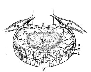
  • Posture and Pain

    Posture and Pain

    Analogies that are often used by chiropractors include:

    • If one walks on the edge of their shoe, the shoe sole will wear out at the weight-bearing portion quicker than other parts of the sole.
    • If an automobile wheel alignment is not optimal, then the excessively stressed wheel will wear out quicker than the other wheels.

    The chiropractic analysis of the human frame involves a postural assessment. The chiropractor is assessing human alignment. Chiropractic interventions, including spinal adjustments (specific correctional manipulations), are often intended to correct postural alignments.

    The alignment issue is well explained by James Oschman, PhD, in his book Energy Medicine, The Scientific Basis (1):

    Gravity is the most potent physical influence in any human life.

    “Gravity pervades our bodies and our environment and affects our every activity. All of the structures around us – our homes, furniture, buildings, machinery, plant, and animal, – and our own bodies, are designed to function in a world dominated by gravity.”

    “To introduce the therapeutic significance of gravity, we summarize the work of Joel E. Goldthwait and his colleagues at Harvard Medical School.” “A surgeon in Boston and founder of the orthopaedic clinic at the General Hospital, Goldthwait developed a successful therapeutic approach to chronic disorders. The aim of his therapies was to get his patients to sit, stand, and move with their bodies in a more appropriate relationship with the vertical. After years of treating patients with chronic problems, he concluded that many of these problems arise because parts of the body become misaligned with respect to the vertical.”

    “Goldthwait documented with X-rays a build-up of calcium deposits around the vertebrae of individuals with chronic arthritis, and observed that these deposits can diminish when the individual acquires a more vertical stance. His therapeutic approach corrected many difficult problems without the use of drugs. He viewed the human body from a mechanical engineering perspective, in which alignment of parts is essential to reduce wear and stress. He pleaded with physicians to recognize and correct misalignments to prevent long-term harmful effects.”

    “The basic principle of gravitational biology is known to any child who plays with blocks. The center of gravity of each block must be vertically above the center of gravity of the one below, to have a stable, balanced arrangement. If the center of gravity of one block lies outside of the gravity line, stability is compromised.”

    “Likewise, there is only one stable, strain-free arrangement of the parts of the human body. Any variation from this orientation will require corresponding compensations in other parts of the support system.”

    “Misalignment of any part will affect the whole system, and that restoration of verticality is a way to address a wide variety of clinical problems.”

    Dr. Goldthwait pleaded for everyone to pay more attention to the ways they hold and move their bodies in relation to the gravitational field.

    Chiropractors consider abnormal postural alignments to have three primary orthopedic consequences that are related to a patient’s pain. They are:

    • Weight
    • Load
    • Chronic Muscle Counterbalance

    Weight

    A recent study (February 10, 2020) from the journal Menopause, notes that obesity “is positively associated with low back pain.” The authors state (2):

    “Higher body mass is thought to increase pressure on the lumbar vertebrae, discs, and other back structures, causing low back pain.”

    The weight premise is simple and logical. The spinal structures that bear weight are the same structures that initiate the pain signal to the brain (3, 4). These structures are primarily the intervertebral disc and the facet joints. More weight increases the stress on these structures, increasing irritation, inflammation, and pain.

    Weight reduction is a simple and proven strategy for spine pain reduction. Weight reduction advice to patients is universal from health care providers. Yet, achieving weight reduction is elusive. On March 3, 2020, the World Health Organization presented the grim global statistics (5):

    • Worldwide obesity has nearly tripled since 1975.
    • In 2016, more than 1.9 billion adults, 18 years and older, were overweight. Of these over 650 million were obese.
    • 39% of adults aged 18 years and over were overweight in 2016, and 13% were obese.
    • Most of the world’s population live in countries where being overweight and obese kills more people than being underweight.
    • 40 million children under the age of 5 were overweight or obese in 2018.
    • Over 340 million children and adolescents aged 5-19 were overweight or obese in 2016.

    The obesity statistics from the United States are more grim than the global numbers. An Associated Press headline from February 18, 2020 notes (6):

    U.S. Obesity Rate 42% and Rising

    This article makes these points:

    • “Obesity – which means not merely overweight, but seriously overweight – is considered one of the nation’s leading public health problems.”
    • About 4 in 10 American adults are obese.
    • About 1 in 10 American adults are severely obese.
    • The obesity rate in 2016 was 40%.
    • Adult obesity rates are trending up.
    • “Half a century ago, about 1 in 100 American adults were severely obese. Now it’s 10 times more common.”
    • The obesity rate has risen about 40% in the last two decades.
    • Increased obesity means that “more Americans will get diabetes, heart disease, and cancer.”

    Successful weight reduction would undoubtedly help spinal pain as well as other public health concerns. Although weight is clearly an issue in back pain, it is not the most important issue. More important than weight is load, as discussed below.

    Load

    Upright posture is a first-class lever mechanical system, such as a teeter-totter or seesaw (7, 8).

    first-class lever

    In the first-class lever, the fulcrum is located between the weight and the effort.

    Notice that the weight is not positioned directly above the fulcrum. Rather, it is located at a distance away from the fulcrum. This distance is called a lever arm.

    The effective weight on the fulcrum is the actual weight multiplied by the lever arm. An actual weight of 10 pounds positioned at a distance of 10 inches away from the fulcrum will impart to the fulcrum an effective weight of 100 pounds. The effective weight at the fulcrum is also known as the load. The load is far more influential on the fulcrum than is the weight.

    The weight-bearing joints of the spinal column are the intervertebral disc and the facet joints (9). There is an optimal pivot motion between these spinal joints known as the instantaneous axis of rotation. Any abnormality of the instantaneous axis of rotation is associated with increased spinal pain syndromes (10, 11, 12). Chiropractors refer to abnormal instantaneous axis of rotation between spinal joints as a spinal subluxation or subluxation complex. All changes in load influence the instantaneous axis of rotation.

    A great discussion pertaining to weight and load is found in Katy Bowman’s 2017 book Move Your DNA. Ms. Bowman is a biomechanics specialist, and she notes (13):

    “Our bodies respond to mechanical input.”

    The conversion of mechanical input to “biochemical processes is called mechanotransduction.”

    “Gravity is one force your body responds to constantly.”

    “The loads created by gravity depend upon our physical position relative to the gravitational force.”

    The load created by gravity differs depending on alignment with the “perpendicular force of gravity.”

    “We experience load 100 percent of the time.”

    “Every unique joint configuration, and the way that joint configuration is positioned relative to gravity, and every motion created, and the way that motion was initiated, creates a unique load that in turn creates a very specific pattern of strain in the body.” This is called “load profile.”

    Every load creates a “unique cellular deformation.”

    “It’s not the weight that breaks you down, it’s the load created by the way you carry it.”

    “Loads are often oversimplified to ‘weight’ because it makes them easier to understand, but there is much more going on with your sore knee (or foot, or back, or pelvic floor) than your weight.”

    “Weight is not the be-all and end-all of loads. When you want to improve your health, it’s much more important to consider how you carry your weight than to spend hours contemplating the lone data point that is Your Weight.”

    “No matter the activity, when it comes to health, of utmost importance is the loads created.”

    “Every rate, size, and angle at which a force is being applied creates a unique environment for your cells.”

    “The timing and rates of loads are important because loads are occurrences over a period of time.”

    Each tissue type responds differently to a load, yet “they are all connected, which means that a load you perceive as only happening in one part of your body is actually affecting all other parts of you, and affecting each part uniquely.”

    “We should recognize our lack of health as a sign of a broken (mechanical) environment.”

    “You can eat the perfect diet, sleep eight hours a night, and use only baking soda and vinegar to clean your house, but without the loads created by natural movement, all of these worthy efforts are thwarted on a cellular level, and your optimal wellness level remains elusive.”

    “Human diseases are repeatedly explained to us in terms of their chemical or genetic makeup; meanwhile, we’ve completely ignored the load profile that the function of our body depends upon.”

    “Whether out of convenience or ignorance, we have failed to address the habitat [“habitual position relative to gravity”] in which our genes dwell, and the impact of the way we move on the state of our health.”

    “Movement, like food, is not optional.”

    “Movement is what most humans are missing more than any other factor, and the bulk of the scientific community has dropped the ball.”

    “A decrease in movement is associated with decreases in muscle size, vascularization, and the sensitivity in your proprioceptive system.”

    “Movement, position, and resting state of our musculoskeletal system are huge influencers of our mechanical environment.”

    “All movement and lack of movement create subtle differences in outcome in individuals and their genes.”

    The amount your joints move is integrated by your sensory system. “Movement provides information for the body. Movement is an environmental or epigenetic factor. Our movement environment has been polluted.”

    “If you want your health to change, you must change the way you move.” Even tiny adjustments to your loading “can be worth millions in unspent healthcare dollars and bring about tremendous relief from your load-induced ailments.”

    “Tissues that spend most of their time in a fixed position will adapt to that position by making alterations that are fairly permanent.”

    “An under-moved area of the body will experience increases in the connective tissues.” The author calls these “extra-connected” areas of the body “sticky spots.”

    Immobility-induced connective tissue growth creates a binding and “behaves much like scar tissue.”

    “On the cellular level, a sticky spot interferes with the transmission of forces throughout your tissues—mechanical signals that give cells context about loads placed upon them as well as position.”

    When a joint has a sticky spot, “you compensate by moving other joints,” which may “come with a heavy dose of damage.” Areas just outside of the sticky spot “experience unnaturally high loads.”

    Exercise cannot come close to restoring the tissues already adapted. “Exercise is good, but not good enough.”

    “We need a tool to measure the loads, both on the whole body and on every body part. The tool I use is alignment.”

    Muscles become physically shorter in response to “chronic joint positioning.”

    This discussion by Bowman explains much of chiropractic:

    • Load is clinically more important than weight. Overweight/obese patients have a lot of trouble trying to lose weight, but chiropractors can quickly and effectively improve the load on the joints by improving postural alignment.
    • Soft tissues adapt to abnormal loads over time. Bowman refers to these soft tissue changes as “sticky spots.” Others refer to them as an “abnormal instantaneous axis of rotation.” Chiropractors refer to them as the “subluxation.”

    Chronic Muscle Counterbalance

    Understanding that upright human posture is a first-class lever system is important. Understanding that load is more important than weight is also important. Of equal importance is the realization that load (weight multiplied by a lever arm) is only half the story.

    Abnormal postural alignment creates a lever arm stress on the spinal fulcrum, which are the intervertebral disc and the facet joints. But since upright posture is a first-class lever, a counterbalancing force must also be generated or the human would fall over. This counterbalancing force is generated by the muscles. This counterbalance contraction of the muscles is both fatiguing to the muscle and increases the compressive loads on the fulcrum tissues.

    fulcrum

    Rene Cailliet, MD, uses an example where a patient has unbalanced forward head posture (14). Dr. Cailliet assigns the head a weight of 10 lbs. and displaces the head’s center of gravity forward by 3 inches. The required counter balancing muscle contraction on the opposite side of the fulcrum (the vertebrae) would be 30 lbs. (10 lbs. X 3 inches):

    When the first-class lever of upright posture alignment is altered, for any reason, there is an increased mechanical load born by the fulcrum, i.e. the spinal intervertebral discs and facet joints. This increased mechanical load accelerates degenerative joint disease (7, 15).

    In their 1990 book Clinical Biomechanics of the Spine (8), White and Panjabi state:

    “The load on the discs is a combined result of the object weight, the upper body weight, the back muscle forces, and their respective lever arms to the disc center.”

    The constant muscle contraction required to counterbalance postural distortions creates muscle fatigue and myofascial pain syndromes. Rene Cailliet, MD states “This increase [in muscle tension] not only is fatiguing, but acts as a compressive force on the soft tissues, including the disk.” (7).

    Dr. Cailliet explains how the constant contraction in the counterbalancing muscles creates a cascade that leads to muscle fatigue, inflammation, fibrosis, and eventually to chronic musculoskeletal pain syndromes (14):

    Summary

    Being obese and overweight are epidemic problems in the modern world, and especially so in the United States. Much of the attention associated with being overweight/obese pertains to diabetes, heart/artery disease, and cancer. Another very important problem associated with being overweight/obese is increased wear and tear on the musculoskeletal joints, including the spinal joints.

    Increased spinal joint wear and tear is associated with intervertebral disc and facet joint degradation and arthritic changes. The consequent irritations and inflammations cause spinal pain syndromes.

    Overweight/obese problems are difficult to improve. At best, improvement requires significant dietary and exercise habit changes over a period of months. However, for the musculoskeletal system, more important than weight is load. Load is weight multiplied by the lever-arm distance from the fulcrum (the intervertebral disc and facet joints). Since the load profile can be improved very quickly and efficiently by chiropractic care, chiropractic is notorious for helping patients with spine pain syndromes.

    Spinal pain is proven to be articular (disc and facet joints), not muscular. Yet, the muscle system is involved, especially if it is called upon to supply chronic contraction in order to keep the human upright in a gravity environment. Chronic muscle contraction adds to (doubles) the load profile stress on the spinal joints. In addition, chronic muscle contraction themselves become painful as a consequence of internal tissue ischemia, retained metabolites, and inflammation.

    The chiropractic improvement of postural alignment distortions help patients with pain syndromes through multiple mechanisms.

    References:

    1. Oschman J; Energy Medicine, The Scientific Basis; Churchill Livingstone; 2000.
    2. Hao-Wei Xu HW, Yi YY, Zhang SB, Hu T, Wang SJ, Zhao WD, Wu DS; Does Vitamin D Status Influence Lumbar Disc Degeneration and Low Back Pain in Postmenopausal Women? A Retrospective Single-center Study; Menopause; February 10, 2020; Vol. 27; No. 5; [epub].
    3. Kuslich S, Ulstrom C, Michael C; The Tissue Origin of Low Back Pain and Sciatica: A Report of Pain Response to Tissue Stimulation During Operations on the Lumbar Spine Using Local Anesthesia; Orthopedic Clinics of North America; Vol. 22; No. 2; April 1991; pp.181-187.
    4. Bogduk N, Aprill C; On the nature of neck pain, discography and cervical zygapophysial joint blocks; Pain; August 1993; Vol. 54; No. 2; pp. 213-217.
    5. https://www.who.int/news-room/fact-sheets/detail/obesity-and-overweight. Accessed March 11, 2020.
    6. Stobbe M; U.S. Obesity Rate 42% and Rising, Survey Finds; Associated Press; February 28, 2020.
    7. Cailliet R; Low Back Pain Syndrome; 4th edition; FA Davis Company; 1981.
    8. White AA, Panjabi MM; Clinical Biomechanics of the Spine; Second Edition; Lippincott; 1990.
    9. Kapandji AI; The Physiology of the Joints: The Spinal Column, Pelvic Girdle and Head; Seventh Edition; Handspring Publishing; 2019.
    10. Haher TR, O’Brien M, Felmly WT, Welin D, Perrier G, Choueka J, Devlin V, Vassiliou A, Chow G; Instantaneous axis of rotation as a function of the three columns of the spine; Spine; June 1992; Vol. 17; No. 6 Suppl; pp. S149-154.
    11. Amevo B, Aprill C, Bogduk N; Abnormal instantaneous axis of rotation in patients with neck pain; Spine; July 1992; Vol. 17; No, 7; pp. 748-756.
    12. Bogduk N, Amevo B, Pearcy M; A biological basis for instantaneous centers of rotation of the vertebral column; Proc Inst Mech Eng H; 1995; Vol. 209; No. 3; pp. 177-183.
    13. Bowman K: Move Your DNA: Restore Your Health Through Natural Movement; propriometricspress; 2017.
    14. Cailliet R; Soft Tissue Pain and Disability; 3rd Edition; FA Davis Company; 1996.
    15. Garstang SV, Stitik SP; Osteoarthritis; Epidemiology, Risk Factors, and Pathophysiology; American Journal of Physical Medicine and Rehabilitation; November 2006; Vol. 85; No. 11; pp. S2-S11.

    “Authored by Dan Murphy, D.C.. Published by ChiroTrust® – This publication is not meant to offer treatment advice or protocols. Cited material is not necessarily the opinion of the author or publisher.”

  • Why Some Patients Take Longer to Recover from Whiplash Injuries

    Why Some Patients Take Longer to Recover from Whiplash Injuries

    Pain and the treatment of pain is the biggest health problem in America. In her book, A Nation in Pain, Healing Our Biggest Health Problem, Judy Foreman documents that approximately 50% of American adults suffer from chronic pain (1). A conservative estimate of the direct costs and lost productivity resulting from this pain is up to $635 billion yearly (2).

    The body region most likely to suffer from pain is the low back. The third most common body location for pain is headache. The fourth most common location is the neck (3):

    Lower-Back Pain – 28.1%
    Knee Pain – 19.5%
    Severe Headache – 16.1%
    Neck Pain – 15.1%
    Shoulder Pain – 09.0%

    The primary reason patients go to chiropractors is for spinal pain complaints. This was established in the journal Spine two years ago (4). In rounded numbers, 60% go to chiropractors for low back pain and 33% do so for neck pain.

    Headaches and neck pain often coexist. Neck problems often manifest in headaches. This is because all headaches synapse in the neck, in a region known as the trigeminal-cervical nucleus (5, 6).

    Chiropractic and spinal manipulation are very effective in the treatment of spinal pain syndromes, and levels of patient satisfaction are quite high (4). This is why spinal manipulation has been advocated in official practice guidelines for more than a decade (7, 8, 9, 10, 11).

    Pain perception and pain character, in any body location, is linked to inflammatory chemicals. This concept is nicely reviewed in the journal Medical Hypothesis (12):

    “Every pain syndrome has an inflammatory profile consisting of the inflammatory mediators that are present in the pain syndrome.”

    “The key to treatment of pain syndromes is an understanding of their inflammatory profile.”

    “Our unifying theory or law of pain states: the origin of all pain is inflammation and the inflammatory response.”

    “Irrespective of the type of pain whether it is acute or chronic pain, peripheral or central pain, nociceptive or neuropathic pain, the underlying origin is inflammation and the inflammatory response.”

    “Activation of pain receptors, transmission and modulation of pain signals, neuro-plasticity and central sensitization are all one continuum of inflammation and the inflammatory response.”

    “Irrespective of the characteristic of the pain, whether it is sharp, dull, aching, burning, stabbing, numbing or tingling, all pain arises from inflammation and the inflammatory response.”

    When discussing inflammation as related to pain, three distinct types of inflammation should be distinguished. This classification is important because they are managed differently:

    • Local inflammation
    • Systemic inflammation
    • A combination of local and systemic inflammation

    Local Inflammation

    Local inflammation is caused by trauma, prolonged tissue stress, and/or repetitive tissue strain. The local inflammation in the area of tissue stress generates local pain. Causation is established by history and confirmed with examination. The management is local, directed to the painful tissues.

    Systemic Inflammation

    Systemic inflammation means the entire body is inflamed. The more systemically inflamed a person is, the more likely they will perceive pain secondary to local injury or tissue stress. The best outcome for these individuals requires management of their systemic inflammatory profile.
    Systemic inflammation is suspected by history, but often requires lab work for confirmation. There are a number of causes of systemic inflammation. The two most common are:

    • Suboptimal levels of omega-3 fatty acids
    • A “leaky gut”

    The champion of management of chronic spine pain syndromes with omega-3 fatty acids is Joseph Maroon, MD. Dr. Maroon is a neurosurgeon and works with the Pittsburgh Steelers National Football League team (13).

    A recent vocal champion of systemic inflammation being caused by “leaky gut” is cardiologist Stephen Gundry, MD. Dr. Gundry’s gut approach to the management of systemic inflammation is well described in his 2017 and 2019 books (14, 15).

    A Combination of Local and Systemic Inflammation

    A combination of local and systemic inflammation is probably the typical patient suffering with pain. Optimal management requires both local and systemic interventions.

    Restating, inflammation and pain are linked. Successful pain management parallels successful inflammation management. The primary category of drugs to treat pain are non-steroidal anti-inflammatory drugs, or (NSAIDs). NSAIDs work by blocking a series of enzymes that produce an inflammatory hormone-like molecule called prostaglandin E2. The name of this enzyme is cyclo-oxygenase, abbreviated COX. These anti-inflammatory drugs have many serious side effects:

    The journal Spine notes in 2003 (16):

    “Adverse reactions to non-steroidal anti-inflammatory (NSAID) medication have been well documented.”

    “Gastrointestinal toxicity induced by NSAIDs is one of the most common serious adverse drug events in the industrialized world.”

    In 2006, the journal Surgical Neurology states (13):

    Blockage of the COX enzyme inhibits the conversion of arachidonic acid to the very pro-inflammatory prostaglandins that mediate the classic inflammatory response of pain.

    “More than 70 million NSAID prescriptions are written each year, and 30 billion over-the-counter NSAID tablets are sold annually.”

    “5% to 10% of the adult US population and approximately 14% of the elderly routinely use NSAIDs for pain control.”

    Almost all patients who take the long-term NSAIDs will have gastric hemorrhage, 50% will have dyspepsia, 8% to 20% will have gastric ulceration, 3% of patients develop serious gastrointestinal side effects, which results in more than 100,000 hospitalizations, an estimated 16,500 deaths, and an annual cost to treat the complications that exceeds 1.5 billion dollars.

    “NSAIDs are the most common cause of drug-related morbidity and mortality reported to the FDA and other regulatory agencies around the world.”

    One author referred to the “chronic systemic use of NSAIDs to ‘carpet-bombing,’ with attendant collateral end-stage damage to human organs.”

    NSAIDs always affect the body systemically, resulting in the many diverse multisystem deleterious side effects. For example, taking NSAIDs for a painful sprained ankle will have deleterious influences of the gastrointestinal tract, kidney, liver, and blood vessels. A local treatment that is directed only to the ankle is desired because of the lack of systemic side effects. Such a treatment involves local controlled motion.

    Low back pain emanates primarily from the intervertebral disc (17, 18, 19, 20, 21). Neck pain primarily emanates from the facet joint (22, 23, 24, 25).

    Chiropractic spinal manipulation primarily helps patients with spinal pain for two reasons:

    ONE:

    Motion disperses the accumulation of local inflammatory chemicals. This is well-stated in Dr. Vert Mooney’s 1986 Presidential Address of the International Society for the Study of the Lumbar Spine (19). Dr. Mooney notes:

    “In the United States in the decade from 1971 to 1981, the numbers of those individuals disabled from low-back pain grew at a rate 14 times that of the population growth. This growth occurred in the very decade when there was an explosion of ergonomic knowledge, labor-saving mechanical assistance devices, and improved diagnostic equipment. We apparently could not find the source of pain.”

    “Anatomically the motion segment of the back is made up of two synovial joints and a unique relatively avascular tissue found nowhere else in the body – the intervertebral disc. Is it possible for the disc to obey different rules of damage than the rest of the connective tissue of the musculoskeletal system?”

    “Mechanical events can be translated into chemical events related to pain.”

    “Mechanical activity has a great deal to do with the exchange of water and oxygen concentration” in the disc.

    An important aspect of disc nutrition and health is the mechanical aspects of the disc related to the fluid mechanics.

    The pumping action maintains the nutrition and biomechanical function of the intervertebral disc. Thus, “research substantiates the view that unchanging posture, as a result of constant pressure such as standing, sitting or lying, leads to an interruption of pressure-dependent transfer of liquid. Actually, the human intervertebral disc lives because of movement.”

    “The fluid content of the disc can be changed by mechanical activity.”

    “In summary, what is the answer to the question of where is the pain coming from in the chronic low-back pain patient? I believe its source, ultimately, is in the disc. Basic studies and clinical experience suggest that mechanical therapy is the most rational approach to relief of this painful condition.”

    TWO:

    Motion initiates a neurological sequence of events that “closes” the pain gate. This is well-stated by orthopedic surgeon WH Kirkaldy-Willis, MD, in 1985 (26):

    “Spinal manipulation is essentially an assisted passive motion applied to the spinal apophyseal [facet] and sacroiliac joints.”

    Melzack and Wall proposed the Gate Theory of Pain in 1965, and this theory has “withstood rigorous scientific scrutiny.”

    “The central transmission of pain can be blocked by increased proprioceptive input.” Pain is facilitated by “lack of proprioceptive input.” This is why it is important for “early mobilization to control pain after musculoskeletal injury.”

    The facet capsules are densely populated with mechanoreceptors. “Increased proprioceptive input in the form of spinal mobility tends to decrease the central transmission of pain from adjacent spinal structures by closing the gate. Any therapy which induces motion into articular structures will help inhibit pain transmission by this means.”

    Stretching of facet joint capsules will fire capsular mechanoreceptors which will reflexively “inhibit facilitated motoneuron pools” which are responsible for the muscle spasms that commonly accompany low back pain.

    Why Do Some Whiplash-Injured
    Patients Take Longer to Recover?

    When a traumatic soft tissue injury does not recover as expected, there are two classic explanations:

    • One was discussed above, systemic inflammation. An injury superimposed on a patient already suffering from systemic inflammation is often treatment resistant until the systemic inflammation is addressed.
    • The second explanation is pre-accident degenerative spinal disease. This includes intervertebral disc degeneration, facet arthrosis, and/or uncinate joint arthrosis. Collectively they are often referred to as degenerative joint disease and/or spondylosis.

    •••

    In 1964, whiplash injury expert and pioneer, Ruth Jackson, MD, published an article titled “The Positive Findings in Neck Injuries” in the American Journal of Orthopedics. Dr. Jackson’s conclusions in this article were based on her evaluation of 5,000 injured patients. She notes (27):

    Pre-existing pathological conditions of the cervical spine, when injured, “result in more damage than would be anticipated in a so-called ‘normal’ cervical spine.”

    •••

    In 1977, Samuel Turek, MD, clinical professor from the Department of Orthopedics and Rehabilitation at the University of Miami School of Medicine, and author of the reference text, Orthopaedic Principles and Their Applications, states (28):

    “The injury may be compounded by the presence of degenerative disease of the spine.”

    “With advancing age, especially in the presence of degenerative disease, the tissues become inelastic and are easily torn.”

    •••

    In 1981, Rene Cailliet, MD, professor and rehabilitation specialist from the University of Southern California, and author of the book Neck and Arm Pain, states (29):

    “The pre-existence of degeneration may have been quiescent in that no symptoms were noted, but now minor trauma may ‘decompensate’ the safety margin and symptoms occur.”

    •••

    In 1983, Norris and Watt followed 61 whiplash-injured patients for a minimum of six months in order to establish factors that were prognostic for recovery. Their conclusions include (30):

    “Factors which adversely affect prognosis include the presence of objective neurological signs, stiffness of the neck, [loss of cervical lordosis], and pre-existing degenerative spondylosis.”

    Degenerative spondylosis was detected in 26% of patients with no objective findings, 33% of patients with reduced cervical range of motion, and 40% of patients with neurological loss, indicating that cervical spine degenerative changes are associated with greater injury and worse prognosis for recovery.

    This “study suggests that prognosis is predictable on the basis of the initial presentation of the patient.” “Two features on plain radiographs seem relevant.”

    1) “Pre-existing degenerative changes in the cervical spine, no matter how slight, do appear to affect the prognosis adversely.”

    2) Abnormal curves in the cervical spine “are more common in patients with a poor outcome.”

    “The prognosis may be modified by the presence or absence of degenerative changes, by an abnormality [degeneration] of the cervical spine on the initial radiograph, or by both.”

    •••

    In 1985, Webb in his article titled “Mechanisms and Patterns of Tissue Injury” notes (31):

    “Degenerative joint disease is recognized as a major influence on subsequent tissue damage both in severity and pattern.”

    “In any individual where changes consistent with degenerative joint disease are present, one can expect the injury to be more severe or a very minor injury to produce severe symptoms requiring prolonged treatment.”

    •••

    In 1986, Arthur Ameis, MD, from the University of Toronto, notes (32):

    “For the elderly, neck injury can be very serious. The degenerative spine is biomechanically ‘stiffer’, behaving more like a single long bone than like a set of articulating structures. Deforming forces are less evenly dissipated, and more damage is done.”

    •••

    In 1987, physicians Edward Dunn and Steven Blazar authored “Soft-Tissue Injuries of the Lower Cervical Spine” for the American Academy of Orthopedic Surgeons. In this publication they note (33):

    “If present, degenerative changes should be duly noted as they may affect the prognosis.”

    “…pre-existing degenerative changes adversely affected the outcome.”

    •••

    In 1988, Maimaris and colleagues published a study titled “Whiplash Injuries of the Neck.” They reviewed 102 whiplash-injured patients 2 years after injury. They concluded (34):

    “The analysis of the radiological results showed that pre-existing degenerative changes in the cervical spine are strongly indicative of a poor prognosis.”

    •••

    In October of 1988, physician Hirsch and colleagues published a paper titled “Whiplash Syndrome, Fact or Fiction?” in Orthopedic Clinics of North America. These authors note (35):

    Pre-existing structural changes and degenerative changes are “frequently associated with a more difficult, more prolonged, and less complete recovery.”

    “The films should be inspected especially for evidence of pre-existing structural changes or for alteration, which are frequently associated with a more difficult, more prolonged, and less complete recovery.”

    •••

    In their 1988 reference text on whiplash injuries titled Whiplash Injuries, The Acceleration/Deceleration Syndrome, Steve Foreman and Arthur Croft note (36):

    “…the presence of preexisting degenerative changes, no matter how slight, appears to alter the prognosis adversely.”

    •••

    In 1989, physician Porter published an article in the British Medical Journal titled “Neck Sprains After Car Accidents.” He noted (37):

    “Pre-existing degenerative changes may worsen the prognosis.”

    •••

    In 1991, Watkinson, along with Gargan and Bannister, radiographically reviewed 35 whiplash-injured patients 10.8 years after injury. They concluded (38):

    “Patients with degenerative change initially have more symptoms after 2 years than those with normal radiographs at the time of injury.”

    “Degenerative changes occurred significantly more frequently in patients who had sustained soft tissue injuries than in a control population.”

    Also in 1991, the reference text Painful Cervical Trauma, Diagnosis and Rehabilitative Treatment of Neuromusculoskeletal Injuries, notes (39):

    “The elasticity of tissues decreases with an increase in age. The range of motion in the cervical spine also decreases. In both cases, the potential for injury is increased because the neck is less resilient.”

    •••

    In 1995, physician Jerome Schofferman and colleague Dr. S. Wasserman published in Spine an article titled: “Successful treatment of low back pain and neck pain after a motor vehicle accident despite litigation.” These authors also noted (40):

    “Pre-existing degenerative changes on initial x-rays, no matter how slight, had a worse prognosis.”

    •••

    In 1996, Squires, along with Drs. Martin Gargan and Gordon Bannister, published a 15.5-year follow-up evaluation of 40 patients who had been injured in a motor vehicle collision. They published their results once again in the prestigious British Journal of Bone and Joint Surgery, titled “Soft-tissue Injuries of the Cervical Spine: 15-year Follow-up.” In this article, these authors note (41):

    “The patients who had deteriorated were on average five years older than the rest of the group.”

    “80% of the patients who had deteriorated in the last five years had degenerative changes.”

    “100% of patients with severe ongoing problems had cervical degeneration at 11 years after injury.”

    •••

    In 1999, the reference text Whiplash and Related Headaches, by neurologist Bernard Swerdlow, MD, makes the following point (42):

    Risk factors that may lead to chronicity include “pre-existing degenerative osteoarthritic changes.”

    “Other conditions that may pre-exist the accident that may contribute to a chronic state following the accident are osteoarthritis, degeneration of vertebral body joints, disc degeneration and inflammatory processes.”

    “Studies indicate that pre-existing osteoarthritic changes contributed to alter the prognosis adversely.”

    •••

    In 2002, in their reference text titled Whiplash, Gerard Malanga, MD and Scott Nadler, DO, state (43):

    “Several researchers have associated poor clinical outcomes with spondylosis, reporting a higher prevalence of spondylosis in patients with continued symptoms.”

    “It is certainly theoretically possible that symptoms from a previously asymptomatic cervical spondylosis are precipitated by trauma and are responsible for the continuing pain.”

    •••

    In 2005, physician Schenardi published a study titled “Whiplash injury, TOS and double crush syndrome, Forensic medical aspects.” In this article he addresses the issue of pre-injury cervical spine degeneration by stating (44):

    A substantial percentage of people will have whiplash symptoms for more than a few months, “especially the elderly or those with pre-existing neck problems who may develop chronic long-term problems which may never resolve.”

    •••

    In his 2005 reference text titled Motor Vehicle Collision Injuries, Lawrence Nordhoff notes (45):

    “Patients who have clinically significant pre-existing medical conditions may have more severe injuries, slower recoveries and poorer prognoses.”

    Dr. Nordhoff clearly lists “spinal degeneration” as one such pre-existing medical factor.

    •••

    In 2019, a study published in The Spine Journal followed 121 whiplash-injured subjects prospectively for six months with CT scan. They noted (46):

    “This study demonstrates an increased risk for non-recovery after whiplash trauma for patients with moderate facet joint degeneration as demonstrated on CT scans performed shortly after trauma.”

    “We suggest that one of the underlying mechanisms of WAD may be that trauma triggers a painful clinical manifestation of underlying, previously asymptomatic, cervical facet joint degeneration.”

    In conclusion, pre-existing degenerative joint disease renders those joints less capable of adequately handling and dispersing the forces of a new injury; therefore, injury to these articulations and the surrounding soft tissues is greater; the amount of treatment required for maximum improvement is greater, and there are more long-term subjective, objective, and functional residuals.

    REFERENCES:

    1. Foreman J; A Nation in Pain, Healing Our Biggest Health Problem; Oxford University Press; 2014.
    2. Pho K; USA TODAY, The Forum; September 19, 2011; pg. 9A.
    3. Wang S; Why Does Chronic Pain Hurt Some People More?; Wall Street Journal; October 7, 2013.
    4. Adams J, Peng W, Cramer H, Sundberg T, Moore C; The Prevalence, Patterns, and Predictors of Chiropractic Use Among US Adults: Results From the 2012 National Health Interview Survey; Spine; December 1, 2017; Vol. 42; No. 23; pp. 1810–1816.
    5. Seletz E; Headache of Extracranial Origin; California Medicine; November 1958; Vol. 89; No. 5; pp. 314-317.
    6. Bogduk N; Anatomy and Physiology of Headache; Biomedicine and Pharmacotherapy; 1995; Vol. 49; No. 10; pp. 435-445.
    7. Chou R, Qaseem A, Snow V, Casey D, Cross JT, Shekelle P, Owens DK; Diagnosis and Treatment of Low Back Pain; Annals of Internal Medicine; Vol. 147; No. 7; October 2007; pp. 478-491.
    8. Chou R, Huffman LH; Non-pharmacologic Therapies for Acute and Chronic Low Back Pain; Annals of Internal Medicine; October 2007; Vol. 147; No. 7; pp. 492-504.
    9. Globe G, Farabaugh RJ, Hawk C, Morris CE, Baker G, DC, Whalen WM, Walters S, Kaeser M, Dehen M, DC, Augat T; Clinical Practice Guideline: Chiropractic Care for Low Back Pain; Journal of Manipulative and Physiological Therapeutics; January 2016; Vol. 39; No. 1; pp. 1-22.
    10. Wong JJ, Cote P, Sutton DA, Randhawa K, Yu H, Varatharajan S, Goldgrub R, Nordin M, Gross DP, Shearer HM, Carroll LJ, Stern PJ, Ameis A, Southerst D, Mior S, Stupar M, Varatharajan T, Taylor-Vaisey A; Clinical practice guidelines for the noninvasive management of low back pain: A systematic review by the Ontario Protocol for Traffic Injury Management (OPTIMa) Collaboration; European Journal of Pain; Vol. 21; No. 2 (February); 2017; pp. 201-216.
    11. Qaseem A, Wilt TJ, McLean RM, Forciea MA; Noninvasive Treatments for Acute, Subacute, and Chronic Low Back Pain: A Clinical Practice Guideline from the American College of Physicians; For the Clinical Guidelines Committee of the American College of Physicians; Annals of Internal Medicine; April 4, 2017; Vol. 166; No. 7; pp. 514-530.
    12. Omoigui S; The biochemical origin of pain: The origin of all pain is inflammation and the inflammatory response: Inflammatory profile of pain syndromes; Medical Hypothesis; 2007; Vol. 69; pp. 1169–1178.
    13. Maroon JC, Bost JW; Omega-3 Fatty acids (fish oil) as an anti-inflammatory: An alternative to nonsteroidal anti-inflammatory drugs for discogenic pain; Surgical Neurology; April 2006; Vol. 65; pp. 326– 331.
    14. Gundry S; The Plant Paradox: The Hidden Dangers in “Healthy” Foods that Cause Disease and Weight Gain; 2017.
    15. Gundry S; The Longevity Paradox: How to Die Young at a Ripe Old Age; 2019.
    16. Giles LGF, Muller R; Chronic Spinal Pain: A Randomized Clinical Trial Comparing Medication, Acupuncture, and Spinal Manipulation; Spine; July 15, 2003; Vol. 28; No. 14; pp.1490-1502.
    17. Smyth MJ, Wright V, Sciatica and the intervertebral disc. An experimental study; Journal of Bone and Joint Surgery [American]; December 1958; Vol. 40; No. A-6; pp. 1401-1408.
    18. Nachemson AL; The Lumbar Spine: An Orthopedic Challenge; Spine; Vol. 1; No. 1; March 1976; pp. 59-71.
    19. Mooney V; Where Is the Pain Coming From?; Spine; Vol. 12; No. 8; October 1987; pp. 754-759.
    20. Kuslich S, Ulstrom C, Michael C; The Tissue Origin of Low Back Pain and Sciatica: A Report of Pain Response to Tissue Stimulation During Operations on the Lumbar Spine Using Local Anesthesia; Orthopedic Clinics of North America; Vol. 22; No. 2; April 1991; pp.181-187.
    21. Izzo R, Popolizio T, D’Aprile P, Muto M; Spine Pain; European Journal of Radiology; May 2015; Vol. 84; pp. 746–756.
    22. Bogduk N, Aprill C; On the nature of neck pain, discography and cervical zygapophysial joint blocks; Pain; August 1993; Vol. 54; No. 2; pp. 213-217.
    23. Barnsley L, Lord SM, Wallis BJ; Bogduk N; The prevalence of chronic cervical zygapophysial joint pain after whiplash; Spine; January 1, 1995; Vol. 20; No. 1; pp. 20-25.
    24. Lord SM, Barnsley L, Wallis BJ; Bogduk N; Chronic cervical zygapophysial joint pain after whiplash. A placebo-controlled prevalence study; Spine; August 1, 1996; Vol. 21; No. 15; pp. 1737-1744.
    25. Bogduk N; On Cervical Zygapophysial Joint Pain After Whiplash; Spine
      December 1, 2011; Vol. 36; No. 25S; pp. S194–S199.
    26. Kirkaldy-Willis WH, Cassidy JD; Spinal Manipulation in the Treatment of Low back Pain; Canadian Family Physician; March 1985; Vol. 31; pp. 535-540.
    27. Jackson R; The Positive Findings in Neck Injuries; American Journal of Orthopedics; August-September 1964; pp. 178-187.
    28. Turek S; Orthopaedics Principles and their Applications; Lippincott; 1977; p. 740.
    29. Cailliet R; Neck and Arm Pain; F. A. Davis Company; 1981; p. 103.
    30. Norris SH, Watt I; The Prognosis of Neck Injuries Resulting From Rear-end Vehicle Collisions; The Journal Of Bone And Joint Surgery (British); November 1983; Vol. 65-B.
    31. Webb; Whiplash: Mechanisms and Patterns of Tissue Injury; Journal of the Australian Chiropractors’ Association; June 1985.
    32. Ameis A; Cervical Whiplash: Considerations in the Rehabilitation of Cervical Myofascial Injury; Canadian Family Physician; September 1986.
    33. Dunn EJ, Blazar S; Soft-tissue injuries of the lower cervical spine; Instructional course lectures; 1987;Vol. 36; pp. 499-512.
      Maimaris C, Barnes MR, Allen MJ; ‘Whiplash injuries’ of the neck: A retrospective study; Injury; November 1988; Vol. 19; No. 6; pp; 393-396.
    34. Hirsch SA, Hirsch PJ, Hiramoto H, Weiss A; Whiplash syndrome: Fact or fiction? Orthopedics Clinics of North America; October 1988; Vol. 19; No. 4; pp. 791-795.
    35. Foreman S and Croft A; Whiplash Injuries, The Acceleration/Deceleration Syndrome; Williams & Wilkins; 1988; p. 389 and p. 395.
    36. Porter KM; Neck sprains after car accidents; British Medical Journal; April 15, 1989; 298(6679); pp. 973-974.
    37. Watkinson A, Gargan M, Bannister G; Prognostic factors in soft tissue injuries of the cervical spine; Injury: the British Journal of Accident Surgery; July 1991; pp. 307-309.
    38. Friedmann L, Marin E, Padula P; “Biomechanics of Cervical Trauma” in Painful Cervical Trauma, Diagnosis and Rehabilitative Treatment of Neuromusculoskeletal Injuries; Edited by C. David Tollison and John R. Satterthwaite; Williams and Wilkins; 1991, p. 17.
    39. Schofferman J, Wasserman S; Successful treatment of low back pain and neck pain after a motor vehicle accident despite litigation; Spine; May 1, 1994; Vol. 19; No. 9; pp. 1007-1010.
    40. Squires B, Gargan M, Bannister G; Soft-tissue Injuries of the Cervical Spine, 15-year Follow-up; Journal of Bone and Joint Surgery (British); November 1996, Vol. 78-B, No. 6, pp. 955-7.
    41. Swerdlow B; Whiplash and Related Headaches; CRC press; 1999; p. 1040.
    42. Malanga G and Nadler S; Whiplash; Hanley & Belfus; 2002; p. 91.
    43. Schenardi C; Whiplash Injury, TOS and double crush syndrome, Forensic medical aspects; Acta Neurochirurgica; supplement, Vol. 92; 2005; pp. 25-27.
    44. Nordhoff L; Motor Vehicle Collision Injuries, Biomechanics, Diagnosis, and Management; Second Edition; Jones and Bartlett; 2005, pp. 537-538.
    45. Rydman E, Kasina P, Ponzer S, Jarnbert-Pettersson H; Association Between Cervical Degeneration and Self-perceived Non-recovery After Whiplash Injury; The Spine Journal; December 2019; Vol. 19; No. 12; pp. 1986−1994.

    “Authored by Dan Murphy, D.C.. Published by ChiroTrust® – This publication is not meant to offer treatment advice or protocols. Cited material is not necessarily the opinion of the author or publisher.”

  • Sleep, Awareness, Motor Vehicle Collisions, and Chiropractic Care

    Sleep, Awareness, Motor Vehicle Collisions, and Chiropractic Care

    In the October 31, 2019 issue of the financial-political magazine Forbes, there is a brief article recommending the reading of a book on sleep by Dr. Matthew Walker. The author of the article is John Doerr, a venture capitalist with Kleiner Perkins. Mr. Doerr prefaces his review by noting that “Leaders from the worlds of business, academia and entertainment and politics share what’s on their bedside tables,” and it is this book by Matthew Walker (1).

    Matthew Walker, PhD, is a professor of neuroscience and psychology at the University of California, Berkeley. He is also the director of the Center for Human Sleep Science at UC Berkeley. In 2017, Dr. Walker wrote the book (2):

    Why We Sleep
    Unlocking the Power of Sleep and Dreams

    This is a fascinating book on neurology and brain neurochemistry and how they influence whole-body health. He notes that every organ in the body and every process in the brain is enhanced with optimal sleep. But he also notes that sleep loss is epidemic in modern developed nations and that sleep loss is the greatest public health challenge we face. He states:

    “Sleep is the single most effective thing we can do to reset our brain and body health each day.”

    Much of Dr. Walker’s book pertains to the adverse consequences of too little sleep, including:

    • Demolishes your immune system, doubling one’s risk of cancer
    • Increases Alzheimer’s disease
    • Disrupts blood sugar creating pre-diabetes
    • Damages arteries, increasing blood pressure and the risks of heart attack and stroke
    • Contributes to all major psychiatric conditions, including: depression/anxiety/suicide
    • Makes you hungry, resulting in overeating and obesity
    • Impairs our ability to learn, memorize, make logical decisions and choices
    • Harms our ability to modify (inhibit) painful memories (PTSD)
    • Inhibits creativity
    • Shortens telomeres and therefore shortens both the quantity and quality of life, stating: “The shorter your sleep, the shorter the life span.”

    Pertinent to this discussion, too little sleep causes hundreds of thousands of traffic accidents yearly, exceeding alcohol and drug caused accidents combined. Dr. Walker makes these points pertaining to too little sleep and motor vehicle collisions:

    There are deadly consequences of “getting behind the wheel of a motor vehicle without having sufficient sleep.”

    “Drowsy driving is the cause of hundreds of thousands of traffic accidents and fatalities each year.”

    “Tragically, one person dies in a traffic accident every hour in the United States due to [sleep] fatigue-related error.”

    “There are many ways in which lack of sufficient sleep will kill you. Some take time; others are far more immediate. One brain function that buckles under even the smallest dose of sleep deprivation is concentration. The deadly societal consequences of these concentration failures play out most obviously and fatally in the form of drowsy driving.”

    “In a disturbing study, researchers took two groups of healthy adults, one of whom they got drunk to the legal driving limit (.08 percent blood alcohol), the other of whom they sleep-deprived for a single night. Both groups performed the concentration test to assess attention performance, specifically the number of lapses. After being awake for nineteen hours, people who were sleep-deprived were as cognitively impaired as those who were legally drunk. Said another way, if you wake up at seven a.m. and remain awake throughout the day, then go out socializing with friends until late that evening, yet drink no alcohol whatsoever, by the time you are driving home at two a.m. you are as cognitively impaired in your ability to attend to the road and what is around you as a legally drunk driver. In fact, participants in the above study started their nosedive in performance after just fifteen hours of being awake (10 p.m. in the above scenario).”

    “Car crashes rank among the leading causes of death in most first-world nations. In 2016, the AAA Foundation in Washington, DC, released the results of an extensive study of over 7,000 drivers in the US, tracked in detail over a two-year period. The key finding reveals just how catastrophic drowsy driving is when it comes to car clashes. Operating on less than five hours of sleep, your risk of a car crash increases threefold. Get behind the wheel of a car when having slept just four hours or less the night before and you are 11.5 times
    more likely to be involved in a car accident. Note how the relationship between decreasing hours of sleep and increasing mortality risk of an accident is not linear, but instead exponentially mushrooms. Each hour of sleep lost vastly amplifies that crash likelihood, rather than incrementally nudging it up.”

    “Drunk driving and drowsy driving are deadly propositions in their own right, but what happens when someone combines them? It is a relevant question, since most individuals are driving drunk in the early-morning hours rather than in the middle of the day, meaning that most drunk drivers are also sleep-deprived.”

    “We can now monitor driver error in a realistic but safe way using driving simulators. A group of researchers examined the number of complete off-road deviations in participants placed under four different experimental conditions.”:

    1) 8 hours of sleep: zero to few off-road errors.

    2) 4 hours of sleep: six times more off-road deviations then sober well-rested individuals.

    3) 8 hours of sleep plus alcohol to the point of being legally drunk: six times more off-road deviations then sober well-rested individuals (the same as only 4 hours of sleep) “Driving drunk or driving drowsy were both dangerous, and equally dangerous.”

    4) 4 hours of sleep plus alcohol to the point of being legally drunk:

    “A reasonable expectation was that performance in the fourth group of participants would reflect the additive impact of these two groups: four hours of sleep plus the effect of alcohol (i.e., twelve times more off-road deviations) It was far worse. This group of participants drove off the road almost 30 times more than the well-rested, sober-group.”
    “The heady cocktail of sleep loss and alcohol was not additive, but instead multiplicative. They magnified each other, like two drugs whose effects are harmful by themselves but, when taken together, interact to produce truly dire consequences.”

    “After thirty years of intensive research, we can now answer many of the questions posed earlier. The recycle rate of a human being is around sixteen hours. After sixteen hours of being awake, the brain begins to fail. Humans need more than seven hours of sleep each night to maintain cognitive performance. After ten days of just seven hours of sleep, the brain is as dysfunctional as it would be after going with¬out sleep for twenty-four hours.”

    “The real-life consequences of drowsy driving deserve special mention. This coming week, more than 2 million people in the US will fall asleep while driving their motor vehicle. That’s more than 250,000 every day, with more such events during the week than week¬ends for obvious reasons. More than 56 million Americans admit to struggling to stay awake at the wheel of a car each month.”

    “As a result, 1.2 million accidents are caused by sleepiness each year in the United States. Said another way: for every thirty seconds you’ve been reading this book, there has been a car accident somewhere in the US caused by sleeplessness. It is more than probable that someone has lost their life in a fatigue-related car accident during the time you have been reading this chapter.”

    “You may find it surprising to learn that vehicle accidents caused by drowsy driving exceed those caused by alcohol and drugs combined. Drowsy driving alone is worse than driving drunk. Drunk drivers are often late in braking, and late in making evasive maneuvers. But when you fall asleep, or have a microsleep, you stop reacting altogether.”

    “A person who experiences a microsleep or who has fallen asleep at the wheel does not brake at all, nor do they make any attempt to avoid the accident. As a result, car crashes caused by drowsiness tend to be far more deadly than those caused by alcohol or drugs. Said crassly, when you fall asleep at the wheel of your car on a freeway, there is now a one-ton missile traveling at 65 miles per hour, and no one is in control.”

    “Drivers of cars are not the only threats. More dangerous are drowsy truckers. Approximately 80 percent of truck drivers in the US are over¬weight, and 50 percent are clinically obese. This places truck drivers at a far, far higher risk of a disorder called sleep apnea, commonly associ¬ated with heavy snoring, which causes chronic, severe sleep depriva¬tion. As a result, these truck drivers are 200 to 500 percent more likely to be involved in a traffic accident. And when a truck driver loses his or her life in a drowsy-driving crash, they will, on average, take 4.5 other lives with them.”

    “In actual fact, I would like to argue that there are no accidents caused by fatigue, microsleeps, falling asleep. None whatsoever. They are crashes. The Oxford English Dictionary defines accidents as unexpected events that happen by chance or without apparent cause. Drowsy-driv¬ing deaths are neither chance, nor without cause. They are predictable and the direct result of not obtaining sufficient sleep. As such, they are unnecessary and preventable. Shamefully, governments of most devel¬oped countries spend less than 1 percent of their budget educating the public on the dangers of drowsy driving relative to what they invest in combating drunk driving.”

    “There are many things that I hope readers take away from this book. This is one of the most important: if you are drowsy while driving, please, please stop. It is lethal. To carry the burden of another’s death on your shoulders is a terrible thing. Don’t be misled by the many ineffective tactics people will tell you can battle back against drowsiness while driving. Many of us think we can overcome drowsiness through sheer force of will, but, sadly, this is not true. To assume otherwise can jeopardize your life, the lives of your family or friends in the car with you, and the lives of other road users. Some people only get one chance to fall asleep at the wheel before losing their life.”

    “One of the ironic statistics concerning drowsy driving. When a sleep-deprived [medical] resident finishes a long shift, such as a stint in the ER trying to save victims of car accidents, and then gets into their own car to drive home, their chances of being involved in in a motor vehicle accident are increased by 168 percent because of fatigue.”

    “After twenty-two hours without sleep, human performance is impaired to the same level as that of someone who is legally drunk.”

    “The leading cause of death among teenagers is road traffic accidents.”

    “The leading cause of death among teenagers is road traffic accidents, and in this regard, even the slightest dose of insufficient sleep can have marked consequences.”

    “When the Mahtomedi School District of Minnesota pushed their school start time from 7:30 to 8:00 a.m., there was a 60 percent reduction in traffic accidents in drivers sixteen to eighteen years of age.”

    “Teton County in Wyoming enacted an even more dramatic change in school start time, shifting from a 7:35 a.m. bell to a far more biologically reasonable one of 8:55 a.m. The result was astonishing—a 70 percent reduction in traffic accidents in sixteen- to eighteen-year-old drivers.”

    “These publicly available findings should have swept the education system in an uncompromising revision of school start times. Instead, they have largely been swept under the rug.”

    In his book, Dr. Matthew Walker describes the relationships between drowsy driving, automobile collisions, and loss of life. For chiropractors, critically important to the discussion is automobile collision injuries. As described by Dr. Walker, drowsy driving is more hazardous than drunk driving. Drunk driving slows protective responses, increasing the risks of collisions. Drowsy driving causes microsleep or full-on sleep, eliminating all protective responses.

    During microsleep or full-on sleep the driver is caught completely unaware of the impending collision:

    In 1990, an article published in the journal of the Society of Automotive Engineers, titled Whiplash in Low Speed Vehicle Collisions notes (3):

    “If the passenger is aware of and anticipates a collision, and makes his neck muscle tense, he can tolerate more severe impact.”

    In 1992, the book Painful Cervical Trauma notes (4):

    “Injury results because the neck is unable to adequately compensate for the rapidity of head and torso movement resulting from the acceleration forces generated at the time of impact. This is particularly true when the impact is unexpected and the victim is unable to brace for it.”

    In 1993, an article published in the journal Trial Talk, titled The Physics, Biomechanics and Statistics of Automobile Rear Impact Collisions, notes (5):

    “Research has shown that an occupant aware of an impending impact may possess sufficient muscle control to prevent hyperflexion and hyperextension during low velocity impacts.”

    In 1993, the journal/book Spine: State of the Art Reviews, Cervical Flexion-Extension/Whiplash Injuries, notes (6):

    In the whiplash acceleration-deceleration injury, muscle response might arrest, limit, or control the movements of a cervical motion segment. “Without muscle control the normal arcuate movement of a cervical motion segment must be disturbed, and the forces to which individual segments are subjected can be resisted only by passive ligamentous elements or bony contact. This sets the scene for a variety of possible injuries.”

    In the same 1993 journal/book, another author notes (7):

    “…when the impact is unexpected and the victim is unable to brace.”

    In 1994, a research article evaluating 137 whiplash-injured subjects was published in Neurology, titled Presenting Symptoms and Signs after Whiplash Injury: The influence of Accident Mechanism, notes (8):

    “Patients struck when they were unprepared for the impact had a significantly higher frequency of multiple symptoms, higher headache intensity, and shorter latency of headache onset.

    The state of preparedness “proved to be the first significant factor with respect to initial injury findings.”

    Also in 1994, research published in the journal Injury, titled Neck Strain in Car Occupants: Injury Status After 6 Months and Crash-related Factors, notes (9):

    “…awareness appears to have a strong protective influence and may prove to be a useful prognostic indicator in clinical settings.

    …subjects who were unaware of the impending collision had a greatly increased likelihood of experiencing persisting symptoms and/or signs of neck strain, compared to those who were aware.

    Subjects who were unaware of the impending collision were 15 times more likely to have a persisting condition than those who were aware.”

    In 1995, a follow-up to the 137 whiplash-injured study (#8) was published in the Journal of Neurology, titled The Effect of Accident Mechanism and Initial Findings on the Long-term Course of Whiplash Injury, again notes

    “…unpreparedness at the time of impact…” is the most significant in poor outcome from a whiplash injury (10):

    In 1998, primary research published in the journal Archives of Physical Medicine and Rehabilitation, titled Clinical Response of Human Subjects to Rear-end Automobile Collisions, notes that if the patient is caught by surprise during a rear-end collision, the threshold for injury begins at a change in velocity of only 2.5 mph (11).

    In 2014, an article published in the journal PET and SPECT in Neurology, titled Whiplash: Real or Not Real?: A Review and New Concept, notes (12):

    In some patients who experience an unexpected rear-end collision, the “symptoms persist for years.”

    An essential factor in chronic whiplash symptoms is the “unexpectedness of the accident.”

    “Whether or not the driver in a rear-end crashed car is aware of the impending collision is extremely important.”

    “The awareness or expectancy of the incoming collision is crucial in the whiplash process;” 70-80% of the patients suffering from chronic whiplash “were unaware of the incoming collision.”

    “A correlation exists between being unaware of the incoming collision and a poor recovery.”

    A crucial factor in determining the extent of a whiplash mechanism injury is the “expectancy of the incoming collision.”

    The biological concepts are simple and not controversial. Joints are injured when the muscles that cross the joint do not optimally protect them. Historically, being “caught by surprise” prior to a motor vehicle collision offers no joint protection from the muscles. Chiropractors have known for decades that for many injured patients, perhaps the most important prognostic factor, is understanding the patient’s state of awareness prior to the collision. As such, chiropractors routinely ask about and record the “state of awareness” question on their patients.

    Similarly, experiencing a microsleep or full-on sleep prior to an automobile collision would eliminate all joint protection from the muscles, increasing joint injury. Perhaps inquiring about sleep status the night prior to and just before an automobile collisions would add valuable injury and prognostic information.

    Thirty percent of patients who initially seek chiropractic care do so because of neck pain, and their level of satisfaction with their care is quite high (13). Studies support the value of spinal manipulation for the treatment of neck pain and neck injured patients. The biological rationale for spinal manipulation in the treatment of whiplash-injury neck pain includes:

    A. The primary injury from whiplash biomechanics is to the facet joints.

    B. The primary source of both acute and chronic whiplash injury pain is the facet joints.

    C. Spinal adjusting (specific joint manipulation) primarily affects the facet joints.

    In support, four studies are briefly presented here:

    In 1996, clinicians from the University Department of Orthopaedic Surgery, Bristol, United Kingdom, published a study in the journal Injury, titled (14):

    Chiropractic Treatment of Chronic ‘Whiplash’ Injuries

    They authors note that 43% of patients will suffer long-term symptoms following ‘whiplash’ injury, for which no conventional treatment has proven to be effective. Consequently, they performed a retrospective study to determine the effects of chiropractic spinal manipulation in a group of 28 patients who were suffering with chronic ‘whiplash’ syndrome.

    The 28 patients in this study had initially been treated with anti-inflammatories, soft collars and physiotherapy. These patients had all become chronic, and were referred for chiropractic at an average of 15.5 months (range was 3–44 months) after their initial injury. At the initial evaluation and prior to chiropractic treatment, 27/28 (96%) of the patients were classified as having intrusive or disabling symptoms.

    Following the chiropractic treatment, 93% of the patients had improved. The authors concluded:

    “The encouraging results from this retrospective study merit the instigation of a prospective randomized controlled trial to compare conventional with chiropractic treatment in chronic ‘whiplash’ injury.”

    “The results of this retrospective study would suggest that benefits can occur in over 90% of patients undergoing chiropractic treatment for chronic whiplash injury.”

    In 1999, the same group of clinicians from the University Department of Orthopaedic Surgery, Bristol, United Kingdom, published a study in the Journal of Orthopaedic Medicine, titled (15):

    A Symptomatic Classification of Whiplash Injury
    and the Implications for Treatment

    This study involved 93 consecutive whiplash-injured patients who were chronic and referred for chiropractic spinal manipulation. Patients underwent a mean of 19.3 treatments (range 1-53), over a period of 4.1 months. The authors note:

    “Conventional treatment of patients with whiplash symptoms is disappointing.”

    “In chronic cases, no conventional treatment has proved successful.”

    “The results from this study provide further evidence that chiropractic is an effective treatment for chronic whiplash symptoms.”

    “Chiropractic is the only proven effective treatment in chronic [whiplash] cases.”

    In 2004, a group of physiotherapists, physicians, and professors from the Rey Juan Carlos University, Spain, published a study in the Journal of Whiplash & Related Disorders, titled (16):

    Manipulative Treatment vs. Conventional Physiotherapy
    Treatment in Whiplash Injury: A Randomized Controlled Trial

    The objective of this clinical trial was to compare the results obtained from a manipulative protocol with the results obtained from a conventional physiotherapy treatment in patients suffering from whiplash injury. This is the first controlled experimental trial documenting the effects of the manipulative protocol used in this study. It was a randomized controlled trial using 380 acute whiplash injury (less than 3 months duration) subjects. The authors note:

    “Patients who had received manipulative treatment needed fewer sessions to complete the treatment than patients who had received physiotherapy treatment.”

    “Patients of manipulative group needed an average of 9 sessions to complete the treatment, whereas physiotherapy group needed an average of 23 sessions.”

    “Results showed that the manipulative group had more benefits than the physiotherapy group.”

    “Our clinical experience with these [whiplash-injured] patientshas demonstrated that manipulative treatment gives better results than conventional physiotherapy treatment.”

    “This clinical trial has demonstrated that head and neck pain decrease with fewer treatment sessions in response to a manipulative treatment protocol as compared to a physiotherapy treatment protocol among patients diagnosed with acute whiplash injury.”

    “Manipulation is “effective in the management of whiplash injury.”

    “Manipulative treatment is more effective in the management of whiplash injury than conventional physiotherapy treatment.”

    In 2015, researchers and clinicians from the Orthopedic University Hospital Balgrist, University of Zurich, Switzerland, published a study in the Journal of Manipulative and Physiological Therapeutics, titled (17):

    Prognostic Factors for Recurrences in
    Neck Pain Patients Up to 1 Year After Chiropractic Care

    This is a prospective cohort study assessing 545 neck pain patients. After a course of chiropractic spinal manipulation, they were followed up for one year regarding recurrence of their neck pain.

    The results of this study are impressive:

    Fifty-four (54) participants (11%) were identified as “recurrent.”
    Four hundred ninety one (491) participants (89%) were not recurrent.

    The authors state:

    “89% of neck pain patients had recovered from their neck pain episode up to 1 year after receiving chiropractic care.”

    “The results of this study suggest that recurrence of neck pain within 1 year after chiropractic intervention is low.”

    REFERENCES

    1. Doerr J; “Book Value”; Forbes; October 31, 2019; p. 26.
    2. Walker S; Why We Sleep: Unlocking the Power of Sleep and Dreams; Scribner; 2017.
    3. Emori RI, Horiguchi J; Whiplash in Low Speed Vehicle Collisions; SAE; February 1990; p. 108.
    4. Teasell RW, McCain GA; in Painful Cervical Trauma; Williams and Wilkins; 1992; p. 293.
    5. Smith JJ; The Physics, Biomechanics and Statistics of Automobile Rear Impact Collisions; Trial Talk; June 1993; pp. 0-14.
    6. Lord S; in Spine: State of the Art Reviews: “Cervical Flexion-Extension/Whiplash Injuries”; Hanley & Belfus; September 1993; p. 360.
    7. Teasell RW, in Spine: State of the Art Reviews: Cervical Flexion-Extension/Whiplash Injuries”; Hanley & Belfus; September 1993; p. 374.
    8. Sturzenegger M, DiStefano G, Radanov BP, Schnidrig A; Presenting symptoms and signs after whiplash injury: The influence of accident mechanism; Neurology; April 1994; pp. 688-693.
    9. Ryan GA, Taylor GW, Moore VM, Dolinis J; Neck strain in car occupants: Injury status after 6 months and crash-related factors; Injury; September 1994; pp. 533-537.
    10. Sturzenegger M, Radanov BP, Di Stefano G; The effect of accident mechanism and initial findings on the long-term course of whiplash injury; Journal of Neurology; 1995; pp. 443-449.
    11. Brault JR, Wheeler JB; Clinical response of human subjects to rear-end automobile collisions; Archives of Physical Medicine and Rehabilitation; 1998, Vol. 79; No. 1; pp. 72-80.
    12. Garcia DV, Dierckx RAJP, Otte A; Whiplash: Real or Not Real?: A Review and New Concept; PET and SPECT in Neurology; 2014; pp. 947-963.
    13. Adams J, Peng W, Cramer H, Sundberg T, Moore C; The Prevalence, Patterns, and Predictors of Chiropractic Use Among US Adults: Results From the 2012 National Health Interview Survey; Spine; December 1, 2017; Vol. 42; No. 23; pp. 1810–1816.
    14. Woodward MN, Cook JCH, Gargan MF, and Bannister GC; Chiropractic treatment of chronic ‘whiplash’ injuries; Injury; Vol. 27; No. 9; November 1996; pp. 643-645.
    15. Khan S, Cook J, Gargan M, Bannister G; A symptomatic classification of whiplash injury and the implications for treatment; The Journal of Orthopaedic Medicine; Vol. 21; No. 1; 1999; pp. 22-25.
    16. Fernández-de-las-Peñas C, Fernández-Carnero J, Palomeque del Cerro L, Miangolarra-Page JC; Manipulative Treatment vs. Conventional Physiotherapy Treatment in Whiplash Injury: A Randomized Controlled Trial; Journal of Whiplash & Related Disorders; 2004; Vol. 3; No. 2.
    17. Langenfeld A, Humphreys K, Swanenburg J, Cynthia K. Peterson CK; Prognostic Factors for Recurrences in Neck Pain Patients Up to 1 Year After Chiropractic Care; Journal of Manipulative and Physiological Therapeutics; September 2015; Vol. 38; No. 7; pp. 458-464.

    “Authored by Dan Murphy, D.C.. Published by ChiroTrust® – This publication is not meant to offer treatment advice or protocols. Cited material is not necessarily the opinion of the author or publisher.”

  • The Biomechanics of the Spine in the Treatment of Spine Pain Syndromes

    The Biomechanics of the Spine in the Treatment of Spine Pain Syndromes

    The Management of Spine Pain Syndromes Through the
    Understanding and Treatment of the Spine as a Single Functioning Unit

    It is convenient to divide and discuss the human spinal column into four regions:

    • Cervical spine (neck)
    • Thoracic spine (middle back)
    • Lumbar spine (low back)
    • Pelvis (the base of the spinal column)

    Although this classification is convenient, it is also misleading and occasionally harmful. The discussion presented here reminds us that the four convenient regions of the spinal column are interrelated both biomechanically and neurologically. In short, the entire spinal column is a single functioning unit.

    Biomechanics

    Ninety-three percent of chiropractic patients seek care for low back (63%) and/or neck pain (30%) (1).

    The most common tissue source of chronic low back pain is the intervertebral disc (2). The most common tissue source for chronic neck pain is the facet joint capsules (3).

    Human bipedal upright posture is a three-dimensional first class mechanical lever system (4, 5):

    first class lever

    In the first class lever mechanical system, the fulcrum is located between the load and the effort. In human posture, the load is the weight multiplied by the distance away from the fulcrum. The effort to maintain a balanced structure is supplied by the muscles. All postural imbalances increase the load on the fulcrum and require muscle effort to prevent tipping over.

    The fulcrum is the structure that bears the greatest mechanical stress. The fulcrum is the “weakest link” for mechanical degradation. Overly large loads with equalizing counterbalancing efforts will cause the structure to break at the fulcrum. An example would be using one’s hands to break a stick on one’s knee.

    In the spine, the fulcrum of the first class lever of upright posture is primarily the vertebral body/disc, and the two facet joints. When the first class lever of upright posture is altered, for any reason, there is an increased mechanical load on the fulcrum, i.e. the spinal intervertebral discs and facet joints. Such increased mechanical loads accelerate degenerative joint disease and inflammation, altering the pain thresholds (4, 5, 6).

    Postural distortions that cause spinal fulcrum stress may begin at the head. Or, postural distortions may also begin at the base of the spine, the pelvis. Pelvic leveling problems may be attributed to problems in the pelvis, but also in the feet, knees, hips, etc. The notorious “Leaning Tower of Pisa” is an example of a mechanical problem caused by an unleveled base.

    The bottom line is that the primary tissues responsible for spinal pain (the disc and the facet) are the same tissues that function as the fulcrum of the first-class level of upright posture.

    •••••

    In the reference text The Spine, chapter 2 is titled (7):

    “Applied Anatomy of the Spine”

    This chapter is written by Wesley Parke, PhD. At the time of publication, Dr. Parke was Professor and Chairman, Department of Anatomy, University of South Dakota School of Medicine. In this chapter, Dr. Parke writes:

    “Although the 23 or 24 individual motor segments must be considered in relation to the spinal column as a whole, no congenital or acquired disorder of a single major component of a unit can exist without affecting first the functions of the other components of the same unit and then the functions of other levels of the spine.”

    The reference text Disorders of the Cervical Spine is written by rheumatologist John Bland, MD. Dr. Bland is a Professor of Medicine at the University of Vermont College of Medicine. Dr. Bland writes (8, 9):

    “We tend to divide the examination of the spine into regions: cervical, thoracic, and lumbar spine clinical studies.

    This is a mistake.

    The three units are closely interrelated structurally and functionally – a whole person with a whole spine.

    The cervical spine may be symptomatic because of a thoracic or lumbar spine abnormality, and vice versa!

    Sometimes treating a lumbar spine will relieve a cervical spine syndrome, or proper management of the cervical spine will relieve low backache.”

    For these biomechanical reasons, chiropractors often evaluate and treat regions of the spine that are asymptomatic yet functionally related to the region of primary complaint. As an example, improving the postural alignment of the head will reduce fulcrum spinal stress in the low back, reducing low back pain.

    Neurology

    By now (2019), nearly everyone is aware of the narcotic epidemic in the United States. Narcotics are pain drugs. Narcotics include both opiates and opioids (10):

    Opiates are compounds that are purified directly from the opium poppy plant.

    Opioids are a synthetic form of opium, made in a chemistry lab, like Fentanyl.

    Both opiates and opioids suppress pain by attaching to receptors. Sadly, opiate receptors adapt quickly to these narcotic drugs and the pain suppression benefits are soon lost. Compensation for this receptor adaptation is increasing the dose or adding a second narcotic product. More sadly, increasing narcotic dose has numerous side effects, including addiction, mood changes, drowsiness, mental clouding, nausea, vomiting, constipation, and even death (11). A study published in the journal Pain Therapy in 2018 from Harvard Medical School states (12):

    “Tragically, opioids claimed over 64,000 lives just last year [2017].”

    Perhaps even more concerning is that 80% of heroin addicts in the United Stated begin their addiction with a narcotic prescribed for pain (13).

    •••••

    The understanding and management of chronic pain was significantly advanced in 1973 when Candace Pert (d. 2013) discovered the opiate receptor (14). Dr. Pert was an American neuroscientist and pharmacologist who was awarded a Ph.D. the following year (1974), from Johns Hopkins University School of Medicine.

    By 1977, researchers determined that the opiate receptors in the upper brain stem (mesencephalon) were responsible for controlling pain throughout the body (15). The specific location for these opiate receptors was the grey matter surrounding the cerebral aqueduct, known as periaqueductal gray matter. The authors inserted electrodes (an invasive and risky endeavor) into the periaqueductal gray matter of the mesencephalon of six cancer patients suffering from intractable pain. They state (15):

    “Relief of intractable pain was produced in six human patients by stimulation of electrodes permanently implanted in the periventricular and periaqueductal gray matter.”

    In his 1979 book, The Brain, The Last Frontier, Richard Restak, MD, reviews this research, stating (16):

    “Within the periaqueductal gray, a deep-seated brainstem area lying along the floor of the third ventricle, neurosurgeons at the University of California in San Francisco placed indwelling stimulating electrodes for pain relief in six patients afflicted with chronic, unremitting pain. Whenever the patients began to experience pain, they were able to shut it off via the activation of a battery-operated stimulator about the size of a pack of cigarettes. After activating the stimulator, all six patients—in accordance with earlier findings in other pain patients—experienced dramatic, long-lasting, and repeatable pain relief.”

    “In order to test the hypothesis that pain relief was genuine and not just an example of a ‘placebo response,’ one patient was outfitted with a stimulator containing a ‘dead’ battery. The patient, a fifty-one-year-old woman with severe back and leg pain caused by cancer of the colon, anxiously reported that her pain had returned and the stimulator ‘wasn’t working.’ Replacement of a new battery led to immediate pain relief.”

    Periaqueductal gray matter stimulation for pain control initiates what is know as activation of the Descending Pain Inhibitory Control System, and it has continued to receive reference text support ever since (17, 18, 19, 20). These studies indicate the following:

    • Opiate receptors, when activated, inhibit pain.
    • The periaqueductal gray matter of the mesencephalon is densely populated with opiate receptors.
    • Electrical stimulation of the periaqueductal gray matter opiate receptors causes quick and complete pain relief throughout the body.

    However, electrical stimulation of the periaqueductal gray matter opiate receptors for pain suppression is quite invasive and has logistical risks. Consequently, narcotic drugs, as noted above, have become increasingly used for pain control since the 1980s (21, 22). This approach to pain control has resulted in a national nightmare, well described in the political magazine Time that states (23):

    “They’re the most powerful painkillers ever invented. And they’re creating the worst addiction crisis America has ever seen.”

    “…9.4 million Americans take opioids for long-term pain.”

    “Doctors so frequently prescribe the drugs known as opioids for chronic pain from conditions like arthritis, migraines and lower back injuries that there are enough pills prescribed every year to keep every American adult medicated around the clock for a month.”

    “Now 4 of 5 [80%] heroin addicts say they came to the drug from prescription painkillers.”

    “The American Academy of Neurology last year concluded that the risks of long-term opioid treatment for headaches and chronic low-back pain likely outweigh the benefits.”

    Spinal Manipulation Science for Pain Evolves

    Over the past century, chiropractors have observed that spinal manipulation in one region of the body would often alleviate pain in a different, untreated region of the body. Consequently, chiropractors adjust (specific manipulation) all documented biomechanical lesions, regardless of the patient’s pain presentation.

    In addition to these biomechanical considerations, published studies show a neurological explanation for the effect of spinal adjusting in the relief of pain in non-treated body areas. Interestingly, these explanations involve the activation of the periaqueductal gray matter descending pain inhibitory control system.

    In 1996, a study was published in the journal Pain, titled (24):

    The Initial Effects of a Cervical Spine Manipulative
    Physiotherapy Treatment on the Pain and
    Dysfunction of Lateral Epicondylalgia

    Importantly, these authors were treating elbow pain in patients, who were not suffering from radiculopathy or referred pain, without touching or treating the offending elbow. In contrast, treatment was directed towards the dysfunctional joints of the patient’s cervical spine. The most commonly found biomechanical dysfunction was hypomobility of the joints of the lower cervical spine. The authors made the following comments:

    “This study has demonstrated a clear hypoalgesic effect of a manipulative therapy technique [applied to the cervical spine] in the period immediately following its application in a group of patients with lateral epicondylalgia.”

    “A significant treatment effect beyond placebo or control was demonstrated.”

    “The beneficial effects of treatment [cervical manipulation] may continue after its application.”

    The author’s theoretical model to explain their results involved manipulative therapy activation of the hypoalgesic effects of the endogenous supraspinal pain inhibitory systems.

    “The [manipulative] treatment technique used in this study provided a non-noxious sensory input at the cervical spine which resulted in a reduction of elbow pain that outlasted the duration of its application.” “This is thought to activate the descending pain inhibitory system as a major component of their pain-relieving effects.”

    The descending pain inhibitory system is activated by stimulation of the periaqueductal gray (PAG).

    “These findings indicate that manipulative therapy may constitute an adequate physical stimulus for activating the descending pain inhibitory system.”

    “Manipulative therapy [may] recruit the descending pain inhibitory system, through which it exerts a portion or all of its pain-relieving effects. That is, manipulative therapy applied to the cervical spine produces a sensory input which could be sufficient to activate descending pain inhibitory system.”

    “In a group of patients with lateral epicondylalgia, a manipulative therapy treatment technique applied to the lower cervical spine produced hypoalgesia at the elbow as manifest by increased pressure pain threshold, increased grip strength, improved neurodynamics and reduced pain over a 24 h period. This finding substantiates clinical observations that manipulative therapy is capable of producing improvements in pain and function immediately following application.”

    Another study indicating that spinal manipulation is capable of activating the descending pain control system was published in the Journal of Back Musculoskeletal Rehabilitation in 2014, and titled (25):

    The role of the Descending Inhibitory Pain Mechanism
    in Musculoskeletal Pain Following High-Velocity,
    Low Amplitude Thrust Manipulation: A Review of the Literature

    The objective of this review was to investigate the role of the Descending Inhibitory Pain Mechanism in musculoskeletal pain following high-velocity, low amplitude thrust manipulation, as well as to identify the pain-relieving importance of this technique within clinical practice. The authors of this article make the following comments and conclusions:

    “Although the antinociceptive effect of high-velocity, low amplitude thrust manipulation has been recognized by numerous systematic reviews, the underlying mechanism for manipulation-related pain relief remains poorly understood. An increasing number of studies have explored its analgesic mechanism suggesting that the excitation of the descending inhibitory pain mechanism might play the most important role for musculoskeletal pain relief.”

    “Findings from current literature support that high-velocity, low amplitude thrust manipulation has a profound influence on nociceptive stimulus via the possible activation of the descending inhibitory pain mechanism. It seems that the application of this technique activates the periaqueductal gray region area of the midbrain, stimulates the noradrenergic descending system and at the level of the spinal cord, the nociceptive afferent barrage is reduced and mechanical hypoalgesia is induced.”

    “The clinical importance of the activation of the descending inhibitory pain mechanism should not be ignored since the resulted analgesic effect of this technique can provide a window of opportunity to restore impaired physical performance and disability.”

    In April 2019, researchers from Stanford University, the University of Colorado, the University of Sydney, and the University of Illinois College of Medicine, published a study in the official journal of the American Pain Society, the Journal of Pain, titled (26):

    Decreased Neurologic Pain Signature Activation
    Following Thoracic Spine Manipulation in Healthy Volunteers

    These authors used functional magnetic resonance imaging in 10 healthy volunteers to study the effect of spinal manipulation on pain-related activity within brain regions predictive of physical pain. The induced pain was a noxious mechanical stimulation of the right index finger cuticle before and after thoracic spinal manipulation. Functional magnetic imaging was performed with 5 minutes of noxious mechanical stimulation before and after thoracic spinal manipulation.

    The index finger cuticle is innervated by the sixth cervical nerve. Hence, any changes in pain perception or pain imaging would not be as a consequence of a thoracic spinal manipulation; if such pain reduction was noted, a plausible explanation would be that the thoracic manipulation activated the descending pain inhibitory control system.

    The authors documented a significant decrease in brain pain markers following spinal manipulation. They noted that spinal manipulation’s therapeutic action may be “mediated at the level of the central nervous system.”

    In October 2019, the same research group (from reference #26) extended their investigations in patients with neck pain. This study was published in the journal NeuroImage: Clinical, and titled (27):

    Evidence for Decreased Neurologic Pain Signature
    Activation Following Thoracic Spinal Manipulation
    in Healthy Volunteers and Participants with Neck Pain

    Once again, these authors used functional magnetic resonance imaging (fMRI) to investigate the effect of thoracic spinal manipulation on pain-related brain activity in 16 subjects with acute/subacute neck pain and compare them to 10 healthy volunteers. The aim of the authors was to extend their investigation to determine if spinal manipulation’s therapeutic action is mediated within the central nervous system. Their primary determination was to document if thoracic spinal manipulation could make meaningful reductions in neck pain.

    These investigators found that pain activation decreases following spinal manipulation, and that spinal manipulation alters the processing of pain-related brain activity. They state:

    “The findings provide evidence that spinal manipulation may alter pain-related brain activity within brain regions specific to the processing of physical pain, supporting a possible central mechanism of spinal manipulation.”

    “A growing body of evidence is pointing towards neurophysiologic mechanisms of action underlying the pain modulating effects of spinal manipulation including both spinal and supraspinal mechanisms.”

    “The findings provide evidence that spinal manipulation may alter the processing of pain-related brain activity within specific pain-related brain regions.”

    Neck pain and brain pain region activation decreased following thoracic spinal manipulation. These findings were not noted following sham thoracic spinal manipulation. The results suggest a decrease in the nociceptive information reaching supraspinal brain areas.

    Most recently (November 2019), an important study on the topic of spinal manipulation activating the brain’s pain descending inhibitory system was published in the Irish Journal of Medical Science, and titled (28):

    The Effect of Spinal Manipulation on Brain Neurometabolites
    in Chronic Nonspecific Low Back Pain Patients:
    A Randomized Clinical Trial

    These authors note that in patients with chronic nonspecific low back pain, brain function changes due to the neuroplastic changes. Therefore, the objective of this study was to evaluate the brain metabolite changes after spinal manipulation. They used using proton magnetic resonance spectroscopy for their analysis.

    Twenty-five patients with chronic nonspecific low back pain, aged 20–50 years were enrolled in the study. Patients were randomly assigned lumbo-pelvic manipulation or sham.

    These patients were evaluated before and 5 weeks after treatment using:

    • Numerical Rating Scale
    • Oswestry Disability Index
    • Proton Magnetic Resonance Spectroscopy

    The authors state:

    “After treatment, severity of pain and functional disability were significantly reduced in the treatment group vs. sham group.”

    The authors also found significant important changes in brain metabolites following low back spinal manipulation that was not found in the sham group. The authors concluded:

    “In the patient with low back pain, spinal manipulation affects the central nervous system and changes the brain metabolites. Consequently, pain and functional disability are reduced.”

    ••••••••••

    These studies support the notion that spinal adjusting activates the supra-segmental descending inhibitory pain system. This means that one may not need to adjust the painful spinal segmental level to obtain meaningful pain reduction. Activating this system with narcotic drugs is very problematic. Activating this system through spinal adjusting is plausible and effective for many patients. Recent evidence suggests that chiropractic spinal adjusting for spinal pain syndromes can reduce narcotic use by 64% (29).

    REFERENCES

    1. Adams J, Peng W, Cramer H, Sundberg T, Moore C; The Prevalence, Patterns, and Predictors of Chiropractic Use Among US Adults: Results From the 2012 National Health Interview Survey; Spine; December 1, 2017; Vol. 42; No. 23; pp. 1810–1816.
    2. Kuslich S, Ulstrom C, Michael C; The Tissue Origin of Low Back Pain and Sciatica: A Report of Pain Response to Tissue Stimulation During Operations on the Lumbar Spine Using Local Anesthesia; Orthopedic Clinics of North America; Vol. 22; No. 2; April 1991; pp. 181-187.
    3. Bogduk N; On Cervical Zygapophysial Joint Pain After Whiplash; Spine; December 1, 2011; Vol. 36; No. 25; pp. S194–S199.
    4. White AA, Panjabi MM; Clinical Biomechanics of the Spine, Second Edition; Lippincott; 1990.
    5. Cailliet R; Soft Tissue Pain and Disability; 3rd Edition; FA Davis Company; 1996.
    6. Cailliet R; Low Back Pain Syndrome, 4th edition; F A Davis Company; 1981.
    7. Parke WW; “Applied Anatomy of the Spine” chapter 2 in Rothman and Simeone; The Spine, second edition; WB Saunders Company; 1982.
    8. Bland J; Disorders of the Cervical Spine; WB Saunders Company; 1987.
    9. Bland J; Disorders of the Cervical Spine; Second edition; WB Saunders Company; 1994.
    10. Offit PA; Pandora’s Lab: Seven Stories of Science Gone Wrong; National Geographic; 2017.
    11. Bear M, Connors B, Paradiso M; Neuroscience: Exploring the Brain; Second Edition; Lippincott Williams & Wilkins; 2001.
    12. Jones MR, Viswanath O, Peck J, Kaye AD, Gill JS, Simopoulos TT; A Brief History of the Opioid Epidemic and Strategies for Pain Medicine; Pain Therapy; June 2018; Vol. 7; No. 1; pp. 13-21.
    13. Irving D; Opioid Rising; Rand Review; Nov/Dec 2015.
    14. Pert CB, Snyder SH; Opiate receptor: demonstration in nervous tissue; Science; 1973 Mar 9;179(4077); pp. 1011-1014.
    15. Hosobuchi Y, Adams JE, Linchitz R; Pain relief by electrical stimulation of the central gray matter in humans and its reversal by naloxone; Science; 1977 Jul 8;197(4299); pp. 183-186.
    16. Restak R; The Brain, The Last Frontier; Warner Books; 1979.
    17. Kiernan JA: Barr’s The Human Nervous System, An Anatomical Viewpoint; Lippincott-Raven; 1998; pp351-352.
    18. Nolte J; The Human Brain, An Introduction To Its Functional Anatomy; Mosby; 1999; pp. 270-271; p. 274.
    19. Kandel E, et.al; Principles of Neural Science; 2000; pp. 482-485.
    20. Steward O; Functional Neuroscience; Springer; 2000; pp. 218-219.
    21. Porter J, Jick H; Addiction rare in patients treated with narcotics; New England Journal of Medicine; Jan 10, 1980; Vol. 302; No. 2; p. 123.
    22. Portenoy RK, Foley KM; Chronic use of opioid analgesics in non-malignant pain: report of 38 cases.; Pain; May 1986; Vol. 25; No. 2; pp.171-86.
    23. Calabresi M; “They’re the most powerful painkillers ever invented. And they’re creating the worst addiction crisis America has ever seen”; Time;
      June 15, 2015.
    24. Vicenzino B, Collins D, Wright A; The Initial Effects of a Cervical Spine Manipulative Physiotherapy Treatment on the Pain and Dysfunction of Lateral Epicondylalgia; Pain; November 1996; Vol. 68; No. 1; pp. 69-74.
    25. Savva C, Giakas G, Efstathiou M; The role of the descending inhibitory pain mechanism in musculoskeletal pain following high-velocity, low amplitude thrust manipulation: a review of the literature; Journal of Back Musculoskeletal Rehabilitation; 2014; Vol. 27; No. 4; pp. 377-382.
    26. Weber K, Wager T, Elliott J, Mackey S, Liu W, Sparks C; Decreased Neurologic Pain Signature Activation Following Thoracic Spine Manipulation in Healthy Volunteers; The Journal of Pain; April 2019; Vol. 20; No. 4; Supplement; p. S69.
    27. Weber K, Wager T, Elliott J, Mackey S, Liu W, Sparks C; Evidence for Decreased Neurologic Pain Signature Activation Following Thoracic Spinal Manipulation in Healthy Volunteers and Participants with Neck Pain; NeuroImage: Clinical; October 18, 2019; Vol. 24; 102042.
    28. Didehdar D, Kamali F, Yoosefinejad AK, Lotfi M; The effect of spinal manipulation on brain neurometabolites in chronic nonspecific low back pain patients: a randomized clinical trial; Irish Journal of Medical Science; November 2019; [epub].
    29. Corcoran KL, DC, Bastian LA, Gunderson CG, Steffens C, Brackett MA, Lisi AJ; Association Between Chiropractic Use and Opioid Receipt Among Patients with Spinal Pain: A Systematic Review and Meta-analysis; Pain Medicine; September 2019; [epub].

    “Authored by Dan Murphy, D.C.. Published by ChiroTrust® – This publication is not meant to offer treatment advice or protocols. Cited material is not necessarily the opinion of the author or publisher.”