Category: Advanced

  • Temporomandibular Joint Disorders, the Cervical Spine, and Spinal Manipulation

    Temporomandibular Joint Disorders, the Cervical Spine, and Spinal Manipulation

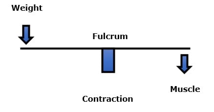
    In biomechanics, there is a rule that notes that the regions of the body that have the greatest mobility have the least stability; and reduced stability is coupled with greater injury and stress risk. Joints that have multiple planes of motion are particularly prone to increased stress and injury risk.

    The jaw not only open and closes, it can move side-to-side as well as protrude forward and retract backwards. These diverse planes of motion increase the risk for biomechanical problems.

    The jaw is used in talking, chewing, swallowing, kissing, yawning, and in facial expressions. It is vulnerable to injury during trauma such as whiplash exposure, boxing, and other impact/collision-types of sports or activities. Certain types of psychometric stress can cause night grinding, stressing the jaw joint.

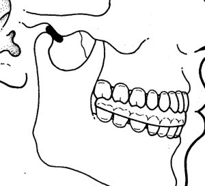
    The jawbone is attached to the skull. Anatomically, the jawbone is known as the mandible. The skull is comprised of many different bones that are tightly bound together. The skull bone where the mandible attaches is anatomically known as the temporal bone. The joint between the mandible and the temporal bone is the temporomandibular joint, commonly abbreviated TMJ.

    Disorders of the temporomandibular joint (TMJ), are referred to as the temporomandibular dysfunction, commonly abbreviated TMD.

    Dental and facial experts have long understood that the function of the TMJ is inherently linked to the biomechanical function of the neck (the cervical spine). This is because of shared neurology.

    temporomandibular joint side view


    In a unique anatomical occurrence, the sensory nerve (mandibular cranial V) that innervates the TMJ, the motor nerves that move the TMJ, and the sensory nerves of the upper neck (cervicals 1-2-3), all communicate with each other in a “box,” depicted above, called the trigeminocervical nucleus. Functionally, this means that neck problems that involve the upper cervical spine nerve roots (C1, C2, C3) have the ability to influence the motor nerves that control the muscles that cross the TMJ. Again, upper neck problems can activate the four muscles that move the TMJ (temporalis, masseter, medial and lateral pterygoid muscles). A muscular imbalance of the TMJ can cause TMJ biomechanical stress and inflammation. The consequence of this is depolarization of the sensory nerves that innervate the TMJ, causing pain and other symptoms (discussed below). This TMJ sensory disturbance sends afferent information into the trigeminocervical nucleus, creating a positive feedback loop (see above drawing), ultimately resulting in temporomandibular dysfunction (TMD).

    The TMJ, like all other moveable joints, has two primary categories of nerves: sensory and motor. The sensory nerves provide to the brain information such as pain, temperature, and position. The motor nerves move the joint by directing the contraction of the muscles that cross the TMJ. The motor and sensory nerves to the TMJ largely travel and exist together in the mandibular branch of the trigeminal nerve (cranial nerve V).

    There are four muscles that cross the TMJ and control the movement of this joint. They are (also noted in the drawing above):

    • Temporalis
    • Masseter
    • Medial Pterygoid
    • Lateral Pterygoid

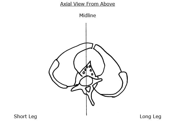
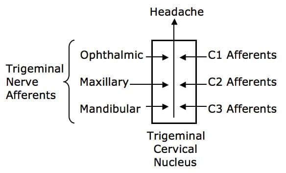
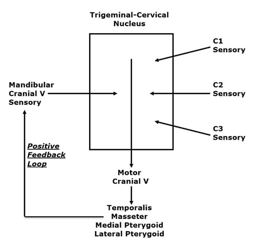
    Please look at the drawing above. In a unique anatomical occurrence, the sensory nerve (mandibular cranial V) that innervates the TMJ, the motor nerves that move the TMJ, and the sensory nerves of the upper neck (cervicals 1-2-3), all communicate with each other in a “box,” depicted above, called the trigeminocervical nucleus. Functionally, this means that neck problems that involve the upper cervical spine nerve roots (C1, C2, C3) have the ability to influence the motor nerves that control the muscles that cross the TMJ. Again, upper neck problems can activate the four muscles that move the TMJ (temporalis, masseter, medial and lateral pterygoid muscles).

    A muscular imbalance of the TMJ can cause TMJ biomechanical stress and inflammation. The consequence of this is depolarization of the sensory nerves that innervate the TMJ, causing pain and other symptoms (discussed below). This TMJ sensory disturbance sends afferent information into the trigeminocervical nucleus, creating a positive feedback loop (see above drawing), ultimately resulting in temporomandibular dysfunction (TMD).

    The pertinent question for this review is:

    Can biomechanical treatment, specifically chiropractic spinal manipulation, of the upper cervical spine that reduces aberrant sensory input from the upper cervical spinal nerves, be successful in the management of TMD?

    •••••

    A noted authoritative reference text pertaining to the jaw is the 1991 book titled (1):

    Temporomandibular Disorders, Diagnosis and Treatment

    The lead author is Andrew Kaplan, DMD. Dr. Kaplan’s credentials include:

    • Director, Temporomandibular Disorder/Facial Pain Clinic, The Mount Sinai Hospital
    • Assistant Clinical Professor, The Mount Sinai Hospital
    • Coordinator, Department of Dentistry, The Mount Sinai Hospital
    • Clinical Instructor, Department of Medicine, Hospital Division, New York University

    Co-author Leon Assael, DMD, has an equally impressive resume:

    • Associate Professor and Residency Program Director, Department of Oral and Maxillofacial Surgery, University of Connecticut School of Dental Medicine
    • Associate Professor of Surgery, School of Medicine, University of Connecticut
    • Associate Chief of Staff, John Dempsey Hospital
    • Attending Oral and Maxillofacial Surgeon, Hartford Hospital, Hartford, Connecticut

    Their book has 31 distinguished contributing authors, 35 chapters, and 754 pages.

    Drs. Kaplan and Assael’s text discusses scenarios where altered neuro-biomechanical function of the cervical spine can put stress on the TMJ causing TMD. They note that temporomandibular joint pain may occur as a consequence of forward head posture.

    The most common TMD symptom is pain just in front of the tragus of the ear. Other common symptoms include clicking, crepitation, transient locking, locking with restricted mandibular movements, and mandibular deflection. TMD is also related to headache, dizziness, earache, and auditory disturbances.

    It is increasingly being accepted that disorders of the cervical spine can cause temporomandibular disorders, even in the absence of direct injury or stress to the temporomandibular joint. In such cases, although the symptoms may be attributed to the jaw, the treatment is to the cervical spine.

    •••••

    In 1998, the journal Clinical Oral Investigations, published a study titled (2):

    Correlation Between Cervical Spine
    and Temporomandibular Disorders

    The authors evaluated 31 consecutive patients with symptoms of temporomandibular disorders and 30 controls with a standardized clinical examination of the masticatory system, evaluating range of motion of the mandible, temporomandibular joint function and pain of the temporomandibular joints and masticatory muscles. Afterwards subjects were referred for clinical examination of the cervical spine, evaluating segmental limitations, tender points upon palpation of the muscles, hyperalgesia, and hypermobility.

    The results indicated that segmental limitations, especially at the occiput-C3 levels, and tender points, especially in the sternocleidomastoid and trapezius muscles, were significantly more present in patients than in controls.

    The authors concluded that the “temporomandibular system and the cervical spine function as a single entity.”

    •••••

    In 2004 the European Journal of Oral Sciences published a study titled (3):

    Deranged Jaw–Neck Motor Control
    in Whiplash-Associated Disorders

    The authors noted that jaw movements are the result of activation of the jaw as well as the neck muscles, leading to simultaneous movements in the temporomandibular, atlanto-occipital and cervical spine joints. Therefore, injury to the neck would disturb natural jaw function. Anatomical, biomechanical, neuroanatomical, neurophysiological and clinical studies indicate that the mandibular and the craniocervical regions are functionally linked. Natural jaw activities require a healthy state of both the mandibular and the head–neck motor systems. Natural jaw actions require a healthy state not only of the temporomandibular joint but also of the atlanto-occipital and cervical spine joints. Neck trauma can be an etiological factor behind temporomandibular disorders. Neck injury from whiplash is associated with deranged control of mandibular movements which compromise jaw function.

    The authors present evidence that supports that whiplash injuries to the cervical spine alter the normal function of the TMJ. Injury to the upper cervical spine causes a reflex to the TMJ muscles, causing TMD.

    •••••

    In 2008, the Swedish Dental Journal published a study titled (4):

    Impaired Jaw Function and Eating Difficulties
    in Whiplash-Associated Disorders

    These authors evaluated 50 whiplash-injured patients with pain and dysfunction in the jaw-face region and 50 healthy age- and sex-matched controls without any history of neck injury. For the whiplash-injured group, there were significantly differences in jaw pain, jaw dysfunction, and eating behavior after the accident. The whiplash-injured patients after the accident reported pain and dysfunction during mouth opening, biting, chewing, swallowing, and yawning and felt fatigue, stiffness, and numbness in the jaw-face region. A majority also reported avoiding tough food and big pieces of food, and taking breaks during meals.

    These authors noted that jaw opening-closing movements are the result of coordinated activation of both jaw and neck muscles, with simultaneous movements in the temporomandibular, atlanto-occipital and cervical spine joints. Consequently, pain or dysfunction in any of the three joint systems involved could impair jaw activities.

    •••••

    In 2015, CRANIO, The Journal of Craniomandibular & Sleep Practice, published a study titled (5):

    Upper Cervical Range of Motion is Impaired
    in Patients with Temporomandibular Disorders

    The authors note that clinicians are increasingly suggesting assessment and treatment of the cervical spine in patients with temporomandibular dysfunction (TMD). The purpose of this study was to investigate whether patients with TMD pain present with upper cervical spine impairment when compared with asymptomatic subjects.

    The authors evaluated 37 subjects with TMD and compared them to 20 asymptomatic control subjects by comparing cervical range of motion (ROM) measures.

    The authors found significant differences in upper the cervical spine range of motion between the TMD group and the control group. They concluded:

    “This study provides evidence for the importance of examination of upper cervical mobility in patients who suffer from TMD.”

    •••••

    In 2016, the journal Manual Therapy published a study titled (6):

    Do Subjects with Acute/Subacute Temporomandibular Disorder
    Have Associated Cervical Impairments: A Cross-Sectional Study

    The authors note that there is evidence of cervical musculoskeletal impairment in some temporomandibular disorder (TMD) pain states. Therefore they conducted a cross-sectional survey to determine whether people with TMD have more cervical signs of dysfunction than healthy subjects. Ninety-nine TMD subjects were evaluated for signs of cervical musculoskeletal impairment and disability as compared to a control group of 45 subjects.

    The authors found that the control group without TMD was consistently less impaired in cervical spine function. The authors note:

    “The more dysfunction and pain is identified in the temporomandibular region, the greater levels of dysfunction is observable on a number of cervical musculoskeletal function tests.”

    “These findings provide evidence that TMD is strongly related with certain cervical spine musculoskeletal impairments which suggests the cervical spine should be examined in patients with TMD as a potential contributing factor.”

    •••••

    A recent study was published in June 2018 in the journal Manuelle Medizin [The European Journal of Manual Medicine], titled (7):

    Spinal High-Velocity Low-Amplitude Manipulation with Exercise
    in Women with Chronic Temporomandibular Disorders:
    A Randomized Controlled Trial Comparing to Patient Education

    This study is the first prospective randomized controlled clinical trial to directly compare the effectiveness of upper cervical manipulation, sham manipulation, and education in patients with temporomandibular disorders.

    The objective of this study was to investigate the effects of spinal high-velocity low-amplitude manipulation with exercise compared to patient education in patients with chronic temporomandibular disorders (TMD). Fifty-five female patients (age 18-50 years) with temporomandibular disorders were randomized to three groups:

    • Cervical spinal manipulation plus neck exercise (CSM + NE)
    • Sham manipulation plus neck exercise (SM + NE)
    • Patient education only (PE)

    Patient assessments were done at baseline, post-treatment (a minimum of 6 treatment sessions), and at a 1-month follow-up. Assessments were performed using:

    • Numeric rating scale / NRS for pain intensity
    • Pressure pain thresholds / PPT masseter/temporalis muscles
    • Pain-free maximum mouth opening / MMO in millimeters
    • Short Form 36 / SF-36 for quality of life

    The cervical manipulations were applied to the upper cervical spine. The authors state:

    “Cervical spinal manipulation was performed using a segment-specific technique for segmental dysfunctions of the upper cervical spine.”

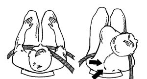
    The manipulation applied in this study appeared to be a sitting rotational maneuver. The physician’s middle finger was placed on the C1 transverse process. The manipulation was done posterior to anterior. Slack taken up to “exclude contraindications (such as pain or dizziness during the test) against an impulse.”

    The sham spine manipulation was performed using a high-velocity low-amplitude manipulation to “give the patient the same mechanical and acoustic sensations.” It was applied to the cervico-thoracic junction by an “impulse to the spinous process of C7.”

    The authors note that “temporomandibular disorders are musculoskeletal conditions characterized by painful conditions and dysfunctions in the muscles of mastication, the temporomandibular joint, and related-tissue components.”

    The prevalence of temporomandibular disorders is between 3% and 15% of the population. They primarily affect middle-aged adults. Women are affected twice as often as men.

    Classic TMD disorders symptoms are headache and neck pain. The authors note that because of the “neuroanatomical convergence of trigeminal and upper cervical afferents in the medullary dorsal horn of the spinal cord (trigeminal caudal nucleus),” that:

    “Cervical dysfunctions can influence the temporomandibular system.”

    The authors found:

    “Significant differences were observed in the cervical spinal manipulation plus neck exercise group vs. the sham manipulation plus neck exercise and patient education only groups post-treatment.”

    There was “significantly increased [in pressure pain thresholds] in the upper cervical manipulation group, while there were no changes in the pressure pain thresholds in both the sham manipulation and education groups.”

    There were significant increases of pain-free maximum mouth opening post-treatment in the cervical spinal manipulation groups.

    There were significant increases in the Short Form-36 scores post-treatment in the cervical spinal manipulation group.

    There was significant improvement in the Numeric Rating Scale in the cervical spinal manipulation group.

    The authors concluded:

    “It is highly likely that functional integration exists between jaw and cervical movements.”

    “This prospective randomized controlled trial demonstrates that in the presence of temporomandibular disorders, the high-velocity low-amplitude manipulation of the upper cervical spine combined with a neck exercise program reduced jaw pain intensity and increased the pressure pain thresholds of masseter and temporalis muscles as well as pain-free maximum mouth opening; moreover, it improved quality of life in women with temporomandibular disorders after treatment and at the 1-month follow up.”

    “Our study suggests that high-velocity low-amplitude manipulation of the upper cervical spine with neck exercise can be effective for treatment of pain and dysfunction in patients with chronic temporomandibular disorders.”

    “It seems reasonable to add cervical manipulation to the rehabilitation program.”

    “Cervical manipulation might positively influence cervical movements, which can affect temporomandibular movements if cervical dysfunction is present.”

    “Our data provide new evidence about the efficacy of treatment for temporomandibular disorders focused on the upper cervical region.”

    “High-velocity low-amplitude manipulation andneck exercise was used for the management of patients with chronic temporomandibular disorders.”

    “Our study proves that upper cervical spine manipulations play an important role in the treatment of pain and dysfunction in the presence of temporomandibular disorders by interruption of the nociceptive vice versa effects of trigemino-cervical convergence.”

    •••••

    SUMMARY

    TMD problems and symptoms are common within the population. It is often assumed that TMD problems are attributed to dysfunctions of the TMJ. This review notes that there is an intimate neurological functional relationship between the TMJ and the upper cervical spine. It is proposed that the best management of TMD involves both evaluation and treatment of the upper cervical spine, and that optimal treatment involves upper cervical spinal manipulation.

    In patients with upper neck, jaw, face or head pain, both the cervical spine and temporomandibular joints should be evaluated. This review implies that perhaps an explanation for chronic TMD is failure to evaluate and treat the upper cervical spine.

    REFERENCES:

    1. Kaplan A, Assael L; Temporomandibular Disorders, Diagnosis and Treatment; WB Saunders Company; 1991.
    2. De Laat A, Meuleman H, Stevens A, Verbeke G; Correlation Between Cervical Spine and Temporomandibular Disorders; Clinical Oral Investigations; June 1998; Vol. 2; No. 2; pp. 54-57.
    3. Eriksson PO, Zafar H, Haggman-Henrikson B; Deranged jaw–neck motor control in whiplash-associated disorders; European Journal of Oral Sciences; February, 2004; Vol. 112; pp. 25–32.
    4. Grönqvist J, Häggman-Henrikson B, Eriksson PO; Impaired jaw function and eating difficulties in whiplash-associated disorders; Swedish Dental Journal; 2008; Vol. 32; No. 4; pp.171-177.
    5. Grondin F, Hall T, Laurentjoye M, Ella B; Upper cervical range of motion is impaired in patients with temporomandibular disorders; CRANIO, The Journal of Craniomandibular & Sleep Practice; April 2015 Vol. 33; No. 2; pp. 91-99.
    6. von Piekartz H, Pudelko A, Danzeisen M, Hall T, Ballenberger N; Do subjects with acute/subacute temporomandibular disorder have associated cervical impairments: A cross-sectional study; Manual Therapy; December 2016; Vol. 26; pp. 208-215.
    7. Corum M, Basoglu C, Topaloglu M, Dıracoglu D; Aksoy C; Spinal High-Velocity Low-Amplitude Manipulation with Exercise in Women with Chronic Temporomandibular Disorders: A Randomized Controlled Trial Comparing to Patient Education; Manuelle Medizin [The European Journal of Manual Medicine]; June 2018; Vol. 56; No. 3; pp. 230–238.

    “Authored by Dan Murphy, D.C.. Published by ChiroTrust® – This publication is not meant to offer treatment advice or protocols. Cited material is not necessarily the opinion of the author or publisher.”

  • Neck Manipulation Facts

    Neck Manipulation Facts

    In October 2016, a former model died from a stroke, and the media attributed her injury to neck manipulation.

    Rebuttals to this manipulation/stroke claim were a combination of both offense and defense. An edited version of one such rebuttal is presented here:

    Earlier this year the British Medical Journal published a study noting that medical errors in hospitals kill 251,000 Americans yearly (the upper range was 440,000). Assuming that medical error deaths outside of the hospital (extended care facilities, nursing homes, at home, etc.) results in an equal number of deaths, an estimated total number of yearly medical error deaths would be about 502,000. [1]

    Some years back, the Journal of the American Medical Association published a study indicating that in the hospital, taking the correct drug for the correct diagnosis in the correct dosage resulted in the death of 106,000 Americans per year (the upper range was 137,000). These are considered non-error deaths as the drug, diagnosis, and dosage were all correct. The article notes that this number constitutes the 4th to 6th leading cause of yearly death in the US. Again, assuming that a similar number of deaths occur from taking the correct drug in the correct dose for the correct problem outside of the hospital setting (extended care facilities, nursing homes, at home, etc.), the number of yearly non-error deaths from medical care would be approximately 212,000. [2]

    Adding the error deaths and the non-error deaths from medical care would total approximately 714,000 yearly.

    Interestingly, from the Journal of the American Medical Association article, 2,216,000 Americans suffer serious adverse reactions from correctly taken drugs in the hospital yearly, but don’t die. The authors defined a serious adverse reaction as one that requires a hospital stay to recover and/or an event that resulted in a lifelong disability. [2]

    In comparison, chiropractic is exceedingly safe. There are about 70,000 practicing chiropractors in the United States, and over 10,000 in California. In a typical year, chiropractic healthcare results in no deaths, and when one is alleged, it tends to make headline news. There are studies comparing chiropractic to the best pain drugs for chronic neck and/or back pain, published in the best journals, concluding that chiropractic is better than 5 times more effective than drugs; the chiropractic care had zero adverse events, while those taking the drugs had more adverse events that were benefited. [3] One of the drugs in that study was Vioxx. Vioxx was only on the market for 5 years, from 1999 to 2004. It was pulled off the market after is was realized that it was responsible for more American deaths in those 5 years (about 60,000) than the Vietnam war killed in 10 years (about 58,000).

    Another example is the regular consumption of non-steroidal anti-inflammatory drugs (NSAIDs) for pain relief. Researchers from Stanford’s Medical School published an article in the world’s most prestigious medical journal, the New England Journal of Medicine, indicating that the taking of prescription NSAIDs resulted in fatal gastrointestinal bleeding 16,500 times (people) yearly, making that the 15th leading cause of yearly death in the US. [4] These same drugs are linked to increased risk of Alzheimer’s disease, deep vein thrombosis, end stage renal disease, liver damage, hearing loss, atrial fibrillation, erectile dysfunction, and more.

    The young lady who is alleged to have died following a chiropractic adjustment is problematic on multiple fronts. The press release on her death mentions 2 different arteries, the carotid and the vertebral artery. Chiropractic spinal adjusting has never been linked to injury to the carotid artery. Which artery was it?

    It is documented that when both the professional and lay press ascribe a manipulative injury to the vertebral artery that they apply the words “chiropractic” and “manipulation” as being synonymous; they are not. Many people “manipulate” and yet they are not chiropractors. Published studies have documented neck manipulations by lay people (barber, masseuse, hair dresser, kung fu instructor, untrained family member, etc.), resulting in vertebral artery injury, and attributing the injury to “chiropractic manipulation” when in fact it was not. [5, 6, 7] Only one type of adjustment has the potential to injure the vertebral artery, and trained-licensed chiropractors are taught not to perform that maneuver; a lay manipulator is not trained and hence would be associated with an increased risk of injury. Who did the manipulation in the case? Was it a chiropractor or a lay untrained manipulator being called a chiropractor by the press?

    Recent studies, published in the best journals, have attempted to quantify the risk of a vertebral artery injury as a consequence of a chiropractic neck adjustment. One such study was published this year from researchers at John Hopkins’s Medical School. [8] These studies are suggesting that there is no risk. In contrast, they are suggesting that it appears that the patient is having a post-injury or spontaneous vertebral artery dissection, causing symptoms that bring them to a chiropractic office, and that the chiropractic adjustment has nothing to do with it. Ironically, one study, in the best medical journal, suggested that being adjusted by a chiropractor actually reduced the chances of the dissection progressing to a stroke as compared to those that had similar pathophysiology and symptoms and went to a medical doctor [9]; importantly, that study included 109 million-person years of follow-up to make their conclusions. Another study from last year with a similar conclusion evaluated about 39 million people; the point is that these are the best and biggest studies on the topic, and they are concluding that there is no stroke risk from a properly delivered chiropractic adjustment. [10] It is more probable that the injury that brought the young lady to the chiropractor’s office was responsible for her artery injury than anything the chiropractor did to try and help her (if in fact it was a chiropractor).

    Even if these studies are incorrect and there is a stroke risk from a chiropractic adjustment, it is so rare that the incidence cannot be quantified. Good studies have suggested that risk might be 1 in every 3 million adjustments, which would mean that a typical chiropractor would have to be in clinical practice for literally hundreds of years to statistically be associated with a single such event, and the majority of chiropractors will never see such an event. Even so, modern chiropractors are trained to recognize the signs and symptoms of a spontaneous or traumatic vertebral artery dissection walking into their office and are educated that such a presentation is an emergency and the patient should be referred to the hospital emergency room. My partner has made 2 such referrals in the past 13 years, to the amazement of the hospital personnel and a credit to her education and experience.

    Every incidence of driving one’s car is more dangerous than seeing a chiropractor.

    •••••••••••

    In the first nine months of this year (2019), three new studies from prestigious medical journals have added to the understanding and statistics of the risks of a cervical artery injury (including the vertebral artery) from cervical spinal manipulation. They are reviewed here:

    •••••

    The first reviewed study was published in March in the journal Annals of Medicine and titled (11):

    A Risk–Benefit Assessment Strategy to
    Exclude Cervical Artery Dissection in Spinal Manual Therapy:
    A Comprehensive Review

    The authors are from the Head and Neck Research Group, Akershus University Hospital, University of Oslo, Norway. This study has 93 references.

    These authors note that cervical spine mobilization and/or manipulation have been suspected to be able to trigger cervical artery dissection. However, these assumptions are based on case studies which are unable to establish direct causality. The controversy relates to the chicken and the egg discussion, i.e. whether the cervical artery dissection (symptoms lead the patient to seek cervical manual-therapy or whether the cervical manual-therapy provoked cervical artery dissection.) The authors note that the controversy is a nearly impossible causality hypothesis.

    The authors note that cervical artery dissection is primarily thought to occur spontaneously, but that it may also be related to physical trauma to the neck. The neck positions that are thought to be most at risk are a coupling of “hyperextension and rotation.” The authors reiterate that cervical artery dissection is very rare, and that manipulation induced cervical artery dissection is extremely rare. They note that conducting sufficiently powered clinical manual-therapy randomized controlled trials to evaluate causality is nearly impossible. Their best estimate is that cervical artery dissection may be as few as 1 per 8.1 million chiropractic office visits and 1 per 5.9 million cervical manipulations by practicing chiropractors.

    The authors believe that the cervical artery most vulnerable to abnormal stresses is the vertebral artery. They state:

    “The vertebral artery is thought to be most susceptible to injury due to extreme rotatory head movements, especially in the transverse foramen in the first cervical vertebrae, as the vertebral artery abruptly transitions from a vertical path to a horizontal orientation.”

    “All people execute several different head and neck movements every day, including side-to-side neck rotations that consequently stretch the vertebral artery. Fortunately, this usually does not trigger cervical artery dissection.”

    Key comments from these authors in this comprehensive review include:

    “The lack of established causality relates to the chicken and egg discussion, i.e. whether the cervical artery dissection symptoms lead the patient to seek cervical spinal manipulative therapy or whether the cervical spinal manipulative therapy provokes cervical artery dissection along with the non- cervical artery dissection presenting headache and/or neck complaint.”

    “Several extensive cohort studies and meta-analyses have found no excess risk of cervical artery dissection resulting in secondary ischaemic stroke for chiropractic spinal manipulative therapy compared to primary care follow-up.”

    “There is no strong evidence in the literature that manual therapy provokes cervical artery dissection.”

    “The assumption that the cervical manual-therapy intervention triggers cervical artery dissection in rare cases has been dominated by single-case reports and retrospective case series or surveys from neurologists who naturally lack substantial methodological quality to establish definitive causality.”

    “There is no firm scientific basis for direct causality between cervical SMT and cervical artery dissection.”

    “No serious adverse events were reported in a large prospective national survey conducted in the UK that assessed all adverse events in 28,807 chiropractic treatment consultations, which included 50,276 cervical spine manipulations.”

    Manual therapists, including chiropractors are taught to perform pre-manipulation provocative vertebral artery assessment testing in order to ensure that such manipulation is “safe” for the patient. However, over the last fifteen years, there has been criticism of these vertebral artery tests because of an excessive incidence of both false positives and false negatives. These author state:

    “There is no sufficient evidence to support cervical vertebral artery tests to identify patients with a higher risk, and the validity and reliability of these tests are low.”

    These authors end their study with a discussion that advises that if a patient has signs and/or symptoms or other warning indications of a cervical artery vascular compromise, that it is best to refer the patient for a more detailed medical vascular evaluation. Chiropractors are so taught and concur.

    •••••

    The second study on this topic this year was published in May in the journal BMJ Open and titled (12):

    Effect of Cervical Manipulation on Vertebral Artery and
    Cerebral Haemodynamics in Patients with Chronic Neck Pain:

    A Crossover Randomised Controlled Trial

    The authors are from Canadian Memorial Chiropractic College, the University of Ontario Institute of Technology, McMaster University Faculty of Engineering, and the University of Toronto. This study cites 67 references. This study took place in the Imaging Research Centre at St. Joseph’s Hospital in Hamilton, Ontario, Canada.

    These authors aimed to determine whether cervical spine manipulation is associated with meaningful changes in vertebral artery and cerebrovascular hemodynamics measured with 3-Telsa MRI compared with neutral neck position and maximum neck rotation in patients with chronic neck pain.

    Twenty subjects aged 23–66 years were enrolled in the study. The cervical manipulation procedure was a supine high velocity, low amplitude impulse, with targeted contact at C1–C2 on the side of most discomfort as elicited on palpation. The subject’s head was in combined axial rotation, flexion and lateral flexion postures. The authors note that this is the first study to measure cerebral blood flow, vertebral artery blood flow and velocity in patients undergoing neck manipulation for neck pain.

    The authors note that patients with neck pain frequently consult chiropractors and manipulation of the cervical spine is commonly performed for symptomatic relief. This, coupled with the fact that vertebrobasilar artery stroke secondary to vertebral artery dissection is so rare, that “very little is known about the risk factors for vertebrobasilar artery stroke.”

    Key findings and comments from these authors include:

    “We found no significant changes within the cerebral haemodynamics following cervical manipulation or maximal neck rotation.”

    “The changes observed were found to not be clinically meaningful and suggests that cervical manipulation may not increase the risk of cerebrovascular events through a haemodynamic mechanism.”

    “When compared with neutral neck position, maximal neck rotation and cervical manipulation did not significantly alter cerebral perfusion within the posterior cerebrum or cerebellum.” [This is the territory supplied by the vertebral arteries].

    “Our work is the first to show that cervical manipulation does not result in brain perfusion changes compared with a neutral neck position or maximal neck rotation.”

    “None of the participants during any of the experimental procedures reported, or were observed by the investigators, to have any signs or symptoms of neurological compromise.”

    “No major adverse events were reported.”

    “Together with previous work, our results support the position that the association between cervical manipulation and stroke is due to protopathic bias.” [Protopathic Bias is when a treatment for the symptoms of a disease or injury appears to cause the outcome].

    “In conclusion, we found no significant change in blood flow in the posterior cerebrum or cerebellum in chronic neck pain participants after maximum head rotation and cervical manipulation.”

    “Our study adds to a growing body of knowledge regarding the impact of head position and cervical manipulation on vascular and neural activity in patients with neck pain.”

    “Our study does not support the hypothesis that neck manipulation or neck rotation are associated with vasospasm of the vertebral artery.”

    In an interesting irony, the authors suggest that their research showed that physiological head/neck rotation was more likely to alter cervical and cranial blood flow than the manipulative thrust. They note:

    “Given the changes in vertebral artery haemodynamics are more pronounced following maximal head rotation compared with cervical manipulation, specifically in contralateral flow, the changes may be the result of the head turning rather than the effect of the thrust associated with cervical manipulation.”

    “This assumption is supported by those who suggest that cervical manipulation imposes less stretch to the vertebral artery than the turning of the head.”

    This unique study adds to the evidence that cervical manipulation is quite safe and unlikely to cause vertebral artery injury. It should be emphasized that, chiropractors are trained to not deliver manipulation thrust to C1-C2 in maximum rotation and extension, and if cranial vascular signs or symptoms occur prior, during, or after any patient contact, that additional evaluation and perhaps referral may be in the patient’s interest.

    •••••

    The third study on this topic this year was published in July (as an epub) in the Journal of Orthopaedic & Sports Physical Therapy and titled (13):

    Effects of Head and Neck Positions on Blood Flow in the
    Vertebral, Internal Carotid and Intracranial Arteries:

    A Systematic Review

    The authors are from Hanze University, Groningen, The Netherlands; University of Groningen, The Netherlands; Rotterdam University, Rotterdam, The Netherlands; HAN University of Applied Sciences, Nijmegen, The Netherlands; University of Nottingham, UK. This study cites 59 references.

    This study is a systematic review of the literature that initially identified 1,453 studies and used 31 that met their inclusion criteria. In total, they used studies involved 2,254 participants. The mean age of these participants was 55 years ranging from 18–98 years.

    The objective was to investigate the effects of cranio-cervical positions and movements (manual therapy interventions) on hemodynamic changes (blood flow velocity and/or volume) of cervical and cranio-cervical arteries, including the vertebral arteries. Blood flow was tested in multiple patient positions (supine, sitting, prone), but for the vertebral artery, maximum rotation and the combination of maximum rotation and extension were the positions tested most frequently.

    Manual therapy is used for the management of people with head and neck pain. Manual therapy is performed utilizing various positions and movements of the craniocervical region. Cervical manual therapy interventions have “rarely been associated with adverse events,” but the exact incidence rates of such adverse events are unknown.

    One of the most frequently claimed adverse events following cervical treatment techniques is arterial dissection. Yet, “studies have been unable to identify specific variables which relates to the increase or mediation of risk for adverse events.”

    A commonly described symptom of cervical arterial dysfunction is neck or head pain. These patients may seek assistance from a manipulative therapist for evaluation and treatment for relief of pain and improvement of function. It is plausible that a cervical arterial dysfunction is not an adverse event of the treatment itself, but exists in situ prior to treatment.

    Key findings and comments from these authors in this systemic review include:

    Most of the studies in this review “mentioned no significant hemodynamic changes during maximal rotation.”

    “Three studies focused on high velocity thrust positioning and movement, all reported no hemodynamic changes.”

    “The positions and movements utilized in high velocity thrust techniques do not seem to alter blood flow.”

    “Based on these data it is unlikely that head and neck movement alone, even if forceful, could mechanistically explain the aetiology of adverse events which have conventionally been purported to be related to therapeutic interventions.”

    “Conventional thought within the domain of manual therapy has been that rapid, forceful interventions such as high velocity thrust techniques are considered to constitute a higher risk for neuro-vascular events resulting from cervical arterial compromise. However, we found that studies which focused specially on high velocity thrust reported no hemodynamic changes.”

    “The synthesized data suggest that in the majority of people most positions and movements of the cranio-cervical region do not have an effect on blood flow.”

    “The data synthesized from 31 experimental and quasi-experimental studies suggest that in most people cranio-cervical positions and movements had no effect on blood flow.”

    “A clinical implication from this review is that the relationship between cranio-cervical movement and alterations in blood flow does not seem to be as obvious as previous data suggested.”

    This study “suggests that adverse events related to cervical spine interventions might be the result of something other than the therapeutic positioning or movement of the head and neck.”

    “Conclusion: Our results suggest that in most people, healthy as well as patients with vascular pathologies, cranio-cervical positions do not alter cervical blood flow. This includes vascular test positions, pre-manipulative positions and manipulations.”

    “A key clinical implication from this review is that the relationship between cranio-cervical movement and blood flow does not seem to be as previously suggested.”

    These authors also point out that pre-manipulative vascular integrity tests (functional positional tests) are unable to establish a relationship between vertebral artery blood flow changes and symptom reproduction. “Therefore, the rationale and value of the tests should be questioned.”

    These authors end their study with a discussion claiming that there may be small sub-groups of the population with underlying arterial pathology where the small hemodynamic changes may be sufficient to induce or exacerbate a neuro-vascular compromise. They suggest that it might be wise to initially use treatment techniques with less than 45 degrees of cervical rotation.

    •••••

    Taken together, these studies support these points:

    • Cervical spine vascular accidents occur spontaneously. Linking cervical vascular accidents to cervical manipulation may be nonexistent at best and at worse overstated in prior publications.
    • Cervical spine vascular accidents occur so rarely that it is essentially impossible to study their occurrence and mechanisms.
    • The available evidence indicates that that cervical adjusting (specific manipulation) is extremely safe, especially in the hands of trained professionals, like chiropractors.
    • Some patients may have elevated risk factors for cervical spine vascular injury, and these risk factors are essentially impossible to ascertain.
    • Patients who are suffering from cervical artery compromise often present to chiropractic offices for treatment.
    • Pre-manipulation positional cervical artery testing is probably not valid.
    • It may be prudent to not rotate the head/neck in excess of 45 degrees when initially manipulating the cervical spine.
    • It is probably ill advised to combine C1-C2 rotation/extension/thrust manipulation.

    References:

    1. Makary MA, Daniel M; Medical Error: The Third Leading Cause of Death in the United States; British Medical Journal; May 3, 2016; Vol. 353; i2139.
    2. Lazarou J, Pomeranz BH, Corey PN; Incidence of Adverse Drug Reactions in Hospitalized Patients: A Meta-analysis of Prospective Studies; Journal of the American Medical Association; Vol. 279; No. 15; April 15, 1998; pp. 1200-1205.
    3. Giles GFL, Muller R; Chronic Spinal Pain: A Randomized Clinical Trial Comparing Medication, Acupuncture, and Spinal Manipulation; Spine; July 15, 2003; Vol. 28; No. 14; pp. 1490-1502.
    4. Wolfe MM, Lichtenstein DL, Singh G; Gastrointestinal Toxicity of Nonsteroidal Anti-inflammatory Drugs; The New England Journal of Medicine; June 17, 1999; Vol. 340; No. 24; pp. 1888-1899.
    5. Terrett AG; Misuse of the literature by medical authors in discussing spinal manipulative therapy injury; Journal of Manipulative and Physiological Therapeutics; May 1995; Vol. 18; No. 4; pp. 203-210.
    6. Weban A, Beck J, Raabe A, Dettmann E. Seifert V; Misuse of the terms chiropractic and chiropractor; J Neurol Neurosurg Psychiatry; May 2004; Vol. 75; No. 5; p. 794.
    7. Weban A; Inappropriate use of the title ‘chiropractor’ and term ‘chiropractic manipulation’ in the peer-reviewed biomedical literature; Chiropractic and Osteopathy; August 2006; Vol. 22; Vol. 14; p. 16.
    8. Church EW, Sieg EP, Zalatimo O, Hussain NS, Glantz M, Harbaugh RE; Systematic Review and Meta-analysis of Chiropractic Care and Cervical Artery Dissection: No Evidence for Causation; Cureus; February 16, 2016; Vol. 8; No. 2; e498.
    9. Cassidy, J David DC, PhD; Boyle, Eleanor PhD; Côté, Pierre DC, PhD; He, Yaohua MD, PhD; Hogg-Johnson, Sheilah PhD; Silver, Frank L. MD; Bondy, Susan J. PhD; Risk of Vertebrobasilar Stroke and Chiropractic Care: Results of a Population-Based Case-Control and Case-Crossover Study; Spine; Vol. 33(4S); February 15, 2008; pp. S176-S183.
    10. Kosloff TM, Elton D, Tao J, Wade M Bannister WM; Chiropractic Care and the Risk of Vertebrobasilar Stroke: Results of a Case–control Study in U.S. Commercial and Medicare Advantage Populations; Chiropractic & Manual Therapies 2015; Vol. 23; No. 19; pp. 1-10.
    11. Chaibi A, Russell MJ; A Risk–benefit Assessment Strategy to Exclude Cervical Artery Dissection in Spinal Manual Therapy: A Comprehensive Review; Annals of Medicine; March, 2019; Vol. 19; pp. 1-10.
    12. Moser N, Mior S, Noseworthy M, Cote P, Wells G, Behr M, Triano J; Effect of Cervical Manipulation on Vertebral Artery and Cerebral Haemodynamics in Patients with Chronic Neck Pain: A Crossover Randomised Controlled Trial; BMJ Open; May 28, 2019; Vol. 9; No. 5; pp. e025219.
    13. Kranenburg R, Tyer R, Schmitt M, Luijckx GJ, van der Schans C, Hutting N, Kerry R; Effects of Head and Neck Positions on Blood Flow in the Vertebral, Internal Carotid and Intracranial Arteries: A Systematic Review; Journal of Orthopaedic & Sports Physical Therapy; July 2019 [epub].

    “Authored by Dan Murphy, D.C.. Published by ChiroTrust® – This publication is not meant to offer treatment advice or protocols. Cited material is not necessarily the opinion of the author or publisher.”

  • Chiropractic and Sagittal Posture

    Chiropractic and Sagittal Posture

    Chiropractors are primary health care providers. This means that patients who seek chiropractic care do not need a referral from another health care provider. Patients may self-refer themselves for chiropractic assessment and treatment.

    The large majority (93%) of patients who initially present themselves for chiropractic care do so for the complaints of spinal pain (1). A typical chiropractic assessment of these patients would often include:

    History of the complaint; this would include information such as causative factors, duration, location, severity, factors that help or aggravate it, etc.

    This type of information helps the chiropractic to determine if the condition is the type of problem that chiropractic typically helps.

    Orthopedic and neurological tests; these tests are often specific for the complaint and history.

    These tests help the chiropractor establish a working diagnosis as the clinical reasons for the complaints. The results can both rule-out or rule-in various pathophysiological processes.

    Imaging studies, which is an optional clinical call by the chiropractor.

    This would typically involve x-rays or perhaps advanced imaging such as a magnetic resonance imaging (MRI) or even computed tomography (CT).

    A chiropractic examination.

    Chiropractors primarily assess and treat patients mechanically. A spinal complaint and problem that a medical doctor might prescribe medication for would be managed quite differently by a chiropractor. The chiropractic examination is to determine if a mechanical problem might be the cause of the patient’s complaints. A common rhetorical scenario would be:

    If your hand was caught is a closed door resulting in severe pain, how would you prefer to be treated?

    Prescribed the best drug available to mask the pain?

    OR

    Ask a chiropractor to come by and open the door?

    The chiropractic mechanical examination typically involves two approaches:

    Segmental Spinal Examination

    The spinal column is comprised of 24 individual bones (vertebra) plus the skull and the pelvis:
    7 neck bones (cervical spine)
    12 mid back bones (thoracic spine)
    5 low back bones (lumbar spine)

    Each vertebra has multiple joints, both above and below (and with the ribs in the thoracic spinal area). Each spinal joint has an optimal ability to move and an optimal position.

    The chiropractic segmental spinal examination assesses the integrity of these vertebra and joints.

    Postural Spinal Examination

    The overall postural alignment of the spinal column and pelvis is an important component of the chiropractic mechanical examination. This typically involves observations of the alignment of ears, jaw, head, shoulders, ribs, and pelvis.

    Spinal x-rays are often used and helpful at documenting and measuring both segmental and postural mechanical problems.

    Posture is important to health and physiology. Postural distortions are three-dimensional. Clinicians often simplify postural distortions by categorizing them into the coronal plane (side-to-side) and the sagittal plane (front-to-back). Evidence is accumulating that forward postural distortion in the sagittal plane is particularly deleterious, increasing pain, reducing health, and enhancing disability. This evidence is reviewed below in this paper.

    There are four primary reasons for forward postural distortion in the sagittal plane:

    1. Forward head: This is often accompanied with increased cervical spine lordosis.
    2. Cervical spine kyphosis (reversal of the normal cervical spine lordotic curve).
    3. Hyperkyphosis of the thoracic spine.
    4. Loss of lumbar lordosis.

    Within the chiropractic profession there are a number of accepted and proven techniques to treat the segmental and postural mechanical problems that are found during the chiropractic mechanical examination. These techniques are taught at both the chiropractic university/college level as well as in post-graduate classes.

    The best documented chiropractic postural technique is Chiropractic Biophysics (CBP). This CBP group has an impressive number of published works pertaining to ideal posture and methods for postural corrections. Currently the group has an excess of 200 studies in the peer-reviewed scientific literature (2).

    Entire medical texts and chapters in medical texts are dedicated to posture and its influences (3, 4). An early description of the importance of good posture on physiology is described by James Oschman, PhD, in his 2000 book titled Energy Medicine, The Scientific Basis (5):

    Joel E. Goldthwait and his colleagues at Harvard Medical School “developed a successful therapeutic approach to chronic disorders. The aim of his therapies was to get his patients to sit, stand, and move with their bodies in a more appropriate relationship with the vertical. After years of treating patients with chronic problems, he concluded that many of these problems arise because parts of the body become misaligned with respect to the vertical.”

    “Goldthwait’s therapeutic approach corrected many difficult problems without the use of drugs. He viewed the human body from a mechanical engineering perspective, in which alignment of parts is essential to reduce wear and stress. He pleaded with physicians to recognize and correct misalignments to prevent long-term harmful effects.”

    “Misalignment of any part will affect the whole system, and that restoration of verticality is a way to address a wide variety of clinical problems.”

    •••••

    Upright posture is a first-class lever mechanical system (6, 7). An example would be a teeter-totter or seesaw. In the first-class lever mechanical system, the fulcrum is the pivot in the center. The fulcrum is where the mechanical loads are the greatest.

    In upright human posture, the fulcrum is the spinal vertebra (vertebral body, intervertebral disc, facet joints). When a person has a forward postural distortion in the sagittal plane, they would literally fall onto their face if the muscles on the opposite side of the fulcrum did not contract, maintaining balance. This counterbalance contraction of the muscles is both fatiguing to the muscle and increases the compressive loads on the fulcrum tissues. The counterbalancing muscles are the posterior spinal muscles; they exist from the back of the skull and neck, all the way down the spinal column to the pelvis.

    fulcrum

    The posterior counterbalancing muscles are in a constant struggle with forward weight and gravity. Constant muscle contraction causes muscle fatigue. Weight and gravity do not fatigue. Weight and gravity eventually win the struggle. Postural distortion and its deleterious consequences tend to become worse over time unless there is an appropriate intervention.

    Rene Cailliet, MD, uses an example where a patient has unbalanced forward head posture (8). Dr. Cailliet assigns the head a weight of 10 lbs. and displaces the head’s center of gravity forward by 3 inches. The required counter balancing muscle contraction on the opposite side of the fulcrum (the vertebrae) would be 30 lbs. (10 lbs. X 3 inches).

    In her 2017 book Move Your DNA, biomechanist Katy Bowman describes how chronic postural distortions cause adverse tissue adaptations that become fixed and rigid unless, again, there are reversing therapeutic interventions (9). Often the beginning of these commonly found forward postural distortion in the sagittal plane are sedentary computer-based occupations and leisure activities. Perhaps the worst offending activity is the use of cellular phones (10).

    •••••

    In 1978, Swedish neurosurgeon Alf Breig, MD authored a book titled Adverse Mechanical Tension in the Central Nervous System (11). In this book, based upon his own surgical observations, Dr. Breig asserts that loss of cervical lordosis tethers the spinal cord creating myelomalacia and spinal cord atrophy. He states, “Patients with long-standing kyphotic deformities are at risk for progression of myelopathy with resultant permanent damage to the spinal cord.”

    •••••

    In 2004, physician Deborah Kado, MD and colleagues published a study in the Journal of the American Geriatrics Society, titled (12):

    Hyperkyphotic Posture Predicts Mortality in
    Older Community-Dwelling Men and Women: A Prospective Study

    Thoracic spine hyperkyphosis is frequently observed in older persons. The objective of this study was to determine the association between hyperkyphotic thoracic posture and the rate of mortality and cause-specific mortality in older persons. It was a prospective cohort study that included 1,353 participants.

    Study participants were followed for an average of 4.2 years, with mortality and cause of death confirmed using a review of death certificates.
    Persons with hyperkyphotic posture had a 44% increased rate of mortality.
    “Hyperkyphotic posture was specifically associated with an increased rate of death due to atherosclerosis.” The authors note that interventions specifically targeted at improving hyperkyphotic posture could result in reduced mortality rates.

    •••••

    In 2005, physician Steven Glassman, MD, and colleagues, published a study in the journal Spine titled (13):

    The Impact of Positive Sagittal Balance in Adult Spinal Deformity

    These authors measured the pain, systemic health, and disability status of 298 individuals and compared them to a full-spine lateral radiographic measurement of sagittal postural balance. A plum line was dropped from the body of the C7 vertebrae and measured with respects to the articulating surface of L5 with the sacral base. This measurement was specifically used to quantify forward postural distortion in the sagittal plane.

    All measures of health status showed significantly poorer scores as C7 plumb line deviation increased in the forward direction (anterior to the sacral base). The authors note:

    “Patients with relative kyphosis in the lumbar region had significantly more disability than patients with normal or lordotic lumbar sagittal measures.”

    “This study shows that although even mildly positive sagittal balance is somewhat detrimental, severity of symptoms increases in a linear fashion with progressive sagittal imbalance.”

    “The results also show that kyphosis is very poorly tolerated in the lumbar spine.”

    “There was clear evidence of increased pain and decreased function as the magnitude of positive [forward] sagittal balance increased.”

    “All measures of health status showed significantly poorer scores as C7 plumb line deviation increased [forward].”

    “This study shows that although even mildly positive [forward] sagittal balance is somewhat detrimental, severity of symptoms increase in a linear fashion with progressive [forward] sagittal imbalance.”

    spine sagittal view

    •••••

    In May 2009, Dr. Deborah Kado and colleagues published a follow-up study on thoracic kyphosis and mortality rates. This article appeared in the Annals of Internal Medicine, and is titled (14):

    Hyperkyphosis Predicts Mortality Independent
    of Vertebral Osteoporosis in Older Women

    This study was a prospective cohort study involving 610 women, aged 67 to 93 years. Their thoracic kyphosis was measured, and mortality was assessed an average of 13.5 years later. The authors concluded that each standard deviation increase in kyphosis carried a 14% increased risk for death. The authors note:

    In older women “increased kyphosis predicts increased risk for all-cause mortality independent of the extent and severity of the underlying spinal osteoporosis.”

    “Other large epidemiologic studies have demonstrated that kyphotic posture may be associated with worse health, including impaired pulmonary function, poor physical function, inferior quality of life, injurious falls, fractures, and death.”

    “We postulate that the phenotype of hyperkyphosis is an easily assessable clinical marker of accelerated physiologic aging or frailty.”

    “These results add to a growing literature that suggests that hyperkyphosis is a clinically important finding.”

    “Because it is readily observed and is associated with ill health in older persons, hyperkyphosis should be recognized as a geriatric syndrome—a ‘multifactorial health condition that occurs when the accumulated effect of impairments in multiple systems renders a person vulnerable to situational challenges.’”

    •••••

    In November 2009, researchers from the Department of Orthopaedics and Rehabilitation Medicine, Fukui University, Japan, published a study in the Journal of Neurosurgery: Spine, titled (15):

    Cervical Spondylotic Myelopathy Associated
    with Kyphosis or Sagittal Sigmoid Alignment

    These authors assessed the records of 476 patients to determine the effects of kyphotic forward sagittal alignment of the cervical spine in terms of neurological morbidity, and specifically on the development of cervical spondylotic myelopathy.

    These authors concluded that cervical spine forward sagittal kyphotic deformity plays an important role in neurological dysfunction. This was especially true when the kyphotic deformity exceeded 10 degrees. They state:

    “Loss of lordosis or kyphotic alignment of the cervical spine and spinal cord may contribute to the development of myelopathy, and in patients with cervical kyphotic deformity, the spinal cord could be compressed by tethering over the apical vertebra or intervertebral disc.”

    “We conclude that the [cervical spine] sagittal kyphotic deformity related to flexion mechanical stress may be a significant factor in the development of cervical spondylotic myelopathy.”

    •••••

    In April 2013, another article was published in the Journal of Neurosurgery: Spine, titled (16):

    Relationship Between Degree of Focal Kyphosis Correction
    and Neurological Outcomes for Patients
    Undergoing Cervical Deformity Correction Surgery

    These authors performed a retrospective review of 36 patients with myelopathic symptoms who underwent cervical deformity correction surgery. They note:

    “The normal lordotic curvature of the cervical spine is critical to maintaining sagittal alignment and spinal balance.”

    “It is believed that the neurological symptoms seen in cervical kyphosis are a result of deformity-induced anatomical changes that apply pressure to the spinal cord and nerve roots.”

    “The reversal of normal cervical curvature, as seen in kyphosis, can occur through a variety of mechanisms and can lead to mechanical pain, neurological dysfunction, and functional disabilities.”

    “Kyphosis of the cervical spine can be a debilitating condition that leads to significant neurological dysfunction.”

    •••••

    In July 2013, a study was published in The Journals of Gerontology: Series A: Biological Sciences, and titled (17):

    Spinal Posture in the Sagittal Plane Is Associated
    with Future Dependence in Activities of Daily Living

    These authors noninvasively measured spinal postures in a community-based prospective cohort of older adults (804 participants: 338 men, 466 women, age range 65–94 years) to determine if any such postures were associated with the need for future assistance in Activities of Daily Living (ADL). They found that lumbar kyphosis pitched the body and head forward, and this postural distortion was significantly associated with the need for future assistance in the person’s activity of daily living. These authors state:

    “Accumulated evidence shows how important spinal posture is for aged populations in maintaining independence in everyday life.”

    “Spinal posture changes with age, but accumulated evidence shows that continued good spinal posture is important in allowing the aged to maintain independent lives.”

    “The gravity line moves further anterior as inclination of the trunk increases.” “Even mildly positive sagittal balance is somewhat detrimental, the decline in health status increases in a linear fashion with progressive sagittal imbalance.”

    Spinal “inclination is associated with future dependence in ADL among older adults and warrants wider attention.”

    The “results indicate that attention needs to be paid to inclination in spinal posture to identify elderly people at high risk of becoming dependent in ADL.”

    •••••

    In 2019, a study was published in the Asian Spine Journal, and titled (18):

    Hypertension Is Related to Positive Global Sagittal Alignment:
    A Cross-Sectional Cohort Study

    The purpose of this study is to investigate the relationship between hypertension and spinal-pelvic sagittal alignment in middle-aged and elderly individuals. The study used 655 participants (262 men and 393 women; mean age, 72.9 years; range, 50–92 years). Whole spine and pelvic x-rays were taken, and thoracic kyphosis, lumbar lordosis, pelvic tilt, sacral slope, pelvic incidence, and sagittal vertical axis (SVA) were measured. Analysis was done using image-analysis software.

    Hypertension was defined as a systolic blood pressure of ≥140 mm Hg or a diastolic blood pressure of ≥90 mm Hg, and participants who regularly took antihypertensive medication were also considered to be hypertensive.

    Postural changes start in the 30s in women and the 50s in men. By middle-age, spinal postural deformities contribute to poor health-related quality of life (QOL). The authors specifically note that when “global sagittal alignment shifts forward, it causes deterioration of the health-related QOL.”

    The authors develop this model of systemic wellness, forward posture and disc degeneration:

    Insulin resistance is the most important
    health problem in developed countries

    Insulin resistance is the precursor to diabetes,
    obesity, and arteriosclerosis

    Arteriosclerosis is the precursor to hypertension

    Hypertension impairs muscle circulation

    Impaired muscle circulation causes muscle fatigue

    Muscle fatigue causes a forward shift
    in sagittal spinal alignment

    A forward shift in sagittal spinal alignment
    accelerates disc degeneration

    The authors note that forward global spinal sagittal alignment is associated with poor health-related quality of life. They make these specific comments:

    “We found that hypertension was significantly related to a forward shift in the global sagittal alignment in middle-aged and elderly individuals.”

    “This study showed that hypertension was associated with forward-shifted global sagittal alignment.”

    This study supports that “postural anomalies occur before disc degeneration in people with hypertension.”

    •••••

    Chiropractors have always understood the adverseness of postural distortions and especially a forward shift in sagittal spinal alignment. A number of chiropractic techniques are primarily concerned with assessing, preventing, and changing these and other postural distortions. The chiropractic interventions used often involve combinations of certain spinal adjustments (specific manipulations), ergonomic advice, spinal exercises, and extension (mirror image reversal) traction.

    References

    1. Adams J, Peng W, Cramer H, Sundberg T, Moore C; The Prevalence, Patterns, and Predictors of Chiropractic Use Among US Adults: Results From the 2012 National Health Interview Survey; Spine; December 1, 2017; Vol. 42; No. 23; pp. 1810–1816.
    2. www.idealspine.com
    3. Kendall HO, Kendall FP, Boynton DA; Posture and Pain, Williams and Wilkins, 1985.
    4. Mennell JM; “The Forward Head Syndrome” in The Musculoskeletal System, Differential Diagnosis from Symptoms and Physical Signs; Aspen; 1992.
    5. Oschman J; Energy Medicine, The Scientific Basis; Churchill Livingstone; 2000.
    6. Cailliet R; Low Back Pain Syndrome; 4th edition; FA Davis Company; 1981.
    7. White AA, Panjabi MM; Clinical Biomechanics of the Spine; Second Edition; Lippincott; 1990.
    8. Cailliet R; Soft Tissue Pain and Disability; 3rd Edition; F A Davis Company; 1996.
    9. Bowman K; Move Your DNA: Restore Your Health Through Natural Movement; 2017.
    10. Hansraj KK; Assessment of Stresses in the Cervical Spine Caused by Posture and Position of the Head; Neuro and Spine Surgery, Surgical Technology International; November 2014; Vol. 25; pp. 277-279.
    11. Breig A; Adverse Mechanical Tension in the Central Nervous System; Almqvist and Wiksell; 1978.
    12. Kado DM, Huang MH, Karlamangla AS, MD, PhD, Elizabeth Barrett-Connor E, Greendale GA; Hyperkyphotic Posture Predicts Mortality in Older Community-Dwelling Men and Women: A Prospective Study; Journal of the American Geriatrics Society; October 2004; Vol. 52; No. 10; pp. 1662-1667.
    13. Glassman SD, Bridwell K, Dimar JR, Horton W, Berven S, Schwab F; The Impact of Positive Sagittal Balance in Adult Spinal Deformity; Spine; Vol. 30; No. 18; September 15, 2005; pp. 2024-2029.
    14. Kado DM, Lui LY, Ensrud KE, MD; Fink HA, MD, MPH; Karlamangla AS, Cummings SR; Hyperkyphosis Predicts Mortality Independent of Vertebral
      Osteoporosis in Older Women; Annals of Internal Medicine; May 19, 2009; Vol. 150; No. 10; W-121; pp. 681-687.
    15. Uchida K, Nakajima H, Sato, Yayama T, Mwaka ES, Kobayashi S, Baba H; Cervical Spondylotic Myelopathy Associated with Kyphosis or Sagittal Sigmoid Alignment: Outcome after Anterior or Posterior Decompression; Journal of Neurosurgery: Spine; November 2009; Vol. 11; pp. 521-528.
    16. Grosso M, Hwang R, Mroz T, Benzel E, Steinmetz, M MD; Relationship between degree of focal kyphosis correction and neurological outcomes for patients undergoing cervical deformity correction surgery; Journal of Neurosurgery: Spine; June 2013; Vol. 18; No. 6; pp. 537-544.
    17. Kamitani K, Michikawa T, Iwasawa S, Eto N, Tanaka T, Takebayashi T, Nishiwaki T; Spinal Posture in the Sagittal Plane Is Associated With Future Dependence in Activities of Daily Living: A Community-Based Cohort Study of Older Adults in Japan; The Journals of Gerontology: Series A: Biological Sciences; July 2013; Vol. 68; No. 7; pp. 869-875.
    18. Arima H, Togawa D, Hasegawa T, Yamato YGo Yoshida G, Kobayashi S, Yasuda T, Banno T, Oe S, Mihara Y, Ushirozako H, Hoshino H, Matsuyama Y; Hypertension Is Related to Positive Global Sagittal Alignment: A Cross-Sectional Cohort Study; Asian Spine Journal; July 9, 2019; [epub].

    “Authored by Dan Murphy, D.C.. Published by ChiroTrust® – This publication is not meant to offer treatment advice or protocols. Cited material is not necessarily the opinion of the author or publisher.”

  • Motor Vehicle Crashes and Low Back Pain

    Motor Vehicle Crashes and Low Back Pain

    Physical events in a gravity environment have always caused injuries. The mathematics and physics of these injuries were not formalized until the year 1687 when Sir Isaac Newton wrote the book Mathematical Principles of Natural Philosophy (1). In this publication, Newton details the principles of inertia.

    Inertia is the resistance of a physical object to any change in its state of motion or to its state of rest. An object in motion will remain in motion unless an outside force acts upon that object. Likewise, an object at rest will remain at rest unless an outside force acts upon that object.

    These Laws of Inertia apply to the human body. Different parts of the human body have different inertias between them. It is these inertial differences between the different parts of the body that increase their risk of injury.

    The greatest inertial difference in the body is between a human’s trunk and head. The neck (cervical spine) connects these two distinct inertial masses and therefore the neck has a great vulnerability to injury.

    The incidence of inertial injury to the cervical spine has dramatically increased with advances in wheeled transportation. Whatever the wheeled transportation, the person is probably sitting in the vehicle. All quick movements of the vehicle will also move the trunk of the person sitting in the vehicle. Since the trunk inertia is separate from head inertia, the neck is vulnerable to inertial injury.

    Trunk Movement Neck Inertial Injury

     Trunk Movement Neck Inertial Injury

    An early scientific/medical publication describing cervical spine inertial injuries caused by wheeled vehicle accidents was published in 1867, titled (2):

    On Railway and Other Injuries of the Nervous System

    As noted in the title, these injuries were from train accidents, occurring decades prior to the first automobiles.

    Within a few decades of automobiles becoming commonplace in the society, cervical spine inertial injuries also became commonplace. In 1928, physician Harold Crowe, MD, presented a lecture at the Western Orthopedic Association convention in San Francisco, CA. In his presentation he introduced the word “whiplash” to describe these cervical inertial injuries, and the terminology is still standard today (3, 4).

    Cervical spine inertial injuries from motor vehicle crashes are well documented and accepted. They are well described in reference texts pertaining to whiplash injuries (5, 6, 7).

    However, it is much more controversial as to whether motor vehicle collisions can cause injury to the low back. The mechanisms for these low back injuries are poorly understood and therefore such injuries are often viewed with skepticism or rejected. Yet, this topic has been explored for decades and a number of studies have quantified their incidence.

    It is established that the primary reason people go to chiropractors is for low back pain (63%), and the level of satisfaction from this chiropractic care is very high (8). Spinal manipulation and chiropractic have a long history of successful clinical outcomes for treating low back pain (9, 10, 11, 12, 13, 14, 15).

    Early documented cases of low back injury during motor vehicle collisions involved the restraint devices, primarily lap seat belts (16, 17, 18, 19, 20).

    The lap belt, when used alone, does not stop the forward inertial motion of the trunk above the belt or the pelvis and legs below the belt. Lap belts concentrate the inertial forces to the small cross-sectional dimension of the belt, allowing the belt to function as a fulcrum. The stresses at the fulcrum (the belt) are magnified, resulting in increased injury to the tissues behind the lap belt. This includes a flexion-type injury to the low back.

    The lap belt, when used alone, does not stop the forward inertial motion of the trunk above the belt or the pelvis and legs below the belt. Lap belts concentrate the inertial forces to the small cross-sectional dimension of the belt, allowing the belt to function as a fulcrum. The stresses at the fulcrum (the belt) are magnified, resulting in increased injury to the tissues behind the lap belt. This includes a flexion-type injury to the low back.

    The addition of a unilateral shoulder harness will further introduce a rotational component of stress to the low back (21). This combination of low back flexion and rotation increases the risk of low back injury.

    The addition of a unilateral shoulder harness will further introduce a rotational component of stress to the low back (21). This combination of low back flexion and rotation increases the risk of low back injury.
    (From #21)

    Other mechanisms (other than restraint device use) of low back injury caused by motor vehicle collisions may occur. Some of these are mentioned in the studies reviewed below.

    Over the decades, a number of medical/scientific studies have attempted to quantify the incidence of low back injury from motor vehicle collisions:

    • In 1994, a study from Iceland was published in the journal Cephalalgia and titled (22):

    Extra-Cervical Symptoms After Whiplash Trauma

    The study involved 38 patients with chronic whiplash syndrome. They were investigated with regard to symptoms that could be confirmed with the criteria for a specific diagnosis:

    • 65.8% were women
    • 34.2% were men
    • Ages were 17 to 52 years with a mean 33 years
    • Time from accident to investigation was 6 to 44 months (mean 17 months)

    All patients had been healthy before the accident and had sustained a whiplash injury without head injury, and had no signs of radiculopathy or myelopathy.

    The authors found that 47.7% of the patients complained of low back pain, and that 13.2% were diagnosed with chronic mechanical low back pain. Importantly, 5.3% were diagnosed with segmental instability of the lumbar spine. The authors state:

    “Reported incidence of low back pain (LBP) after whiplash injury is 40-60%, and the incidence of 47.7% in the present study is in concordance with these earlier results.”

    “Mechanical injury to the low back can be caused by the initial extension of the low back followed by flexion when the pelvis and the legs are thrown forward while the body is held in place by the safety belt or stopped by the steering wheel.”

    • A 2001 study from Sweden was published in the Journal of Clinical Epidemiology and titled (23):

    The Association Between Exposure to a
    Rear-End Collision and Future Health Complaints

    Over a period of 7 years, the authors compared 232 whiplash-injured patients to 204 subjects who were involved in motor vehicle crashes but not injured. Both groups were compared to 4,088 control subjects.

    The whiplash-injured patients had an increased risk of low back pain at 7 years by 270% compared to the other groups.

    • In 2002, an American study was published in the American Journal of Physical Medicine & Rehabilitation and titled (24):

    Lumbar Spinal Strains Associated with Whiplash Injury:
    A Cadaveric Study

    The authors note that up to half of those involved in a whiplash accidents may develop low back pain. Therefore, they measured the forces on the lumbar spines of human embalmed cadavers subjected to a number of low speed vehicle collision events.

    These authors proved that there are both horizontal and vertical spinal forces that result from a strictly horizontal force applied experimentally. The authors state:

    “Frequently, seemingly implausible claims of lumbar injuries are mentioned after low-speed rear-end collisions, even when little vehicular damage is reported.”

    “Apparently, rear-end collisions lead to soft-tissue injuries with paucity of findings on physical examination, negative radiograph studies, profuse symptomatology, and prolonged disability.”

    The forces generated during simulated whiplash collisions induce biphasic lumbar spinal motions (increased-decreased lordosis) that “may be sufficient to cause soft-tissue injuries.”

    • A 2003 study from Canada was published in the journal Spine and titled (25):

    Low Back Pain After Traffic Collisions:
    A Population-Based Cohort Study

    The study is a population-based, incidence cohort study involving 4,473 subjects. The authors concluded:

    “Low back pain is a common traffic injury with a prolonged recovery.”

    • In 2010, a study from the United Kingdom was published in the journal Injury and titled (26):

    Can Patients with Low Energy Whiplash Associated Disorder
    Develop Low Back Pain?

    The study analyzed 800 consecutive patients for symptomatology of whiplash injury including the presence of low back pain. The authors found that back injury occurred in approximately 40% of the subjects. These injuries occurred in all directions of impact (rear, frontal and side impact), and these injuries occurred in low, medium and high levels of collision impacts. Almost all of these subjects with low back injury also injured their necks, and the majority of them had a past medical history of back pain. The authors stated:

    “We were surprised that patients with next to no car damage had the same incidence of back pain as those involved in more violent crashes when biomechanically unlikely.”

    • Another Canadian study was published in 2010 in the Journal of Occupational Environmental Medicine and titled (27):

    Whiplash Injury is More than Neck Pain:
    A Population-Based Study of Pain Localization after Traffic Injury

    This study was a cross-sectional analysis of a cohort of 6,481 patients, and 60% were found to have low back pain.

    • Again in 2010, the Society of Automotive Engineers published a study titled (28):

    Lumbar Loads in Low to Moderate Speed Rear Impacts

    The authors note that although most of the research on vehicular rear impacts has focused on the neck, there is currently increasing concern about the lumbar spine. They produced a series of low impact collisions involving anthropomorphic test devices and measured the stresses imparted to the lumbar spines. The authors concluded:

    The lumbar spines may experience overall compressive and bending loads.

    “Spinal bending superimposed with sudden spinal compression has been suggested as a mechanism of creating acute herniations on the rare occasion in which low back pain associated with an intervertebral disc herniation was reported.”

    “The spine might experience some transient bending and compression, which could be consistent with the proposed mechanism of acute disc herniation.”

    • A 2011 study from Norway was published in the journal BMC Musculoskeletal Disorders and titled (29):

    Headache and Musculoskeletal Complaints Among Subjects
    with Self-Reported Whiplash Injury

    This study was a cross-sectional study on 59,104 subjects. The authors found a positive association between a history of a whiplash injury and low back pain in both men and women. The increased risk in men was 210%. The increased risk in women was 380%.

    • A 2013 study from Japan was published in the European Spine Journal and titled (30):

    Prevalence of Low Back Pain and Factors Associated
    with Chronic Disabling Back Pain in Japan

    This was a cross-sectional survey in Japan on 65,496 adults. It found a positive association between a traffic injury and chronic disabling low back pain. Specifically, motor vehicle crashes increased the risk of low back pain by 181%.

    • A 2014 study from the United States (Massachusetts, Florida, Michigan, and New York) was published in the journal Pain and titled (31):

    Pain Location and Duration Impact Life Function Interference
    During the Year Following Motor Vehicle Collision

    The study involved a cohort of 948 patients who had initially been seen in the emergency department and subsequently assessed a year later. The authors found that 37% of the patients had moderate or severe low back pain. The author stated:

    “Pain across all body regions accounted for nearly twice as much of the variance in pain interference as neck pain alone. These findings suggest that studies of post-MVC pain should not focus on neck pain alone.”

    • In 2018, a Canadian study was published in the European Spine Journal and titled (32):

    The Association Between a Lifetime History of Low Back Injury
    in a Motor Vehicle Collision and Future Low Back Pain:
    A Population-Based Cohort Study

    The purpose of this population-based cohort study of 789 subjects was to investigate the association between a lifetime history of a low back injury in a motor vehicle collision (MVC) and future troublesome low back pain. Troublesome LBP was measured at 6 and 12 months following the MVC.

    The authors found that in those with a history of low back pain, MVC injury increased the development of future troublesome LBP by 176%. The authors stated:

    “LBP is common after a motor vehicle collision.”

    “This study supports prior research on the hypothesis that a past history of a low back injury in a MVC may be a determinant of future LBP.”

    “Our analysis suggests that a history of low back injury in a MVC is a risk factor for developing future troublesome LBP.”

    “Our analysis provides the public, clinicians, government and insurers with evidence that a low back injury in a MVC may have a role in the development of future low back pain.”

    “The consequences of a low back injury in a MVC can predispose individuals to experience recurrent episodes of low back pain.”

    • In 2019, another Canadian study, published in the journal Traffic Injury Prevention and titled (33):

    Low-Velocity Motor Vehicle Collision Characteristics
    Associated with Claimed Low Back Pain

    The objective of this investigation was to characterize the physical circumstances of low-velocity motor vehicle collisions that resulted in claims of low back pain (LBP). The authors assessed 83 injury cases.

    The study found that 77% of the cases reported low back pain. The authors state:

    “The most common pre-existing medical condition was prior LBP or evidence of disc degeneration.”

    “It was found that pre-existing LBP and lumbar spine disc degeneration were particularly common in those with LBP.”

    “The development of future LBP is higher in individuals with a past self-reported low back injury resulting from a motor vehicle collision compared to those without.”

    This data set showed a “very high correlation between LBP claims and WAD claims.”

    “These observations point to the likelihood of the rear-end impact crash configuration being a relevant factor in LBP reporting.”

    “For individuals with a history of LBP, the risk of redeveloping LBP doubles, putting these individuals at greater risk of reporting LBP in the future.”

    As noted, many studies have investigated the incidence of low back pain following motor vehicle crashed. These studies span many decades, multiple scientific journals, and multiple countries. Collectively, they involve tens of thousands of subjects. They all conclude that motor vehicle crashes do increase the risk of low back injury and pain. Any suggestion that motor vehicle crashes cannot cause low back pain appear to be in error, or perhaps self-serving. Chiropractors should be confident that complaints of low back pain following vehicle crashes are credible, and treatment is reasonable, necessary, and appropriate.

    References:

    1. Newton I. Principia Mathematica; July 5, 1687.
    2. Erichsen JE; On Railway and Other Injuries of the Nervous System; Philadelphia, PA; Henry C. Lea; 1867.
    3. Crowe H; A New Diagnostic Sign in Neck Injuries; California Medicine; January 1964; Vol. 100; No. 1; pp. 12-13.
    4. Todman D; Whiplash Injuries: A Historical Review; The Internet Journal of Neurology; Vol. 8; No 2; 2006.
    5. Jackson R; The Cervical Syndrome; Thomas; 1978.
    6. Foreman S, Croft A; Whiplash Injuries: The Cervical Acceleration/Deceleration Syndrome; Williams & Wilkins; 1988.
    7. Cailliet R; Whiplash Associated Diseases; American Medical Association; 2006.
    8. Adams J, Peng W, Cramer H, Sundberg T, Moore C; The Prevalence, Patterns, and Predictors of Chiropractic Use Among US Adults: Results From the 2012 National Health Interview Survey; Spine; December 1, 2017; Vol. 42; No. 23; pp. 1810–1816.
    9. Edwards BC; Low back pain and pain resulting from lumbar spine conditions: a comparison of treatment results; Australian Journal of Physiotherapy; Vol. 15; 104; 1969.
    10. Kirkaldy-Willis WH; Managing Low Back Pain; Churchill Livingston; (1983 & 1988).
    11. Kirkaldy-Willis WH, Cassidy JD; Spinal Manipulation in the Treatment of Low-Back Pain; Canadian Family Physician; March 1985; Vol. 31; pp. 535-40.
    12. Meade TW, Dyer S, Browne W, Townsend J, Frank OA; Low back pain of mechanical origin: Randomized comparison of chiropractic and hospital outpatient treatment; British Medical Journal; Vol. 300; June 2, 1990; pp. 1431-7.
    13. Stern PJ, Côté P, Cassidy JD; A series of consecutive cases of low back pain with radiating leg pain treated by chiropractors; Journal of Manipulative and Physiological Therapeutics; 1995 Jul-Aug; Vol. 18; No, 6; pp. 335-342.
    14. Giles LGF; Muller R; Chronic Spinal Pain: A Randomized Clinical Trial Comparing Medication, Acupuncture, and Spinal Manipulation; Spine July 15, 2003; Vol. 28; No. 14; pp. 1490-1502.
    15. Muller R, Giles LGF; Long-Term Follow-up of a Randomized Clinical Trial Assessing the Efficacy of Medication, Acupuncture, and Spinal Manipulation for Chronic Mechanical Spinal Pain Syndromes; Journal of Manipulative and Physiological Therapeutics; January 2005; Vol. 28; No. 1; pp. 3-11.
    16. Chance GQ; Note on a type of flexion fracture of the spine; British Journal of Radiology; September 1948; Vol. 21(249); p. 452.
    17. Marsh HO, Bailey D; Seat Belt fractures: Chance fractures caused by seat belts: Presentation of three cases; J Kans Med Soc; September 1970; Vol. 71; No. 9:361-365.
    18. Rogers LF; The roentgenographic appearance of transverse or chance fractures of the spine: The seat belt fracture; Am J Roentgenol Radium Ther Nucl Med; April 1971; Vol. 111; No. 4; pp. 844-849.
    19. Rutherford WH; The Medical Effects of Seat-Belt Legislation in the United Kingdom: a critical review of the findings; Archives of Emergency Medicine; 1985; Vol. 2; pp. 221-223.
    20. Thompson NS, Date R, Charlwood AP, Adair IV, Clements WD; International Journal of Clinical Practice; Seat-belt syndrome revisited; October 2001; Vol. 55; No. 8; pp. 573-5.
    21. Anrig C, Plaugher G; Pediatric Chiropractic; Williams & Wilkins; 1998.
    22. T Magnússon T; Extra-cervical Symptoms After Whiplash Trauma; Cephalalgia; June 1994; Vol. 14; No. 3; pp. 223-227.
    23. Berglund A, Alfredsson L, Jensen I, Cassidy JD, Nygren Å; The association between exposure to a rear-end collision and future health complaints; Journal of Clinical Epidemiology; November 2001; Vol. 54; No. 11; pp. 851–856.
    24. Fast A, Sosner J, Begeman P, Thomas MA, Chiu T; Lumbar Spinal Strains Associated with Whiplash Injury: A Cadaveric Study; American Journal of Physical Medicine & Rehabilitation; September 2002; Vol. 81; No. 9; pp. 645-650.
    25. Cassidy JD, Carroll L, Cote P, Berglund A, Nygren A; Low Back Pain After Traffic Collisions: A Population-Based Cohort Study; Spine; May 15, 2003; Vol. 28; No. 10; pp. 1002-1009.
    26. Beattie N, Lovell ME; Can patients with low energy whiplash associated disorder develop low back pain?; Injury; February 2010; Vol. 41; No. 2; pp 144-146.
    27. Hincapie C, Cassidy JD, Carroll LJ, Cote P, Guzman J (2010); Whiplash injury is more than neck pain: a population-study of pain localization after traffic injury; Journal of Occupational Environmental Medicine; Vol. 52; No. 4; pp. 434–440.
    28. Gates, D, Bridges, A, Welch, T, Lam, T et al.; Lumbar Loads in Low to Moderate Speed Rear Impacts; SAE Technical Paper 2010-01-0141, 2010.
    29. Myran R, Hagen K, Swebak S, Nygaard O, Zwart JA; Headache and musculoskeletal complaints among subjects with self-reported whiplash injury: the HUNT-2 study; BMC Musculoskeletal Disorders; June 8, 2011; Vol. 12:129.
    30. Fujii T, Matsudaira K; Prevalence of low back pain and factors associated with chronic disabling back pain in Japan; European Spine Journal; Vol. 22; No. 2; pp. 432–438.
    31. Bortsov AV, Platts-Mills TF, Peak DA, Jones JS, Swor RA, Domeier RM, Lee DC, Rathlev NK, Hendry PL, Fillingim RB, McLean SA; Pain Location and Duration Impact Life Function Interference During the Year Following Motor Vehicle Collision; Pain; September 2014; Vol. 155; No. 9; pp. 1836-1845.
    32. Nolet PS, Kristman VL, Cote P, Carroll LJ, CassidyJD; The Association Between a Lifetime History of Low Back Injury in a Motor Vehicle Collision and Future Low Back Pain: A Population-Based Cohort Study; European Spine Journal; January 2018; Vol. 27; No. 1; pp. 136–144.
    33. Fewster KM, Parkinson RJ, Callaghan JP; Low-Velocity Motor Vehicle Collision Characteristics Associated with Claimed Low Back Pain; Traffic Injury Prevention; 2019; Vol. 20; No. 4; pp. 419-423.

    “Authored by Dan Murphy, D.C.. Published by ChiroTrust® – This publication is not meant to offer treatment advice or protocols. Cited material is not necessarily the opinion of the author or publisher.”

  • The Short Leg, Levers, and Low Back Pain

    The Short Leg, Levers, and Low Back Pain

    Chiropractic Analysis
    and Management

    For decades, chiropractic and spinal manipulation has proven to be very effective for the management of low back pain (1, 2, 3, 4, 5, 6, 7, 8, 9, 10, 11, 12, 13, 14, 15, 16, 17). Yet, chiropractic does not fix or improve all patients. Some patient’s problems are more complex making their case more difficult. The objective of this presentation is to share some of the critical thinking and clinical management for stubborn cases of low back pain.

    Studies indicate that chronic low back pain is primarily discogenic (18, 19, 20, 21). It is also understood that the intervertebral disc is particularly intolerant to rotational stress (22).

    The fibers of the annulus (outer portion) of the intervertebral disc are arranged in layers, and each layer is crossed in opposite directions. During chronic rotational stress on the disc, half of the annular fibers become tense, and the other half become lax. Since rotational stress applied to the annulus is resisted by only half of the annular fibers, the disc is operating at only half strength. This increases the vulnerability of the disc to injury and degenerative disease.

    Crossed Annular Fibers of the Intervertebral Disc   Crossed Annular Fibers of the Intervertebral Disc

    An appropriate clinical question pertaining to a patient with stubborn chronic back pain is:

    “Is there a chronic rotational stress on a low back disc that may be causing the chronic pain?”

    A common differential assessment of such a patient would include evaluation for a clinically relevant “short leg syndrome,” because the short leg syndrome causes a chronic rotational stress on the low back intervertebral discs (23).

    In a study by Ora Friberg (23), standing radiographs of the pelvis and lumbar spine in 288 consecutive patients with chronic low back pain and in 366 asymptomatic controls were exposed. His findings showed that 73% of the subjects assessed had meaningful inequality of a lower limb (>5 mm shortness). The incidence of leg length inequality in LBP patients was significantly higher than in asymptomatic controls (more than twice as much).

    Friberg emphasized the counter-rotational stresses on the L5-S1 disc:

    counter-rotational stresses on the L5-S1 disc

    • The L5 spinous process will rotate to the right of midline, towards the side of the long leg. This causes a counterclockwise rotation of the L5-S1 intervertebral disc.
    • The pubic symphysis and pelvis will also rotate to the right of midline, also towards the side of the long leg. Because the pubic symphysis is in the anterior, this causes a clockwise rotation of the pelvis and sacrum, and a clockwise rotation of the L5-S1 intervertebral disc.
    • This results in “significant” counter-rotational stresses, primarily at the L5-S1 intervertebral disc. The consequences of these counter-rotational stresses at the L5 disc are accelerated disc degeneration and degradation, back pain and sciatica.

    ••••••••••

    Levers

    Pisa is a town in Italy. Pisa is notorious for its “Leaning Tower.” Groundbreaking for the Leaning Tower of Pisa was in the year 1173, and it was completed in 1372. The angle of the lean was 4 degrees and increasing over the centuries, putting the tower at risk of falling over. Modern technology and efforts have reduced the lean and hence have reduced the risk of falling over. The Leaning Tower of Pisa is solely mechanical (as opposed to biomechanical).

    When humans lean, in any direction, and at angles much greater than 4 degrees, they do not fall over. This is because humans are biomechanical. When humans lean, our muscles contract in a manner to counter-balance the lean, keeping us upright (24, 25). Humans are capable of doing this because upright human posture is a first-class lever mechanical system (24, 25).

    In the first-class lever, the fulcrum is between the load and the effort. The fulcrum of a first-class lever is the place where the load is the greatest. In humans, the fulcrum is the weight-bearing portions of the spinal column, which includes the intervertebral disc

    first-class lever

    Examples of a first-class lever include a teeter-totter or crowbar.

    The load experienced at the fulcrum (spine/disc) of a first-class lever system is dependent upon three factors:

    • The magnitude of the weight
    • The distance the weight is away from the fulcrum (lever arm)
    • The addition of the counterbalancing effort required to remain balanced

    In humans, the greater the postural distortion (lean), the greater the load on the weight-bearing spine (fulcrum/disc) and the greater the required counter-balancing muscle contraction (effort) to maintain upright posture.

    •••••

    In his book Energy Medicine, The Scientific Basis, James Oschman, PhD, notes that gravity is the most potent physical influence in any human life (26). He states:

    “The basic principle of gravitational biology is known to any child who plays with blocks. The center of gravity of each block must be vertically above the center of gravity of the one below, to have a stable, balanced arrangement. If the center of gravity of one block lies outside of the gravity line, stability is compromised.”

    “There is only one stable, strain-free arrangement of the parts of the human body. Any variation from this orientation will require corresponding compensations in other parts of the support system. Misalignment of any part will affect the whole system.”

    •••••

    In her book Move Your DNA: Restore Your Health Through Natural Movement, biomechanist Katy Bowman, in agreement with Dr. Oschman, notes “Gravity is one force your body responds to constantly.” (27) Ms. Bowman clearly explains the difference between weight and load, emphasizing that the real problem is the load, which is the weight multiplied by a lever distance from a fulcrum, and of course adding the counter-balancing efforts of muscles. She states:

    “The loads created by gravity depend upon our physical position relative to the gravitational force.”

    The load created by gravity differs depending on alignment with the “perpendicular force of gravity.”

    “Every unique joint configuration, and the way that joint configuration is positioned relative to gravity, and every motion created, and the way that motion was initiated, creates a unique load that in turn creates a very specific pattern of strain in the body.” This is called “load profile.”

    “It’s not the weight that breaks you down, it’s the load created by the way you carry it.”

    “Loads are often oversimplified to ‘weight’ because it makes them easier to understand, but there is much more going on with your sore knee (or foot, or back, or pelvic floor) than your weight.”

    “Weight is not the be-all and end-all of loads. When you want to improve your health, it’s much more important to consider how you carry your weight than to spend hours contemplating the lone data point that is Your Weight.”

    Ms. Bowman also emphasizes the importance of time. A temporary change in postural positioning affects the body quite differently than a chronic postural distortion. She states:

    “The timing and rates of loads are important because loads are occurrences over a period of time.”

    •••••

    The Short Leg Syndrome, Postural Distortions,
    Chronic Increased Loads, Counter-rotational
    Disc Stress, and Low Back Pain

    In 1946, Lieutenant Colonel Weaver Rush and Captain Howard Steiner meticulously exposed upright lumbosacral x-rays on 1,000 soldiers for the specific purpose of measuring differences in their leg lengths and to determine if leg length inequality was a factor in the incidence of back pain. They published their results in the American Journal of Roentgenology and Radium Therapy in an article titled (28):

    A Study of Lower Extremity Length Inequality

    The authors constructed a spinal fixation and stabilization device to ensure the accuracy of upright measurements of leg length and their effects on spinal alignment. The 1,000 soldiers in this study were consecutive, non-selected cases who were sent to the roentgen department because of a low back complaint. The authors concluded, “it is possible to accurately measure differences in lower extremity lengths as manifested by a difference in the heights of the femoral heads.” They noted:

    23% of the soldiers had legs of equal length.
    77% of the soldiers had unequal length of their legs.

    Leg length differences exceeding 5 mm were associated with the greatest low back pain or disability, and therefore 5 mm is labeled as being a “marked difference.” The authors stated:

    “For this reason, it is our opinion that the existence of such a condition [a short leg exceeding 5 mm] is significant from the standpoint of symptomatology and disability.”

    They also noted that the short leg was associated with a tilt of the pelvis and a scoliosis, stating:

    Whenever there is a pelvic tilt, “there exists coincidentally a scoliosis of the lumbar spine.”

    “Because this scoliosis, in all instances, compensates for the tilt of the pelvis, it is referred to by us as compensatory scoliosis.”

    “The existence of this compensatory scoliosis in the presence of a tilted pelvis due to shortening of one or the other lower extremity is believed by us to have clinical significance.”

    “It was a general consistent observation that the degree of scoliosis was proportionate to the degree of pelvic tilt. An individual who has a shortened leg will have to compensate completely if he intends to hold the upper portion of his body erect or in the midsagittal plane.”

    “A consistent observation which has been made is that in those cases with a shortened leg there is a corresponding tilt of the pelvis and a compensatory scoliosis of the lumbar spine.”

    The short leg creates the “leaning tower.” The compensatory scoliosis is the counter-balance by muscle contraction so that the patient does not fall over. The Problem:

    • This significantly increases the load at the fulcrum, including the intervertebral disc.
    • As noted by Friberg above (23), the disc is subjected to chronic counter-rotational stress.

    •••••

    Perhaps the best known short leg syndrome and chronic low back pain subject is former US President John F. Kennedy (b. May 29, 1917). President Kennedy’s back problems significantly worsened in August of 1943 during the sinking of his boat PT-109 in the Pacific during WWII. Kennedy’s back problems never fully recovered from that war injury, for which he was awarded the Purple Heart.

    In 1954, then Senator Kennedy underwent an attempted spinal fusion operation, and it went badly; it was his second spinal surgery for his persistent low back pain. He nearly died, and his recovery took 8 months. The following year, Kennedy came under the care of myofascial pain expert Janet Travell, MD.

    When Dr. Travell first saw Senator Kennedy in May of 1955, he was non-ambulatory. He had suffered from 2 devastating spinal surgeries, yet he continued to suffer from debilitating back spasm and left leg pain. Things were so bad that Senator Kennedy was “questioning his ability to continue his political career.”(29) Dr. Travell treated Senator Kennedy. His improvement was so impressive that Dr. Travell’s daughter wrote (30):

    “Senator Kennedy received so much relief of pain from my mother’s medical treatments that he had ‘new hope for a life free from crutches if not from backache’.”

    In 2003, James Bagg wrote this, pertaining to Dr. Travell (29):

    “Jack Kennedy saw a great many physicians over the course of his short life, but one of them, according to his brother Bobby, enabled Jack to become President of the United States.”

    Dr. Travell “discovered that one of [Senator] Kennedy’s legs was shorter than the other and made heel lifts for all of his left shoes to counter that additional source of stress on his back.” “Dr. Travell had a workbench in her office and made lifts for both patients and family members. ‘One of the first things I did for him [Kennedy] was to institute a heel lift—a correction for the difference in leg length’.”(29)

    •••••

    Over the decades, numerous studies have continued to document the relationship between the anatomical short leg syndrome, pelvic unleveling, and chronic back pain (31, 32, 33, 34, 35, 36, 37, 38, 39).

    •••••

    A recent (2018) study pertaining to the biomechanical consequences of an anatomical short leg was published in the Journal of Bone and Joint Surgery Reviews and titled (40):

    Leg-Length Discrepancy, Functional Scoliosis, and Low Back Pain

    The authors are from the Hospital for Special Surgery, New York. They note that leg-length discrepancy results in a functional scoliosis when the lumbar spine compensates for pelvic obliquity to maintain shoulder balance.

    The authors define a functional scoliosis as one that corrects when an appropriate heel lift is placed under the shorter leg:

    “When evaluating a patient with leg length discrepancy and scoliosis radiographically, a true functional scoliosis will correct completely or partially when the pelvis is leveled with blocks placed under the short leg.”

    They note that in addition to the functional scoliosis, patients with leg length discrepancy also often suffer from lower back pain. They note that the standard evaluation of these patients is radiographic. These radiographs are taken both initially when the leg length discrepancy is posturally suspected, and again after the use of heel lifts to assess correction. They state:

    “Patients with leg length discrepancy, low back pain, and functional scoliosis should undergo radiographic evaluation with the pelvis leveled using blocks placed under the shorter limb.”

    The authors note, that as a general rule, leg length discrepancy patients are managed with the use of a “shoe lift,” but if the discrepancy exceeds 20 mm (slightly more than ¾ inch), a surgical correction may be necessary.

    Leg length discrepancy cause asymmetries of weight-bearing. In children, asymmetries of weight-bearing adversely affect the growth plates of the weight-bearing bones, especially the legs and the spinal vertebra. This means that leg length discrepancies tend to become greater with growth and age (41). However, in children, these growth asymmetries tend to be asymptomatic. Yet, they may be the real cause of adult symptomatology and adult degenerative changes. This would suggest that asymptomatic children should be evaluated for growth asymmetries and correction is advised to reduce incidences of adult biomechanical problems. These authors state:

    “While children typically do not complain of low back pain in the setting of leg length discrepancy, the available evidence suggests that long-standing leg length discrepancy may cause permanent changes in lumbar spine biomechanics, predisposing these patients to future low back pain and degenerative scoliosis.”

    Long standing leg length discrepancy and asymmetries of weight bearing are associated with acceleration of articular degenerative changes as well as histological and functional changes in the counter-balancing musculature. These authors note the following:

    “Pelvic tilt and compensatory, or functional, scoliosis in the short term resulted in asymmetrical loading of the intervertebral discs and facet joints in the lumbar spine.”

    “Patients with leg length discrepancy had a significantly increased prevalence of degenerative joint disease at the L5/S1 spinal motion segment and at the L4/L5 segment compared with cohorts without leg length discrepancy.”

    “Long-standing leg length discrepancy may result in degenerative changes of the lumbar spine, altered gait mechanics, and low back pain.”

    “Long-standing abnormal spinal biomechanics were thought to result in degenerative disc disease and permanent changes in the lumbar spine.”

    “Superimposed functional scoliosis is likely to result in accelerated disc degeneration.”

    “When L5/S1 segment degeneration was evaluated as a function of age, men >50 years old with leg length discrepancy had a significantly increased prevalence of degenerative changes compared with an age-matched cohort without leg length discrepancy.”

    “There is evidence to support leg length discrepancy as a cause of persistent biomechanical changes in the lumbar spine, and those biomechanical abnormalities may result in permanent degenerative changes to the vertebral bodies.”

    “Long-duration functional scoliosis may result in permanent biomechanical changes in the lumbar spine.”

    “Leg length discrepancy of >10 mm [slightly less than ½ inch] can generate substantial changes in gait, with greater differences in leg length having greater impact.”

    These authors note that the best initial approach for the management of patients with leg length discrepancy, symptomatology, and/or disability is the insertion of an appropriate shoe lift. They reference a study in which correction of leg length discrepancy with a shoe lift for 6 months resulted in “91% reported either decreased or resolved symptoms.” They note that the positive response was greatest in those who spent substantial time standing and those with spondylolysis and/or spondylolisthesis.

    The authors reference a second study in which a small group (twelve subjects) of patients with low back pain and chronic leg length discrepancy, found that the use of shoe lifts was associated with significant improvement in terms of general pain, pain with standing, and disability.

    Low back surgery is commonplace and often disabling (42). These authors suggest that acknowledgement and correction of leg length discrepancy may reduce the incidence of lumbar spine surgery. They state:

    “There is a correlation, if not a cause-and-effect relationship, between pelvic obliquity and degenerative scoliosis resulting in lumbar spine surgery.”

    SUMMARY

    These concepts and studies support a rationale for why all people should be evaluated chiropractically for leg length discrepancy, pelvic asymmetry and functional scoliosis. This is especially appropriate for those with chronic low back pain and low back pain that is treatment resistant.

    There is evidence that correction of leg length discrepancy with appropriate shoe lift therapy can successfully help those with chronic low back pain and/or back pain disabilities. Additionally, correction of pelvic obliquity and functional scoliosis early in life may prevent low back pain and disc disease/herniation; it may reduce the need for future spinal surgery and may improve surgical outcomes.

    For individuals suffering from chronic low back pain, the combination of shoe lifts to compensate for leg length discrepancy, and chiropractic spinal adjusting (specific manipulations) to the spinal joints appears to be a biologically sound management approach.

    REFERENCES

    1. Ramsey RH; Conservative Treatment of Intervertebral Disk Lesions; American Academy of Orthopedic Surgeons, Instructional Course Lectures; Volume 11; 1954; pp. 118-120.
    2. Mathews JA, Yates DAH; Reduction of Lumbar Disc Prolapse by Manipulation; British Medical Journal; September 20, 1969; No. 3; pp. 696-697.
    3. Edwards BC; Low back pain and pain resulting from lumbar spine conditions: a comparison of treatment results; Australian Journal of Physiotherapy; Vol. 15; 104; 1969.
    4. Turek S; Orthopaedics, Principles and Their Applications; JB Lippincott Company; 1977; page 1335.
    5. Kuo PP, Loh ZC; Treatment of Lumbar Intervertebral Disc Protrusions by Manipulation; Clinical Orthopedics and Related Research; No. 215; February 1987; pp. 47-55.
    6. Kirkaldy-Willis WH; Managing Low Back Pain; Churchill Livingston; (1983 & 1988).
    7. Kirkaldy-Willis WH, Cassidy JD; Spinal Manipulation in the Treatment of Low-Back Pain; Canadian Family Physician; March 1985; Vol. 31; pp. 535-40.
    8. Quon JA, Cassidy JD, O’Connor SM, Kirkaldy-Willis WH; Lumbar intervertebral disc herniation: treatment by rotational manipulation; Journal of Manipulative and Physiological Therapeutics; June 1989; Vol. 12; No. 3; pp. 220-227.
    9. White AA, Panjabi MM; Clinical Biomechanics of the Spine; Second edition; JB Lippincott Company; 1990.
    10. Meade TW, Dyer S, Browne W, Townsend J, Frank OA; Low back pain of mechanical origin: Randomized comparison of chiropractic and hospital outpatient treatment; British Medical Journal; Vol. 300; June 2, 1990; pp. 1431-7.
    11. Cassidy JD, Thiel HW, Kirkaldy-Willis WH; Side posture manipulation for lumbar intervertebral disk herniation; Journal of Manipulative and Physiological Therapeutics; February 1993; Vol. 16; No. 2; pp. 96-103.
    12. Stern PJ, Côté P, Cassidy JD; A series of consecutive cases of low back pain with radiating leg pain treated by chiropractors; Journal of Manipulative and Physiological Therapeutics; 1995 Jul-Aug; Vol. 18; No, 6; pp. 335-342.
    13. Giles LGF; Muller R; Chronic Spinal Pain: A Randomized Clinical Trial Comparing Medication, Acupuncture, and Spinal Manipulation; Spine July 15, 2003; Vol. 28; No. 14; pp. 1490-1502.
    14. Muller R, Giles LGF; Long-Term Follow-up of a Randomized Clinical Trial Assessing the Efficacy of Medication, Acupuncture, and Spinal Manipulation for Chronic Mechanical Spinal Pain Syndromes; Journal of Manipulative and Physiological Therapeutics; January 2005; Vol. 28; No. 1; pp. 3-11.
    15. Santilli V, Beghi E, Finucci S; Chiropractic manipulation in the treatment of acute back pain and sciatica with disc protrusion: A randomized double-blind clinical trial of active and simulated spinal manipulations; The Spine Journal; March-April 2006; Vol. 6; No. 2; pp. 131–137.
    16. Leemann S, Peterson CK, Schmid C, Anklin B, Humphreys BK; Outcomes of Acute and Chronic Patients with Magnetic Resonance Imaging–Confirmed Symptomatic Lumbar Disc Herniations Receiving High-Velocity, Low Amplitude, Spinal Manipulative Therapy: A Prospective Observational Cohort Study With One-Year Follow-Up; Journal of Manipulative and Physiological Therapeutics; March/April 2014; Vol. 37; No. 3; pp. 155-163.
    17. Adams J, Peng W, Cramer H, Sundberg T, Moore C; The Prevalence, Patterns, and Predictors of Chiropractic Use Among US Adults; Results From the 2012 National Health Interview Survey; Spine; December 1, 2017; Vol. 42; No. 23; pp. 1810–1816.
    18. Nachemson AL; The Lumbar Spine, An Orthopedic Challenge; Spine; Vol. 1; No. 1; March 1976, pp. 59-71.
    19. Mooney V; Where Is the Pain Coming From?; Spine; Vol. 12; No. 8; 1987; pp. 754-759.
    20. Kuslich S, Ulstrom C, Michael C; The Tissue Origin of Low Back Pain and Sciatica: A Report of Pain Response to Tissue Stimulation During Operations on the Lumbar Spine Using Local Anesthesia; Orthopedic Clinics of North America; Vol. 22; No. 2; April 1991; pp. 181-7.
    21. Izzo R, Popolizio T, D’Aprile P, Muto M; Spine Pain; European Journal of Radiology; May 2015; Vol. 84; pp. 746–756.
    22. Kapandji IA; The Physiology of the Joints; Volume 3; The Trunk and the Vertebral Column; Churchill Livingstone; 1974.
    23. Friberg O; Clinical Symptoms and Biomechanics of Lumbar Spine and Hip Joint in Leg Length Inequality; Spine; September 1983; Vol. 8; No. 6; pp. 643-651.
    24. White AA, Panjabi MM; Clinical Biomechanics of the Spine; Second Edition; Lippincott; 1990.
    25. Cailliet R; Soft Tissue Pain and Disability; 3rd Edition; FA Davis Company; 1996.
    26. Oschman J; Energy Medicine, The Scientific Basis; Churchill Livingstone; 2000.
    27. Bowman K; Move Your DNA: Restore Your Health Through Natural Movement, Propriometrics Press; 2017.
    28. Rush WA, Steiner HA; A Study of Lower Extremity Length Inequality; American Journal of Roentgenology and Radium Therapy; Vol. 51; No. 5; November 1946; pp. 616-623.
    29. Bagg JE; The President’s Physician; Texas Heart Institute Journal; 2003; Vol. 30; No. 1; pp. 1–2.
    30. Wilson V; Janet G. Travell, MD: A Daughter’s Recollection; Texas Heart Institute Journal; 2003; Vol. 30; No. 1; pp. 8–12.
    31. Sicuranza B, Richards J, Tisdall L; The Short Leg Syndrome in Obstetrics and Gynecology; American Journal of Obstetrics and Gynecology; May 15, 1970; Vol. 107; No. 2; pp. 217-219.
    32. Giles LG, Taylor JR; Low-back pain associated with leg length inequality; Spine; 1981 Sep-Oct; Vol. 6; No. 5; pp. 510-251.
    33. Friberg O; Clinical symptoms and biomechanics of lumbar spine and hip joint in leg length inequality; Spine; 1983 Sep; Vol. 8; No. 6; pp. 643-651.
    34. Gofton JP; Persistent Low Back Pain and Leg Length Disparity; Journal of Rheumatology; Vol. 12, No. 4; August 1985; pp. 747-750.
    35. Helliwell M; Leg Length Inequality and Low Back Pain; The Practitioner; May 1985; Vol. 229; pp. 483-485.
    36. Defrin R, Benyamin SB, Dov Aldubi R, Pick CG; Conservative Correction of Leg-Length Discrepancies of 10 mm or Less for the Relief of Chronic Low Back Pain; Archives of Physical Medicine and Rehabilitation; November 2005; Vol. 86; No. 11; pp. 2075-2080.
    37. Golightly YM, Tate JJ, Burns CB, Gross MT; Changes in Pain and Disability Secondary to Shoe Lift Intervention in Subjects With Limb Length Inequality and Chronic Low Back Pain; Journal of Orthopaedic & Sports Physical Therapy; Vol. 37; No. 7; July 2007; pp. 380-388.
    38. Balik SM, Kanat A, Erkut A, Ozdemir B, Batcik OE; Inequality in Leg Length is Important for the Understanding of the Pathophysiology of Lumbar Disc Herniation; Journal of Craniovertebral Junction; Spine; April-June 2016; Vol. 7; No. 2; pp. 87-90.
    39. Cambron JA, Dexheimer JM, Duarte M, Freels S; Shoe Orthotics for the Treatment of Chronic Low Back Pain: A Randomized Controlled Trial; Archives of Physical Medicine and Rehabilitation; September, 2017; Vol. 98; No. 9; pp. 1752-1762.
    40. Sheha ED; Steinhaus ME; Kim HJ; Cunningham ME; Fragomen AT; Rozbruch SR; Leg-Length Discrepancy, Functional Scoliosis, and Low Back Pain; Journal of Bone and Joint Surgery Reviews; August 8, 2018; Vol. 6; No. 8; pp. e6.
    41. Travell J, Simons D; Myofascial Pain and Dysfunction, The Trigger Point Manual; New York: Williams & Wilkins, 1983.
    42. Keeney BJ, Fulton-Kehoe D, Turner JA, Wickizer TM, Chan KCG; Franklin GM; Early Predictors of Lumbar Spine Surgery after Occupational Back Injury: Results from a Prospective Study of Workers in Washington State; Spine; May 15, 2013; Vol. 38; No. 11; pp. 953-964.

    “Authored by Dan Murphy, D.C.. Published by ChiroTrust® – This publication is not meant to offer treatment advice or protocols. Cited material is not necessarily the opinion of the author or publisher.”

  • Chiropractic Manipulation for Migraine Headache: The Evidence Continues to Grow

    Chiropractic Manipulation for Migraine Headache: The Evidence Continues to Grow

    The total sum of suffering caused by migraine headache is higher than any other kind of headache (1). About 38 million adults in the United States are migraine sufferers, and 91% experience migraine-associated disability (2, 3, 4).

    Migraine headache is often incapacitating, with considerable impact on social activities and work, and may lead to significant consumption of drugs (1). Commonly prescribed rescue medications (analgesics, ergots, triptans, and opioids) may increase the risk of medication overuse headaches, allodynia, and dependence (5).

    An article appearing in the Business Section of the San Francisco Chronicle newspaper on July 20, 2014, notes that the migraine market in developed countries will grow to about $5.4 billion in 2022 (6). The author, Stephanie Lee, notes that current treatments are not very effective and they may have dangerous side effects:

    “Frustrated patients often seek out opioids in the emergency room, but opioids can be dangerous. In a year, … 20,000 patients in California developed chronic migraines because of opioid overuse, and 3,000 became addicted.”

    It is ironic that opioids taken for migraines cause chronic migraines in many patients. Ms. Lee concludes (6):

    “The demand for safe and effective alternatives [for migraine headaches] is urgent.”

    Approximately 40% of those with episodic migraines have unmet treatment needs, including dissatisfaction with treatment and moderate or severe headache-related disability (7). Evidence is mounting, (presented here), that chiropractic and spinal manipulation may provide a sought-after safe and effective alternative to pharmacology and reduce the incidence of unmet treatment needs.

    ••••••••••

    Perhaps his most important contribution to the understanding of the clinical anatomy of headache, including migraine headache, appeared in the journal Biomedicine and Pharmacotherapy in 1995. The article is titled (8):

    Anatomy and Physiology of Headache

    In this article, Dr. Nikolai Bogduk notes that “all headaches have a common anatomy and physiology,” including migraine headache. They are all “mediated by the trigeminocervical nucleus.” The trigeminocervical nucleus is located at the upper aspect of the neck. Consequently, all headaches, including migraine headaches, synapse in the upper aspect of the neck. Anatomically, this presents a potential mechanism by which upper neck manipulation could have an influence on migraine headaches.

    The trigeminocervical nucleus is “defined by its afferent fibers.” The primary afferent fibers to the nucleus are from the Trigeminal Nerve (Cranial Nerve V), and from the upper three cervical nerves (C1, C2, C3). All headaches synapse in the trigeminocervical nucleus. Second order afferent neurons arising in the trigeminocervical nucleus ascend to create an electrical signal in the brain that is interpreted as “headache.”

    Upper cervical spine afferents may be a source of the electrical signal that is interpreted as headache in the brain, including migraine headache. Consequently, structures that are innervated by C1, C2, and C3, can cause headaches, including migraine, when irritated and/or inflamed.

    Review of Recent Clinical Studies

    In 1978, an early study was published in the Australian and New Zealand Journal of Medicine titled (9):

    A Controlled Trial of Cervical Manipulation of Migraine

    The efficacy of cervical manipulation for migraine was evaluated in a six-month trial using 85 migraine sufferers. The cervical manipulation was randomly performed by a medical practitioner, a physiotherapist, or chiropractor. The authors stated:

    “For the whole sample, migraine symptoms were significantly reduced.”

    “Chiropractic patients did report a greater reduction in pain associated with their attacks.”

    •••••

    In 2000, a study was published in the Journal of Manipulative and Physiological Therapeutics titled (10):

    A Randomized Controlled Trial of
    Chiropractic Spinal Manipulative Therapy for Migraine

    This randomized controlled trial was of 6 months duration. It used 127 subjects. The outcome measures used included frequency, intensity (visual analogue score), duration, disability, and use of medication for each.

    The chiropractic manipulation treatment group “showed statistically significant improvement in migraine frequency, duration, disability, and medication use when compared with the control group.”

    “Twenty-two percent of participants reported more than a 90% reduction of migraines as a consequence of the 2 months of spinal manipulative therapy.”

    “The results of this study support previous results showing that some people report significant improvement in migraines after chiropractic spinal manipulative therapy.”

    In June 2014, a study was published in the journal Headache, titled (11):

    Cervical Referral of Head Pain in Migraineurs:
    Effects on the Nociceptive Blink Reflex

    This study assessed the pain intensity and nociceptive blink reflex in 15 migraine subjects between times of symptoms with passive movements of the occipital and upper cervical spinal segments. The authors note that anatomically and neurophysiologically there is a functional convergence (see above drawing) of trigeminal and cervical afferent pathways.

    This study showed that passive manual intervertebral movement between the occiput and the upper cervical spinal joints decreases excitability of the trigeminocervical nucleus. They note that manual cervical modulation of this pathway is of potential benefit in migraine sufferers. The authors concluded:

    Ongoing noxious sensory input arises from biomechanically dysfunctional spinal joints. Mechanoreceptors including proprioceptors (muscle spindles) within deep paraspinal tissues react to mechanical deformation of these tissues. Manual mechanical deformation can cause “biomechanical remodeling” with restoration of zygapophyseal joint mobility and joint “play.” “Biomechanical remodeling resulting from mobilization may have physiological ramifications, ultimately reducing nociceptive input from receptive nerve endings in innervated paraspinal tissues.”

    These findings “corroborate previous results related to anatomical and functional convergence of trigeminal and cervical afferent pathways in animals and humans, and suggest that manual modulation of the cervical pathway is of potential benefit in migraine.”

    This article supports the clinical anatomical perspective of Dr. Bogduk. It also supports the concept for the inclusion of manual/manipulative therapy of the upper cervical spine in the management of migraine headaches.

    •••••

    In 2015, the journal Complementary Therapies in Medicine, published a study titled (12):

    Clinical Effectiveness of Osteopathic Treatment in Chronic Migraine:
    3-Armed Randomized Controlled Trial

    The authors of this study note that this manipulative therapy trial is the largest ever conducted on adult migraine patients. The authors assessed the effectiveness of manipulative treatment on 105 chronic migraine patients.

    The authors note that manipulation in migraine could reduce the release of pro-inflammatory substances that have an effect on the nociceptive nervous system. Key findings from this study include:

    • At the end of the study period, the manipulation group was statistically improved from the control and sham group.
    • Manipulation “significantly reduced the frequency of migraine.”
    • Manipulation “significantly reduced the number of subjects taking medications.”
    • Manipulation “showed a significant improvement in the migraineurs’ quality of life.”
    • No study participant reported any adverse effects of the manipulation.
    • The use of manipulative therapy as an “adjuvant therapy for migraine patients may reduce the use of drugs and optimize the clinical management of the patients.”
    • Manipulation “may be considered a clinically valid procedure for the management of patients with migraine.”

    •••••

    In 2017, researchers from Akershus University Hospital, Oslo, Norway, and the Department of Chiropractic, Macquarie University, NSW, Australia, published a study in the journal Musculoskeletal Science and Practice titled (13):

    Adverse Events in a Chiropractic Spinal Manipulative Therapy:
    Single-blinded, Placebo, Randomized Controlled Trial for Migraineurs

    The primary objective of this study was to report on all adverse events in a prospective chiropractic spinal manipulative therapy, single-blinded, placebo, randomized controlled trial for migraineurs.

    The authors note that migraines are a common worldwide challenge, and pharmacological management is often the first treatment of choice. However, migraine drugs can have serious and/or undesirable side effects. In contrast, manual-therapy is a non-pharmacological migraine treatment option that appears to have a similar effect as common drugs on migraine frequency, migraine duration, and migraine intensity.

    This study prospectively reported all adverse events in a chiropractic spinal manipulative therapy randomized controlled trial. It is a prospective assessment; 70 migraineurs were randomized to chiropractic manipulation (Gonstead full-spine adjusting) or a placebo, with 12 intervention sessions over three months. The Gonstead chiropractic spinal adjusting method is a specific contact, high-velocity, low-amplitude, short-lever spinal manipulation directed to spinal biomechanical dysfunction.

    The subjects attended 12 intervention sessions over 12 weeks with follow-up at 3, 6, and 12-months post-treatment. The authors concluded:

    This study “showed significant differences between the chiropractic spinal manipulation group and the control group [drug group] at all post-treatment time points.”

    “Adverse events in migraine prophylactic pharmacological randomized clinical trials are common. The risk for adverse events during manual-therapy [is] substantially lower than the risk accepted in any medical context for both acute and prophylactic migraine medication.”

    Non-pharmacological management of migraines has the advantage of having mild and transient adverse events, “whereas pharmacological adverse events tend to be continuous.”

    “Chiropractic spinal manipulation applying the Gonstead technique appears to be safe for the management of migraine headache and presents few mild and transient adverse events.”

    In this study, Gonstead chiropractic adjusting had the best long-term clinical results.

    •••••

    In 2019, a study was published in the journal Headache, titled (14):

    The Impact of Spinal Manipulation on Migraine Pain and Disability:
    A Systematic Review and Meta-Analysis

    This study is a systematic review and meta-analysis of published randomized clinical trials to evaluate the evidence regarding spinal manipulation as an alternative or integrative therapy in reducing migraine pain and disability. The authors identified 6 randomized clinical trials eligible for their analysis that included 677 subjects. The spinal manipulations were performed by a chiropractor in 3 studies, an osteopath in 2 studies, and undetermined in 1 study.

    The intervention duration ranged from 2 to 6 months, and the number of treatments ranged from 8 to 16. Outcomes included number of migraine days (primary outcome), migraine pain/intensity, and migraine disability.

    The authors include a discussion pertaining to problems with pharmacology for migraine headaches and the need for non-drug approaches to the migraine issue. They note:

    “The limitations to current pharmacological therapies have highlighted the need to explore alternative or integrative treatments for migraine.”

    There is a need for evidence-based non-pharmacological approaches to treat migraines, and to “understand whether spinal manipulation, an integral component to chiropractic care, is an effective non-pharmacological approach for the treatment of migraine headaches.”

    “Spinal manipulation may be an effective therapeutic technique to reduce migraine days and pain/intensity.”

    “One potential non-pharmacological approach to the treatment of migraine patients is spinal manipulation, a manual therapy technique most commonly used by doctors of chiropractic.”

    Pertaining to chiropractic and spinal manipulation, the authors note that 94% of spinal manipulation for which reimbursement is sought in the United States is delivered by chiropractors, and:

    “In the United States, approximately 12% of patients seeking treatment from a chiropractor report headache as their chief complaint.”

    Approximately 15.4% of individuals with migraines have used chiropractic care in the past 12 months.

    Fifty three percent of chiropractors reported managing patients with migraines “often.”

    Forty one percent of chiropractors reported managing patients with migraines “sometimes.”

    The conclusion of this meta-analysis is that spinal manipulation had a greater impact on reducing the number of migraine days compared to controls. The authors state:

    “In this meta-analysis, spinal manipulation was associated with significant reductions in migraine days compared to those in active control groups which suggests that the results seen for the intervention group are not solely due to attention or expectation.”

    “Results from this preliminary meta-analysis suggest that spinal manipulation may reduce migraine days and pain/intensity.”

    Importantly, the authors note that their review focused on spinal manipulation, yet they acknowledged that chiropractic clinical practice often uses a multimodal approach which may also be beneficial, including but not limited to education, spinal stabilization exercises, soft tissue manipulation, breathing training, stretching techniques, nutrition, and ergonomic modifications.

    The authors also address the issue of potential adverse events that might arise from cervical spine manipulation. They were particularly mindful of cervical artery dissection because of concerns that cervical manipulation may be associated with cervical artery dissection and because there is an increased risk of cervical artery dissection among migraineurs. The authors found:

    “Few adverse events were observed and none were considered serious or severe.”

    •••••

    Lastly, and importantly, this same group published a study in the journal Global Advances in Health and Medicine in March 2019, titled (15):

    Integrating Chiropractic Care into the Treatment of
    Migraine Headaches in a Tertiary Care Hospital: A Case Series

    These authors are from Brigham and Women’s Hospital, Boston, and Harvard Medical School. They are specifically including chiropractic in the treatment of migraine headache. They state:

    In the past decade, there has been a “rise in the number of chiropractors working collaboratively with medical practitioners and many are integrated into medical facilities, including at the Harvard Medical School Osher Center for Integrative Medicine.”

    “Many medical practitioners are often unaware of the treatment approaches within the chiropractic profession and the efficacy of these approaches because chiropractors have practiced as a separate health-care system for over a century.”

    “This article serves to inform medical colleagues of the scope and depth of chiropractic training and treatment and how integration of care might be beneficial to a neurology practice.”

    The rationale for using chiropractic care for these migraine patients includes these points:

    • Migraines “can be managed, but there is no cure, and many medications used to treat migraines have disabling side effects.”
    • “Patients often prefer to avoid daily medication and seek nonpharmacologic options, including complementary and integrative therapies.”
    • In 2007, the Harvard Medical School Osher Center for Integrative Medicine established the Osher Clinical Center for Complementary and Integrative Therapies based at Brigham and Women’s Hospital. A central focus of the Osher Clinical Center is treatment of musculoskeletal pain, and the “most widely used modality is chiropractic.”
    • “Our patients have expressed the desire for integrated models of care, particularly to minimize medication use.”
    • “Over 75% of migraine patients report associated neck pain, and many note musculoskeletal complaints, such as neck stiffness, muscle tension, or problems with jaw function.”
    • “Typical allopathic treatments for such neck pain include muscle relaxants, nonsteroidal anti-inflammatory drugs, and physical therapy, but these are often inadequate in addressing the underlying dynamics of these complaints and individuals with migraines frequently report unmet treatment needs.”

    Chiropractors are licensed to administer nonsurgical and nonpharmacological therapies for health restoration and maintenance. The chiropractic approach to patients suffering migraine headaches may include combinations of spinal manipulative therapy, soft tissue therapies (myofascial release, massage, trigger point therapies, etc.), rehabilitation/exercises, ergonomic advice, lifestyle management, and nutritional counseling. Chiropractic has “been shown to be efficacious for a wide-range of musculoskeletal conditions including neck pain and temporomandibular pain.” The goals of chiropractic therapy are to optimize neuromusculoskeletal health and reduce the patient’s overall pain burden.

    The authors present a three migraine patient case series that illustrates an integrated model of care for migraine patients that combines standard neurological care with chiropractic treatment. Each patient initially presented to a neurologist and was subsequently referred to a chiropractic colleague. A summary of the presented cases follows:

    Case 1:

    42-year old female with migraine headaches 3 to 4 times weekly.
    She also had temporomandibular joint pain and neck pain.

    With chiropractic care, including spinal manipulation, there was a “nearly immediate reduction in headache and neck pain and a reduced headache frequency to 1 per month.”

    Case 2:

    34-year old female with daily migraines for 12 years 7-8/10 intensity.
    Multiple medications provided limited relief.

    After 9 months of chiropractic, headaches reduced to 10 per month with average pain rating of 3/10. At 10 months, she experienced her first headache free month.

    Case 3:

    29-year old female with migraines nearly daily for 16 years.

    There was a near immediate positive response to chiropractic care in headache and neck pain intensity and frequency. There was a 50% reduction in headache intensity.

    Findings from the 3 presented case studies integrating neurologic and chiropractic care for the treatment of migraine included:

    • Improvement in pain scores
    • Increase in pain-free days
    • Decreased medication usage
    • Patient reported decreased anxiety/dysthymia

    The authors state the most promising migraine patient candidates for integration of chiropractic care include:

    • Patients seeking non-drug approaches or wanting to reduce drug usage.
    • Nonresponsive patients in need of concurrent multipronged care.
    • Patients who have reached a therapeutic plateau in need of sequential care.

    The authors stated:

    “All patients reported greater therapeutic benefits with the addition of the integrative approach.”

    “Our case series highlights the promise of and the need to further evaluate integrated models of chiropractic and neurologic care.”

    Summary

    These studies document what essentially every chiropractor has observed for more than a century:

    Improvement of the mechanical function of the upper cervical spine with spinal manipulation and other adjunctive mechanical interventions is effective and safe intervention for patients suffering from migraine headache.

    The benefits of chiropractic spinal manipulation is not only that it is effective and safe, but that it helps patients avoid problems associated with pharmacological treatment.

    REFERENCES

    1. Olsen J, Tfelt-Hansen P, Welch KMA; The Headaches, second edition; Lippincott Williams & Wilkins; 2000.
    2. Hu XH, Markson LE, Lipton RB, Stewart WF, Berger ML; Burden of migraine in the United States: Disability and economic costs; Archives of Intern Medicine; April 26, 1999; Vol. 159; No. 8; pp.813-818.
    3. Lipton RB, Stewart WF, Diamond S, Diamond ML, Reed M; Prevalence and burden of migraine in the United States: Data from the American Migraine Study II; Headache; July-August 2001; Vol. 41; No. 7; pp. 646-657.
    4. Lipton RB, Bigal ME, Diamond M, et al.; Migraine prevalence, disease burden, and the need for preventive therapy; Neurology; January 30, 2007; Vol. 68; No. 5; pp. 343-349.
    5. Thorlund K, Sun-Edelstein C, Druyts E, et al.; Risk of medication overuse headache across classes of treatments for acute migraine; Journal of Headache Pain; December 2016; Vol. 17; No. 1; p. 107.
    6. Lee, SM; Huge Headache of a Problem; Mastering Migraines Still a Challenge for Patients, Scientists; San Francisco Chronicle; July 20, 2014; pp. D1 and D5.
    7. Lipton RB, Buse DC, Serrano D, Holland S, Reed ML; Examination of unmet treatment needs among persons with episodic migraine: Results of the American Migraine Prevalence and Prevention (AMPP) study; Headache; September 2013; Vol. 53; No. 8; pp. 1300-1311.
      Bogduk N; Anatomy and Physiology of Headache; Biomedicine and Pharmacotherapy; 1995; Vol. 49; No. 10; pp. 435-445.
    8. Parker GB, Tupling H, Pryor DS; A controlled trial of cervical manipulation of migraine; Australian and New Zealand Journal of Medicine; December 1978; Vol. 8; No. 6; pp. 589-593.
    9. Tuchin PJ, Pollard H, Bonello R. A randomized controlled trial of chiropractic spinal manipulative therapy for migraine; Journal of Manipulative and Physiological Therapeutics; 2000; Vol. 23; pp. 91-95.
    10. Watson DH, Drummond PD; Cervical Referral of Head Pain in Migraineurs: Effects on the Nociceptive Blink Reflex; Headache 2014; Vol. 54; pp. 1035-1045.
    11. Cerritelli F, Ginevri L, Messi G, Caprari E, Di Vincenzo M, Renzetti C, Cozzolino V, Barlafante G, Foschi N, Provincial L; Clinical Effectiveness of Osteopathic Treatment in Chronic Migraine: 3-Armed Randomized Controlled Trial; Complementary Therapies in Medicine; April 2015; Vol. 23; No. 2; pp. 149—156.
    12. Chaibi A, Benth JS, Tuchin PJ, Russell MB; Adverse Events in a Chiropractic Spinal Manipulative Therapy Single-blinded, Placebo, Randomized Controlled Trial for Migraineurs; Musculoskeletal Science and Practice; March 2017; Vol. 29; pp. 66-71.
    13. Rist PM, Hernandez A, Bernstein C, Kowalski M, Osypiuk K, Vining R, Long CR, Goertz C, Song R, Wayne PM; The Impact of Spinal Manipulation on Migraine Pain and Disability: A Systematic Review and Meta-Analysis; Headache; April 2019; Vol. 59; No. 4; pp. 532-542.
    14. Bernstein C, Wayne PM, Rist PM, Osypiuk K, Hernandez A, Kowalski M; Integrating Chiropractic Care into the Treatment of Migraine Headaches in a Tertiary Care Hospital: A Case Series; Global Advances in Health and Medicine; March 28, 2019; [epub]

    “Authored by Dan Murphy, D.C.. Published by ChiroTrust® – This publication is not meant to offer treatment advice or protocols. Cited material is not necessarily the opinion of the author or publisher.”

  • Spinal Manipulation and Vascular Risk Safety: The Safety Evidence Continues to Grow

    Spinal Manipulation and Vascular Risk Safety: The Safety Evidence Continues to Grow

    Both health care providers and lay people may manipulate the spine. The health care provider group most likely to employ spinal manipulation is chiropractic. A recent (2017) in-depth analysis indicates that there are approximately 70,000 practicing chiropractors in the United States, the majority of whom use spinal manipulation in their clinical practice (1). There are eighteen government accredited chiropractic colleges located throughout the United States. Many of them have achieved university status, granting academic degrees in addition to chiropractic degrees (DC degree).

    The lay press and occasionally peer-reviewed journals claim that cervical spine manipulation may cause injury to a cervical artery. This claim is exceptionally controversial because proof of such an occurrence is impossible and the alleged incidence is so incredibly rare that definitive conclusions are also impossible.

    The most recent credible evidence (March 2019) estimates the claim of cervical artery injury to be as few as 1 per 8.1 million chiropractic office visits and 1 per 5.9 million cervical manipulations by practicing chiropractors (2). Assuming that a typical chiropractor has 20 patient visits per day, 100 patient visits per week, 5,000 patient visits per year, and a 40-year career, this would total approximately 200,000 patient visits. Such a chiropractor would have to be in clinical practice, at that volume, for 1,200 years before statistically seeing a single alleged vascular injury event.

    Although cervical artery dissection primarily occurs spontaneously, physical trauma to the neck, especially traumas involving hyperextension and rotation, has been suspected to trigger them. This is especially noted with whiplash-type injury mechanisms (3, 4, 5, 6, 7, 8, 9, 10, 11, 12, 13).

    Despite this incredibly low incidence of risk, students in chiropractic college are taught about such risks out of an abundance of caution; chiropractors are licensed health care professionals. The cervical arteries at risk are the internal carotid artery and the vertebral artery.

    The artery that has the greatest risk for injury is the vertebral artery. This is because the vertebral artery ascends in the cervical vertebrae, in a hole called the foramen transversarium.

    The atlas-axis (C1-C2) vertebral articulation of the cervical spine is mechanically unique. It is designed for the function of rotational motion. Theoretically, such rotation places the vertebral artery at increased risk of tractional stress, increasing the potential for injury. Students in chiropractic college are taught not to apply a rotational manipulative thrust of C1 on C2. Furthermore, biomechanical studies indicate that the vertebral artery will experience additional tractional stress if the upper cervical spine is in extension. Chiropractic college students are taught not to use the combination of C1—C2 rotation—extension—thrust manipulations (adjustments).

    Cervical Artery Injury Terminology

    Dissection:
    A disruption or tearing of the inner layer of an artery.

    Embolus (singular) or emboli (plural):
    An arterial dissection may form a clot that is capable of breaking free and traveling distal from the source artery.

    Stroke:
    When a traveling embolus plugs and disrupts arterial blood flow, it is known as a stroke. Strokes result in a variety of neurological signs and symptoms.

    The signs and symptoms that would warn of a possible vertebral artery dissection with ischemia are summarized as the 5 Ds And the 3 Ns (14):

    Dizziness (vertigo, light-headedness)
    Drop attacks
    Diplopia (or other visual problems)
    Dysarthria [Speech Disorder]
    Dysphagia [Difficult or Painful Swallowing]
    Ataxia of gait (Hemiparesis)
    Nausea (possibly with vomiting)
    Nystagmus
    Numbness (hemianesthesia)

    A history that would warn of a possible acute vertebral artery dissection with ischemia involves a sudden onset of severe head and/or neck pain, which is like no other pain the patient has previously suffered. This is especially important if the patient can isolate the pain to the suboccipital region (14).

    A number of non-manipulative mechanical events have been linked to vertebral artery dissections. These mechanical events usually involve rotation and/or extension, and include (14):

    Childbirth
    By Surgeon or Anesthetist During Surgery
    Calisthenics, Athletics, Fitness Exercise
    Yoga
    Overhead Work, Painting a Wall
    Hanging Out the Wash
    Neck Extension during Radiography
    Neck Extension for a Bleeding Nose
    Turning the Head while Driving a Vehicle
    Tonic Clonic Convulsive Seizure
    Amusement Park Ride
    Protracted Dental Work
    Archery
    Sneezing/Nose Blowing/Coughing
    Wrestling
    Emergency Resuscitation
    Star Gazing, Watching Aircraft
    Sleeping Position
    Swimming
    Break Dancing
    Football
    Beauty Parlor Stroke, Sitting in a Barber’s Chair
    Tai Chi
    Sexual Intercourse

    Peer-reviewed journals that ascribe cervical artery dissections to chiropractic manipulation have been proven to be incorrect (15). When untrained laypersons or physicians performed a manipulation resulting in a reported adverse event, authors would claim that the manipulation was performed by a chiropractor, despite knowing that it was not the case. The list of discovered manipulators included:

    A Blind Masseur
    An Indian Barber
    A Wife
    A Kung-Fu Practitioner
    Self-Manipulation
    Medical Doctor
    Osteopath
    Naturopath
    Physical Therapist

    Several other peer-reviewed studies have confirmed a similar misrepresentation of chiropractic in the literature (16, 17).

    The Major Controversy: Correlation/Association v. Causation

    These questions are perpetually debated:

    • Did the manipulation cause an artery injury? If yes, it is causation.
    • Did the patient already have an artery injury that produced symptoms and the patient subsequently went to a provider that manipulates to treat the symptoms? In other words, the manipulation did not cause the injury because it pre-existed. If yes, it is correlation/association, and NOT causation.

    ••••••••••

    Selective Review of Credible Publications

    In 2002, Dr. Scott Haldeman from the Department of Neurology, University of California, Irvine, and colleagues, published a study in the journal Spine titled (18):

    Unpredictability of Cerebrovascular Ischemia Associated
    with Cervical Spine Manipulation Therapy:
    A Review of Sixty-four Cases After Cervical Spine Manipulation

    These authors conclude:

    “This study was unable to identify factors from the clinical history and physical examination of the patient that would assist a physician attempting to isolate the patient at risk of cerebral ischemia after cervical manipulation.”

    “Cerebrovascular accidents after manipulation appear to be unpredictable and should be considered an inherent, idiosyncratic, and rare complication of this treatment approach.”

    •••••

    In 2008, Dr. David Cassidy and colleagues published a comprehensive study pertaining to the risk of vertebral artery dissection as related to chiropractic cervical spine manipulation. The article was published in Spine, and titled (19):

    Risk of Vertebrobasilar Stroke and Chiropractic Care:
    Results of a Population-Based Case-Control and Case-Crossover Study

    Key points from this article include:

    “Vertebrobasilar artery stroke is a rare event in the population.”

    “We found no evidence of excess risk of vertebral artery stroke associated with chiropractic care.”

    “Neck pain and headache are common symptoms of vertebral artery dissection, which commonly precedes vertebral artery stroke.”

    “The increased risks of vertebral artery stroke associated with chiropractic and primary care physicians visits is likely due to patients with headache and neck pain from vertebral artery dissection seeking care before their stroke.”

    “Because patients with vertebrobasilar artery dissection commonly present with headache and neck pain, it is possible that patients seek chiropractic care for these symptoms and that the subsequent vertebral artery stroke occurs spontaneously, implying that the association between chiropractic care and vertebral artery stroke is not causal.”

    “Our results suggest that the association between chiropractic care and vertebral artery stroke found in previous studies is likely explained by presenting symptoms attributable to vertebral artery dissection.”

    •••••

    In 2010, Dr. Donald Murphy published an article in the journal Chiropractic and Osteopathy, titled (20):

    Current Understanding of the Relationship
    Between Cervical Manipulation and Stroke:
    What Does it Mean for the Chiropractic Profession?

    In this article, Dr. Murphy states:

    “The understanding of the relationship between cervical manipulative therapy (CMT) and vertebral artery dissection and stroke (VADS) has evolved considerably over the years.”

    “In the beginning the relationship was seen as simple cause-effect, in which CMT was seen to cause VADS in certain susceptible individuals. This was perceived as extremely rare by chiropractic physicians, but as far more common by neurologists and others.”

    “Recent evidence has clarified the relationship considerably, and suggests that the relationship is not causal, but that patients with VADS often have initial symptoms which cause them to seek care from a chiropractic physician and have a stroke some time after, independent of the chiropractic visit.”

    •••••

    In 2011, the Journal of Manipulative and Physiological Therapeutics published a population-based case series using administrative health care records of all Ontario, CAN, residents hospitalized with vertebral artery stroke between April 1, 1993, and March 31, 2002, titled (21):

    A Population-based Case-series of Ontario Patients Who Develop a Vertebrobasilar Artery Stroke After Seeing a Chiropractor

    These authors note:

    “The current evidence suggests that association between chiropractic care and vertebrobasilar artery (VBA) stroke is not causal. Rather, recent epidemiological studies suggest that it is coincidental and reflects the natural history of the disorder.”

    “Because neck pain and headaches are symptoms that commonly precede the onset of a VBA stroke, these patients might seek chiropractic care while their stroke is in evolution.”

    •••••

    Also in 2011, The Open Neurology Journal published an editorial by Drs. Dean Smith and Gregory Cramer, titled (22):

    “Spinal Manipulation is Not an Emerging Risk Factor for Stroke Nor is it Major Head/Neck Trauma. Don’t Just Read the Abstract!”

    Dean L. Smith is Clinical Faculty, Department of Kinesiology and Health, Miami University, Oxford, Ohio, and Gregory D. Cramer is Professor and Dean of Research, National University of Health Sciences, Lombard, Illinois. Their editorial includes:

    We would like to address two points in this letter:

    1) The current best-evidence indicates no causal relationship between spinal manipulation (‘chiropractic maneuver’ in the paper) and vertebrobasilar artery (VBA) stroke.

    2) Spinal manipulation or ‘chiropractic maneuvers’ are not major head/neck trauma as suggested in the abstract of this article.

    “Evidence is mounting that the association between spinal manipulation and stroke is coincidental rather than causal and reflects the natural history of the disorder.”

    “The prevailing hypothesis is that patients with vertebral artery dissections often have initial symptoms that cause them to seek care from a chiropractic or medical physician and the stroke is independent of their visit.”

    “The latest scientific evidence questions whether spinal manipulation is a risk factor at all for cervical artery dissection.”

    “Chiropractic spinal manipulations may very well be a demerging risk factor for stroke since there may not be any risk.”

    “Spinal manipulative treatments produce stretches of the vertebral artery that are much smaller than those that are produced during normal everyday movements, and thus they appear harmless.”

    •••••

    In 2012, Dr. Walter Herzog and colleagues published a study in the Journal of Electromyography and Kinesiology titled (23):

    Vertebral Artery Strains During High-speed, Low Amplitude
    Cervical Spinal Manipulation

    This study presents the first ever data on the mechanics between C1/C2 during cervical SMT performed by chiropractic clinicians. The authors compared the results of human VA strains during high-speed, low-amplitude SMTs administered by qualified chiropractic clinicians and compared them to the strains encountered during full range of motion (ROM) tests. They used a total of 3,034 segment strains obtained during SMTs and 2,380 segment strains obtained during full ROM testing, making this is an extensive study. The authors conclude:

    “VA strains obtained during SMT are significantly smaller than those obtained during diagnostic and range of motion testing, and are much smaller than failure strains.”

    “We conclude from this work that cervical SMT performed by trained clinicians does not appear to place undue strain on VA, and thus does not seem to be a factor in vertebro-basilar injuries.”

    “The maximal strain values for the ROM testing at each segmental level were always greater than the corresponding strain values for the SMTs, suggesting that neck SMTs impose less stretch than turning your head, or extending your neck while looking up at the sky.”

    “Therefore, based on the mechanical tests performed here, one should be able to conclude that stretching of VA during neck SMTs does not cause any damage of the VAs.”

    “The VA is never really strained during spinal manipulative treatments but that the VA is merely taking up slack as the neck and head are moved during SMT, but that there is no stress and thus no possibility for microstructural damage.”
     
    “The results from this study demonstrate that average and maximal VA strains during high-speed low-amplitude cervical spinal manipulation are substantially less than the strains that can be achieved during ROM testing for all vertebral artery segments.”

    “We conclude that cervical spinal manipulations, as tested here, are safe from a mechanical point of view for normal, healthy VA.”

    •••••

    An extensive review of the literature pertaining to the relationship between cervical spine manipulation and cervical artery dissection was published in the journal Stroke in 2014 (24). Stroke is the official journal of the American Heart Association. The title of the article is:

    Cervical Arterial Dissections and Association
    With Cervical Manipulative Therapy

    Key points from this publication include:

    • Cervical artery “dissections can be either spontaneous or traumatic.”
    • “Current biomechanical evidence is insufficient to establish the claim that cervical manipulation causes cervical artery dissection.”
    • “The underlying pathogenesis responsible for spontaneous cervical artery dissections is unknown.”
    • The vertebral artery (VA) between C1-C2, where most of cervical spine rotation occurs, “is most susceptible to injury.”
    • “Current biomechanical evidence is insufficient to establish the claim that spinal manipulation causes cervical artery dissection, including data from a canine model showing no significant changes in VA lesions before and after cervical manipulation.”
    • “Because patients with VAD commonly present with neck pain, it is possible that they seek therapy for this symptom from providers, including cervical manipulation practitioners, and that the VAD occurs spontaneously, implying that the association between cervical manipulation and VAD/vertebrobasilar artery stroke is not causal.”
    • There is no gold standard diagnostic test for cervical artery dissection.

    •••••

    In 2015, Dr. Thomas Kosloff and colleagues published a study in the journal Chiropractic & Manual Therapies titled (25):

    Chiropractic Care and the Risk of Vertebrobasilar Stroke:
    Results of a Case–control Study in U.S. Commercial
    and Medicare Advantage Populations

    The main purpose of this study was to replicate the case–control epidemiological design study published by Cassidy, et al. in 2008 (19), and to investigate the association between chiropractic care and vertebral artery stroke. The authors assessed commercially insured and Medicare Advantage (MA) health plan members in the U.S. The data set included health plan members located in 49 of 50 states (excluded North Dakota) and encompassed national health plan data for 35,726,224 commercial and 3,188,825 MA members. Hence, this study looked at approximately 39 million people, making this the largest case–control study to investigate the association between chiropractic manipulation and vertebral artery stroke. These authors concluded:

    “There was no association between chiropractic visits and VBA stroke found for the overall sample, or for samples stratified by age.”

    “We found no significant association between exposure to  chiropractic care and the risk of vertebral artery stroke. We conclude that manipulation is an unlikely cause of vertebral artery stroke.”

    •••••

    In 2016, authors from the Department of Neurosurgery, Penn State Hershey Medical Center, and the Department of Neurosurgery, Johns Hopkins University School of Medicine, published a study in the journal Cureus titled (26):

    Systematic Review and Meta-analysis of Chiropractic Care and Cervical Artery Dissection: No Evidence for Causation

    These authors evaluated the evidence related to this topic by performing a systematic review and meta-analysis of published data on chiropractic manipulation and cervical artery dissection (CAD). They state:

    “We found no evidence for a causal link between chiropractic care and CAD. This is a significant finding because belief in a causal link is not uncommon, and such a belief may have significant adverse effects such as numerous episodes of litigation.”  

    “Excellent peer reviewed publications frequently contain statements asserting a causal relationship between cervical manipulation and CAD. We suggest that physicians should exercise caution in ascribing causation to associations in the absence of adequate and reliable data. Medical history offers many examples of relationships that were initially falsely assumed to be causal, and the relationship between CAD and chiropractic neck manipulation may need to be added to this list.”

    “There is no convincing evidence to support a causal link, and unfounded belief in causation may have dire consequences.”  

    “The association between a chiropractor visit and dissection may be explained by” understanding that “patients with cervical artery dissection more frequently have headache and neck pain” and understanding that “patients with headache and neck pain more frequently visit chiropractors.”

    “Because (on average) patients with headache and neck pain visit chiropractors more frequently, and patients with cervical artery dissection more frequently have headache and neck pain, it appears that those who visit chiropractors have more cervical artery dissections.”

    •••••

    As noted at the beginning of this article, additional evidence has been added to the topic of cervical artery injury and cervical spine manipulation. The article was published March 2019 in the journal Annals of Medicine, and titled (2):

    A Risk–benefit Assessment Strategy to Exclude Cervical Artery Dissection in Spinal Manual Therapy: A Comprehensive Review

    The authors note that conducting sufficiently powered clinical manual-therapy randomized controlled trials to evaluate causality of spinal manipulation and cervical artery dissection is “nearly impossible” because “to scientifically establish the prevalence of CAD as a direct trigger of cervical mobilization and/or manipulation intervention, a prospective study would need to include 1000 manual therapists treating the cervical spine 100 times per week for 52 weeks.”

    Overall, key concepts from this study include:

    “There is no strong evidence in the literature that manual therapy provokes cervical artery dissection.”

    “It is highly unlikely that mobilization and manipulative techniques that tend to be specific with minimal force and amplitude affect the internal carotid arteries.”

    “All people execute several different head and neck movements every day, including side-to-side neck rotations that consequently stretch the VA. Fortunately, this usually does not trigger cervical artery dissection.”

    “The assumption that the cervical manual-therapy intervention triggers cervical artery dissection in rare cases has been dominated by single-case reports and retrospective case series or surveys from neurologists who naturally lack substantial methodological quality to establish definitive causality.”

    “The reality is, there is no firm scientific basis for direct causality between cervical spinal manipulative therapy and cervical artery dissection.”

    •••••

    Chiropractic students and chiropractors are extensively trained in the anatomy and biomechanics of the upper cervical spine. They are also extensively trained in the science and art of spinal adjusting (specific directional manipulation). Vertebral artery injury ascribed to chiropractic manipulation in the peer-reviewed literature is often inappropriate as the actual manipulation was done by an untrained layperson or untrained healthcare provider.

    All therapeutic interventions have risk. The risk of cervical artery dissection from cervical manipulation is extremely low, and may be nonexistent. The symptoms associated with spontaneous vertebral artery dissection may bring the patient into chiropractic offices.

    Despite the uncertainty of the relationship between cervical manipulation and cervical artery injury, informing each patient of the potential risk is prudent. If any of the 5 Ds And the 3 Ns are present, either before or after spinal manipulation, an appropriate referral should be made.

    REFERENCES

    1. Adams J, Peng W, Cramer H, Sundberg T, Moore C; The Prevalence, Patterns, and Predictors of Chiropractic Use Among US Adults; Results From the 2012 National Health Interview Survey; Spine; December 1, 2017; Vol. 42; No. 23; pp. 1810–1816.
    2. Chaibi A, Russell MB; A Risk–benefit Assessment Strategy to Exclude Cervical Artery Dissection in Spinal Manual Therapy: A Comprehensive Review; Annals of Medicine; March, 2019; Vol. 19; pp. 1-10.
    3. Jackson R; The Cervical Syndrome; Thomas; 1978.
    4. Seletz E; Whiplash Injuries, Neurophysiological Basis for Pain and Methods Used for Rehabilitation; Journal of the American Medical Association; November 29, 1958; pp. 1750–1755.
    5. Friedman D, Flanders A, Thomas C, Millar W; Vertebral artery injury after acute cervical spine trauma: rate of occurrence as detected by MR angiography and assessment of clinical consequences; AJR Am J Roentgenol; 1995 Feb;164(2):443-7.
    6. Viktrup L, Knudsen GM, Hansen SH; Delayed onset of fatal basilar thrombotic embolus after whiplash injury; Stroke; 1995 Nov;26(11):2194-6.
    7. Nibu K, Cholewicki J, Panjabi MM, Babat LB, Grauer JN, Kothe R, Dvorak J; Dynamic elongation of the vertebral artery during an in vitro whiplash simulation; Eur Spine J. 1997;6(4):286-9.
    8. Chong CL, Ooi SB; Neck pain after minor neck trauma, Is it always neck sprain? Eur J Emerg Med 2000 Jun;7(2):147-9.
    9. Chung YS, Han DH; Vertebrobasilar dissection: a possible role of whiplash injury in its pathogenesis; Neurol Res. 2002 Mar;24(2):129-38.
    10. Beaudry M, Spence JD; Motor Vehicle Accidents: The Most Common Cause of Traumatic Vertebrobasilar Ischemia; Canadian Journal of Neurological Sciences; November 2003; Volume 30, No. 4; pp. 320-325.
    11. Haneline M, Triano J; Cervical artery dissection. A comparison of highly dynamic mechanisms: manipulation versus motor vehicle collision; J Manipulative Physiol Ther. 2005 Jan;28(1):57-63.
    12. Endo K, Ichimaru K, Komagata M, Yamamoto K; Cervical vertigo and dizziness after whiplash injury; Eur Spine J. 2006 Jun;15(6):886-90.
    13. Hauser V, Zangger P, Winter Y, Oertel W, Kesselrin J; Late Sequelae of Whiplash Injury with Dissection of Cervical Arteries; European Neurology August 18, 2010, Vol. 64, No. 4, pp. 214–218.
    14. Terrett AGJ; Current Concepts in Vertebrovascular Complications Following Spinal Manipulation; Second Edition; NCMIC Group, 2001.
    15. Terrett AG; Misuse of the literature by medical authors in discussing spinal manipulative therapy injury; Journal of Manipulative and Physiological Therapeutics; 1995 May;18(4):203-10.
    16. Weban A, Beck J, Raabe A, Dettmann E. Seifert V; Misuse of the terms chiropractic and chiropractor J Neurol Neurosurg Psychiatry. 2004 May; 75(5): 794.
    17. Weban A; Inappropriate use of the title ‘chiropractor’ and term ‘chiropractic manipulation’ in the peer-reviewed biomedical literature. Chiropractic and Osteopathy; 2006 Aug 22;14:16.
    18. Haldeman S, Kohlbeck FJ, McGregor M; Unpredictability of cerebrovascular ischemia associated with cervical spine manipulation therapy: a review of sixty-four cases after cervical spine manipulation; Spine; 2002 Jan 1;27(1):49-55.
    19. Cassidy, J David DC, PhD; Boyle, Eleanor PhD; Côté, Pierre DC, PhD; He, Yaohua MD, PhD; Hogg-Johnson, Sheilah PhD; Silver, Frank L. MD; Bondy, Susan J. PhD; Risk of Vertebrobasilar Stroke and Chiropractic Care: Results of a Population-Based Case-Control and Case-Crossover Study; Spine; Volume 33(4S), February 15, 2008 pp. S176-S183.
    20. Murphy DR; Current understanding of the relationship between cervical manipulation and stroke: what does it mean for the chiropractic profession?; Chiropractic and Osteopathy; 2010 Aug 3;18:22.
    21. Choi S, Boyle E, Cote P, Cassidy JD. A population-based case-series of Ontario patients who develop a vertebrobasilar artery stroke after seeing a chiropractor. J Manipulative Physiol Ther 2011; 34(1): 15-22.
    22. Smith DL, Cramer GC; LETTER TO THE EDITOR: Spinal Manipulation is Not an Emerging Risk Factor for Stroke Nor is it Major Head/Neck Trauma. Don’t Just Read the Abstract!; The Open Neurology Journal, 2011, 5, 46-47
    23. Herzog W, Leonard TR, Symons B, Tang C, Wuest S; Vertebral artery strains during high-speed, low amplitude cervical spinal manipulation; Journal of Electromyography and Kinesiology; Oct;22(5):740-6.
    24. Biller J, Sacco RL, Albuquerque FC, Demaerschalk BM, Fayad P, Long PH, Noorollah LD, Panagos PD, Schievink WI, Schwartz NE, Shuaib A, Thaler DE, Tirschwell DL; on behalf of the American Heart Association Stroke Council; Cervical Arterial Dissections and Association With Cervical Manipulative Therapy: A Statement for Healthcare Professionals From the American Heart Association/American Stroke Association; Stroke; October 2014; 45(10):3155-74.
    25. Thomas M Kosloff, David Elton, Jiang Tao and Wade M Bannister; Chiropractic Care and the Risk of Vertebrobasilar Stroke: Results of a Case–control Study in U.S. Commercial and Medicare Advantage Populations; Chiropractic & Manual Therapies 2015; 23:19; pp. 1-10.
    26. Church EW, Sieg EP, Zalatimo O, Hussain NS, Glantz M, Harbaugh RE; Systematic Review and Meta-analysis of Chiropractic Care and Cervical Artery Dissection: No Evidence for Causation; Cureus; February 16, 2016; Vol. 8; No. 2; e498.


    “Authored by Dan Murphy, D.C.. Published by ChiroTrust® – This publication is not meant to offer treatment advice or protocols. Cited material is not necessarily the opinion of the author or publisher.”

  • Chiropractic Care for  Military Patients with Spine Pain

    Chiropractic Care for Military Patients with Spine Pain

    BACKGROUND (1)

    Opium is from the poppy plant.

    The term opiates are applied to compounds that are purified directly from opium without modification, like morphine and codeine.

    The term opioid refers to compounds that are synthetic forms of opium, like Fentanyl. Fentanyl is the deadliest drug in America.

    •••••

    Members of our military and military veterans are plagued with musculoskeletal pain problems. In addition, for a number of years, the public has been informed of the depth of problems in the health care delivery for members of our military and our veterans. A recent Defense Department report (December 2018) indicated that 25% of active-duty service members had at least one prescription for an opioid in 2017 (2).

    Opioids, especially with long-term use, increase the risks of addiction, overdose and death. According to the Centers for Disease Control, opioid use disorder and overdose have been a growing problem throughout the U.S. The amount of opioids prescribed has been dramatically increasing since the 1990s, and the number of overdoses and deaths from prescription opioids has also increased. A study last year (2018) authored by physicians from Harvard, Johns Hopkins, and Louisiana State University stated (3):

    “Tragically, opioids claimed over 64,000 lives just last year [2017].”

    These problems are particularly affecting active-duty military personnel and military veterans. Consequently, the Department of Defense is establishing a comprehensive, standardized pain management model for the system to provide consistent, quality and safe care for patients with pain (2).

    There is evidence that military personnel in combat zones suffer increased spinal and upper extremity complaints/injuries as a consequence of prolonged wearing of body armor (4).
    Chiropractic services for musculoskeletal conditions were introduced to Veterans Health Administration in 2004.

    A noted leader in helping our active duty military and veterans with improved healthcare access, outcomes, and safety, is Anthony J. Lisi, DC. Dr. Lisi is Chiropractic Section Chief, VA Connecticut Healthcare System, West Haven, CT; Assistant Clinical Professor, Yale Center for Medical Informatics, Yale University School of Medicine, New Haven, CT.

    In 2010, Dr. Lisi published the first published data on chiropractic services for Operation Iraqi Freedom and Operation Enduring Freedom (OIF/OEF) veterans. It is a retrospective review of 31 consecutive cases consulted at a Veterans Health Administration chiropractic clinic. It was published in Journal of Rehabilitation Research & Development, and titled (5):

    Management of Operation Iraqi Freedom
    and Operation Enduring Freedom Veterans
    in a Veterans Health Administration Chiropractic Clinic:
    A Case Series

    In this study, Dr. Lisi notes that the two most frequent diagnoses seen in military veterans receiving Veterans Health Administration services are musculoskeletal system and connective tissue disorders. Not surprisingly, Operation Iraqi Freedom/Operation Enduring Free¬dom (OIF/OEF) veterans now commonly seek care for musculo-skeletal complaints in Veterans Health Administration (VHA) facilities.

    The Veterans Health Administration established chiropractic clinics at 26 facilities beginning in 2004. By 2010, the number of clinics increased from 26 to 36, a 38% increase. Also, the number of veterans seen at these clinics increased from more than 4,000 to more than 13,000, an increase of 225%.

    Spinal manipulation is a central component of chiropractic treat¬ment, and it may provide benefit in cases of low back and neck pain.

    In the 31 patients reviewed for this study, the most common reasons for seeking chiropractic care were low back pain with or without leg pain (48%), and mid back (thoracic) pain (32%). The duration of complaint was on average 14 months (range 2 to 72 months).

    The onset of pain in these subjects was as a result of military trauma and blast injuries. Six of the thirty-one cases (19%) were diagnosed with traumatic brain injury.

    All cases in this series were chronic and had failed to adequately respond to prior treatment interventions, including:

    • NSAIDs 100%
    • Muscle relaxants 65%
    • Physical therapy 61%
    • Opioids 39%

    Chiropractic Services included:

    • Education on natural history and self-care
    • Manual spinal manipulation
    • Therapeutic exercise
    • Seventy-four percent of cases received physical modalities and manual myofascial release.
    • Patients were seen for an average of 6.1 visits (range 2 to 19).
    • Fifty-five percent of cases reported no adverse events.
    • Forty-five percent of cases reported increase soreness, but in all cases “the increase in pain self-resolved within 4 to 48 hours.”
    • Treatment duration: no improvement over two to four consecutive visits during the trial typically indicated the end point.
    • Pain intensity was measured using the 11-point numeri¬cal rating scale (NRS); a predetermined mini¬mum clinically important difference of 2 points was established. Patients who reached resolution of pain (0/10 on the NRS) were discharged.
    • Sixty-one percent of the subjects reported a pain decrease that exceeded the threshold for minimally important change (2 points).

    Dr. Lisi concluded:

    “Veterans Health Administration primary care and specialty providers may consider chiropractic services when managing musculo¬skeletal conditions in OIF/OEF patients.”

    “This report shows that chiropractic management was safe and may be effective in reducing pain in some cases.”

    “Chiropractic management was safe in these cases, and the results sup¬port the hypothesis that chiropractic management may be effective in certain OIF/OEF veterans.”

    Dr. Lisi also noted that sixty-eight percent of the subjects screened positive for Posttraumatic Stress Disorder (PTSD). PTSD and chronic pain often co-occur and “may interact in such a way as to negatively impact the course of either disorder.” The high prevalence of PTSD in this sample may have reduced outcome.

    •••••

    Six years later, in 2016 Lisi and colleague updated the status in the trends of the use of chiropractic services in the Department of Veterans Affairs. The authors presented a serial cross-sectional analysis of the VA administrative data from the first record of chiropractic services in the VA in 2004 through September 30, 2015. Their findings included (6):

    • From October 1, 2004, through September 30, 2015, the annual number of different patients seen in VA chiropractic clinics increased from 4,052 to 37,349, which is an increase of 822%.
    • From October 1, 2004, through September 30, 2015, the annual number of chiropractic visits increased from 20,072 to 159,366, which is an increase of 694%.
    • The typical VA chiropractic patient is male, is between the ages of 45 and 64, is seen for low back and/or neck conditions, and receives chiropractic spinal manipulation and evaluation and management services.
    • The total number of VA chiropractic clinics grew from 27 to 65, and the number of chiropractic employees grew from 13 to 86.
    • The typical VA chiropractic employee is a 46-year-old male who has worked in VA for 4.5 years, and receives annual compensation of $97,860.
    • During this period of time, the VA also purchased care from private sector chiropractors, growing to 159,533 chiropractic visits for 19,435 patients at a cost of $11,155,654 annually.

    The authors concluded:

    “Use of chiropractic services and the chiropractic workforce in VA have grown substantially over more than a decade since their introduction.”

    •••••

    In February 2018, a group of researchers published a study in the journal Complementary Therapies in Clinical Practice, and titled (7):

    Changes in Female Veterans’ Neck Pain Following
    Chiropractic Care at a Hospital for Veterans

    The objective of this study was to determine if US female veterans had demonstrable improvements in neck pain after chiropractic management at a Veterans Affairs (VA) hospital. Thirty-four veterans met the inclusion criteria and received a mean of 8.8 chiropractic treatments.

    At the time of this study, no randomized controlled trials had been conducted evaluating chiropractic management for neck pain in the veteran population and there was no literature specifically about chiropractic care for female veterans with neck pain.

    The type of manual therapy typically included spinal manipulative therapy (SMT), spinal mobilization, flexion-distraction therapy, and/or myofascial release.

    • SMT was a manipulative procedure involving a high-velocity, low-amplitude thrust to the cervical spine.
    • Spinal mobilization was manually assisted passive motion involving repetitive joint oscillations at the end of joint play and without the application of a high-velocity, low-amplitude thrust.
    • Flexion-distraction therapy is a gentle form of spinal manipulation using traction and manual pressure applied to the neck in a prone position.

    The authors note that neck pain is a common complaint among U.S. military active duty personnel and veterans. Musculoskeletal conditions are the leading cause of morbidity for female veterans. There has been a 400% increase in overdose deaths since 1999 for women, and that 1 in 10 suicides by women in the United States involves prescription opioids or narcotics. They state:

    “One potential non-pharmacological treatment option for musculoskeletal pain is chiropractic care.”

    “Female veterans in the present study receiving chiropractic management for neck pain had demonstrable improvement which was statistically and clinically significant.”

    “Chiropractic management may be an effective treatment strategy for female veterans with neck pain complaints.”

    “No significant adverse events were reported for any of the patients in the sample.”

    •••••

    In May 2018, a group of researchers published a study in the Journal of the American Medical Association Network Open, and titled (8):

    Effect of Usual Medical Care Plus Chiropractic Care
    vs Usual Medical Care Alone on Pain and Disability
    Among US Service Members With Low Back Pain:
    A Comparative Effectiveness Clinical Trial

    The lead author of this study is Christine M. Goertz, DC, PhD, from Palmer Chiropractic College.

    The objective of this study was to determine whether the addition of chiropractic care to usual medical care (UMC) for low back pain results in better pain relief and pain-related function when compared with usual medical care alone. The study used 750 active-duty US service members aged 18 to 50 years with low back pain from a musculoskeletal source. The intervention period was 6 weeks. Clinical status was assessed at 6 weeks and 12 weeks. This is the largest trial evaluating usual medical care with chiropractic care vs usual medical care alone.

    The usual medical care (UMC) included:

    • Self-care
    • Medications
    • Physical Therapy
    • Pain clinic referral

    The chiropractic care included:

    • Spinal manipulation in the low back and adjacent regions
    • Other manual therapies
    • Rehabilitative exercise
    • Cryotherapy
    • Superficial heat

    The outcome measures used included:

    • Numerical Rating Scale (NRS)
    • Roland Morris Disability Questionnaire
    • Medication use

    The authors note that musculoskeletal disorders are the second leading cause of disability worldwide, led by low back pain (LBP). They state:

    “In the US military, LBP is one of the most common reasons members seek medical care and one of the most likely conditions to interrupt combat duty.”

    “Common medical therapies for LBP, including nonsteroidal anti-inflammatory drugs, opioids, spinal fusions, and epidural steroid injections, demonstrate limited effectiveness; furthermore, many of these treatments have unacceptably high risk profiles.”

    “The US opioid crisis creates an urgent need to evaluate cost-effective and low-risk non-pharmacological treatments.”

    “It is critically important to evaluate the effect of non-pharmacological treatments on low back pain and associated disability.”

    The authors note that chiropractic care has been integrated into more than half of military treatment facilities across the United States, and that “spinal manipulation or chiropractic care is recommended as a first line of treatment for pain.” They also note that chiropractic care is an option to the opiate crisis, and that current clinical guidelines recommend the use of spinal manipulative therapy and/or chiropractic care for low back pain.

    Chiropractic care provides conservative care focused on diagnosis, treatment, co-management, or referral for musculoskeletal conditions, including low back pain. The primary therapeutic procedure used by doctors of chiropractic is spinal manipulative therapy.

    In this study, the primary chiropractic service was spinal manipulation in the low back and adjacent regions. No serious related adverse events were reported. Reported outcomes included:

    “Chiropractic care, when added to usual medical care, resulted in moderate short-term treatment benefits in both LBP intensity and disability, demonstrated a low-risk of harms, and led to high patient satisfaction and perceived improvement in active-duty military personnel.”

    “Participants receiving usual medical care with chiropractic care had significantly better global perceived improvement at 6 weeks at all sites.”

    “Those receiving usual medical care with chiropractic care had significantly greater mean satisfaction with care at 6 weeks at all sites.”

    “Participants allocated to receive usual medical care with chiropractic care self-reported significantly less pain medication use than those receiving usual medical care alone at week 6 [by 27%] and week 12 [by 24%].”

    Odds ratios “were statistically significant in favor of usual medical care plus chiropractic care overall for perceived improvement and self-reported pain medication use.”

    “This trial provides additional support for the inclusion of chiropractic care as a component of multidisciplinary health care for low back pain, as currently recommended in existing guidelines.”

    “Patients who received usual medical care plus chiropractic care reported a statistically significant moderate improvement in low back pain intensity and disability at 6 weeks compared with those who received usual care alone.”

    “The changes in patient-reported pain intensity and disability as well as satisfaction with care and low risk of harms favoring usual medical care with chiropractic care found in this pragmatic clinical trial are consistent with the existing literature on spinal manipulative therapy in both military and civilian populations.”

    “This trial provides additional support for the inclusion of chiropractic care as a component of multidisciplinary health care for LBP, as currently recommended in existing guidelines.”

    “Our findings further support existing guidelines that recommend non-pharmacological treatments as a first line of treatment for LBP.”

    “This is a critically important issue as the US health care delivery system struggles to adequately address the challenges of managing LBP and the opioid epidemic.”

    This study generated an Invited Commentary by Daniel Cherkin, MD, PhD (9):

    Innovating to Improve Care for Low Back Pain
    in the Military Chiropractic Care Passes Muster

    After reviewing the study’s outcomes, Dr. Cherkin notes:

    An explanation as to why chiropractic care improved outcomes is:

    • Chiropractors are specialists in back problems and enjoy seeing patients with low back pain.

    Dr. Cherkin concludes:

    “True integration of chiropractic care into the military health care system involving professional communication and referrals between chiropractors and medical personnel has the potential for more effectively and efficiently serving patients and for providing models for other integrated health care systems in civilian settings to follow.”

    •••••

    In September 2018, a group of researchers, many of whom we have referenced in prior studies, published another study in the journal Pain Medicine, and titled (10):

    Opioid Use Among Veterans of Recent Wars
    Receiving Veterans Affairs Chiropractic Care

    This study was funded by the US Department of Veterans Affairs, the Palmer College Foundation, and the NCMIC Foundation. It explores the relationship between timing of chiropractic care and receipt of an opioid drug prescription in Veterans of Operations Enduring Freedom/Iraqi Freedom/New Dawn (2004-2014).

    For military and veteran populations, the Department of Health and Human Services, the Department of Defense, and the Department of Veterans Affairs (VA) have “identified non-pharmacological approaches to pain management as a national research priority.” For the VA in particular, treatments for pain recommend broader use of a group of evidence-based non-pharmacological therapies, including spinal manipulation, massage, acupuncture, and exercise. These types of interventions are standard in chiropractic clinical practice, especially spinal manipulation.

    A reduction in opioid use remains a national priority. Apart from the potential to reduce pain and improve function in patients with musculoskeletal conditions, chiropractic care may have an impact on opioid use in such patients. In private sector populations, an increase in chiropractic care is correlated with reduced opioid use.

    The outcomes from this study include:

    “Nearly one-third of veterans receiving VA chiropractic services also received an opioid prescription, yet the frequency of opioid prescriptions was lower after the index chiropractic visit than before.”

    “The percentage of veterans receiving opioid prescriptions was lower in each of the three 30-day time frames assessed after the index chiropractic visit than before.”

    The percentages of veterans receiving opioid prescriptions was higher before the chiropractic visit than after the visit. The delivery of chiropractic care may have been a substitute for opioid use. These authors suggest that “chiropractic care is more likely to be a replacement for, rather than an addition to, opioid therapy for chronic musculoskeletal pain conditions in the VA.”

    These authors concluded:

    “Our results, along with the previous literature, suggest that expanding access to chiropractic care should be a key policy consideration for the VA, congruent with national initiatives aimed to increase the use of evidence-based non-pharmacological treatments for chronic musculoskeletal pain.”

    SUMMARY

    Media reports little good news pertaining to the crisis in healthcare delivery and options for active duty military and military veterans. The studies reviewed here, although rarely reviewed in the lay media, are from a pubmed (www.pubmed.gov) search of the US National Library of Medicine. They represent a rare bit of good news for our military personnel. In short, they indicate that for musculoskeletal pain and dysfunction, especially for back and neck complaints, chiropractic care, including principally spinal manipulation, is effective, cost effective, safe, reduces narcotic drug use, and has high levels of patient satisfaction. These outcomes are good news for all concerned, and the recommendation is that government and non-government entities should be increasing the utilization of chiropractic services.

    REFERENCES

    1. Paul Offit P; Pandora’s Lab: Seven Stories of Science Gone Wrong; National Geographic; 2017.
    2. Jowers K; One in Four troops have an opioid prescription in a given year; Military Times; December 4, 2018.
    3. Jones MR, Viswanath O, Peck J, Kaye AD, Gill JS, Simopoulos TT; A Brief History of the Opioid Epidemic and Strategies for Pain Medicine; Pain Therapy; June 2018; Vol. 7; No. 1; pp. 13-21.
    4. Konitzer LN, Fargo MV, Brininger TL, Lim Reed M; Association between back, neck, and upper extremity musculoskeletal pain and the individual body armor; Journal of Hand Therapy; 2008; Vol. 21; No. 2; pp. 143–48.
    5. Lisi AJ; Management of Operation Iraqi Freedom and Operation Enduring Freedom veterans in a Veterans Health Administration chiropractic clinic: A Case Series; Journal of Rehabilitation Research & Development; 2010; Vol. 47; No. 1; 2010; pp. 1–6.
    6. Lisi AJ, Brandt CA; Trends in the Use and Characteristics of Chiropractic Services in the Department of Veterans Affairs; Journal of Manipulative and Physiological Therapeutics; June 2016; Vol. 39; No. 5; pp. 381-386.
    7. Corcoran KL, Dunn AS, Green BN, Formolo LR, Beehler GP; Changes in Female Veterans’ Neck Pain Following Chiropractic Care at a Hospital for Veterans; Complementary Therapies in Clinical Practice; February 2018; Vol. 30; pp. 91-95.
    8. Goertz CM, Long CR, Vining RD, Pohlman KA, Walter J, Coulter I; Effect of Usual Medical Care Plus Chiropractic Care vs Usual Medical Care Alone on Pain and Disability Among US Service Members With Low Back Pain: A Comparative Effectiveness Clinical Trial; Journal of the American Medical Association Network Open; May 18, 2018; Vol. 1; No. 1; e180105.
    9. Cherkin DC; Invited Commentary; Innovating to Improve Care for Low Back Pain in the Military Chiropractic Care Passes Muster; Journal of the American Medical Association Network Open; May 18, 2018; Vol. 1; No. 1.
    10. Lisi AJ, DC, Corcoran KL,DeRycke EC, Bastian LA, Becker WC, MD, Edmond SN, Goertz CM, Goulet JL, Haskell SG, Higgins DM, Kawecki T, Kerns RD, Mattocks K, Ramsey C, Ruser CB, Brandt CA; Opioid Use Among Veterans of Recent Wars Receiving Veterans Affairs Chiropractic Care; Pain Medicine; September 1, 2018; Vol. 19 supplemental; pp. S54–S60.
  • Herniated Low Back Disc and Low Back Manipulation

    Herniated Low Back Disc and Low Back Manipulation

    A Review of Concepts and Evidence

    Manipulation of the joints of the lumbar spine usually involves some degree of segmental rotation. Historically, lumbar spine manipulations that are primarily rotational in nature are discouraged because it is assumed that such maneuvers are associated with an increased risk of injury to the annulus of the intervertebral disc. Such an annular disc injury would increase the risk for disc herniation and compressive neuropathy (radiculopathy), a problem that may require a surgical management.

    The traditional caution pertaining to rotational manipulations to the low back is based upon an understanding of the anatomy of the annulus of the disc. The collagen fibers that comprise the annulus of the disc are arranged in layers, and each layer is crossed in opposite directions.

    During disc rotational movements, half of the annular collagen fibers become tense, and the other half become lax. Therefore it is argued that rotational stress applied to the annulus of the disc is resisted by only half of the annular collagen fibers, the half that become tense. It is argued that the disc is operating at only half strength during rotationally applied stress, increasing its vulnerability to injury, herniation, compressive radiculopathy and surgical intervention.

    Despite these arguments, there is evidence that the lumbar disc cannot be injured by rotational manipulations, and that rotational manipulations are safe when applied by an appropriately trained provider.

    In 1981, a study was published in the journal Spine, titled (1):

    The Relevance of Torsion to the
    Mechanical Derangement of the Lumbar Spine

    The authors of this study observed the mechanics of applied rotational stresses that were applied to the lumbar spine discs of cadavers. They noted that the limit of lumbar spinal segmental rotation was not created by the disc, but rather by the facet joint. During rotational stress, the compression facet is the first structure to yield at the limit of torsion, and this occurs after about 1-2° of rotation. The authors state:

    “Much greater angles are required to damage the intervertebral disc, so torsion seems unimportant in the etiology of disc degeneration and prolapse.”

    “Because of the protection offered by the compression facet, the intervertebral disc is subjected to relatively small stresses and strains in the physiologic range of torsion. By the time the facets are damaged, the disc is rotated only about one-third to one-tenth of its maximum angle and is bearing a small fraction of the torque required to rupture it.”

    “Except in cases of extreme trauma and as a sequel to crushing of the apophyseal joints, axial rotation can play no major part in the mechanical derangement of the intervertebral disc in life.”

    Two years later, in 1983, the same group published an updated cadaver studies in journal Spine, titled (2):

    The Mechanical Function of the Lumbar Apophyseal Joints

    Based upon their experiments, the authors concluded that the facet joints “prevent excessive movement from damaging the discs: the posterior annulus is protected in torsion by the facet surfaces and in flexion by the capsular ligaments.” They note that the facets only allow at most 2° of rotation, and also note that the disc will completely recover from all rotational stresses that are less then 3°. The authors state:

    “In flexion, as in torsion, the apophyseal joints protect the intervertebral disc.”

    “The function of the lumbar apophyseal joints is to allow limited movement between vertebrae and to protect the discs from shear forces, excessive flexion, and axial rotation.”

    In 1995, a third updated article was published by this group and published in the journal Clinical Biomechanics, titled (3):

    Recent Advances in Lumbar Spinal Mechanics
    and their Clinical Significance

    Once again, these authors note that rotational loading of the lumbar spinal motor unit will always damage the facet joints “long before the disc.” If the facet joints are removed, rotational forces will damage the disc if subjected to rotational loads between 10-20°.

    Despite the supposition that lumbar spinal manipulation, and especially primary rotational manipulation, may injure the intervertebral disc, these cadaver biomechanical studies indicate that such injuries are not biomechanically possible.

    Can Rotational Manipulation Help Patients
    with Proven Lumbar Disc Herniation?

    There is evidence that lumbar spine rotational manipulations are effective in the treatment of low back pain, including the management of disc herniation.

    In 1954, there was a study in the Instructional Course Lectures of the American Academy of Orthopedic Surgeons, titled (4):

    Conservative Treatment of Intervertebral Disk Lesions

    The author advocates the use of conservative treatment, including spinal manipulation, for patients suffering with low back disc herniations. He states:

    “The conservative management of lumbar disk lesions should be given careful consideration because no patient should be considered for surgical treatment without first having failed to respond to an adequate program of conservative treatment.”

    “From what is known about the pathology of lumbar disk lesions, it would seem that the ideal form of conservative treatment would theoretically be a manipulative closed reduction of the displaced disk material.”

    “We limit the use of manipulation almost entirely to those patients who do not seem to be responding well to non-manipulative conservative treatment and who are anxious to have something else done short of operative intervention.”

    “The patient lies on his side on the edge of the table facing the surgeon and the leg that is up is allowed to drop over the side of the table, tending to swing the up-side of the pelvis forward. The arm that is up is allowed to drop back behind the patient, tending to pull the shoulder back. The surgeon then places one hand on the patient’s shoulder and his opposite forearm on the patient’s iliac crest. Simultaneously, the shoulder is thrust suddenly back, rotating the torso in one direction while the iliac crest is thrust down and forward, rotating the pelvis in the opposite direction. This gives the lumbar spine a twist that frequently causes an audible and palpable crunch. This procedure is then repeated with the patient on his other side. The patient is then turned on his back and his hips and knees are hyperflexed sufficiently to forcibly flex the lumbar spine which tends to open up the disk spaces posteriorly.”

    The manipulation described here is rotational in nature, using the term “twist.”

    •••••

    In 1969, a study was published in the British Medical Journal, titled (5):

    Reduction of Lumbar Disc Prolapse by Manipulation

    These authors evaluated a number of patients that presented with an acute onset of low back and buttock pain that did not respond to rest. Diagnostic epidurography showed a clinically relevant small disc protrusion, along with antalgia and positive lumbar spine nerve stretch tests.

    These patients were treated with long-lever rotation manipulations of the lumbar spine, using the shoulder and iliac crest as levers. These lumbar spine manipulations were clearly accompanied with a thrust maneuver. The manipulations were repeated until abnormal symptoms and signs had disappeared. Following the manipulations there was resolution of signs, symptoms, antalgia, and reduction in the size of the protrusions. The authors state:

    “Manipulation of the lumbar spine has been used as an empirical treatment of low backache since antiquity. The persistence and popularity of this type of treatment was based on the clinical impression that it is beneficial.”

    “The lumbar spine was rotated away from the painful side to the limit of its range, the buttock or thigh of the painful side being used as a lever; a firm additional thrust was made in the same direction. This manoeuver was repeated until abnormal symptoms and signs had disappeared, progress being assessed by repeated examination.”

    “Rotation manipulations apply torsion stress throughout the lumbar spine. If the posterior longitudinal ligament and the annulus fibrosus are intact, some of this torsion force would tend to exert a centripetal force, reducing prolapsed or bulging disc material.”

    “The results of this study suggest that small disc protrusions were present in patients presenting with lumbago and that the protrusions were diminished in size when their symptoms had been relieved by manipulations.”

    These authors concluded “it seems likely that the reduction effect [of the disc protrusion] is due to the manipulating thrust used.”

    This article clearly describes the manipulations used as “forceful,” “thrust,” and “rotation.” Their explanation for the successful treatment was credited to a reduction in the size of the disc protrusion from the rotational component of the manipulation: rotation tightens up intact aspects of the annular ring, pulling the nuclear protrusion towards the center and away from the nervous system.

    •••••

    Another study was published in 1969 in the Australian Journal of Physiotherapy, titled (6):

    Low Back Pain and Pain Resulting from Lumbar Spine Conditions:
    A Comparison of Treatment Results

    The author compared the effectiveness of heat/massage/exercise to spinal manipulation in the treatment of 184 patients that were grouped according to the presentation of back and leg pain. His results were clearly summarized in White and Panjabi’s Clinical Biomechanics of the Spine, in 1990. Drs. White and Panjabi state (7):

    “A well-designed, well executed, and well-analyzed study.”

    In the group with central low back pain only, “the results were acceptable in 83% for both treatments. However, they were achieved with spinal manipulation using about one-half the number of treatments that were needed for heat, massage, and exercise.”

    In the group with pain radiating into the buttock, “the results were slightly better with manipulation, and again they were achieved with about half as many treatments.”

    In the groups with pain radiation to the knee and/or to the foot, “the manipulation therapy was statistically significantly better,” and in the group with pain radiating to the foot, “the manipulative therapy is significantly better.”

    “This study certainly supports the efficacy of spinal manipulative therapy in comparison with heat, massage, and exercise. The results (80 – 95% satisfactory) are impressive in comparison with any form of therapy.”

    It is usual for pain that travels further down an extremity to be associated with greater compression, or a larger disc protrusion. In this study, manipulation worked excellently in patients with leg pain radiation.

    •••••

    In 1977, the third edition of Orthopaedics, Principles and Their Applications was published. The author, Samuel Turek, MD (d. 1986), was a Clinical Professor, Department of Orthopedics and Rehabilitation at the University of Miami School of Medicine. His text encompasses 1,574 pages. In the section pertaining to the protruded disc, Dr. Turek states (8):

    Treatment of Intervertebral Disc Herniation With Manipulation

    “Manipulation. Some orthopaedic surgeons practice manipulation in an effort at repositioning the disc. This treatment is regarded as controversial and a form of quackery by many men. However, the author has attempted the maneuver in patients who did not respond to bed rest and were regarded as candidates for surgery. Occasionally, the results were dramatic.

    Technique. The patient lies on his side on the edge of the table facing the surgeon, and the uppermost leg is allowed to drop forward over the edge of the table, carrying forward that side of the pelvis. The uppermost arm is placed backward behind the patient, pulling the shoulder back. The surgeon places one hand on the shoulder and the other on the iliac crest and twists the torso by pushing the shoulder backward and the iliac crest forward. The maneuver is sudden and forceful and frequently is associated with an audible and palpable crunching sound in the lower back. When this is felt, the relief of pain is usually immediate. The maneuver is repeated with the patient on the opposite side.”

    The manipulation maneuver described by Dr. Turek is the classic description of the rotational manipulation of the lumbar spine. His comments are in the aspect of his book pertaining to the treatment of the protruded lumbar intervertebral disc.

    •••••

    In February 1987, a study was published in the journal Clinical Orthopedics and Related Research, titled (9):

    Treatment of Lumbar Intervertebral Disc Protrusions by Manipulation

    In this study, the authors performed a series of eight manipulations on 517 patients with protruded lumbar discs and clinically relevant signs and symptoms. Their outcomes were quite good, with 84% achieving a successful outcome and only 9% not responding. Only 14 % suffered a reoccurrence of symptoms at intervals ranging from two months to twelve years. The manipulation was described as follows:

    “The patient is placed on the sound side first with the hip and knee of the painful side flexed and the sound side straight. The operator rests one hand in front of the shoulder and the other hand on the buttock. By simultaneously pulling the shoulder backwards and pushing the buttock forwards, a snap or click can usually be heard or felt. This manipulation may then be repeated on the other side as required.”

    The authors state:

    “Manipulation of the spine can be effective treatment for lumbar disc protrusions.” “Most protruded discs may be manipulated.”

    “Manipulation usually begins with preparatory movements of the vertebral joints to their extreme and then rotation is carried out.”

    “During manipulation a snap may accompany rotation. Subjectively it has dramatic influence on both patient and operator and is thought to be a sign of relief.”

    “If derangement of the facets or subluxation of the posterior elements near the protruded disc occurs, the rotation may have caused reduction, giving remarkable relief.”

    “Gapping of the disc on bending and rotation may create a condition favorable for the possible reentry of the protruded disc into the intervertebral cavity, or the rotary manipulation may cause the protruded disc to shift away from pressing on the nerve root.”

    These authors stress that rotation is the most critical component of the manipulation to enhance successful outcome.

    •••••

    In 1989, the Journal of Manipulative and Physiological Therapeutics published a case study of a patient with an “enormous central herniation lumbar disc” who underwent a course of side posture manipulation (10). The patient improved considerably with only 2 weeks of treatment. The authors state:

    “It is emphasized that manipulation has been shown to be an effective treatment for some patients with lumbar disc herniation.”

    •••••

    In 1993, a “Review Of The Literature” article was published and titled (11):

    Side Posture Manipulation for Lumbar Intervertebral Disk Herniation

    These authors were from the Department of Orthopaedics, Royal University Hospital, Saskatoon, Saskatchewan, Canada. Based upon their review of the literature and their own experiences, these authors state:

    “The treatment of lumbar disk herniation by side posture manipulation is not new and has been advocated by both chiropractors and medical manipulators.”

    “The treatment of lumbar intervertebral disk herniation by side posture manipulation is both safe and effective.”

    •••••

    In 1995, a study was published and titled (12):

    A Series of Consecutive Cases of Low Back Pain
    with Radiating Leg Pain Treated by Chiropractors

    The authors retrospectively reviewed the outcomes of 59 consecutive patients complaining of low back and radiating leg pain, and were clinically diagnosed as having a lumbar spine disk herniation. Ninety percent of these patients reported improvement of their complaint after chiropractic manipulation. The authors concluded:

    “Based on our results, we postulate that a course of non-operative treatment including manipulation may be effective and safe for the treatment of back and radiating leg pain.”

    •••••

    In 2006, a study was published in The Spine Journal, titled (13):

    Chiropractic Manipulation in the Treatment of Acute Back Pain
    and Sciatica with Disc Protrusion

    The purpose of this study was to assess the short- and long-term effects of spinal manipulations on acute back pain and sciatica with disc protrusion. It is a randomized double-blind trial that used 102 patients.

    The authors noted the following observations:

    “Active manipulations have more effect than simulated manipulations on pain relief for acute back pain and sciatica with disc protrusion.”

    “Patients receiving active manipulations enjoyed significantly greater relief of local and radiating acute LBP, spent fewer days with moderate-to-severe pain, and consumed fewer drugs for the control of pain.”

    “No adverse events were reported.”

    The authors concluded that chiropractic spinal “manipulations may relieve acute back pain and sciatica with disc protrusion.”

    •••••

    In 2014, a group of multidisciplinary researchers and chiropractic clinicians presented a prospective study involving 148 patients with low back and leg pain. The study was published in the Journal of Manipulative and Physiological Therapeutics and titled (14):

    Outcomes of Acute and Chronic Patients with
    Magnetic Resonance Imaging–Confirmed Symptomatic Lumbar Disc Herniations
    Receiving High-Velocity, Low-Amplitude, Spinal Manipulative Therapy

    The purpose of this study was to document outcomes of patients with confirmed, symptomatic lumbar disc herniations and sciatica that were treated with chiropractic side posture high-velocity, low-amplitude, spinal manipulation at the level of the disc herniation. The authors make the following statements:

    “The proportion of patients reporting clinically relevant improvement in this current study is surprisingly good, with nearly 70% of patients improved as early as 2 weeks after the start of treatment. By 3 months, this figure was up to 90.5% and then stabilized at 6 months and 1 year.”

    “A large percentage of acute and importantly chronic lumbar disc herniation patients treated with high-velocity, low-amplitude side posture spinal manipulative therapy reported clinically relevant ‘improvement’ with no serious adverse events.”

    “Spinal Manipulative therapy is a very safe and cost-effective option for treating symptomatic lumbar disc herniation.”

    This study shows that patients with proven lumbar intervertebral disc herniation and compressive neuropathology that receive traditional chiropractic side-posture manipulation is both safe and effective. The ultimate clinical effectiveness of about 90% is impressive when compared to any form of therapy, and with no reported serious side effects.

    What is the Best Evidence Assessing the Safety of
    Spinal Manipulation as Related to Lumbar Disc Herniation?

    In July 2018, a team of Canadian researchers from multiple universities and health care facilities published an article on this topic in the European Spine Journal, titled (15)

    Chiropractic Care and Risk for Acute Lumbar Disc Herniation:
    A Population-based Self-controlled Case Series Study

    This study is important and impressive. The objective was to investigate the association between chiropractic care and acute lumbar disc herniation and contrast this with the association between primary care physician care and acute lumbar disc herniation. The authors note that:

    “To date, no valid epidemiologic assessment of the risk for acute disc herniation following chiropractic treatment is available in the scientific literature.”

    “This study is the first population-based epidemiologic investigation of the association between chiropractic care and acute lumbar disc herniation.”

    This most impressive aspect of this study is that the study population included the entire population registered in Ontario’s provincial healthcare system over an 11-year period, representing over 100 million person-years of observation. The authors were able to identify all surgically managed cases of acute lumbar disc herniation, visits to chiropractors and to primary care providers.

    The full understanding of this study requires a discussion of protopathic bias:

    Protopathic bias is when a treatment for the first symptoms of a disease appear to cause or accelerate a deteriorating outcome, when, in fact, the disease process was following a natural progression, and the treatment intervention had nothing to do with the deteriorating outcome. It is a potential bias when there is a lag time from the first symptoms and start of treatment before actual diagnosis is understood or determined.

    One of the most recognizable initial presentations of lumbar disc herniation is low back pain without leg pain or extremity findings. This initial symptom of back pain commonly precedes extremity symptoms/signs and eventually a lumbar disc herniation diagnosis confirmed. Many patients initially present with low back pain alone, which “then progresses to radicular leg pain with or without neurologic signs.”

    Symptomatic lumbar disc herniation tends to follow this course:

    • Individuals in the early phase of a symptomatic lumbar disc herniation often complain only of low back pain.
    • As the condition naturally progresses, most patients will develop sciatica/leg symptoms/signs.
    • At different points in time along this course, these patients may seek healthcare for assessment and intervention.

    These authors state:

    “If chiropractic treatment occurs before a lumbar disc herniation progresses to radiculopathy or neurologic deficit and is thus diagnosed, then the [chiropractic] treatment itself can be erroneously blamed for causing the lumbar disc herniation.”

    “This systematic error—known as protopathic bias—is a type of reverse-causality bias due to processes that occur before a diagnosed or measured outcome event.”

    “Given that deteriorating outcome can initially present as low back pain, it is possible that these patients seek chiropractic care in the prodromal phase of deteriorating outcome, implying that an observed association between chiropractic care and acute deteriorating outcome may not be causal.”

    “Since patients also commonly see primary care physicians for back pain and this healthcare encounter is unlikely to cause disc herniation, an observed association between PCP visits and acute deteriorating outcome could be attributed to care seeking for the initial symptoms of deteriorating outcome (protopathic bias).”

    “The risk for acute lumbar disc herniation with early surgery associated with chiropractic visits was no higher than the risk associated with primary care physician visits.”

    “Our analysis suggests that patients with prodromal back pain from a developing disc herniation likely seek healthcare from both chiropractors and primary care physicians before full clinical expression of acute lumbar disc herniation.”

    “We found no evidence of excess risk for acute lumbar disc herniation with early surgery associated with chiropractic compared with primary medical care.”

    The analysis “suggested a positive safety profile for chiropractic care relative to the baseline risk represented by primary care physician care.”

    This study presents the best evidence to date that chiropractic spinal adjusting does not increase the risk or incidence of lumbar disc herniation.

    SUMMARY

    The information presented here supports these points:

    • Rotational manipulation cannot injure the intervertebral disc.
    • Chiropractic rotational manipulation is often effective treatment for lumbar disc herniation.
    • Evidence concludes that chiropractic rotational manipulation does not herniate the lumbar disc.

    REFERENCES

    1. Adams MA, Hutton WC; The Relevance of Torsion to the Mechanical Derangement of the Lumbar Spine; Spine; Vol. 6, No. 3; May/June 1981; pp. 241-248.
    2. Adams MA, Hutton WC; The Mechanical Function of the Lumbar Apophyseal Joints; Spine; Vol. 8; No. 3; April 1983; pp. 327-330.
    3. Adams MA, Dolan P; Recent advances in lumbar spinal mechanics and their clinical significance; Clinical Biomechanics; Vol. 10; No. 1; 1995; pp. 3-19.
    4. Ramsey RH; Conservative Treatment of Intervertebral Disk Lesions; American Academy of Orthopedic Surgeons, Instructional Course Lectures; Volume 11; 1954; pp. 118-120.
    5. Mathews JA, Yates DAH; Reduction of Lumbar Disc Prolapse by Manipulation; British Medical Journal; September 20, 1969; No. 3; pp. 696-697.
    6. Edwards BC; Low back pain and pain resulting from lumbar spine conditions: a comparison of treatment results; Australian Journal of Physiotherapy; Vol. 15; 104; 1969.
    7. White AA, Panjabi MM; Clinical Biomechanics of the Spine; Second edition; JB Lippincott Company; 1990.
    8. Turek S; Orthopaedics, Principles and Their Applications; JB Lippincott Company; 1977; page 1335.
    9. Kuo PP, Loh ZC; Treatment of Lumbar Intervertebral Disc Protrusions by Manipulation; Clinical Orthopedics and Related Research; No. 215; February 1987; pp. 47-55.
    10. Quon JA, Cassidy JD, O’Connor SM, Kirkaldy-Willis WH; Lumbar intervertebral disc herniation: treatment by rotational manipulation; Journal of Manipulative and Physiological Therapeutics; June 1989; Vol. 12; No. 3; pp. 220-227.
    11. Cassidy JD, Thiel HW, Kirkaldy-Willis WH; Side posture manipulation for lumbar intervertebral disk herniation; Journal of Manipulative and Physiological Therapeutics; February 1993; Vol. 16; No. 2; pp. 96-103.
    12. Stern PJ, Côté P, Cassidy JD; A series of consecutive cases of low back pain with radiating leg pain treated by chiropractors; Journal of Manipulative and Physiological Therapeutics; 1995 Jul-Aug; Vol. 18; No, 6; pp. 335-342.
    13. Santilli V, Beghi E, Finucci S; Chiropractic manipulation in the treatment of acute back pain and sciatica with disc protrusion: A randomized double-blind clinical trial of active and simulated spinal manipulations; The Spine Journal; March-April 2006; Vol. 6; No. 2; pp. 131–137.
    14. Leemann S, Peterson CK, Schmid C, Anklin B, Humphreys BK; Outcomes of Acute and Chronic Patients with Magnetic Resonance Imaging–Confirmed Symptomatic Lumbar Disc Herniations Receiving High-Velocity, Low Amplitude, Spinal Manipulative Therapy: A Prospective Observational Cohort Study With One-Year Follow-Up; Journal of Manipulative and Physiological Therapeutics; March/April 2014; Vol. 37; No. 3; pp. 155-163.
    15. Hincapié CA, Tomlinson GA, Côté P, Rampersaud YR, Jadad AJ, Cassidy JD; Chiropractic Care and Risk for Acute Lumbar Disc Herniation: A Population-based Self-controlled Case Series Study; European Spine Journal; July 2018; Vol. 27; No. 7; pp. 1526–153.
  • Neck Tongue Syndrome and Chiropractic Care

    Neck Tongue Syndrome and Chiropractic Care

    Health science professors and clinicians often note that the most important aspect of healthcare education is anatomy. Anatomy holds the integration of pathology, complaint, and eventually treatment.

    Diagnostic Concepts

    Biology We May Not Know / Biology We May Have Forgotten
    (1, 2, 3, 4, 5, 6)

    • Compression on nerve roots and/or peripheral nerves does not cause pain. Rather, compression produces paresthesia [abnormal sensations such as tingling, numbness] and/or weakness and/or paralysis.

    “Pressure on a nerve blocks conduction without causing stimulation.” [i.e., pain] (#4)

    • Prolonged nerve compression will cause demyelination and scar/fibrosis. These changes may result in permanent nerve injury and permanent loss of nerve function.
    • Repeated injury to a nerve will cause demyelination and scar/fibrosis. These changes may result in permanent nerve injury and permanent loss of nerve function.
    • Pain afferents (nociceptors) have small (the smallest) diameter neuron fibers.

    Mechanoreceptor afferents (proprioceptors) have large diameter (the largest) neuron fibers.

    Nerve compression affects large diameter fibers (proprioceptors) before affecting small diameter (nociceptive/pain) fibers.

    As such, nerve compression will adversely affect proprioception before it will adversely affect nociception.

    • Compression of proprioceptive afferents results in the perception of numbness. This is a Key Point.

    Clinical Protocols

    How Do Clinicians Approach The Making Of A Diagnosis?

    The clinical diagnosis is the clinician’s “best guess” as to what is wrong with a patient. When additional information is collected, the diagnosis may change or be updated. Establishing the initial “working diagnosis” involves these steps:

    • Listen, record, detail, etc. the patient’s symptoms.
      Obtain an adequate history to establish the cause/onset of the patient’s symptoms.
      Perform examinations that confirm or rule-out suspected causations.
      Order special tests, such as labs or imaging, to confirm or rule-out suspected diagnoses.

    The Nervous System

    There are several ways to categorize the nervous system. For this discussion a distinction is made between the cranial nerves and the spinal nerves.

    Cranial refers to “head.” Classically, cranial nerves arise from the brain and/or from the brainstem. There is one noteworthy exception. This exception is the eleventh cranial nerve, cranial nerve XI, called the spinal accessory nerve. The spinal accessory nerve is fully formed in the neck from cervical roots C2-C3-C4-C5-C6. After it is formed it enters the skull through the foramen magnum but then quickly exits the skull through the jugular foramen to become the motor (movement) innervation to the trapezius and sternocleidomastoid muscles. Although this nerve is referred to as being a cranial nerve, its origin is in the neck. It is fully formed in the neck. Technically, strictly, it is therefore a neck nerve, not a cranial nerve. Yet, after exiting the skull through the foramen magnum, it is known as cranial nerve XI, the spinal accessory nerve.

    Officially, there are twelve cranial nerves. They are numbered with Roman numerals:

    Cranial Nerve Name Location Basic Function
    I Olfactory Brain Smell
    II Optic Brain Vision
    III Oculomotor Brainstem Mesencephalon Eye Movement
    IV Trochlear Brainstem Mesencephalon Eye Movement
    V Trigeminal Brainstem

    Pons

    Facial Sensation

    Jaw Movement

    VI Abducens Brainstem

    Pons

    Eye Movement
    VII Facial Brainstem

    Pons

    Muscles of face expression (smile, frown)
    VIII Vestibulo-Cochlear Brainstem

    Pons

    Hearing

    Balance

    IX Glossopharyngeal Brainstem

    Medulla

    Throat Function
    X Vagus Brainstem

    Medulla

    Viscera Control (heart, lungs, gastrointestinal)
    XI Spinal Accessory The Neck Motor (movement) to trapezius and sternocleidomastoid
    XII Hypoglossal Brainstem

    Medulla

    Tongue Movement

    In contrast to cranial nerves, spinal nerves do not arise or exit from the skull. Spinal nerves arise from the spinal cord and exit from the spaces between the spinal vertebrae. For this discussion, the spinal nerve of greatest importance is the first spinal nerve, known as C1. C1 exists between the skull and the atlas (the first cervical vertebrae).

    C1 is exclusively a sensory nerve. This means it only feel things (brings sensory electrical signals to the brain), and it does not control any muscles (movement).

    An important anatomical relationship is that the C1 nerve (a spinal nerve) travels with the hypoglossal nerve (cranial nerve XII), which is the motor nerve to the tongue. It is the nerve that controls tongue movement:

    course of hypoglossal nerve

    Additionally, when the tongue is moved to a specific location, we know (sense) where it is. This sense is known as proprioception. Importantly, tongue proprioception enters the spinal cord and brain via nerve roots C1, C2, and C3.

    The take-home clinical message from all of this is that problems (irritations, inflammations, compressions) of nerve roots C1, C2, and/or C3 can result in tongue symptoms. These irritations, inflammations, compressions can be foraminal (nerve root) or peripheral (facet, at the capsule, or at the inferior oblique muscle).

     The take-home clinical message from all of this is that problems (irritations, inflammations, compressions) of nerve roots C1, C2, and/or C3 can result in tongue symptoms. These irritations, inflammations, compressions can be foraminal (nerve root) or peripheral (facet, at the capsule, or at the inferior oblique muscle).

    It is counter-intuitive to think that mechanical problems in the neck can cause symptoms in the tongue because we are taught that the tongue is innervated by cranial nerve XII, the hypoglossal nerve, a cranial nerve. Yet, clinicians have observed the relationship between the neck and the tongue for some time. The scientific literature has reviewed the relationship a number of times beginning in 1980. The specifics of these relationships and the supportive scientific studies remain unknown by the vast majority of clinicians. Several of these studies are reviewed below.

    The first noteworthy study was published in the Journal of Neurology, Neurosurgery, and Psychiatry in 1980, and titled (7):

    Neck-Tongue Syndrome on Sudden Turning of the Head

    The authors were from the Division of Neurology, The Prince Henry Hospital, Little Bay, and the School of Medicine, University of New South Wales, Sydney, Australia.

    These authors report on 4 cases of Neck-Tongue Syndrome from their clinical practice. All 4 patients were young, ages 15, 15, 17, and 26 years. Most of their symptoms initially started with a trauma and all were aggravated by neck rotation.

    Symptoms included “sharp pain in the upper neck, occiput, or both areas together on sharp rotation of the neck, accompanied by numbness of the tongue on the same side and other [hand/arm] symptoms.” The authors state:

    Neck-Tongue Syndrome is a “syndrome of unilateral upper nuchal or occipital pain, with or without numbness in these areas, accompanied by simultaneous ipsilateral numbness of the tongue is explicable by compression of the second cervical root in the atlantoaxial space on sharp rotation of the neck.”

    The authors suggest that the site of irritation is the 2nd and/or 3rd cervical nerve root, which communicates with the 1st cervical nerve root. The 1st cervical nerve root travels with the Hypoglossal nerve (Cranial Nerve XII). They note that the 2nd cervical root is “particularly vulnerable to compression in its course between atlas and axis during excessive rotation of the neck.”

    The authors note that the hypoglossal nerve is connected with a loop, the Ansa Cervicalis, with the 1st, 2nd, and 3rd cervical roots. Hypoglossal proprioceptive afferent fibers enter the central nervous system by the 1st, 2nd, and 3rd cervical roots. They state:

    Afferent fibers from the tongue “enter the central nervous system through the second cervical root and appear to have a proprioceptive function, so that it is surprising to find that sudden compression of these fibers in man gives rise to a sensation described as ‘numbness’ by the patients.”

    “The mechanical disability of the upper cervical spine that induces compression of the second cervical root on sudden rotation of the neck almost certainly involves some degree of unilateral subluxation of the facets of the atlanto-axial joint.”

    The neck-tongue syndrome is “attributable to instability of the upper cervical spine causing unilateral compression of the second (and possibly third) cervical root.”

    The neck-tongue syndrome may be “caused by asymmetrical slipping of the facets, compressing a root but not the spinal cord.”

    In brief, this study notes that:

    • Tongue proprioceptive afferents travel in the sensory roots of C1, C2, and C3.
    • Asymmetrical slipping of the upper cervical facets can compress/irritate the nerve roots, causing Neck-Tongue Syndrome.

    ••••••••••

    The following year, 1981, Nikolai Bogduk, MD, PhD, from the Division of Neurology, The Prince Henry Hospital, and the School of Medicine, University of New South Wales, Sydney, Australia published an article in the same Journal of Neurology, Neurosurgery, and Psychiatry, titled (8):

    An Anatomical Basis for the Neck-Tongue Syndrome

    Dr. Bogduk dissected the C2 nerve roots and rami in five cadavers to explore the pathogenesis of Neck-Tongue Syndrome. Dr. Bogduk makes these points:

    “The most likely cause of the simultaneous occurrence of suboccipital pain and ipsilateral numbness of the tongue is an abnormal subluxation of one lateral atlanto-axial joint with impaction of the C2 ventral ramus against the subluxated articular processes.”

    It is argued that Neck-Tongue Syndrome symptoms are “due to compression of the second cervical nerve root in the atlanto-axial space; the numbness of the tongue was caused by compression of proprioceptive fibers coursing from the tongue through the ansa hypoglossi, the cervical plexus, and finally the second cervical dorsal root.”

    “In the Neck-Tongue Syndrome, numbness of the tongue and suboccipital pain are triggered by rotation of the head. The site of pain and the precipitating maneuver clearly implicate an abnormality at upper cervical levels.”

    It is “well established that proprioceptive fibers from the tongue do pass via the ansa hypoglossi to the C2 dorsal root.”

    “A more satisfying explanation is that patients with Neck-Tongue Syndrome suffer a temporary abnormal subluxation of their lateral atlanto-axial joint which strains the joint capsule, thus causing pain.”

    The tongue numbness “is explicable in terms of compression of the proprioceptive fibers.”

    ••••••••••

    More recently (2016) an article from Georgetown University Hospital, Department of Neurology, was published in the journal Current Pain Headache Reports, and titled (9):

    Neck-Tongue Syndrome

    The authors note that the Neck-tongue syndrome is a “headache disorder often initiated by rapid axial rotation of the neck resulting in unilateral neck and/or occipital pain and transient ipsilateral tongue sensory disturbance.” They state:

    “The anatomical basis of neck-tongue syndrome centers on the C1-C2 facet joint, C2 ventral ramus, and inferior oblique muscle in the atlanto-axial space.”

    ••••••••••

    The most comprehensive review of the Neck Tongue Syndrome was published in the journal Cephalagia in 2018 (10). The authors were an international team of experts from the:

    University of California, San Francisco
    Boston Children’s Hospital
    University of California, Davis
    University of Newcastle, Australia
    King’s College London, London

    The article was titled:

    Neck-Tongue syndrome: A Systematic Review

    These authors undertook a systematic review of the literature to identify all reported cases in order to phenotype clinically the disorder and subsequently formulate clinical diagnostic criteria. They state that the Neck-Tongue syndrome is “characterized by brief attacks of neck or occipital pain, or both, brought out by abrupt head turning and accompanied by ipsilateral tongue symptoms.” The authors concluded:

    “Neck-Tongue syndrome typically has pediatric or adolescent onset, suggesting that ligamentous laxity during growth and development may facilitate transient subluxation of the lateral atlantoaxial joint with sudden head turning.” 

    The authors also suggest that there may be a genetic predisposition in some individuals.

    ••••••••••

    A text with a review chapter on the Neck-Tongue Syndrome suggests that the syndrome often becomes chronic unless effective treatment is administered (11). There are a number of studies that document the effectiveness of spinal manipulation for the syndrome, including these:

    A study was published in the journal The Pain Clinic in 1986 and titled (12):

    Treatment of Neck-Tongue Syndrome by Spinal Manipulation

    The authors present three cases of neck-tongue syndrome that were successfully treated by rotational manipulation of the cervical spine. They also note that:

    • There is often a history of head trauma before onset of symptoms.
    • The symptoms commonly begin at a very young age, 8-14 years.
    • The symptoms can be chronic, with documented cases lasting 18 years or longer.

    CASE #1:

    A 48-year old woman began having symptoms of neck tongue syndrome as a child, noting sharp right suboccipital pain with right head rotation.

    “The patient was given a two-week course of daily manipulations directed towards mobilizing the upper and lower cervical spine on the right side. She tolerated the treatment quite well, and reported that she was symptom free after these treatments. She was reviewed at two weeks, three months, one year and two years after therapy and reported no further trouble. On each occasion, she demonstrated a full painless range of motion in the cervical spine with no numbness of the tongue.”

    CASE #2:

    A 28-year old woman had been suffering from left-sided neck-tongue syndrome for a year. The syndrome began one day following a whiplash injury, and physiotherapy had not improved her condition.

    “The patient was given a two-week regimen of daily spinal manipulations directed towards mobilizing the upper cervical spine. The first two treatments caused an increase in neck discomfort, but by the end of the first week she was very much improved. After two weeks of treatment the tongue numbness was gone and could not be provoked.” She had no further episodes of tongue numbness at three months and six months after treatment.

    CASE #3:

    A 57-year old woman had been suffering from left-sided neck-tongue syndrome for years. There was no history of trauma.

    “The patient was given a two-week course of daily spinal manipulations directed towards the upper cervical spine on the left side. She improved rapidly and was asymptomatic by the second week. She reported no further episodes of neck pain or tongue numbness when reviewed after two and six months.”

    These authors conclude:

    “We have been able to treat [neck-tongue syndrome] successfully with rotational manipulation of the cervical spine. We therefore suggest a short regimen of cervical manipulation for patients with neck-tongue syndrome before considering operative intervention.”

    ••••••••••

    Several other published studies document the benefit of spinal manipulation in the treatment of neck-tongue syndrome (13, 14, 15). The most recent study on the topic showing a favorable response to chiropractic spinal manipulation was published in December 2018 in the journal BMJ Case Reports.

    In this study, clinicians from the Chiropractic and Physiotherapy Department, New York Medical Group, Hong Kong, presented a case report, in which a 47-year-old man, who fulfilled the International Classification of Headache Disorders criteria for a diagnosis of neck–tongue syndrome, “was treated successfully with a chiropractic approach.”

    This patient had been suffering for three months. Neither gabapentin [a drug used to treat neuralgia] nor diclofenac [a prescription NSAID] improved his symptoms.

    The initial chiropractic treatment involved a regimen to mobilize the restricted joints and release a suspected impacted nerve. The treatment frequency was three treatments per week for a duration of four weeks  (a total of twelve chiropractic visits). Following this approach, the patient’s  “pain frequency and intensity had significantly decreased.”

    After this, a second phase of treatment that was designed to relax hypertonic muscles and strengthen weak muscles began. The frequency and duration of this phase was also three times per week for four weeks.

    Following the total of 24 chiropractic visits, there was “no further trouble.” The patient remained stable at a three-month follow-up.
    The authors state:

    “Neck-tongue syndrome is an under-recognized condition that can be debilitating for patients and challenging for the treating physicians.”

    “The most likely cause of this clinical entity is a temporary subluxation of the lateral atlantoaxial joint with impaction of the C2 ventral ramus against the articular processes on head rotation.”

    “Our patient above benefitted from cervical adjustment and this appears to support that cervical adjustment could be an effective approach for some cases of neck-tongue syndrome.”

    “Chiropractic care for uncomplicated neck–tongue syndrome appears highly effective.”

    ••••••••••

    There is strong evidence that spinal manipulation is an effective treatment for Neck Tongue Syndrome. At the very least, spinal manipulation should be the initial treatment of choice for this syndrome.

    References

    1. Fontana F, in 1797; Cited by Meek WJ, Leaper WE; The effect of pressure on conductivity of nerve and muscle; American Journal of Physiology; 1911; 11; pp. 308-322. Found in The Upper Cervical Syndrome; Edited by Vernon H; Williams & Wilkins; 1988.
    2. Luttges MW, Gerren RA; Compression physiology: nerves and roots; In Modern Developments in the Principles and Practice of Chiropractic; Haldeman S; Appleton-Century-Crofts; 1980.
    3. Kelly M; Is pain due to pressure on nerves? Spinal tumors and intervertebral disc; Neurology; 1956; pp. 32-36.
    4. Vernon H, editor; The Upper Cervical Syndrome; Williams & Wilkins; 1988.
    5. Nolte J; The Human Brain; Mosby Year Book; 1993.
    6. Kandel E, Schwartz J, Jessell T; Principles of Neural Science; McGraw-Hill; 2000.
    7. Lance JW; Anthony M; Neck-Tongue Syndrome on Sudden Turning of the Head; Journal of Neurology, Neurosurgery, and Psychiatry; February 1980; Vol. 43; No. 2; pp. 97-101.
    8. Bogduk N; An Anatomical Basis for the Neck-Tongue Syndrome; Journal of Neurology, Neurosurgery, and Psychiatry; March 1981; Vol. 44; No. 3; pp. 202-208.
    9. Hu N, Dougherty C; Neck-Tongue Syndrome; Current Pain Headache Reports; April 2016; Vol. 20; No. 4; pp. 27.
    10. Gelfand AA, Johnson H, Lenaerts ME, Litwin JR, De Mesa C, Bogduk N, Goadsby PJ: Neck-Tongue syndrome: A systematic review; Cephalalgia; February 2018; Vol. 38; No. 3; pp. 374-382.
    11. Terrett AGJ; Neck-tongue syndrome and spinal manipulative therapy, In: Vernon H ed. Upper Cervical Syndrome: Chiropractic Diagnosis And Treatment; Williams and Wilkins; 1988; pp. 223-229.
    12. Cassidy JD, Diakow PRP, De Korompay VL, Munkacsi I, Yong-Hing K; Treatment of Neck-Tongue Syndrome by Spinal Manipulation; The Pain Clinic; Vol. 1; No. 1; 1986; pp. 41-46.
    13. Borody C; Neck-tongue syndrome; Journal of Manipulative and Physiological Therapeutics; 2004; Vol. 27; No. 5; pp. e8.
    14. Niethamer L, Myers R. Manual therapy and exercise for a patient with neck-tongue syndrome: A case report; Journal of Orthopedic and Sports Physical Therapy; 2016; Vol. 46; pp. 217–224.
    15. Roberts C; Chiropractic Management of a Patient with Neck-Tongue Syndrome: A Case Report; Journal of Chiropractic Medicine; December 2016; Vol. 15; pp. 321-324.
    16. Chu ECP, Lin AFC; Neck–Tongue Syndrome; BMJ Case Reports; December 4, 2018; Vol. 11; No. 1; e227483.