Category: Back Pain

  • Low Back And Leg Pain

    Low Back And Leg Pain

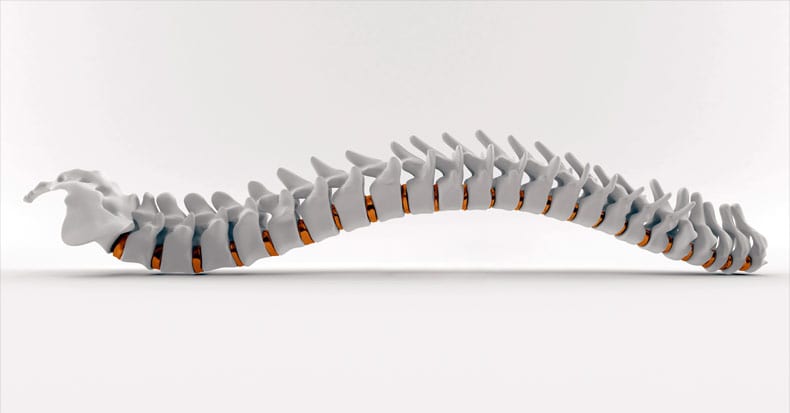
    Spinal Manipulation for Lumbar Intervertebral Disc Syndrome with Radiculopathy

    For thirty years (since 1985), it has been acknowledged that spinal manipulation is successful in the treatment of the majority of patients with low back pain, and that “there is a scientific basis for the treatment of back pain by manipulation.” (1) However, the consensus pertaining to the use of spinal manipulation for the treatment of intervertebral disc syndrome with radiculopathy is less investigated. Consequently, there is the potential for an opinion that spinal manipulation may be inappropriate for patients with low back intervertebral disc syndrome and symptoms/signs of radiculopathy.  This publication will review a number of articles on this topic, spanning six decades (1954-2015).

    ••••••••••

    In 1954, RH Ramsey, MD, published a study titled (2):

    Conservative Treatment of Intervertebral Disk Lesions

    Dr. Ramsey’s study appeared in the Instructional Course Lectures of the American Academy of Orthopedic Surgeons. Dr. Ramsey states:

    “The conservative management of lumbar disk lesions should be given careful consideration because no patient should be considered for surgical treatment without first having failed to respond to an adequate program of conservative treatment.”

    “If after a fair trial of conservative treatment, the pain and disability continue and the symptoms are of sufficient gravity to warrant surgery, the patient is advised that he should be operated upon and the offending disk lesion should be removed.”

    Dr. Ramsey advocated a number of conservative treatments for this syndrome, including spinal manipulation. Pertaining to manipulation, Dr. Ramsey makes the following comments:

    “From what is known about the pathology of lumbar disk lesions, it would seem that the ideal form of conservative treatment would theoretically be a manipulative closed reduction of the displaced disk material.”

    “Many forms of manipulation are carried out by orthopaedic surgeons and by cultists and this form of treatment will probably always be a controversial one.”

    “We limit the use of manipulation almost entirely to those patients who do not seem to be responding well to non-manipulative conservative treatment and who are anxious to have something else done short of operative intervention.”

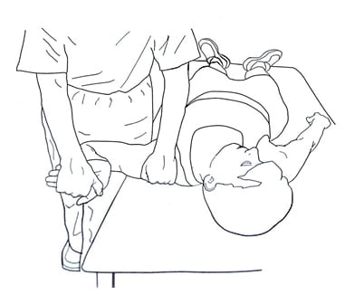
    “The patient lies on his side on the edge of the table facing the surgeon and the leg that is up is allowed to drop over the side of the table, tending to swing the up-side of the pelvis forward. The arm that is up is allowed to drop back behind the patient, tending to pull the shoulder back. The surgeon then places one hand on the patient’s shoulder and his opposite forearm on the patient’s iliac crest. Simultaneously, the shoulder is thrust suddenly back, rotating the torso in one direction while the iliac crest is thrust down and forward, rotating the pelvis in the opposite direction. This gives the lumbar spine a twist that frequently causes an audible and palpable crunch. This procedure is then repeated with the patient on his other side. The patient is then turned on his back and his hips and knees are hyperflexed sufficiently to forcibly flex the lumbar spine which tends to open up the disk spaces posteriorly.”

    “The patient should be cautioned beforehand that forceful manipulation may possibly make his symptoms worse although many patients will get marked relief.”

    Dr. Ramsey notes that the manipulation is “forceful” and associated with an “audible and palpable crunch.” Although he cautions that the manipulation may make the patient worse, “many patients will get marked relief.”

    ••••••••••

    Fifteen years later (in 1969), physicians JA Mathews and DAH Yates from the Department of Physical Medicine, St. Thomas’ Hospital, London, published a study titled (3):

    Reduction of Lumbar Disc Prolapse by Manipulation

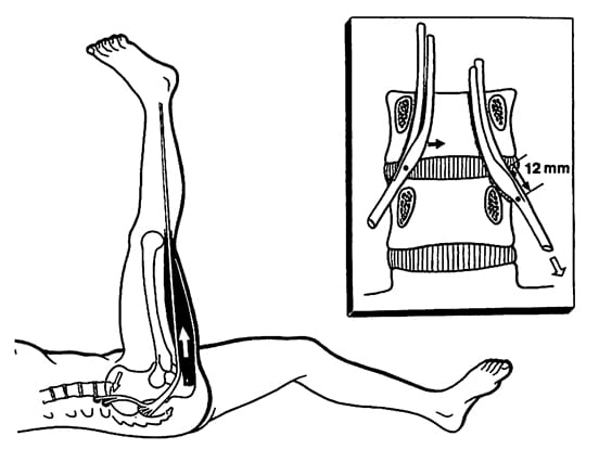
    This study appeared in the September 20, 1969 issue of the British Medical Journal. These authors evaluated a number of patients that presented with an acute onset of low back and buttock pain that did not respond to rest. Diagnostic epidurography showed a clinically relevant small disc protrusion, along with antalgia and positive lumbar spine nerve stretch tests. These patients were then treated with long-lever rotation manipulations of the lumbar spine, using the shoulder and iliac crest as levers. These lumbar spine manipulations were clearly accompanied with a thrust maneuver. The manipulations were repeated until abnormal symptoms and signs had disappeared. Following the manipulations there was resolution of signs, symptoms, antalgia, and reduction in the size of the protrusions. Drs. Mathews and Yates state:

    “The frequent accompaniment of acute onset low back pain by spinal deformity suggests a mechanical factor, and the accompanying abnormality of straight-leg raise or femoral stretch test suggests that the lesion impinges on the spinal dura matter of the dural nerve sheaths.”

    “The lumbar spine was rotated away from the painful side to the limit of its range, the buttock or thigh of the painful side being used as a lever; a firm additional thrust was made in the same direction. This manoeuver was repeated until abnormal symptoms and signs had disappeared, progress being assessed by repeated examination.”

    “Rotation manipulations apply torsion stress throughout the lumbar spine. If the posterior longitudinal ligament and the annulus fibrosus are intact, some of this torsion force would tend to exert a centripetal force, reducing prolapsed or bulging disc material.” 

    “The results of this study suggest that small disc protrusions were present in patients presenting with lumbago and that the protrusions were diminished in size when their symptoms had been relieved by manipulations.”

    These authors conclude: “it seems likely that the reduction effect [of the disc protrusion] is due to the manipulating thrust used.”

    ••••••••••

    In another study published in 1969, BC Edwards compared the effectiveness of heat/massage/exercise to spinal manipulation in the treatment of 184 patients that were grouped according to the presentation of back and leg pain, as follows (4):

    Group

    Treatment

    Acceptable Outcome

    Central Low Back Pain Only

    heat/massage/exercise

    83%

    spinal manipulation

    83%

    Pain Radiation to Buttock

    heat/massage/exercise

    70%

    spinal manipulation

    78%

    Pain Radiation Down Thigh to Knee

    heat/massage/exercise

    65%

    spinal manipulation

    96%

    Pain Radiation down Leg to Foot

    heat/massage/exercise

    52%

    spinal manipulation

    79%

    This study by Edwards was published in the Australian Journal of Physiotherapy.

    This study by Edwards was reviewed by Augustus A. White, MD, and Manohar M. Panjabi, PhD, in their 1990 book, Clinical Biomechanics of the Spine (5). Drs. White and Panjabi make the following points pertaining to the Edwards article:

    “A well-designed, well executed, and well-analyzed study.”

    In the group with central low back pain only, “the results were acceptable in 83% for both treatments. However, they were achieved with spinal manipulation using about one-half the number of treatments that were needed for heat, massage, and exercise.”

    In the group with pain radiating into the buttock, “the results were slightly better with manipulation, and again they were achieved with about half as many treatments.”

    In the groups with pain radiation to the knee and/or to the foot, “the manipulation therapy was statistically significantly better,” and in the group with pain radiating to the foot, “the manipulative therapy is significantly better.”

    “This study certainly supports the efficacy of spinal manipulative therapy in comparison with heat, massage, and exercise. The results (80 – 95% satisfactory) are impressive in comparison with any form of therapy.”

    ••••••••••

    In 1977, the third edition of Orthopaedics, Principles and Their Applications was published. The author, Samuel Turek, MD (d. 1986), was a Clinical Professor, Department of Orthopedics and Rehabilitation at the University of Miami School of Medicine. In the section pertaining to the protruded disc, Dr. Turek makes the following observations (6):

    Treatment of Intervertebral Disc Herniation With Manipulation

    “Manipulation.  Some orthopaedic surgeons practice manipulation in an effort at repositioning the disc.  This treatment is regarded as controversial and a form of quackery by many men.  However, the author has attempted the maneuver in patients who did not respond to bed rest and were regarded as candidates for surgery.  Occasionally, the results were dramatic.

    Technique. The patient lies on his side on the edge of the table facing the surgeon, and the uppermost leg is allowed to drop forward over the edge of the table, carrying forward that side of the pelvis.  The uppermost arm is placed backward behind the patient, pulling the shoulder back.  The surgeon places one hand on the shoulder and the other on the iliac crest and twists the torso by pushing the shoulder backward and the iliac crest forward.  The maneuver is sudden and forceful and frequently is associated with an audible and palpable crunching sound in the lower back.  When this is felt, the relief of pain is usually immediate.  The maneuver is repeated with the patient on the opposite side.”

    “The patient should be cautioned beforehand that the manipulation may make his symptoms worse and that this is an attempt to avoid surgery.”

    ••••••••••

            In February 1987, physicians Paul Pang-Fu Kuo and Zhen-Chao Loh published an important study pertaining to lumbar disc protrusions and rotary spinal manipulation, titled (7):

    Treatment of Lumbar Intervertebral Disc Protrusions by Manipulation

            Their article appeared in the journal Clinical Orthopedics and Related Research. Drs. Paul Pang-Fu Kuo and Zhen-Chao Loh are from the Department of Orthopedic Surgery, Shanghai Second Medical College, and Chief Surgeon, Department of Orthopaedic Surgery, Rui Jin Hospital, Shanghai, China. They note that manipulation has been used in Chinese healthcare for thousands of years, and by the Tang Dynasty (618-907 AD), who noted “manipulation was fully established and became a routine for the treatment of low back pain.”

    In their study, they performed a series of eight manipulations on 517 patients with protruded lumbar discs and clinically relevant signs and symptoms. Their outcomes were quite good, with 84% achieving a successful outcome and only 9% not responding. Only 14 % suffered a reoccurrence of symptoms at intervals ranging from two months to twelve years. These authors state:

    “The patient is placed on the sound side first with the hip and knee of the painful side flexed and the sound side straight. The operator rests one hand in front of the shoulder and the other hand on the buttock. By simultaneously pulling the shoulder backwards and pushing the buttock forwards, a snap or click can usually be heard or felt. This manipulation may then be repeated on the other side as required.”

    “Manipulation of the spine can be effective treatment for lumbar disc protrusions.”   

    “Most protruded discs may be manipulated. When the diagnosis is in doubt, gentle force should be used at first as a trial in order to gain the confidence of the patient.”

    “During manipulation a snap may accompany rotation. Subjectively it has dramatic influence on both patient and operator and is thought to be a sign of relief.”

    “If derangement of the facets or subluxation of the posterior elements near the protruded disc occurs, the rotation may have caused reduction, giving remarkable relief.”

    “Gapping of the disc on bending and rotation may create a condition favorable for the possible reentry of the protruded disc into the intervertebral cavity, or the rotary manipulation may cause the protruded disc to shift away from pressing on the nerve root.”

    In terms of applying manipulation, Drs. Kuo and Loh indicate “practice is necessary to become proficient in spinal manipulation techniques,” and  “expertise plays an important role in the success of manipulation.” The manipulation of disc protrusions should be performed only by trained experts. Additionally, manipulation is contraindicated if the patient is suffering from incontinence or paraplegia.

    ••••••••••

    In 1989, the Journal of Manipulative and Physiological Therapeutics published a case study of a patient with an “enormous central herniation lumbar disc” who underwent a course of side posture manipulation (8). The patient improved considerably with only 2 weeks of treatment. The authors state:

    “It is emphasized that manipulation has been shown to be an effective treatment for some patients with lumbar disc herniation.

    While complications of this form of treatment have been reported in the literature, such incidents are rare.”

    ••••••••••

            In 1993, chiropractor J. David Cassidy, chiropractor Haymo Thiel, and physician (orthopedic surgeon) William Kirkaldy-Willis published a “Review Of The Literature” article titled (9):

    Side posture manipulation for lumbar intervertebral disk herniation

    These authors are from the Department of Orthopaedics, Royal University Hospital, Saskatoon, Saskatchewan, Canada, and their article appeared in the Journal of Manipulative and Physiological Therapeutics.

    In their article, these authors cite studies on human cadavers that show the annulus of the disc is quite resistant to rotational stresses. Specifically, a normal disc did not show failure until 22.6° of rotational stress, and a degenerated disc could withstand an average of 14.3° of rotational stress. They therefore conclude “torsional failure of the lumbar disk first requires fracture of the posterior joints” before there is any annular tearing.

    When performing rotational manipulation in the management of lumbar disc herniation, these authors suggest that it is wise to begin with mobilization prior to performing manipulation to assess the patients responses. Additionally, they state that if positioning increases leg pain, “one should not proceed to manipulation at that particular session.”

    Based upon their review of the literature and their own experiences, these authors state:

    “The treatment of lumbar disk herniation by side posture manipulation is not new and has been advocated by both chiropractors and medical manipulators.”

    “The treatment of lumbar intervertebral disk herniation by side posture manipulation is both safe and effective.”

    ••••••••••

            In 1995, chiropractors PJ Stern, Peter Côté P, and David Cassidy published a study titled (10):

    A series of consecutive cases of low back pain with radiating leg pain treated by chiropractors

            Their article appeared in the Journal of Manipulative and Physiological Therapeutics. The authors retrospectively reviewed the outcomes of 59 consecutive patients complaining of low back and radiating leg pain, and were clinically diagnosed as having a lumbar spine disk herniation. Ninety percent of these patients reported improvement of their complaint after chiropractic manipulation. The maximum complication rate associated with this treatment approach was estimated to be 5% or less. A previous history of low back surgery was a statistically significant predictor of poor outcome. They concluded:

    “Based on our results, we postulate that a course of non- operative treatment including manipulation may be effective   and safe for the treatment of back and radiating leg pain.”

    ••••••••••

            In 2006, physicians Valter Santilli, MD, Ettore Beghi, MD, Stefano Finucci, MD, published an article in The Spine Journal titled (11):

    Chiropractic manipulation in the treatment of acute back pain and sciatica with disc protrusion:
    A randomized double-blind clinical trial of active and simulated spinal manipulations

            The purpose of this study was to assess the short- and long-term effects of spinal manipulations on acute back pain and sciatica with disc protrusion. It is a randomized double-blind trial comparing active and simulated manipulations for these patients. The study used 102 patients. The manipulations or simulated manipulations were done 5 days per week by experienced chiropractors for up to a maximum of 20 patient visits, “using a rapid thrust technique.” Re-evaluations were done at 15, 30, 45, 90, and 180 days.

    The authors list rationales for using manipulation in the treatment of low back pain and sciatica to include:

    • Reduction of a bulging disc
    • Correction of disc displacement
    • Release of adhesive fibrosis surrounding prolapsed discs or facet joints
    • Release of entrapped synovial folds
    • Inhibition of nociceptive impulses
    • Relaxation of hypertonic muscles
    • Unbuckling displaced motion segments

    The authors noted the following observations:

    “Active manipulations have more effect than simulated manipulations on pain relief for acute back pain and sciatica with disc protrusion.”

    “At the end of follow-up a significant difference was present between active and simulated manipulations in the percentage of cases becoming pain-free (local pain 28% vs. 6%; radiating pain 55% vs. 20%).”

    “Patients receiving active manipulations enjoyed significantly greater relief of local and radiating acute LBP, spent fewer days with moderate-to-severe pain, and consumed fewer drugs for the control of pain.”

    “No adverse events were reported.”

    The authors concluded that chiropractic spinal “manipulations may relieve acute back pain and sciatica with disc protrusion.”

    Real Manipulations

    Simulated Manipulations

    # of Subjects

    53

    49

    % of Local Pain

    Free Subjects

    28%

    6%

    % of Radiation Pain Free Subjects

    55%

    20%

    ••••••••••

            In 2014, an interdisciplinary group of physicians, chiropractors, and researchers published a study in the Annals of Internal Medicine, titled (12):

    Spinal Manipulation and Home Exercise With Advice  for Subacute and Chronic Back-Related Leg Pain

            This study was funded by the United States Department of Health and Human Services. It included 192 patients who were suffering from back-related leg pain for least 4 weeks. The number of subjects in the study gave it good statistical power. The subjects were randomized into either:

    • Chiropractic spinal manipulation + home exercise and advice, or
    • Home exercise and advice alone

    The treatment lasted 12 weeks. The authors concluded:

    “For leg pain, spinal manipulative therapy plus home exercise and advice had a clinically important advantage over home exercise and advice (difference, 10 percentage points) at 12         weeks.”


    “Spinal manipulative therapy with home exercise and advice improved self-reported pain and function outcomes more than exercise and advice alone at 12 weeks.”

    “Spinal manipulative therapy combined with home exercise and advice can improve short-term outcomes in patients with back-related leg pain.”

    “For patients with subacute and chronic back-related leg pain, spinal manipulative therapy in addition to home exercise and advice is a safe and effective conservative treatment approach,        resulting in better short-term outcomes than home exercise and   advice alone.”

    “No serious treatment-related adverse events or deaths occurred.”

    ••••••••••

            In another 2014 study, a group of multidisciplinary researchers and chiropractic clinicians from Switzerland presented a prospective study involving 148 patients with low back and leg pain. The study was published in the Journal of Manipulative and Physiological Therapeutics and titled (13):

    Outcomes of Acute and Chronic Patients with Magnetic Resonance Imaging–Confirmed Symptomatic Lumbar Disc Herniations Receiving High-Velocity, Low-Amplitude, Spinal Manipulative Therapy:

    A Prospective Observational Cohort Study With One-Year Follow-Up

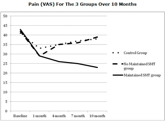
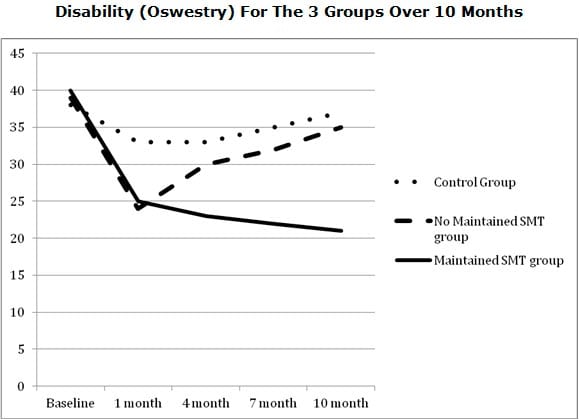
    The purpose of this study was to document outcomes of patients with confirmed, symptomatic lumbar disc herniations and sciatica that were treated with chiropractic side posture high-velocity, low-amplitude, spinal manipulation to the level of the disc herniation. It is important to emphasize that all patients in this study had clear abnormal physical examination findings of radiculopathy, including positive MRI abnormalities that corresponded with their symptoms and physical findings. Their pain was rated using the numerical rating scale and their disability was measured with the Oswestry questionnaire. Evaluations were performed at 2 weeks, 1 month, 3 months, 6 months, and 12 months.

    The outcomes from this study are summarized in the following table:

    Substantial Improvement (rounded)


    2 weeks

    1 month

    3 months

    6 months

    12 months

    Entire group

    70%

    80%

    91%

    89%

    88%

    Acute group

    81%

    85%

    95%

    91%

    86%

    Chronic group

    47%

    71%

    82%

    89%

    89%

    The authors make the following statements:

    “The proportion of patients reporting clinically relevant improvement in this current study is surprisingly good, with nearly 70% of patients improved as early as 2 weeks after the start of treatment. By 3 months, this figure was up to 90.5% and then stabilized at 6 months and 1 year.”

    “A large percentage of acute and importantly chronic lumbar disc herniation patients treated with chiropractic spinal manipulation reported clinically relevant improvement.”

    “Even the chronic patients in this study, with the mean duration of their symptoms being over 450 days, reported significant improvement, although this takes slightly longer.”

    “A large percentage of acute and importantly chronic lumbar disc herniation patients treated with high-velocity, low-  amplitude side posture spinal manipulative therapy reported clinically relevant ‘improvement’ with no serious adverse events.”

    “Spinal Manipulative therapy is a very safe and cost-effective option for treating symptomatic lumbar disc herniation.”

    This study shows that patients with proven lumbar intervertebral disc herniation and compressive neuropathology that receive traditional chiropractic side-posture manipulation is both safe and effective. The ultimate clinical effectiveness of about 90% is impressive when compared to any form of therapy, and with no reported serious side effects.

    This study would suggest that all patients suffering from lumbar intervertebral disc herniation with compressive neuropathology should be treated with chiropractic spinal adjusting.

    REFERENCES

    • Kirkaldy-Willis WH, Cassidy, JD; Spinal Manipulation in the Treatment of Low back Pain; Canadian Family Physician; March 1985, Vol. 31, pp. 535-540.
    • Ramsey RH; Conservative Treatment of Intervertebral Disk Lesions; American Academy of Orthopedic Surgeons, Instructional Course Lectures; Volume 11, 1954, pp. 118-120.
    • Mathews JA and Yates DAH; Reduction of Lumbar Disc Prolapse by Manipulation; British Medical Journal; September 20, 1969, No. 3, 696-697.
    • Edwards BC; Low back pain and pain resulting from lumbar spine conditions: a comparison of treatment results; Australian Journal of Physiotherapy; 15:104, 1969.
    • White AA, Panjabi MM; Clinical Biomechanics of the Spine; Second edition, JB Lippincott Company, 1990.
    • Turek S; Orthopaedics, Principles and Their Applications; JB Lippincott Company; 1977; page 1335.
    • Kuo PP and Loh ZC; Treatment of Lumbar Intervertebral Disc Protrusions by Manipulation; Clinical Orthopedics and Related Research. No. 215, February 1987, pp. 47-55.
    • Quon JA, Cassidy JD, O’Connor SM, Kirkaldy-Willis WH; Lumbar intervertebral disc herniation: treatment by rotational manipulation; Journal of Manipulative and Physiological Therapeutics; 1989 Jun;12(3):220-7.
    • Cassidy JD, Thiel HW, Kirkaldy-Willis WH; Side posture manipulation for lumbar intervertebral disk herniation; Journal of Manipulative and Physiological Therapeutics; February 1993;16(2):96-103.
    • Stern PJ, Côté P, Cassidy JD; A series of consecutive cases of low back pain with radiating leg pain treated by chiropractors; Journal of Manipulative and Physiological Therapeutics; 1995 Jul-Aug;18(6):335-42.
    • Santilli V, Beghi E, Finucci S; Chiropractic manipulation in the treatment of acute back pain and sciatica with disc protrusion: A randomized double-blind clinical trial of active and simulated spinal manipulations; The Spine Journal; March-April 2006; Vol. 6; No. 2; pp. 131–137.
    • Bronfort G, Hondras M, Schulz CA, Evans RL, Long CR, PhD; Grimm R; Spinal Manipulation and Home Exercise With Advice for Subacute and Chronic Back-Related Leg Pain; A Trial With Adaptive Allocation; Annals of Internal Medicine; September 16, 2014; Vol. 161; No. 6; pp. 381-391.
    • Leemann S, Peterson CK, Schmid C, Anklin B, Humphreys BK; Outcomes of Acute and Chronic Patients with Magnetic Resonance Imaging–Confirmed Symptomatic Lumbar Disc Herniations Receiving High-Velocity, Low Amplitude, Spinal Manipulative Therapy: A Prospective Observational Cohort Study With One-Year Follow-Up; Journal of Manipulative and Physiological Therapeutics; March/April 2014; Vol. 37; No. 3; pp. 155-163.
  • The Key To Preventing Spinal Pain: Understanding Levers

    The Key To Preventing Spinal Pain: Understanding Levers

    Pain is an Electrical Signal Interpreted by the Brain Nerves Bring the Pain Electrical Signal to the Brain

    The nerves that bring the pain electrical signal to the brain begin in the various tissues of the body. At the very beginning of the nerve there is a specialized ending call a receptor. The receptor is unique in its ability to initiate the pain electrical signal and send it along the pain nerves (nociceptors) to the brain. The brain interprets the pain electrical signal for a number of parameters (1):

    • Location: the region of the body where the pain receptors begin the pain electrical signal to the brain (head, neck, back, finger, toe, etc.).
    • Character: whether the pain signal is sharp, dull, aching, burning, stabbing, etc.

    The most common cause for the initiation of the pain electrical signal is an inflammatory reaction in the tissues where the pain nerve receptors reside (1).

    “The Origin of all Pain is Inflammation and the Inflammatory Response”

    It is because of this inflammation-pain response that so much pain is treated with anti-inflammatory approaches:

    • Non-steroidal anti-inflammatory drugs (NSAIDs)
    • Steroidal anti-inflammatory drugs
    • Omega-3 fatty acids that are found primarily in fish oil
    • Ice
    • Low-level laser therapy
    • Controlled Motion: disperses the accumulation of inflammatory exudates: chiropractic adjusting, massage, passive motions, active motions, etc.

    Steroidal and non-steroidal anti-inflammatory drugs are associated with many side effects, some serious, and some fatal, especially if consumed chronically, including:

    • Gastrointestinal bleeding, including fatal bleeding
    • End stage renal disease (ESRD)
    • Liver damage (hepatotoxicity)
    • Heart attacks/strokes
    • Dementia, including Alzheimer’s disease
    • Hearing loss
    • Erectile dysfunction
    • Atrial fibrillations

    As noted by Giles and Muller (2):

    “Adverse reactions to non-steroidal anti-inflammatory (NSAIDs) medication have been well documented. Gastrointestinal toxicity induced by NSAIDs is one of the most common serious adverse drug events in the industrialized world.”

    As noted by Maroon and Bost (3):

    “Extreme complications, including gastric ulcers, bleeding, myocardial infarction, and even deaths, are associated with their [NSAIDs] use.”

    Almost all patients who take the long-term NSAIDs will have gastric hemorrhage, 50% will have dyspepsia, 8% to 20% will have gastric ulceration, 3% of patients develop serious gastrointestinal side effects, which results in more than 100,000 hospitalizations, an estimated 16,500 deaths, and an annual cost to treat the complications that exceeds 1.5 billion dollars.

    “NSAIDs are the most common cause of drug-related morbidity and mortality reported to the FDA and other regulatory agencies around the world.”

    One author referred to the “chronic systemic use of NSAIDs as ‘carpet-bombing,’ with attendant collateral end-stage damage to human organs.”

    Chiropractic spinal adjusting reduces pain by using mechanisms that function in addition to the ability to help disperse the accumulation of inflammatory exudates. The most noted of these applies to the mechanical “closing of the ‘Pain Gate’” (4, 5, 6).

    •••••

     Whatever Tissue is Causing Spinal Pain, it Must Have a Nerve Supply

    It has been understood for decades that the articular hyaline cartilage has no nerve supply, and consequently is not capable of initiating the pain electrical signal. This holds true, even when the cartilage is injured. Sadly, injured articular hyaline cartilage degenerates at an accelerated rate (7, 8, 9, 10), creating arthritic changes that often irritate and inflame adjacent tissues, eventually generating the pain electrical signal to the brain. This is the explanation as to why some spinal injuries are initially asymptomatic, but become painful as a function of time as the degenerative process progresses.

    Other tissues that have been shown to be pain insensitive include the fascia (11).

    Stephen Kuslich and colleagues completed an extensive assessment, involving 700 live humans, as to the tissue sources of spinal pain in 1991 (11). These authors performed 700 lumbar spine operations using only local anesthesia to determine the tissue origin of back pain. The tissues they assessed for pain generation included skin, fat, fascia, supraspinous ligament, interspinous ligament, spinous process, muscle, lamina, facet capsule, facet synovium, nerve root, dura, compressed nerve root, normal nerve root, annulus fibrosus of the disc, nucleus of the disc, and vertebral end plate. All of these tissues, except for the fascia, were capable of producing spinal pain. They discovered that the primary “sight” for chronic back pain was the annulus of the intervertebral disc.

    Pertaining to the cervical spine, studies have indicated that the primary tissue that initiates the chronic neck pain signal is the facet joint capsular ligaments (12, 13).

    Interim Summary

    Spinal pain is an electrical signal in the brain. The pain electrical signal is brought to the brain by pain nerves (nociceptive). The pain nerves have receptors that initiate the pain electrical signal, and these receptors exist in most spinal tissues, but the most clinically relevant are in the intervertebral disc and in the facet joint capsular ligaments. The primary “trigger” for the initiation of the pain electrical signal from these tissues is inflammation and the inflammatory response.

    The inflammatory response that initiates the pain electrical signal can have a number of causes, including:

    • Infection
    • Autoimmune disease
    • Injury (trauma)
    • Chronic mechanical stress

    The healthcare provider attempts a differential diagnosis between these different etiologies by assessing factors such as history, family history, systemic factors, etc. The healthcare provider may also rely upon imaging and laboratory findings. Each of these can be influenced by individual genetics, epigenetics, nutrition, prior life events (injuries, pregnancies, etc.), occupation, ergonomics, age, fitness, etc. Successful management depends upon ascertaining a clear (or reasonably probable or suspected) etiology. A prudent healthcare provider will consequently do their best to make an accurate differential diagnosis.

    •••••

    Chiropractic clinical practice specializes in injury (trauma) and/or chronic mechanical stress as the etiology. Chiropractors not only treat the mechanical findings, they also inform the patient as to causation and to continuing causes of their ongoing mechanical problems that are initiating their pain. The chiropractor then coaches the patient on strategies to avoid or minimize the mechanical causes of their tissue inflammatory response. Often, patients can prevent future spinal problems by using this same practical advice. The basic advice requires knowledge of levers.

    The efficiency of human function in a gravity environment is an integration of mechanics and biology, known as biomechanics. Mechanics comprises a group of simple machines, which include:

    • Lever
    • Pulley
    • Screw
    • Wedge
    • Inclined plane
    • Wheel and axle

    The most important of these simple machines as applied to human upright posture and spinal syndromes is the lever. Levers allow for increased efficiency of strength and movement. The increased efficiency of the lever is called mechanical advantage.

    There are three classes of levers. All three types of levers have three common features:

    • Fulcrum (the pivot point)
    • Load (resistance, weight)
    • Effort (applied force)

    It is the sequential arrangement of the three common features that determine the class of the lever:

    First Class Lever:      load            fulcrum                    effort

    First Class Lever

    Examples of a first-class lever include a teeter-totter or crowbar.

    Second Class Lever:  fulcrum              load                          effort

    Second Class Lever

    An example of a second-class lever is a wheelbarrow: The wheel is the fulcrum; the effort is the handlebars; the load is in between.

    Third Class Lever:     fulcrum              effort                        load

    Third Class Lever

    An example of a third-class lever is tweezers, where the effort is applied between the fulcrum and the load.

    •••••

    The strongest mechanical structure is a column. This is why engineers use columns to support bridges and buildings. However, the human spine cannot function as a column. The human spine must be able to allow us to bend, stoop, lift, twist, etc. Consequently, a different mechanical design is required. Upright human posture is a three dimensional first class lever mechanical system, such as a teeter-totter or seesaw (14, 15).

    Recall, in the first class lever, the fulcrum is located between the load and the effort.

    first class lever

    The fulcrum of a first class lever is the place where the load is the greatest: if excessively heavy objects are placed on both ends of the teeter-totter, it will break in the middle, at the fulcrum.

    The force experienced at the fulcrum of a first class lever system is dependent upon three factors:

    The magnitude of the load (weight)

    The distance the load is away from the fulcrum (lever arm)

    The addition of the counterbalancing effort required to remain balanced

    If the load was 10 lbs., and the distance from the fulcrum was 10 inches (the lever arm), the force on the fulcrum would be 100 lbs. (10 X 10). In order to remain balanced, the effort on the opposite side of the fulcrum would have to also be 100 lbs. The effective load applied to the fulcrum would be 200 lbs. Thus an actual load of 10 lbs. would have an effective load on the fulcrum of 200 lbs.

    If the load was 10 lbs., and the distance from the fulcrum was 10 inches (the lever arm), the force on the fulcrum would be 100 lbs. (10 X 10). In order to remain balanced, the effort on the opposite side of the fulcrum would have to also be 100 lbs. The effective load applied to the fulcrum would be 200 lbs. Thus an actual load of 10 lbs. would have an effective load on the fulcrum of 200 lbs.

    The effective load (EF) at the fulcrum is the actual load (AL), multiplied by the lever arm (LA), plus the counterbalancing effort (CBE):

    EL = 10 lbs. (AL) X 10 in. (LA) + 100 lbs. (CBE) = 200 lbs. (EL)

    If the same actual load were closer to the fulcrum, the effective load changes significantly:

    EL = 10 lbs. (AL) X 5 in. (LA) + 50 lbs. (CBE) = 100 lbs. (EL)

    EL = 10 lbs. (AL) X 5 in. (LA) + 50 lbs. (CBE) = 100 lbs. (EL)

    •••••

    In the spine, the fulcrum of the first class lever of upright posture is primarily the vertebral body/disc, and the two facet joints.

    The actual load includes the weight of our own body and the weight of anything we are lifting or carrying.

    The effective load is the actual load multiplied by the length of the lever arm between the actual load and the fulcrum (disc and facets).

    The effort is the required contraction of the spinal muscles, on the opposite side of the fulcrum, to keep the spine balanced and prevent it from tipping over. This muscle contraction adds to the load at the fulcrum.

    This means that when the first class lever of upright posture is altered, for any reason, there is an increased effective mechanical load born by the fulcrum, i.e. the spinal intervertebral discs and facet joints. Such increased mechanical loads accelerate degenerative joint disease and the inflammation, altering the pain thresholds (14, 16). In their 1990 book Clinical Biomechanics of the Spine (15), White and Panjabi state:

    “The load on the discs is a combined result of the object weight, the upper body weight, the back muscle forces, and their respective lever arms to the disc center.”

    Events that increase the actual load at the spinal joints (disc and facets and require counterbalancing muscle contraction include postural distortions, lifting ergonomics (14), and weight problems (15).

    When a person gains abdominal weight (obesity, pregnancy), the first class lever system of upright posture is altered in such a manner that the intervertebral disc and facet joints bear significantly more weight. To counterbalance the weight, the muscles on the other side of the fulcrum (spine) must constantly contract with more force, or the person would fall forward. The black arrow attached to the posterior spinal elements represents the muscle contraction (14).

    When a person gains abdominal weight (obesity, pregnancy), the first class lever system of upright posture is altered in such a manner that the intervertebral disc and facet joints bear significantly more weight. To counterbalance the weight, the muscles on the other side of the fulcrum (spine) must constantly contract with more force, or the person would fall forward. The black arrow attached to the posterior spinal elements represents the muscle contraction

    This constant muscle contraction with more effort, creates muscle fatigue and myofascial pain syndromes. Rene Cailliet, MD states “This increase [in muscle tension] not only is fatiguing, but acts as a compressive force on the soft tissues, including the disk.” (14).

    •••••

    lever
    Rene Cailliet, MD, also uses the first class lever example in his 1996 book Soft Tissue Pain and Disability pertaining to the forward head syndrome (17). The patient has an unbalanced forward head posture. Dr. Cailliet assigns the head a weight of 10 lbs. and displaces the head’s center of gravity forward by 3 inches. The required counter balancing muscle contraction on the opposite side of the fulcrum (the vertebrae) would be 30 lbs. (10 lbs. X 3 inches):

     Rene Cailliet, MD, also uses the first class lever example in his 1996 book Soft Tissue Pain and Disability pertaining to the forward head syndrome (17). The patient has an unbalanced forward head posture. Dr. Cailliet assigns the head a weight of 10 lbs. and displaces the head’s center of gravity forward by 3 inches. The required counter balancing muscle contraction on the opposite side of the fulcrum (the vertebrae) would be 30 lbs. (10 lbs. X 3 inches):

    The constant muscle contraction required to balance postural distortions creates muscle fatigue and myofascial pain syndromes.

    Dr. Cailliet explains how the constant contraction in the counterbalancing muscles creates a cascade that leads to muscle fatigue, inflammation, fibrosis, and eventually to chronic musculoskeletal pain syndromes (17):

    cascade that leads to muscle fatigue, inflammation, fibrosis, and eventually to chronic musculoskeletal pain syndromes

    It is the increased effective loads at the spinal discs and facet joints (the spinal fulcrum) that accelerate degeneration and inflammation and are of particular concern in spinal pain syndromes. One should not discount the contribution of inflammation and pain from the contraction of the counterbalancing muscles and myofascial pain syndromes.

    SUMMARY

    It is much easier to hold a 10 lb. weight against your chest than in an outstretched arm. The reason is leverage. With the first class lever of upright posture, the most vulnerable tissues to mechanical loading stress, breakdown, degeneration, inflammation, and pain are the intervertebral disc and facet joints. This is because they function as the fulcrum. Numerous studies have identified the intervertebral disc and facet joints as the primary generators of chronic spinal pain. Likewise, the leverage stress applied to the fulcrum (disc and facets) must be counterbalanced my muscle contraction (effort), or the patient would fall over. This leads to chronic muscle problems (myofascial pain syndrome).

    Practical Advice

    • Weight: Excess body weight increases the weight to the disc and facet joints (the fulcrum). Increased weight increases compression, degeneration, inflammation, and pain. Also, excess weight is not gained symmetrically. The rule is most weight gain is at the abdomen. This creates a lever arm that multiplies the actual load (weight) to a significantly higher effective load on the fulcrum (disc and facets). The back muscles must then contract to maintain upright posture, further adding to the effective load (weight) to the spinal joints.
    • Posture: Poor posture significantly increases the effective load to the spinal joints and triggers counterbalancing muscle contraction (effort). An easily understood example is the forward head of Cailliet above. Poor spinal posture is routinely addressed by chiropractors.
    • Ergonomics: Individuals with acceptable posture may assume unacceptable postures during required work or leisure activities. Such activities may include “sitting at desk” postures, or “driving a vehicle” postures. During all such activities, the more one is coached to keep the center of masses of the parts of the body in alignment, the smaller the lever arm stress to the joints, and the less counterbalancing muscle contraction required (effort). Chiropractors routinely coach patients on such ergonomic issues.

    Poor posture significantly increases the effective load to the spinal joints and triggering counterbalancing muscle contraction (effort).

    • Bending: Bending forward at the waist to pick up an object, even a light object like a shoe or sock or pencil on the floor, can create significant adverse mechanical loads at the spinal disc and facets (the fulcrum). It is not the weight of the object, but rather it is the weight of the body multiplied by the lever arm, and also the required muscle contraction to become upright again. Bending is always risky for the spine. Stooping with the legs is always preferred when possible.
    • Lifting: Lifting by bending is the same as bending (above) with the addition of the weight of the object being lifted. This is well illustrated by White and Panjabi above.

    REFERENCES

    1. Omoigui S; The biochemical origin of pain: The origin of all pain is inflammation and the inflammatory response: Inflammatory profile of pain syndromes; Medical Hypothesis; 2007, Vol. 69, pp. 1169 – 1178.
    2. Giles LGF; Muller M; Chronic Spinal Pain: A Randomized Clinical Trial Comparing Medication, Acupuncture, and Spinal Manipulation; Spine July 15, 2003; 28(14):1490-1502.
    3. Maroon JC, Jeffrey W. Bost JW; Omega-3 Fatty acids (fish oil) as an anti-inflammatory: an alternative to nonsteroidal anti-inflammatory drugs for discogenic pain; Surgical Neurology; 65 (April 2006) 326–331.
    4. Melzack R, Wall PD; On the nature of cutaneous sensory mechanisms; Brain; 1962 Jun;85:331-56.
    5. Melzack R, Wall PD; Pain mechanisms: a new theory; Science. 1965 Nov 19;150(3699):971-9.
    6. Kirkaldy-Willis WH,  Cassidy JD; Spinal Manipulation in the Treatment of Low back Pain; Canadian Family Physician; March 1985, Vol. 31, pp. 535-540.
    7. Hadley Lee; Anatomical Roentgenographic Studies of the Spine, Thomas; 1972.
    8. Jackson R; The Cervical Syndrome; Thomas; 1978.
    9. Ruch W; Atlas of Common Subluxations of the Human Spine and Pelvis; CRC Press, 1997.
    10. Gargan MR, Bannister GC; The compararive effects of whiplash injuries; The Journal of Orthopaedic Medicine; 19(1), 1997, pp. 15-17.
    11. Kuslich S, Ulstrom C, Michael C; The Tissue Origin of Low Back Pain and Sciatica: A Report of Pain Response to Tissue Stimulation During Operations on the Lumbar Spine Using Local Anesthesia; Orthopedic Clinics of North America, Vol. 22, No. 2, April 1991, pp.181-7.
    12. Bogduk N, Aprill C; On the nature of neck pain, discography and cervical zygapophysial joint blocks; Pain; August 1993;54(2):213-7.
    13. Bogduk N; On Cervical Zygapophysial Joint Pain After Whiplash; Spine; December 1, 2011; Volume 36, Number 25S, pp S194–S199.
    14. Cailliet R; Low Back Pain Syndrome, 4th edition, F A Davis Company, 1981.
    15. White AA, Panjabi MM; Clinical Biomechanics of the Spine, Second Edition, Lippincott, 1990.
    16. Garstang SV, Stitik SP; Osteoarthritis; Epidemiology, Risk Factors, and Pathophysiology; American Journal of Physical Medicine and Rehabilitation; November 2006, Vol. 85, No. 11, pp. S2-S11.
    17. Cailliet R; Soft Tissue Pain and Disability; 3rd Edition; F A Davis Company, 1996.


  • Two Important, Yet Simple Tests For Low Back Pain / Leg Pain

    Two Important, Yet Simple Tests For Low Back Pain / Leg Pain

    The nervous system can be categorized in many different ways. In understanding nervous system physiology, a simple but accurate way of categorization is to view the nervous system as two separate but integrated systems:

    • The MOTOR nerve system
    • The SENSORY nerve system

    The MOTOR nerve system is the nerves that move our muscles (motor), and also control the function of our visceral organs (like heart, lungs, intestines, pancreas, liver, kidneys, etc.). The nerves that send the electrical signal from our brain and spinal cord to our muscles to control their contraction are actually called motor nerves. The nerves that send the electrical signal from our brain and spinal cord to control the function of our visceral organs are called autonomic nerves. This is because they function automatically, without our thinking and even when we are sleeping. Occasionally these autonomic visceral organ nerves are called visceral motor nerves. The motor nerve systems are output nerves, also called efferent nerves.

    The SENSORY nerve system is the nerves that send electrical nerve signals into our spinal cord and brain. Therefore, the sensory nerves travel in the opposite direction of the motor nerves. The sensory nerve systems are input nerves, also called afferent nerves.

    The sensory nerves have special endings (receptors) that can take environmental events, convert these events into an electrical signal, and send the electrical signal along sensory nerves to the brain for interpretation. The sensory nerves create our senses. It is our lifelong sensory experiences that “mold” our brain.

    •••••

    There are six primary sensory inputs into our brains:

    • Sight (vision). Our eye has specific sensory receptors that have the ability to take specific electromagnetic waves in the environment, convert them into an electrical signal, send the electrical signal along a nerve (the optic nerve) to a specific place in the brain for interpretation (the occipital visual cortex).
    • Sound (hearing). Similarly, the ear has specific sensory receptors that have the ability to take specific environmental disturbances, convert them into an electrical signal, send the electrical signal along a nerve (cochlear nerve) to a specific place in the brain for interpretation (the superior gyrus of the temporal lobe).
    • Taste. When molecules from food or drink contact our tongue, again an electrical signal is sent via a sensory nerve to the brain for interpretation.
    • Smell. When certain molecules in the air travel up our nose, an electrical signal is again sent via a sensory nerve to the brain for interpretation.
    • Touch (requires sub categorization). There are special receptors on our skin and in other tissues like muscle, tongue, teeth, and viscera that generate an electrical single when they are mechanically perturbed. This electrical signal is once again sent via sensory nerves to the appropriate place in the brain for interpretation.
    • Proprioception. Proprioception is often referred to as our “sixth” sense. There are special receptors in our skin, muscles, joints, fascia, etc., that generate an electrical signal that lets the brain know where we are in space. These receptors and their sensory nerves inform the brain about changes in the position and movements of the various parts of our bodies. Most of us know where our nose is, even when our eyes are closed, and we can easily touch our nose with the tip of our index finger (we also know where the tip of our finger is, even with eyes closed). With our eyes closed, we cannot see, hear, smell, taste, or touch our nose or fingertip, yet we can easily connect the two. This is proprioception.

    proprioception

    Touch (#5) requires elaboration. Touch (for our purposes) will also include the sensations of pain and temperature (hot/cold). When there is a perturbation on our skin, we readily can distinguish between touch, pain, hot, and/or cold. All of these sensations are electrical signals that travel to various parts of the brain for interpretation.

    The important point is that all perceptions (sight, sound, taste, smell, touch [including pain and temperature] and proprioception) occur in the cortical brain. “All perceptions are cortical.” This means that they occur in our brain. Lay people often believe that their eye sees, their ear hears, their tongue tastes, their nose smells, or that something at their toe or back or neck hurts. But actually these various parts of our body only initiate an electrical signal that is then interpreted in our brain.

    •••••

    The cortical perception of pain is a universal human experience. The electrical signal for the perception of pain in the brain is brought to the brain via special sensory nerves called nociceptive afferents or nociceptors.

    All sensory inputs into the brain begin with a special receptor, except for pain (nociception). The receptor that initiates the electrical signal for sight is different than the receptor that initiates the electrical signal for sound or taste or smell. Pain (nociception) is the great receptor exception in that, for the most part, there is no receptor on the end of the nociceptive nerves. Consequently, the nociceptive nerve beginnings are referred to as free or naked receptors.

    The pain problem in our country (USA) and in the world is astonishingly huge and it is getting worse. In the United States alone, 116 million Americans suffer from chronic daily pain (1). A recent cover article in the newspaper The Wall Street Journal quantifies the anatomical regions for American’s chronic pain (2):

    Hip Pain                       07.1%

    Finger Pain                  07.6%

    Shoulder Pain             09.0%

    Neck Pain                    15.1%

    Severe Headache       16.1%

    Knee Pain                    19.5%

    Lower-Back Pain        28.1%

    The total cost attributed to America’s pain problem, including treatment, lost productivity, and disability, is approaching $1 trillion per year.

    It is useful to discuss pain using the categorizations of C. Chan Gunn, MD (3, 4). Dr. Gunn is a Clinical Professor at the Multidisciplinary Pain Center at the University of Washington Medical School, Seattle, Washington. Dr. Gunn’s pain categories are:

    1) Nociception Pain

    In this category of pain, there is no tissue damage, and therefore no inflammation. This is the type of pain one would experience if someone stepped on your toe; one would have pain but no tissue damage or inflammation. This type of pain does not require a healthcare provider to diagnose the cause of the pain. The cause of the pain is obvious; someone is standing on your toe.

    Likewise, this type of pain does not require healthcare provider treatment. The treatment is obvious; get the person’s foot off your toe. The patient self treats.

    With this type of pain, once the person’s foot is off your toe, you experience immediate and lasting relief. The prognosis is excellent.

    This is the type of pain that most patients (and insurance companies) hope they are experiencing, hoping for instant relief. Sadly, this type of pain rarely makes it into a doctor’s office because it is self diagnosed and treated.

    2) Algogenic Pain

    Suppose that instead of someone stepping on your toe, they smacked your toe with a sledgehammer. Even though the hammer is no longer actually on your toe, your toe still hurts. The hammer added something to the equation, trauma, tissue damage, and inflammation. This disruption of the tissues and blood vessels by the trauma produces and releases inflammatory chemicals that are often collectively called algogenic exudates.

    The inflammatory algogenic chemicals alter the thresholds of the nociceptive afferent system, increasing the pain electrical signal to the brain. Instant relief for this type of pain is not possible. The pain subsides as inflammation resolves and the nociceptive afferents system becomes sub-threshold.

    Individuals suffering from this type of pain often go to healthcare providers for relief. Treatment often involves anti-inflammatory efforts (controlled motion, drugs, omega-3s, ice, electrical modalities, low–level laser therapy, etc.) and efforts to accelerate healing (low-level laser therapy). Depending upon the degree of tissue injury and a myriad of individual unique characteristics, response can last days, week, or months.

    Chronic inflammation, caused by scar tissue, autoimmune responses, infection, etc., can cause chronic algogenic pain.

    3) Neuropathic Pain

    This is pain that persists after all possible tissue healing has occurred. Once again, instant relief for this type of pain is not possible. This is chronic pain that may persist for months, years, or forever.

    •••••

    Lay people often view pain solely as a bad thing, but healthcare professionals recognize pain to be both friend and foe. For example, if one sits on one’s foot for a prolonged period of time, it will eventually begin to hurt. This is an example of nociceptive pain. We simply self diagnose and treat our foot pain by moving it or changing our sitting position, and the pain goes away.

    People are constantly doing things that begin to generate pain, and the pain afferents send a sensory signal to our brain reminding us to stop doing that activity. In this regard, pain keeps us safe, reminding us not to do certain things or to stop doing certain things. Without pain, we would not survive childhood and make it into adulthood.

    Chronic pain is another story.

    •••••

    Of the many structures that make up the spine, most of them are capable of generating pain. All of the spinal structures that can initiate the pain signal to the brain have a common factor: they are innervated by sensory afferent nociceptive neurons that carry the pain electrical signal to the brain. As noted by exceptional spine care pioneer Alf Nachemson, MD, whatever causes spine pain must have a nerve (5). In 1991, Stephen Kuslich, MD, and colleagues clarified and quantified the spinal tissues that were capable of initiating the pain electrical signal to the brain as (6):

    • Skin
    • Superficial Muscles
    • Deep Muscles
    • Intervertebral Disc
    • Facet Joint Capsules
    • Periosteum of the Vertebral Bone
    • Nerve roots

    Any of these tissues are capable of initiating acute spinal pain. Chronic spinal pain perception was primarily attributed to the intervertebral disc and facet capsules, in that order (6). Other studies have primarily attributed chronic neck pain perception to the facet capsules and the intervertebral disc, in that reverse order (7).

    •••••

    For a new incidence of non-traumatic low back or neck pain, it is important for both clinicians and patients to make an initial quick assessment of the severity of the problem. Absent other historical indicators, it is common to assume the pain is algogenic in nature. This means there is an accumulation of algogenic inflammatory exudates that are increasing the sensitivity of the pain sensory nerves. The first step in doing this is to categorize the symptoms into one of three groups:

    Group 1: Spinal pain alone

    Either neck pain or back pain without pain radiation into the arms or legs. In general, algogenic spine pain that does not radiate is not serious. It is usually the consequence of a local inflammatory condition. It can be chronic and even disabling, but it is not dangerous.

    Group 2: Sclerogenic pain; also known as sclerotomic pain or sclerotogenous pain

    Sclerogenic pain radiates from the neck into the arm(s) or from the low back into the leg(s). Classically the pain radiation will not extend below the elbow (from the neck) or below the knee (from the low back).

    As a rule, sclerogenic pain is difficult for the patient to localize. The pain presentation is often described as being deep and dull in character, similar to a toothache.

    In general, sclerogenic pain is not dangerous. It is a form of referred pain that occurs as a consequence of a shared neuromere during embryonic development. In other words, the neurology of the back and the leg, or of the neck and the arm, are shared embryological, which can cause some confusion as to the exact location of the irritation when the electrical signal is sent to the brain. Originally based on the research of JH Kellgren and colleagues in 1938 and 1939, irritations of deep spinal tissues can cause sclerogenic pain referral to the arm or to the leg (8, 9).

    In the sclerogenic pain patient, successful management of the deep spinal tissue irritations will resolve the sclerogenic pain referral. Deep spinal tissue irritations include irritations to the intervertebral disc, the facet joint capsules, and the core stabilization segmental mover muscles. These tissues respond excellently to spinal adjusting.

    Group 3:    Radicular pain, radiculitis, radiculopathy

    The technical definition of radicular pain is that the spinal nerve root is inflamed, and the classic symptomatology is radiating arm or leg pain. In contrast to sclerogenic pain (a deep dull ache), the pain is often sharp and easily localized by the patient. Also, the pain often travels below the elbow (into the hands and fingers) and/or below the knee (into the foot and toes).

    Radicular pain is more serious than sclerogenic pain. It is therefore a good idea to determine (to the best of one’s ability) if the pain is radicular or sclerogenic. History and physical examination can be quite accurate in establishing a differential diagnosis. However, conformation will require advanced diagnostic imaging. The current gold standard in advanced diagnostic imaging is magnetic resonance imaging, or MRI.

    Radicular pain is often caused by compression of the nerve root, the compression causing nerve root irritation and/or inflammation. This pathology is commonly referred to as compressive radiculopathy. Interestingly, the compression itself is not necessarily painful. Rather, the pain arises when the compression initiates irritation and/or inflammation of the nerve root. The degree of nerve root compression and its seriousness is estimated with MRI scans.

    The most classic cause of radicular compression is herniation of the intervertebral disc. Other causes include arthritic changes (degenerative joint disease, degenerative disc disease, spondylosis) causing osseous (bone spurs, hypertrophic changes, osteophytes) narrowing of the intervertebral foramen.

    Each nerve root supplies a specific patch of skin (a dermatome) and a specific muscle (a myotome). Consequently, radicular compression is often associated with specific myotomal muscle weakness and altered sensation in the dermatomal patch of skin (paresthesias).

    The deep tendon reflex is a common component of establishing if the extremity pain is sclerogenic or radicular. With radicular compression, the deep tendon reflex is diminished or possibly even absent. There are three common deep tendon reflexes in the arms (assessing the nerve roots of the neck), and two in the legs (assessing the nerve roots in the low back).

    Radicular compressive pathology can result in permanent death of some of the neurons in the nerve root, resulting in permanent loss of various functions. Consequently, when compressive radiculopathyis suspected, “red flags” of such pathology should be watched for and assessed. These “red flags” include:

    • Progressive myotomal muscle weakness.
    • Atrophy of the muscle.
    • Saddle anesthesia (loss of sensation in the area of the buttocks that would contact a saddle when sitting).
    • Loss of bowel, bladder, and/or sexual function.(difficulty starting, difficulty ending, dripping, loss of sensation, etc.)

    •••••

    There are two (one for the neck and one for the low back) very simple tests that are commonly done by healthcare providers to help determine if radiating pain is sclerogenic referral or as a consequence of radicular compression. These tests can also be easily performed by patients to help determine the seriousness of compression and its progress while under treatment.

    Both tests are stretch tests. If the nerve root is compressed, irritated, and inflamed as it exits the spinal column, stretching it will aggravate the discomfort and the radiation.

    Low Back Pain With Leg Radiation Test

    This test is known as the Straight Leg Raising Test. It is also known as Laseque’s Test, after Charles Laseque who first described the test in 1864 (10). The premise of the test is simple: movement of the leg causes movement of the lower lumbar nerve roots.

    This test is performed by lying flat on one’s back and raising one’s leg up into the air while keeping the knee locked straight. Many normal people can do this to almost 90°. Individuals with lower back (lumbar) spinal radicular compressionwill begin to feel an increased in leg or back symptoms starting at about 35°.

    According to Kapandji (11), when the leg is raised during the Straight Leg Raising Test, the lower lumbar spinal nerve roots will slide out of the nerve root hole (intervertebral foramen) by as much as half an inch (12 mm). If the nerve root is entrapped or compressed, the stretch will aggravate the irritation/inflammation, increasing symptoms.

    Straight Leg Raising Test

    It is accepted that the primary cause of compressive radiculopathy is herniation of the intervertebral disc. Most patients with discogenic compressive radiculopathy obtain symptomatic relief when lying down flat on their back. The probable explanation for this is that the intradiscal pressure is least when in this position (12, 13)

    In contrast, it is established that when one sits down, intradiscal pressures are increased by roughly a factor of 6 (25 psi to 140 psi) (12, 13).

    In contrast, it is established that when one sits down, intradiscal pressures are increased by roughly a factor of 6 (25 psi to 140 psi) (12, 13).

    Therefore it is argued that performing the Straight Leg Raising Test when sitting down is a better indicator of the presence of discogenic compressive radiculopathy. This test is known as Bechterew’s Test. It is performed by sitting up straight, and then straightening out one’s leg until it is parallel with the horizon. An increase in leg or spinal symptomatology is considered to be a positive indicator of the presence of low back/leg compressive radiculopathy.

    Straight Leg Raising Test

    Brachial Plexus Tension Test of Elvey

    The test that is an equivalent to the lower back Straight Leg Raising Test in the neck (cervical spine) is the Brachial Plexus Tension Test of Elvey. This test was originally described by Australian physiotherapist Robert Elvey in 1986 (14). Once again, the premise of the test is simple: movement of the arm causes movement of the lower cervical spine nerve roots.

    The step-by-step procedure for performing the Brachial Plexus Tension Test of Elvey are well described by Quintner in the British Journal of Rheumatology in 1989 (15):

    TO START:

    Put the patient supine.

    Externally rotate the arm and supinate the forearm.

    Flex the fingers, wrist, and elbow.

    Abduct the shoulder joint 110 degrees, so that the elbow is superior to the glenohumeral joint.

    Put the arm behind the coronal plane of the body.

    TO ASSESS:

    Keep the shoulder girdle depressed.

    Keep the forearm supinated.

    Extend the elbow.

    Extend the wrist, supination of the forearm.

    Extend the fingers.

    IF NEGATIVE:

    Reassess with the head/neck laterally flexed to the opposite side.

    Brachial Plexus Tension Test of Elvey

    Summary

    Both cervical spine and lumbar spine compressive radiculopathies are coupled with a worse prognosis for complete recovery. Compressive radiculopathy typically requires more frequent treatment and more prolonged treatment. Compressive radiculopathy patients often have more long-term subjective and objective residuals, and more disability. Patients with compressive radiculopathy often require advanced imaging (such as MRI) for a full assessment of their pathology. Occasionally, patients with compressive radiculopathy will require a surgical decompression. These patients should always be monitored for the emergence of “red flag” signs.

    The Straight Leg Raising Test (Laseque’s) and the Brachial Plexus Tension Test (Elvey) are simple tests to assess the presence of a compressive radiculopathic process.

  • Spinal Pain Exercise, Mobilization, Manipulation

    Spinal Pain Exercise, Mobilization, Manipulation

    Where Does Back Pain Come From?

    In 1934, William Mixter, MD and Joseph Barr, MD, established that herniation of the lumbar disc could put pressure on the nerve root or the cauda equina, resulting in sciatica. Their paper on the topic appeared in 1934 in the New England Journal of Medicine (1) and was titled:

    Rupture of the Intervertebral Disc with Involvement of the Spinal Canal

    Dr. Mixter was the primary author of the paper, and at the time, at age 54, he was considered to be the top surgeon in the United States. Dr. Mixter was born in 1880 and graduated from Harvard Medical School in 1906. He became a prominent surgeon at the Massachusetts General Hospital, and by 1911, along with his father, became the primary neurosurgeon at that institution. By the early 1930s, Dr. Mixter was considered to be one of the nation’s leading experts in spinal surgery, and he went on to become the first chief of the neurosurgery department at Massachusetts General Hospital.

    This article by Drs. Mixter and Barr fundamentally changed the popular understanding of sciatica at that time, and for this work Dr. Mixter is generally credited by his contemporaries as being the man who best clarified the relation between the intervertebral disc and sciatica. Their landmark article helped to establish surgery’s prominent role in the management of sciatica at the time.

    However, this 1934 article by Drs. Mixter and Barr did little to discuss the pathophysiology of low back pain, but rather only discussed the pathophysiology of sciatica. Drs. Mixter and Barr continued to publish studies pertaining to sciatica, and in 1941 they published in the Journal of Bone and Joint Surgery (American) (2) a paper titled:

    Posterior Protrusion of the Lumbar Intervertebral Discs

    In this article, Drs. Barr and Mixter continue their discussions and case series presentations of posterior lumbar spine disc protrusions and consequent sciatica. Importantly, they also offer perhaps the first explanation for reoccurring low back pain in patients who are not suffering from sciatica. They state:

    “A second explanation is that a fissure occurs in the annulus fibrosus as the result of the wear and tear of ordinary use or of degenerative change, and through this fissure there is a slow, gradual extrusion of disc tissue with final resultant symptoms of sciatica when the extruded mass becomes large enough to press upon a nerve root. Clinical cases of this type may have recurring episodes of low backache over many years before the extruded mass becomes large enough to precipitate an attack of sciatica.”

    In this paragraph, Drs. Barr and Mixter suggest that “recurring episodes of low backache over many years” may be the consequence of degenerative changes in the annulus fibrosus of the disc accompanied with fissures.

    The modern era in the understanding of low back pain began in 1976 when internationally respected orthopedic surgeon, Dr. Alf Nachemson, published his detailed review (136 references) in the journal SPINE (3), titled:

    The Lumbar Spine: An Orthopaedic Challenge

    In this article, Dr. Nachemson notes that 80% of us will experience low back pain at some time in our life. He further states that:

    “The intervertebral disc is most likely the cause of the pain.”

    Dr. Nachemson presents 6 lines of reasoning, supported by 17 references, to support his contention that the intervertebral disc is the most likely source of back pain, including the primary research completed by Drs. MJ Smyth and V Wright in

    1958 (4). Drs. Smyth and Wright published their paper in the Journal of Bone and Joint Surgery (American), titled:

    Sciatica and the intervertebral disc:

    An experimental study

    Regarding this work by Smyth and Wright, Dr. Nachemson states:

    “Investigations have been performed in which thin nylon threads were surgically fastened to various structures and around the nerve root. Three to four weeks after surgery these structures were irritated by pulling on the threads, but pain resembling that which the patient had experienced previously could only be registered only from the outer part of the annulus” of the disc.

    In his 1976 review, Dr. Nachemson was noting that a non-herniated disc problem was causing back pain and that the disc itself was a probable source of back pain. Dr. Nachemson notes that the source of back pain must have a nerve supply, but at that time, good studies showing the innervation of the intervertebral disc were lacking.

    Support for Dr. Nachemson’s contention of disc pain came in 1981 when Australian clinical anatomist and physician, Dr. Nikoli Bogduk, published an extensive review of the literature on the topic of disc innervation, along with his own primary research, in the prestigious Journal of Anatomy (5). Dr. Bogduk and colleagues conclude:

    “The lumbar intervertebral discs are supplied by a variety of nerves.”

    and

    “Clinically, the concept of ‘disc pain’ is now well accepted.”

    In 1983, Dr. Bogduk updates his research when he publishes an article in the journal SPINE titled (6):

    The Innervation of the Lumbar Spine

    In this 1983 study, Dr. Bogduk states:

    “The lumbar intervertebral discs are innervated posteriorly by the sinuvertebral nerves, but laterally by branches of the ventral rami and grey rami communicantes. The posterior longitudinal ligament is innervated by the sinuvertebral nerves and the anterior longitudinal ligament by branches of the grey rami. Lateral and intermediate branches of the lumbar dorsal rami supply the iliocostalis lumborum and longissimus thoracis, respectively. Medial branches supply the multifidus, intertransversarii mediales, interspinales, interspinous ligament, and the lumbar zygapophysial joints.”

    “The distribution of the intrinsic nerves of the lumbar vertebral column systematically identifies those structures that are potential sources of primary low-back pain.”

    In 1986, Dr. Vert Mooney’s was the president of the International Society for the Study of the Lumbar Spine. That year, his Presidential Address was delivered at the 13th Annual Meeting of the International Society for the Study of the Lumbar Spine, May 29-June 2, 1986, Dallas, Texas. It was published in the journal SPINE in 1987, and titled (7):

    Where Is the Pain Coming From?

    In this article, Dr. Mooney states:

    “Anatomically the motion segment of the back is made up of two synovial joints and a unique relatively avascular tissue found nowhere else in the body – the intervertebral disc. Is it possible for the disc to obey different rules of damage than the rest of the connective tissue of the musculoskeletal system?”

    “Persistent pain in the back with referred pain to the leg is largely on the basis of abnormalities within the disc.”

    “Mechanical events can be translated into chemical events related to pain.” An important aspect of disc nutrition and health is the mechanical aspects of the disc related to the fluid mechanics.

    “Mechanical activity has a great deal to do with the exchange of water and oxygen concentration” in the disc.

    The pumping action maintains the nutrition and biomechanical function of the intervertebral disc. Thus, “research substantiates the view that unchanging posture, as a result of constant pressure such as standing, sitting or lying, leads to an interruption of pressure-dependent transfer of liquid. Actually the human intervertebral disc lives because of movement.”

    “In summary, what is the answer to the question of where is the pain coming from in the chronic low-back pain patient? I believe its source, ultimately, is in the disc. Basic studies and clinical experience suggest that mechanical therapy is the most rational approach to relief of this painful condition.”

    “Prolonged rest and passive physical therapy modalities no longer have a place in the treatment of the chronic problem.”

    The model presented by Dr. Mooney in this paper includes:

    The intervertebral disc is the primary source of both back pain and referred leg pain. The disc becomes painful because of altered biochemistry, which sensitizes the pain afferents that innervate it. Disc biochemistry is altered because of mechanical problems, especially mechanical problems that reduce disc movement. Therefore, the most rational approach to the treatment of chronic low back pain is mechanical therapy that restores the motion to the joints of the spine, especially to the disc. Prolonged rest is inappropriate management.

    Additional support for the disc being the primary source of back pain was presented by Dr. Stephen Kuslich and colleagues in the prestigious journal Orthopedic Clinics of North America in April 1991 (8). The title of their article is:

    The Tissue Origin of Low Back Pain and Sciatica:

    A Report of Pain Response to Tissue Stimulation During Operations on the Lumbar Spine Using Local Anesthesia

    The authors performed 700 lumbar spine operations using only local anesthesia to determine the tissue origin of low back and leg pain, and they present the results on 193 consecutive patients studied prospectively. Several of their important findings include:

    “Back pain could be produced by several lumbar tissues, but by far, the most common tissue of origin was the outer layer of the annulus fibrosis.”

    The lumbar fascia could be “touched or even cut without anesthesia.”

    Any pain derived from muscle pressure was “derived from local vessels and nerves, rather than the muscle bundles themselves.”

    “The normal, uncompressed, or unstretched nerve root was completely insensitive to pain.”

    “In spite of all that has been written about muscles, fascia, and bone as a source of pain, these tissues are really quite insensitive.”

    In summary, Dr. Kuslich and colleagues found that the outer annulus is “the site” of a patient’s back pain. Irritation of a normal or inflamed nerve root never produced low back pain. Back muscles themselves are not a source of back pain; in fact, the muscles, fascia, and bone are really quite insensitive for pain. Also the inflamed, stretched, or compressed nerve root is the cause of buttock, leg pain and sciatica, but not back pain.

    In 2006, physician researchers from Japan published in the journal SPINE the results of a sophisticated immunohistochemistry study of the sensory innervation of the human lumbar intervertebral disc (9). The article is titled:

    The Degenerated Lumbar Intervertebral Disc is Innervated Primarily by Peptide-Containing Sensory Nerve Fibers in Humans

    These authors note:

    “Many investigators have reported the existence of sensory nerve fibers in the intervertebral discs of animals and humans, suggesting that the intervertebral disc can be a source of low back pain.” “Both inner and outer layers of the degenerated lumbar intervertebral disc are innervated by pain sensory nerve fibers in humans.”

    Pain neuron fibers are found in all human discs that have been removed because they are the source of a patient’s chronic low back pain.

    The nerve fibers in the disc, found in this study, “indicates that the disc can be a source of pain sensation.”

    The perspective offered by these studies from 30 years of publications and research in the best journals is that the annulus of the intervertebral disc is primarily responsible for the majority of chronic low back pain. Acourding to Dr. Vert Mooney (7) above, the pain producing disc lesion is segmental, or pertaining to abnormal mechanical function of an intersegmental “motion segment of the back.”

    Where Does Neck Pain Come From?

    In 1993, Australian physician / clinical anatomist Nikioli Bogduk and American physician / radiologist Charles Aprill thoroughly evaluated the tissue sources for chronic neck pain. They published their findings in the journal Pain (10) in an article titled:

    On the nature of neck pain, discography and cervical zygapophysial joint blocks

    In this study, the authors evaluated the sources of chronic neck pain by using both provocation discography and cervical zygapophysial joint blocks. Their findings include:

    “Both a symptomatic disc and a symptomatic zygapophysial joint were identified in the same segment in 41% of the patients.”

    “Discs alone were symptomatic in only 20% of the sample.”

    “Zygapophysial joints were symptomatic but discs were asymptomatic in 23%.”

    “Only 17% of the patients had neither a symptomatic disc nor a symptomatic zygapophysial joint at the segments studied.”

    Neck muscle injury “does not provide a satisfying model for persistent or chronic neck pain.”

    A summary of the findings of Drs. Bogduk and Aprill from this 1993

    study include:

    The most frequent finding was “both a symptomatic disc and a

    symptomatic zygapophysial joint at the same segment,” seen in 41%. There is important clinical relevance to the finding that the primary source of chronic neck pain was segmental, involving both the intervertebral disc and the facet articulation.

    The second most frequent finding was a symptomatic zygapophysial joint, alone, with no disc involvement, which was found in 23%.

    “This indicated that 64% of the sample had a symptomatic zygapophysial joint.” [41% + 23% = 64%]

    The third most frequent finding was a symptomatic disc alone, with no zygapophysial joint involvement, found at 20%.

    This indicated that 61% of the sample had a symptomatic disc.

    [41% + 20% = 61%]

    The most important finding in this study was that the most common source for chronic neck pain was both articular (facet and disc) and segmental.

    This 1993 study by Bogduk and Aprill was followed by two studies completed by the Australian research team of Leslie Barnsley, Susan Lord, Barbara Wallis, and Nikioli Bogduk. Both studies were published in the journal SPINE, in 1995 (11) and in 1996 (12). Both studies confirm the conclusions of Aprill and Bogduk from 1993: chronic neck pain is primarily segmental and articular, not muscular.

    •••••

    A series of experimental studies involving pigs (13, 14), cats (15) and humans (15), all published in the journal SPINE, established that sensory irritation of the nerve fibers found in the intervertebral disc and/or the facet joint capsule will initiate a reflex contraction of the segmental multifidus muscle. This has great clinical significance because the studies reviewed above document that the primary sources of chronic low back and neck pain are sensory irritations to the disc and/or facet joint capsules. Apparently the same sensory irritation causing the perception of pain also initiates a segmental contraction of the mutlifidus, locking the segmental motor unit into a fixed pattern of position and/or movement. In the chiropractic profession, this segmental locking of the motor unit is referred to as a “vertebral subluxation.”

    This positive feedback loop between the disc/facet and multifidus contraction, affecting segmental motion, can explain cases of chronic spine pain. The clinical significance of this aberrant neurobiomechanics was recognized in 2006 by Dr. Manohar M. Panjabi. Dr. Panjabi, Ph.D. is from the Department of Orthopaedics and Rehabilitation, Yale University School of Medicine. A search of the National Library of Medicine database using the PubMed search engine (December 2009) showed that Dr. Panjabi is credited with an incredible 265 publications. Dr. Panjabi’s article was published in the European Spine Journal, and titled (16):

    A hypothesis of chronic back pain:

    Ligament subfailure injuries lead to muscle control dysfunction

    In this article, Dr. Panjabi presents an explanation for chronic spinal pain as a consequence of subfailure injuries of ligaments (spinal ligaments, disc annulus and facet capsules) that cause chronic segmental muscle control dysfunction. His treatment approach is to treat the articular (disc and facet) mechanoreceptors. It appears that the positive feedback loop causing chronic segmental muscle control dysfunction can be aborted by improving segmental articular mechanical function.

    How Does One Abort

    the Positive Feedback Loop of Segmental Dysfunction?

    Two studies show that the positive feedback loop of segmental dysfunction can be aborted by the stretching of the facet joint capsules at the level of dysfunction, resulting in the firing of facet joint mechanoreceptors. One study involved pigs, and was published in SPINE in 1997 (14). The other study involved humans, and was published in the New England Journal of Medicine in 1994 (17). In the 1997 study, stretching of the facet joint capsules and firing of the capsular mechanoreceptors aborted the contraction of the multifidus muscle. In the 1994 study, stretching of the facet joint capsules and firing of the capsular mechanoreceptors aborted the patient’s pain complaint. In both studies, the stretching of the facet joint capsules and firing of the capsular mechanoreceptors was accomplished by distending the capsule by the injection of a liquid. The exact words from the 1994 New England Journal of Medicine study were:

    “… the patients who derived a benefit from either treatment may have had a condition that was improved by the stretching of the joint capsule during intraarticular injection, irrespective of what was injected.”

    Can exercise initiate adequate stretch to the facet joint capsules to fire the mechanoreceptors, abort the multifitus contraction, improve segmental biomechanics and inhibit chronic pain?

    Apparently, the answer is “No,” or at least not very well. This does not imply that exercise is without value as an aspect of chronic spinal pain management. Recall from Dr. Kirkaldy-Willis that segmental motion has three ranges (18):

    joint range of motion

    According to Dr. Kirkaldy-Willis, the limit of anatomical integrity is created by the capsular ligaments of the facet joints. It is the stretching of these capsular ligaments that fires the mechanoreceptors that abort the deleterious positive feedback loop discussed above. Exercise is less effective in affecting the joint capsular ligaments that other approaches.

    Is there a method to stretch to the facet joint capsules to fire the mechanoreceptors, abort the multifitus contraction, improve segmental biomechanics and inhibit pain?

    Apparently, the answer is “Yes.” As a representative study, in 2002, physical therapist Jan Lucas Hoving published a study in the Annals of Internal Medicine, titled (19):

    Manual Therapy, Physical Therapy, or Continued Care by a General Practitioner for Patients with Neck Pain:

    A Randomized, Controlled Trial

    In this study, “Manual Therapy” was defined as:

    “According to the International Federation of Orthopedic Manipulative Therapies, ‘Orthopedic manipulative (manual) therapy is a specialization within physical therapy and provides comprehensive conservative management for pain and other symptoms of neuro-musculo-articular dysfunction in the spine and extremities.’”

    This study involved 183 patients who were followed for a 7-week period. The physical therapy used consisted primarily of active exercise, therapies exercises, postural exercises, and stretching. At 7 weeks, the success rates were 68.3% for manual therapy, 50.8% for physical therapy exercise, and 35.9% for continued physician care. The authors noted:

    “The success rates for manual therapy were statistically significantly higher than those for physical therapy [exercise].”

    “Manual therapy scored better than physical therapy [exercise] on all outcome measures…”

    “Range of motion improved more markedly for those who received manual therapy or physical therapy [exercise] than for those who received continued care.”

    “Patients receiving manual therapy had fewer absences from work than patients receiving physical therapy [exercise] or continued [physician] care.”

    “Manual therapy and physical therapy [exercise] each resulted in statistically significantly less analgesic use than continued [physician] care.”

    “The postulated objective of manual therapy is the restoration of normal joint motion, was achieved, as indicated by the relatively large increase in the range of motion of the cervical spine.”

    “In our study, mobilization, the passive component of the manual therapy strategy, formed the main contrast with physical therapy [exercise] or continued care and was considered to be the most effective component.”

    These authors concluded:

    “In daily practice, manual therapy is a favorable treatment option for patients with neck pain compared with physical therapy or continued care by a general practitioner.”

    “Primary care physicians should consider manual therapy when treating patients with neck pain.”

    Conclusions

    Chronic spine pain is often articular (disc and/or facet) and segmental. Sensory nerves in the disc and/or facet joint capsules can initiate the perception of the pain signal in the brain; and simultaneously initiate a reflex to the segmental multifidus muscle, locking the segmental motor unit into aberrant and adverse positional or movement patterns. These aberrant and adverse positional or movement patterns further stress the disc and/or facet capsule sensory nerves, creating a positive feedback loop. This adverse positive feedback loop can be aborted by the firing of facet joint capsule mechanoreceptors. The firing of the facet joint capsule mechanoreceptors can be safely accomplished with manual/manipulative therapy, improving local biomechanics, pain and disability. Such manual/manipulative therapy requires education, experience, and skill.

    References

    1) Mixter WJ, Barr JS. Rupture of the Intervertebral Disc with Involvement of the Spinal Canal. New England Journal of Medicine. CCXI, 210, 1934.

    2) Barr JS, Mixter WJ. Posterior Protrusion of the Lumbar Intervertebral Discs. Journal of Bone and Joint Surgery (American). 1941;23:444-456.

    3) Nachemson AL, The Lumbar Spine: An Orthopaedic Challenge. Spine, Volume 1, Number 1, March 1976, pp. 59-71.

    4) Smyth MJ, Wright V, Sciatica and the intervertebral disc. An experimental study. Journal of Bone and Joint Surgery [American];40: 1958, pp. 1401-1408.

    5) Bogduk N, Tynan W, Wilson AS. The nerve supply to the human lumbar intervertebral discs, Journal of Anatomy; 1981, 132, 1, pp. 39-56.

    6) Bogduk N. The innervation of the lumbar spine. Spine. April 1983;8(3): pp. 286-93.

    7) Mooney, V, Where Is the Pain Coming From? Spine, 12(8), 1987, pp. 754-759.

    8) Kuslich S, Ulstrom C, Michael C; The Tissue Origin of Low Back Pain and Sciatica: A Report of Pain Response to Tissue Stimulation During Operations on the Lumbar Spine Using Local Anesthesia; Orthopedic Clinics of North America, Vol. 22, No. 2, April 1991, pp.181-7.

    9) Ozawa, Tomoyuki MD; Ohtori, Seiji MD; Inoue, Gen MD; Aoki, Yasuchika MD; Moriya, Hideshige MD; Takahashi, Kazuhisa MD; The Degenerated Lumbar Intervertebral Disc is Innervated Primarily by Peptide-Containing Sensory Nerve Fibers in Humans; Spine, Volume 31(21), October 1, 2006, pp. 2418-2422.

    10) Bogduk N, Aprill C. On the nature of neck pain, discography and cervical zygapophysial joint blocks; Pain; August 1993;54(2):213-7.

    11) Barnsley L, Lord SM, Wallis BJ, Bogduk N. The prevalence of chronic cervical zygapophysial joint pain after whiplash. Spine. 1995 Jan 1;20(1):20-5.

    12) Lord SM, Barnsley L, Wallis BJ, Bogduk N. Chronic cervical zygapophysial joint pain after whiplash. A placebo-controlled prevalence study. Spine. 1996 Aug 1;21(15):1737-44.

    13) Indahl A, Kaigle A, Reikerås O, Holm S. Electromyographic response of the porcine multifidus musculature after nerve stimulation.Spine. 1995 Dec 15;20(24):2652-8.

    14) Indahl A, Kaigle AM, Reikeras O et al (1997) Interaction between the porcine lumbar intervertebral disc, zygapophysial joints, and paraspinal muscles. Spine 22:2834–2840.

    15) The ligamento-muscular stabilizing system of the spine. Solomonow M, Zhou BH, Harris M, Lu Y, Baratta RV. Spine. 1998 Dec 1;23(23):2552-62.

    16) Panjabi MM. A hypothesis of chronic back pain: ligament subfailure injuries lead to muscle control dysfunction. Eur Spine J. 2006 May;15(5):668-76.

    17) Barnsley L, Lord SM, Wallis BJ, Bogduk N. Lack of effect of intraarticular corticosteroids for chronic pain in the cervical zygapophyseal joints. N Engl J Med. 1994 Apr 14;330(15):1047-50.

    18) Kirkaldy-Willis, W.H., M.D., & Cassidy, J.D.,”Spinal Manipulation in the Treatment of Low-Back Pain,” Can Fam Physician, (1985), 31:535-40.

    19) Hoving JL, Koes BW, de Vet HCW, van der Windt AWM, Assendelft WJJ, van Mameren H, Devillé WLJM, Pool JJM, Scholten RJPM,Bouter LM. Manual Therapy, Physical Therapy, or Continued Care by a General Practitioner for Patients with Neck Pain A Randomized, Controlled Trial. Annals of Internal Medicine, Vol. 136 No. 10, Pages 713-722 May 21, 2002.

  • Chronic Non-Specific Low Back Pain

    Chronic Non-Specific Low Back Pain

    The headlines in the lay press are troubling and disturbing. A front section full-page in the newspaper Wall Street Journal showing a person clenching their back while proclaiming (1):

    “More Than 100 Million American Adults Live with Chronic Pain”

    Another cover study in the Wall Street Journal quantifying the anatomical regions for American’s chronic pain (2):

    Hip Pain 07.1%

    Finger Pain 07.6%

    Shoulder Pain 09.0%

    Neck Pain 15.1%

    Severe Headache 16.1%

    Knee Pain 19.5%

    Lower-Back Pain 28.1%

    An editorial discussion in the newspaper USA Today, referencing the Institutes of Medicine of the United States noting (3):

     “One hundred sixteen million Americans suffer from chronic pain, costing the US up to $635 billion in treatment and lost productivity. Chronic pain even increases the risk of depression and suicide.”

    These appalling numbers indicate that more than a third of all Americans, and more than half of American adults, suffer from chronic daily pain. More than a quarter of this chronic pain is located in the low back.

    ••••••••••

    For decades, conventional wisdom pertaining to Low Back Pain (LBP) has been that the great majority (90%) of this pain will resolve quickly (within two months) with no treatment or with any form of treatment. This “wisdom” became entrenched in the minds of health care providers, insurance companies, government bodies and practice guidelines after it was succinctly stated by the exceptional spine care pioneer Alf Nachemson, MD, PhD, in the debut issue of the journal SPINE in 1976. Dr. Nachemson stated (4):

     “Irrespective of treatment given, 70% of [back pain] patients get well within 3 weeks, 90% within 2 months.”

    A few years later (1979 first edition, 1990 second edition), the authoritative reference text Clinical Biomechanics of the Spine, is published (5). Written by Harvard’s Augustus White, MD, and Yale’s Manohar Panjabi, PhD, the text reiterates Dr. Nachemson’s message, stating:

    “There are few diseases [low back pain] in which one is assured improvement of 70% of the patients in 3 weeks and 90% of the patients in two months, regardless of the type of treatment employed.”

    Therefore, “it is possible to build an argument for withholding treatment.”

    This “quick recovery regardless of treatment conventional wisdom” pertaining to low back pain was fervently challenged in 1998 by Peter R. Croft, PhD, and colleagues. Dr. Croft is a Professor of Primary Care Epidemiology at KeeleUniversity in Staffordshire, UK. Dr. Croft and colleagues published their work in 1998 in the British Medical Journal in an article titled (6):

    Outcome of Low back Pain in General Practice:

    A Prospective Study

    These authors evaluated the statistics on the natural history of low back pain, noting that it is widely believed that 90% of episodes of low back pain seen in general practice resolve within one month. They consequently investigated this claim by prospectively following 463 cases of acute low back pain for a year.

    These researchers discovered that 92% of these low back pain subjects ceased to consult their primary physician about their low back symptoms within three months of onset; they were no longer going to their doctor for low back pain treatment. Yet, most of them still had substantial low back pain and related disability. Only 25% of the subjects who consulted about low back pain had fully recovered 12 months later; 75% had progressed to chronic low back pain sufferers, but they were no longer going to their doctor!

    This study is adamant that NOT seeing a doctor for a back problem does NOT mean that the back problem has resolved. This study showed that 75% of the patients with a new episode of low back pain have continued pain and disability a year later, even though most are not continuing to go to the doctor. They conclude that the belief that 90% of episodes of low back pain seen in general practice resolve within one month is false.

    The belief that most low back pain episodes will be “short lived and that ‘80-90% of attacks of low back pain recover in about six weeks, irrespective of the administration or type of treatment’” is untrue, false. Many patients seeing their general practitioner for the first time in an episode of back pain will still have pain or disability 12 months later but will not be consulting their doctor about it. Low back pain should be viewed as a chronic problem with an untidy pattern of grumbling symptoms and periods of relative freedom from pain and disability interspersed with acute episodes, exacerbations, and recurrences.

    Important quotes from this article include:

     “It is generally believed that most of these episodes [of low back pain] will be short lived and that ‘80-90% of attacks of low back pain recover in about six weeks, irrespective of the administration or type of treatment.’”

     “By three months after the [initial] consultation with their general practitioner, only a minority of patients with low back pain had recovered.”

     “There was little increase in the proportion who reported recovery by 12 months, emphasizing the recurrent and persistent nature of this [low back pain] problem.”

     “The findings of our interview study are in sharp contrast to the frequently repeated assumption that 90% of episodes of low back pain seen in primary care will have resolved within a month.”

     “However, the results of our consultation figures are consistent with the interpretation that 90% of patients presenting in primary care with an episode of low back pain will have stopped consulting about this problem within three months of their initial visit.”

     “The inference that the patients have completely recovered [because they have stopped going to the doctor] is clearly not supported by our data.”

     “We should stop characterizing low back pain in terms of a multiplicity of acute problems, most of which get better, and a small number of chronic long term problems. Low back pain should be viewed as a chronic problem with an untidy pattern of grumbling symptoms and periods of relative freedom from pain and disability interspersed with acute episodes, exacerbations, and recurrences. This takes account of two consistent observations about low back pain: firstly, a previous episode of low back pain is the strongest risk factor for a new episode, and, secondly, by the age of 30 years almost half the population will have experienced a substantive episode of low back pain. These figures simply do not fit with claims that 90% of episodes of low back pain end in complete recovery.”

    ••••••••••

    In 2003, Lise Hestbaek, DC, PhD, and colleagues from the University of Southern Denmark published a study in the European Spine Journal, titled (7):

    Low back pain: what is the long-term course? A review of studies of general patient populations

    These authors performed a comprehensive review of the literature on this topic, noting “it is often claimed that up to 90% of low back pain (LBP) episodes resolve spontaneously within 1 month.” They used 36 articles that met their criteria. The tabulated results showed that on average 62% (range 42-75%) still experienced pain after 12 months. The authors concluded:

     “The overall picture is that LBP does not resolve itself when ignored.”

     “The overall picture is clearly that LBP is not a self-limiting condition. There is no evidence supporting the claim that 80– 90% of LBP patients become pain free within 1 month.”

    ••••••••••

    Ronald Donelson, MD, is a Board Certified Orthopedic Surgeon and the current Vice President of the American Back Society. Dr. Donelson is associated with the State University of New Youk, in Syracuse. In 2102, Dr. Donelson and colleagues published a study in the journal Physical Medicine and Rehabilitation, titled (8):

    Is It Time to Rethink the Typical Course of Low Back Pain?

    The purpose of this study was to determine the frequency and the characteristics of low back pain (LBP) recurrences by asking these questions:

    1) Are low back pain (LBP) recurrences common?

    2) Do episodes worsen with multiple recurrences?

    Questionnaires were given to 589 LBP patients from 30 clinical practices (primary care [7%], physical therapy [67%], chiropractic [19%], and surgical spine [7%]) in North America and Europe. The results were:

    1) Are low back pain (LBP) recurrences common?: [rounded]

    73% had suffered a previous episode of LBP

    54% had experienced ≥10 episodes of prior LBP in their lifetime

    20% had experienced >50 episodes of prior LBP in their lifetime

    27% with a previous episode of LBP had 5 or more episodes of LBP per year

    2) Do LBP episodes worsen with multiple recurrences?: [rounded]

    61% reported in the affirmative

    Dr. Donelson and colleagues are critical of clinical practice guidelines that characterize the typical course of LBP as benign and favorable, stating:

     “It is often stated that LBP is normal; has an excellent prognosis, with 90% of individuals recovering within 3 months of onset in most cases; and is not debilitating over the long term. One guideline states that recovery usually takes place within as little as 6 weeks.”

     “Acute LBP is perceived as largely self-limiting and requiring little if any formal treatment. This benign view justifies what has become the standard clinical guideline recommendation that clinicians often need do nothing more than simply reassure patients that they will likely recover.”

     “In any one year, recurrences, exacerbations, and persistence dominate the experience of low back pain in the community. This clinical picture is very different from what is typically portrayed as the natural history of LBP in most clinical guidelines.”

    They note that few clinicians realize that this positive recovery prognosis was derived from flawed protocols:

    1) When patients with LBP did not return for follow-up assessment, the researchers assumed that the patients had recovered. It is now known that the failure of a patient with acute LBP to return to the same doctor “does not necessarily indicate recovery.” “A patient’s disappearance from the practice is a poor proxy for recovery.” When persistent LBP does not respond to a doctor’s care, the patient tends to drop out of care.

    2) A number of studies used the “ability to return to work” as a proxy for recovery, even if the patient has substantial low back pain.

    Dr. Donelson and colleagues note:

     “Recurrences of back pain are widely recognized as common, reported as occurring in 60%-73% of individuals within 1 year after recovery from an acute episode.”

    “Consistent with many other published studies, the recurrence rate among our respondents with LBP was 73%.”

     “Most persistent disabling back pain is preceded by episodes that, although they may resolve completely, may also increase in severity and duration over time.”

     “Many patients with chronic LBP had prior recurrent episodes that had become longer and more severe until the most recent episode did not resolve and thus became chronic.”

     “The conventional view of the natural history of acute LBP is that it is self-limiting and that 90% of patients experiencing LBP recover within 90 days or less, but there is no evidence to suggest that either of these statements is accurate. In reality, the recovery rates reported in population studies and in our survey data are far less optimistic.”

     “Collectively, our findings, and those of other studies, indicate that it may be inaccurate to characterize LBP as having an excellent prognosis. Recurrences are frequent and are often progressively worse over time. Recovery from acute LBP is not as favorable as is routinely portrayed.”

     “Eventually, there may be no recovery, and the underlying condition may become chronically painful. In light of these characteristics, it seems inappropriate to characterize the natural history of LBP as benign and favorable.”

    ••••••••••

    In 2013, Coen J. Itz, PhD, and colleagues from the Department of Health Service Research, MaastrichtUniversity, The Netherlands, published a study in the European Journal of Pain, titled (9):

    Clinical Course of Non-Specific Low Back Pain: A Systematic Review of Prospective Cohort Studies set in Primary Care

    Dr. Itz and colleagues performed a systematic literature review investigating the clinical course of pain in patients with non-specific acute low back pain that obtained treatment in primary care. All included studies were prospective studies, with follow-up of at least 12 months. Proportions of patients still reporting pain during follow-up were pooled. A total of 11 studies were eligible for evaluation. In the first 3 months, recovery was observed in 33% of patients, but the pooled proportion of patients still reporting pain after 1 year was 71%. These authors state:

     “Non-specific low back pain is a relatively common and recurrent condition for which at present there is no effective cure.”

     “In current guidelines, the prognosis of acute non-specific back pain is assumed to be favorable.”

    These authors conclude:

     “The findings of this review indicate that the assumption that spontaneous recovery occurs in a large majority of patients is not justified.”

    Importantly, these authors emphasize that there should be more focus on intensive follow-up of patients who have not recovered within the first 3 months following an episode of acute low back pain.

    ••••••••••

    Kate Dunn, PhD, is an epidemiologist working at the Arthritis Research UK Primary Care Centre, KeeleUniversity, Staffordshire, UK. In 2013, Dr Dunn and colleagues published a study in the journal Best Practice & Research Clinical Rheumatology, titled (10):

    Low Back Pain Across the Life Course

    Dr. Dunn and colleagues note that people with pain continue to have it on and off for years. They state:

     “Back pain episodes are traditionally regarded as individual events, but this model is currently being challenged in favor of treating back pain as a long-term or lifelong condition. Back pain can be present throughout life, from childhood to older age, and evidence is mounting that pain experience is maintained over long periods.”

    Dr. Dunn and the other articles referenced above all make the same central points. They are, as a rule, acute non-specific low back pain is not self limiting, it is more likely than not to become chronic, when it becomes asymptomatic recurrences are very common, each recurrence tends to become worse, and the solution is to administer a long-term management strategy that alters the pathophysiological process.

    ••••••••••

    SOLUTIONS

    In 2011, Manuel Cifuentes, MD, PhD, and colleagues from the Center for Disability Research at the Liberty Mutual Research Institute for Safety, Hopkinton, MA, USA, published a study in the Journal of Occupational and Environmental Medicine, titled (11):

    Health Maintenance Care in Work-Related Low Back Pain and Its Association With Disability Recurrence

    Dr. Cifuentes and colleagues compared the occurrence of repeated disability episodes across types of health care providers who treat claimants with new episodes of work-related low back pain (LBP). The providers evaluated were medical physicians, physical therapists, and chiropractors. A total of 894 cases were followed for 1-year using workers’ compensation claims data.

    Dr. Cifuentes and colleagues note that low back pain is one of the costliest work-related injuries in the United States in terms of disability and treatment costs. Yet, there has been little success in preventing recurrent LBP. Specifically, as noted in their title, these authors evaluate the efficacy of “heath maintenance” in the prevention of recurrent LBP.

    Health maintenance care is defined as treatment after optimum recorded benefit has already been reached. Health maintenance care can include providing advice, information, counseling, and specific physical procedures. Health maintenance care is “predominantly and explicitly recommended by chiropractors who advocate health maintenance procedures to prevent recurrences.”

    This study showed that in the treatment of Workers Compensation low back injury that:

    1) Chiropractically managed patients are significantly less likely to have a recurrence of low back pain.

    2) Chiropractically managed patients that do have a recurrence of low back pain do so an average of 29 days later than those treated by a physical therapist or medical doctor.

    3) Chiropractically managed patients have shorter periods of disability, meaning they returned to work earlier.

    4) Chiropractic patients had “fewer surgeries, used fewer opioids, and had lower costs for medical care than the other provider groups.”

    5) The reduced recurrence of low back disability is the consequence of “chiropractic treatment.”

    6) Chiropractic patients had “less expensive medical services and shorter initial periods of disability than cases treated by other providers.”

    These authors state:

     “After controlling for demographics and severity indicators, the likelihood of recurrent disability due to LBP for recipients of services during the health maintenance care period by all other provider groups was consistently worse when compared with recipients of health maintenance care by chiropractors.”

     “This clear trend deserves some attention considering that chiropractors are the only group of providers who explicitly state that they have an effective treatment approach to maintain health.”

     “Our results, which seem to suggest a benefit of chiropractic treatment to reduce disability recurrence, imply that if the benefit is truly coming from the chiropractic treatment, there is a mechanism through which care provided by chiropractors improves the outcome.”

     “Our findings seem to support the use of chiropractor services, as chiropractor services generally cost less than services from other providers.”

    Dr. Cifuentes and colleagues speculate that the main advantage of chiropractors could be based on the dual nature of their practice: regular care plus maintenance care.

    ••••••••••

    In 2011, Mohammed K. Senna MD, Shereen A. Machaly, MD, published a study in the journal SPINE, titled (12):

    Does Maintained Spinal Manipulation Therapy for Chronic Nonspecific Low Back Pain Result in Better Long-Term Outcome?

    Randomized Trial

    Drs. Senna and Machaly are “MD certified, well-trained, have been in practice for more than 10 years with good experience in managing LBP, and they are staff members of Rheumatology & Rehabilitation Department, Mansoura University [Egypt].”

    This prospective single blinded placebo controlled study was conducted to assess the effectiveness of spinal manipulation therapy (SMT) for the management of chronic nonspecific low back pain (LBP) and to determine the effectiveness of maintenance SMT in long-term reduction of pain and disability levels associated with chronic low back conditions. The spinal manipulation was defined as a “high velocity thrust to a joint beyond its restricted range of movement.”

    Sixty patients with chronic, nonspecific LBP lasting at least 6 months, were randomized to receive either:

    a) 12 treatments of sham SMT over a 1-month period

    b) 12 treatments consisting of SMT over a 1-month period

    c) 12 SMT treatments over a 1-month period plus maintenance SMT every 2 weeks for the following 9 months

    Follow-up evaluations occurred at 1, 4, 7, and 10-months, assessing:

    a) Pain (Visual Analog Scale [VAS]

    The “VAS is a valid tool to indicate the current intensity of pain.”

    b) Disability [Oswestry Disability Questionnaire]

    The Oswestry disability questionnaire has been shown to be a valid indicator of disability in patients with LBP.

    c) Generic health [SF-36]

    The 36-Item Short Form Health Survey (SF-36) measures eight dimensions: general health perception, physical function, physical role, bodily pain, social functioning, mental health, emotional role, and vitality. “The SF-36 is a valid and reliable instrument widely used to measure generic health status, particularly for monitoring clinical outcomes after medical interventions.”

    Results:

    Patients receiving real manipulation “experienced significantly lower pain and disability scores” than patients receiving sham manipulation at the end of 1-month. Only the group that was given maintenance spinal manipulations showed more improvement in pain and disability scores at the 10-month evaluation. In the non-maintained SMT group, the mean pain and disability scores returned back near to their pretreatment level. The authors concluded:

    “This study confirms previous reports showing that SMT is an effective modality in chronic nonspecific LBP.”

    “SMT is effective for the treatment of chronic nonspecific LBP. To obtain long-term benefit, this study suggests maintenance SMT after the initial intensive manipulative therapy.”

    “One possible way to reduce the long-term effects of LBP is maintenance care (or preventive care).”

    maintained smt group had less pain over time

    maintained smt group had less disability over time

    ••••••••••

    Nonspecific chronic LBP is not attributable to a recognizable, known specific pathology (such as infection, tumor, osteoporosis, fracture, structural deformity, inflammatory disorder, radicular syndrome, or cauda equina syndrome). It represents about 85% of LBP patients seen in primary care. About 10% of these patients will go on to develop chronic, disabling LBP, using the majority of health care and socioeconomic costs. Eighty-four percent of total medical costs for patients with LBP are related to back pain recurrence. The studies presented support using spinal manipulation for both acute and chronic non-specific low back pain, as well as using maintenance spinal manipulation to reduce the incidence of back pain recurrence and its associated costs.

    •••••

     The Chiropractic Impact Report™ is a monthly publication by myself, Dan Murphy, DC. I am a 1978 graduate of WesternStatesChiropracticCollege in Portland, OR. I have managed about 10,000 whiplash-injury cases. In the past 32 years, I have taught more than 500 12-hour post graduate continuing education classes pertaining to whiplash and spinal trauma, including 21 years of coordinating a year-long certification program in spine trauma, certified through the International Chiropractic Association. Additionally, I am board certified in chiropractic orthopedics (DABCO), and I am on the faculty at Life Chiropractic College West in Hayward, CA (28 years).

    The purpose of The Chiropractic Impact Report™ is to keep you updated as to relevant academic concepts pertaining to whiplash-injured patients. The hope is that the information is useful in terms of enhanced understanding, as well as helping the personal injury attorney deal with insurance claim adjusters and adverse medical experts.

    The chiropractor sending you this Report is well versed and trained in these concepts, and can be a valuable asset in personal injury cases in terms of both academics and treatment. Additionally, these expert chiropractors have access to a monthly phone consultation with me to discuss any pertinent issues that they may be facing on a particular case. I hope that you find this Report and the referring chiropractor a valuable resource.

    Sincerely,

    Daniel J. Murphy DC, DABCO

    REFERENCES

    1) Foreman J; Why Women are Living in the Discomfort Zone; More Then 100 Million American Adults Live with Chronic Pain—Most of them Women. What will it take to bring them relief?; January 31, 2014.

    2) Wang S; Why Does Chronic Pain Hurt Some People More?; Wall Street Journal; October 7, 2013.

    3) Pho, K; USA TODAY, The Forum; September 19, 2011; pg. 9A.

    4) Nachemson, Alf, MD, PhD; The Lumbar Spine, An Orthopedic Challenge; SPINE Volume 1, Number 1, March 1976, Pages 59-71.

    5) White AA, Panjabi MM, Clinical Biomechanics of the Spine, Second Edition, J.B. Lippincott Company, 1990.

    6) Croft PR, Macfarlane GJ, Papageorgiou AC, Thomas E, Silman AJ; Outcome of Low Back Pain in General Practice: A Prospective Study; British Medical Journal; May 2, 1998; Vol. 316, pp. 1356-1359.

    7) Hestbaek L, Leboeuf-Yde C, Manniche C; Low back pain: what is the long-term course? A review of studies of general patient populations; European Spine Journal; April 2003; Vol. 12; No 2; pp. 149-65.

    8) Donelson R, McIntosh G; Hall H; Is It Time to Rethink the Typical Course of Low Back Pain?; Physical Medicine and Rehabilitation (PM&R); Vol. 4; No. 6; June 2012, Pages 394–401.

    9) Itz CJ, Geurts JW, van Kleef M, Nelemans P; Clinical course of non-specific low back pain: a systematic review of prospective cohort studies set in primary care; European Journal of Pain; January 2013;Vol. 17; No. 1; pp. 5-15.

    10) Dunn KM, Hestbaek L, Cassidy JD; Low back pain across the life course;Best Practice & Research Clinical Rheumatology; October 2013; Vol. 27; No. 5; pp. 591-600.

    11) Cifuentes M, Willetts J, Wasiak R; Health Maintenance Care in Work-Related Low Back Pain and Its Association With Disability Recurrence; Journal of Occupational and Environmental Medicine; April 14, 2011; Vol. 53; No. 4; pp. 396-404.

    12) Senna MK, Machaly SA; Does Maintained Spinal Manipulation Therapy for Chronic Nonspecific Low Back Pain Result in Better Long-Term Outcome? Randomized Trial; SPINE; August 15, 2011; Volume 36, Number 18, pp. 1427–1437.

    “Authored by Dan Murphy, D.C.. Published ChiroTrust™. This publication is not meant to offer treatment advice or protocols. Cited material is not necessarily the opinion of the author or publisher.”

  • Back Pain Causes And More

    Back Pain Causes And More

    Joint Motion’s Influence On…

    Tissue Response, Infection, Inflammation, Trauma & Healing…

    And The Potential Pitfalls Of Clinical Joint Immobilization

    Back in 1984, orthopedic surgeon Sir James Cyriax, MD, reviewed The Concept Of Motion in his Textbook of Orthopaedic Medicine, Diagnosis of Soft Tissue Lesions (1). In this text, Dr. Cyriax carefully noted that harmful infections create tissue destruction, resulting in inflammation.

    A current prevailing concept in explaining this observation is the body recognizes this inflammation and attempts to “wall off” the infectious pathogens by creating a fibrous response.

    This is in fact in agreement with Dr. William Boyd who states in his pathology text (2):

    “The inflammatory reaction tends to prevent the dissemination of infection. Speaking generally, the more intense the reaction, the more likely the infection to be localized.”

    Physiologist and physician Arthur Guyton (3) provides support for this concept as well in his statement:

    “One of the first results of inflammation is to ‘wall off’ the area of injury from the remaining tissues. This walling-off process delays the spread of bacteria or toxic products.”

    Some interpret this type of response to mean that (in a world prior to the availability of antibiotics, inflammation, with reactive walling-off fibrosis to contain pathogens) it is desirable because it increases survivability of the host.

    As explained by three vaunted researchers Cyriax, Boyd, and Guyton above, the trigger to the walling-off fibrosis response of the body is inflammation.

    Problems appear to only seriously arise when the inflammatory trigger is non-infectious inflammation.

    In such cases, excessive tissue fibrosis creates local impairments in biomechanical function.

    This impairment in local biomechanical function affects performance, can generate pain, and accelerate degenerative changes. These impairments can adversely affect the patient for years or even decades.

    Fortunately, abnormal tissue fibrosis can be minimized with early, persistent, controlled motion. Once established, abnormal tissue fibrosis can be improved with the use of a variety of motion applications.

    Cyriax’s text (1) states the following:

    “The excessive reaction of tissues to an injury is conditioned by the overriding needs of a process designed to limit bacterial invasion.

    If there is to be only one pattern of response, it must be suited to the graver of the two possible traumas. However, elaborate preparation for preventing the spread of bacteria is not only pointless after an aseptic injury, but is so excessive as to prove harmful in itself. The principle on which the treatment of post-traumatic inflammation is based is that the reaction of the body to an injury unaccompanied by infection is always too great.” (Cyriax, p.14)

    Cyriax finds support in the sports trauma text authored by physicians Steven Roy and Richard Irvin (4), who state:

    “It is important to realize that the body’s initial reaction to an injury is similar to its reaction to an infection. The reaction is termed inflammation and may manifest macroscopically (such as after an acute injury) or at a microscopic level, with the latter occurring particularly in chronic overuse conditions.” (Roy, p. 125)

    Additional support for these concepts from Cyriax and Roy/Irvin are the writings of

    physician I. Kelman Cohen and associates (5). In their 1992 text Wound Healing, these authors note:

    “There are two important consequences of being a warm-blooded animal. One is that body fluids make optimal culture media for bacteria. It is to the animal’s advantage, therefore, to heal wounds with alacrity in order to reduce chances of infection.”

    “The prompt development of granulation tissue forecasts the repair of the interrupted dermal tissue to produce a scar.” In addition to providing tensile strength, scars are believed to be a barrier to infectious migration.

    The chronic nature of this scar tissue or fibrosis is expressed in the 1998 article by Thomas Melham and associates (6).

    These authors note that post-traumatic scar tissue can cause pain with activity, pain on palpation, decreased range of motion, and loss of function, and that these problems are resistant to surgery and to conventional physical.

    Excessive scar tissue contributes to chronic soft tissue dysfunction that cause significant disabilities and time lost from work or training activities, and these problems are often difficult to successfully treat. The authors extensively elaborate on the mechanical and neurological adverseness caused by connective tissue fibrosis, noting:

    “Many athletes develop excessive connective tissue fibrosis (scar tissue) or poorly organized scar tissue in and around muscles, tendons, ligaments, joints, and myofascial planes as a result of acute trauma, recurrent microtrauma, immobilization, or as a complication of surgical intervention.”

    “This can lead to soft tissue adhesions, tendonitis, tendonosis, fascial restrictions, and chronic inflammation or dysfunction which in many cases responds poorly to conventional treatments.”

    These authors present an argument that carefully and precisely applied external forces “appear to stimulate connective tissue remodeling through resorption of fibrosis, along with inducing repair and regeneration of collagen secondary to fibroblast recruitment.”

    Pain, Healing, and MOTION…

    As noted above, abnormal tissue fibrosis can be minimized with early, persistent, controlled motion.

    Once established, abnormal tissue fibrosis can be improved with the use of a variety of motion applications.

    Support for the value in using motion to treat soft-tissue injuries has been throughout the literature for decades.

    As an example, Beverly Hills neurosurgeon Emil Seletz, associated with the medical school at the University of California, Los Angeles (UCLA), noted in the Journal of the American Medical Association in 1958, the following, with respects to the management of whiplash soft-tissue injuries (7):

    “During injury, hemorrhage within the capsular ligaments gives rise to swelling of the nerves and eventually adhesions between the dural sleeve and the nerve root; these factors give rise to symptoms that may be prolonged for months or even years after the injury.”

    “In reviewing the types of treatment with a number of specialists in this field, it is found that, while therapy naturally varies to suit the individual need, it consists primarily of local heat in the form of hot wet packs and cervical traction, followed by very gentile massage and manual rotations.”

    “The importance of a carefully planned scheme of treatment must be emphasized to the patient, and treatments must be religiously carried out daily during the first two or three weeks (and then about three times weekly), depending, of course, on the individual case.”

    “Delay or faulty treatment leads to adhesions about the facets and scarring about the capsular ligaments, persistent spasm, congestive lymph edema, and fibrosis of muscles, swelling, and eventual adhesions of nerves within the nerve root canals.”

    “The resultant faulty posture in neglected cases enhances the degeneration of the intervertebral disks, as well as spur formation in the lateral co-vertebral articulations, which on the roentgenogram has come to be known as traumatic arthritis.”

    “I cannot too strongly emphasize the urgency of early and persistent therapy, always by a specialist in this field.”

    “Occasionally, a patient is seen with persistent complaints of head, neck, and shoulder pain, who has had on surgical exposure persistent swelling and adhesions of several nerve roots within the dural sleeve of exit. It is most likely that early, persistent, and adequate therapy by those expertly trained in physical medicine will prevent most patients from developing a surgical condition.”

    On this very same topic, Cyriax’s comments include a review of the 1940 primary research by ML Stearns (8), stating:

    “Her (Stearns) main conclusion on the mechanics of the formation of scar tissue was that external mechanical factors, were responsible for the development of the fibrillary network into orderly layers.

    Within four hours of applying a stimulus, an extensive network of fibrils was already visible around the fibroblasts; during the course of 48 hours this became dense enough to hide the cells almost completely: and in 12 days a heavy layer of fibrils had appeared.

    At first the fibrils developed at random, but later they acquired a definite arrangement, apparently as a direct result of the mechanical factors.

    Of these factors, movement is obviously the most important and equally obvious it is most effective and least likely to cause pain before the fibrils have developed an abnormal firm attachment to neighboring structures.

    When free mobility was encouraged from the onset, the fibers in the scar were arranged lengthwise as in a normal ligament.

    Gentle passive movements do not detach fibrils from their proper formation at the healing breach but prevent their continued adherence at normal sites.

    The fact that the fibrils rapidly spread in all directions provides sufficient reason for beginning movements at the earliest possible moment; otherwise they develop into strong fibrous scars (adhesions) that so often cause prolonged disability after a sprain.” (Cyriax, p. 15)

    Cyriax notes further:

    “When pain is due to bacterial inflammation, Hilton’s advocacy of rest remains unchallenged and is today one of the main principles of medical treatment.

    When, however somatic pain is caused by inflammation due to trauma, his ideas require modification.

    When non-bacterial inflammation attacks the soft tissues that move, treatment by rest has been found to result in chronic disability, later, although the symptoms may temporarily diminish.

    Hence, during the present century, treatment by rest has given way to therapeutic movement in many soft tissue lesions.

    Movement may be applied in various ways: the three main categories are:

    (a) active and resistive exercises:

    (b) passive, especially forced movement: and

    (c) deep massage.” (Cyriax, p.14)

    “Tension within the granulation tissue lines the cells up along the direction of stress.

    Hence, during the healing of mobile tissues, excessive immobilization is harmful.

    It prevents the formation of a scar strong in the important direction by avoiding the strains leading to due orientation of fibrous tissue and also allows the scar to become unduly adherent, e.g. to bone.” (Cyriax, p.15)

    In 1983, sports physicians Steven Roy and Richard Irvin also note (4):

    “The trauma, or initial lesion, leads to an increase of the friction that occurs between moving tissues as well as to a release of chemical mediators, both of which may start the inflammatory process.

    This process may present macroscopically with a number of signs, particularly (a) pain (b) swelling, and (c) redness and warmth. However, microtrauma may not present with any of these signs, particularly during the early stages, even though the inflammation is proceeding at the microscopic level.” (p. 125)

    “The injured tissues next undergo remodeling, which can take up to one year to complete in the case of major tissue disruption.

    The remodeling stage blends with the later part of the regeneration stage, which means that motion of the injured tissues will influence their structure when they are healed.

    This is one reason why it is necessary to consider using controlled motion during the recovery stage.

    If a limb is completely immobilized during the recovery process, the tissues may emerge fully healed but poorly adapted functionally, with little chance for change, particularly if the immobilization has been prolonged.

    Another reason for encouraging controlled motion is that any adhesions that develop will be flexible and will thus allow the tissues to move easily on each other.

    Caution should be observed during the first two weeks, as mentioned previously, as the tensile strength of the tissues may be markedly reduced.” (p.127)

    In 1986, physician John Kellett notes (9):

    Acute inflammation is beneficial when one has acute infection. However, the “acute inflammatory phase of the body’s response to trauma is apparently of no benefit.”

    “The micropathology of acute soft tissue trauma has been investigated. Healing of ligaments and soft tissue injuries in general has been shown to occur by fibrous repair (scar tissue) and not by regeneration of the damaged tissue.”

    “Early mobilization, guided by the pain response, promotes a more rapid return to full activity.”

    “Early mobilization, guided by the pain response, promotes a more rapid return to full functional recovery.”

    “The collagen is remodeled to increase the functional capabilities of the tendon or ligament to withstand the stresses imposed upon it.”

    “It appears that the tensile strength of the collagen is quite specific to the forces imposed on it during the remodeling phase: i.e. the maximum strength will be in the direction of the forces imposed on the ligament.”

    Dr. Kellett summarizes the benefits of early mobilization following soft tissue injury as follows:

    1) Improvement of bone and ligament strength, reducing recurrence of injury.

    2) The strength of repaired ligaments is proportional to the mobility of the ligament, resulting in larger diameter collagen fiber bundles and more total collagen.

    3) “Collagen fiber growth and realignment can be stimulated by early tensile loading of muscle, tendon, and ligament.”

    4) Collagen formation is not confined to the healing ligaments, but adheres to surrounding tissues. The formation of these adhesions between repairing tissues and adjacent structures is minimized by early movement.

    5) With motion, “joint proprioception is maintained or develops earlier after injury, and this may be of importance in preventing recurrences of injuries and in hastening full recovery to competitive fitness.”

    6) The nutrition to the cartilage is better maintained with early mobilization.

    7) Following this acute inflammatory phase and largely guided by the pain response of the patient, early mobilization is commenced, based upon the premise that the stress of movement on repairing collagen is largely responsible for the orientation and tensile strength of the tendons and ligaments.

    Dr. Cohen (5) and associates also comment even further on the value of range of motion exercises in the management of soft tissue injury, by stating:

    “During the phase of wound contraction, the active cellular process is locked into position by increasing amounts of rigid collagenous scar. Frequent, gentle exercise can be used to put an extremity joint through a full range of motion and keep the newly developing scar tissue stretched and remodeled. Frequent use of the range of motion exercises is important to keep the developing and contracting scar tissue from becoming a rigid, fixed scar contracture. Range of motion exercises concentrate on remodeling the newly laid collagen before it develops into a rigid scar contracture.” (p. 110)

    In 1994, Halldor Jonsson and associates (10) performed surgical evaluations of 50 patients with chronic whiplash symptoms, showing a “high incidence of discoligamentous injuries in whiplash-type distortions.” The authors noted:

    “The injured spinal segments had become increasingly stiffer over 5 years, which may reflect healing of unrecognized soft tissue injuries.”

    “The most likely source of radicular symptoms is perineural scarring.

    Therefore, patients with neck distortions after traffic accidents should be mobilized early within the limits of pain to prevent scar transformation of hidden injuries.”

    In 1996, orthopedic surgeon Joseph Buckwalter, MD, from the University of Iowa, adds to the concepts with the following points from an article published in the journal Hand Clinics (11):

    1) Treatment of tissue injuries with prolonged rest delays recovery and can cause irreversible changes in tissue strength and function.

    2) Early motion of tissue injuries maintains the structure and composition of normal bone, tendon, ligament, articular cartilage and muscle.

    3) Immobilization of dense fibrous tissues (tendon, ligament, and joint capsule) causes the tissues to be weaker and stiffer.

    4) Complete restoration of normal ligament insertion structure and mechanical properties require up to one year of activity, which can mean some patients may require a year of management following these injuries.

    5) Ageing decreases the adaptive response to repetitive loading, indicating that older patients do not respond as well to the same treatment delivered to younger patients, and that older patients may require more treatment and have a worse prognosis for complete recovery.

    6) Early motion during the repair and remodeling phases of healing can decrease or prevent adhesions.

    In 1997, US President Bill Clinton tore the tendon of his quadriceps at the attachment to the patella. After surgical repair, President Clinton was put into a passive range of motion device to improve the timing and quality of healing of his injury.

    The device used to treat Mr. Clinton was researched by Canadian orthopedic surgeon Robert Salter. Dr. Salter has published many primary research studies on the physiological effects of passive motion.

    Much of this research is summarized in his 1993 book Continuous passive Motion, A Biological Concept for the Healing and Regeneration of Articular Cartilage, Ligaments, and Tendons; From Origination to Research to Clinical Applications (12).

    It can be reasonably assumed that President Clinton received the best treatment in the world for his injuries. All indications reflect that he enjoyed a speedy and complete recovery.

    Lastly, as presented here, an excellent review on The Concept Of Motion was published in the journal The Physician and Sports Medicine in 2000 by Pekka Kannus, MD, Ph.D. (13).

    Dr. Kannus is chief physician and head of the Accident and Trauma Research Center and sports medicine specialist at the Tampere Research Center of Sports Medicine at the UKK Institute in Tampere, Finland. His article titled “Immobilization or Early Mobilization After an Acute Soft-Tissue Injury?” notes:

    “Experimental and clinical studies demonstrate that early, controlled mobilization is superior to immobilization for primary treatment of acute musculoskeletal soft-tissue injuries and postoperative management.”

    Prolonged inflammation may lead to excessive scarring. Therefore, early, effective treatment seeks to prevent prolonged inflammation and excessive scarring.

    “The current literature on experimental acute soft-tissue injury speaks strongly for the use of early, controlled mobilization rather than immobilization for optimal heating.”

    Experimentally induced ligament tears in animals heal much better with early, controlled mobilization than with immobilization.

    “The superiority of early controlled mobilization has been especially clear in terms of quicker recovery and return to full activity without jeopardizing the subjective or objective long-term outcome.”

    “Controlled experimental and clinical trials have yielded convincing evidence that early, controlled mobilization is superior to immobilization for musculoskeletal soft-tissue injuries. This holds true not only in primary treatment of acute injuries, but also in their postoperative management. The superiority of early controlled mobilization is especially apparent in terms of producing quicker recovery and return to full activity, without jeopardizing the long-term rehabilitative outcome. Therefore, the technique can be recommended as the method of choice for acute soft-tissue injury.”

    Two Additional Supportive Studies…

    Suportive Study #1

    Early Mobilization of Acute Whiplash Injuries (14)

    British Medical Journal

    March 1986

    In this study, 61 whiplash-injured patients were randomized to treatment with either “a period of immobility using a soft collar and simple analgesia before gradual mobilization” (standard treatment), or a alternative treatment involving “daily neck exercises and mobilization.” The authors concluded that:

    “Results showed that eight weeks after the accident the degree of improvement seen in the actively treated group compared with the group given standard treatment was significantly greater for both cervical movement and intensity of pain.”

    “Our results confirmed expectations that initial immobility after whiplash injuries gives rise to prolonged symptoms whereas a more rapid improvement can be achieved by early active management without any consequent increase in discomfort.”

    Supportive Study #2

    Early Intervention in Whiplash-Associated Disorders

    A Comparison of Two Treatment Protocols (15)

    Spine

    July 15, 2000

    This study was designed as a prospective randomized trial in 97 patients with a whiplash injury caused by a motor vehicle collision. Patients were randomly assigned to initial cervical collar or to early active mobilization. The authors concluded:

    “In patients with whiplash-associated disorders caused by a motor vehicle collision, treatment with frequently repeated, active submaximal movements combined with mechanical diagnosis and therapy is more effective in reducing pain than a standard program of initial rest, recommended use of a soft collar, and gradual self-mobilization.”

    “The main finding in this study was that active treatment of whiplash associated disorder resulted in a significantly greater pain reduction than standard [initial immobilization] treatment.”

    “In patients with WAD caused by a motor vehicle collision, early treatment with frequently repeated active submaximal movements combined with mechanical diagnosis and therapy is more effective in reducing pain than treatment with initial rest, recommendation of a soft collar, and a gradual introduction of home exercises.”

    Conclusions

    The discussion and references above support the concept that adverse pathogens cause tissue destruction and subsequent inflammation. The body appears to respond in a manner to wall-off the area of inflammation by over healing the region with a fibrous response.

    The fibrous response appears to be a physical barrier, reducing the ability of the pathogens to spread to other regions of the body, thereby improving the host’s chances for survival.

    However, when inflammation is caused by non-infectious mechanisms, the same fibrotic tissue response occurs. In such cases, without infectious pathogens, the fibrotic tissue response is excessive, resulting in mechanical harm to the host.

    This harmful tissue fibrosis is worsened with early immobilization of the affected tissues.

    This tissue fibrosis is minimized with early persistent controlled mobilization.

    Established harmful tissue fibrosis is best managed with specific controlled motion for purpose of adhesion rupture and remodeling. The motion to treat established harmful fibrotic tissue should be individualized to the needs of the patient.

    Different fibrotic tissues respond optimally to different categories of controlled motion application:

    1) Periarticular tissue fibrosis responds optimally to joint adjustments / specific line-of-drive manipulation.

    2) Muscle fibrosis responds well to active resistive exercise.

    3) Non-contractile tissue (tendon, fascia, ligament, etc.) fibrosis responds best to manually applied tissue friction.

    Most patients have a combination of tissues that are adversely affected, depending on the mechanism of injury or stress.

    Consequently, a combination of these applications of controlled motion, by someone who is expertly trained, is often required to achieve timely, efficient, and long-lasting clinical improvements.

    REFERENCES

    1) Cyriax, James; Textbook of Orthopaedic Medicine, Diagnosis of Soft Tissue Lesions, Bailliere Tindall, Volume 1, eighth edition, 1982.

    2) Boyd, William, Pathology, Lea and Febiger, 1952.

    3) Guyton, Arthur, Textbook of Medical Physiology, Saunders, 1986.

    4) Roy, Steven; Irvin, Richard; Sports Medicine: Prevention, Evaluation, Management, and Rehabilitation, Prentice-Hall, 1983.

    5) Cohen, I. Kelman; Diegelmann, Robert F; Lindbald, William J; Wound Healing, Biochemical & Clinical Aspects, WB Saunders, 1992.

    6) Melham TJ, Sevier TL, Malnofski MJ, Wilson JK, Helfst RK, Chronic ankle pain and fibrosis successfully treated with a new noninvasive augmented soft tissue mobilization technique (ASTM); Medicine Science Sports Exercise, June 1998; 30(3): 801-4.

    7) Seletz E, Whiplash Injuries, Neurophysiological Basis for Pain and Methods Used for Rehabilitation; Journal of the American Medical Association, November 29, 1958, pp. 1750– 1755.

    8) Stearns, ML, Studies on development of connective tissue in transparent chambers in rabbit’s ear; American Journal of Anatomy, vol. 67, 1940, p. 55.

    9) Kellett J, Acute soft tissue injuries–a review of the literature;

    Medicine and Science in Sports and Exercise. Oct. 1986;18(5):489-500.

    10) Jonsson H, Cesarini K, Sahlstedt B, Rauschning W, Findings and Outcome in Whiplash-Type Neck Distortions; Spine, Vol. 19, No. 24, December 15, 1994, pp 2733-2743.

    11) Buckwalter J, Effects of Early Motion on Healing of Musculoskeletal Tissues, Hand Clinics, Volume 12, Number 1, February 1996.

    12) Salter R, Continuous Passive Motion, A Biological Concept for the Healing and Regeneration of Articular Cartilage, Ligaments, and Tendons; From Origination to Research to Clinical Applications, Williams and Wilkins, 1993.

    13) Kannus P, Immobilization or Early Mobilization After an Acute Soft-Tissue Injury?; The Physician And Sports Medicine; March, 2000; Vol. 26 No 3, pp. 55-63.

    14) Mealy K, Brennan H, Fenelon GCC; Early Mobilization of Acute Whiplash Injuries; British Medical Journal, March 8, 1986, 292(6521): 656-657.

    15) Rosenfeld M, Gunnarsson R, Borenstein P, Early Intervention in Whiplash-Associated Disorders, A Comparison of Two Treatment Protocols; Spine, 2000;25:1782-1787.

  • A Review Of The Literature

    A Review Of The Literature

    Back Pain, Acute Soft Tissue Injuries, Mobilization, & Fibromyalgia A Review Of The Literature

    Medicine and Science in Sports and Exercise. Oct. 1986;18(5):489-500.

    John Kellett

    FROM ABSTRACT:

    The pathological processes [of soft tissue injury and repair] at a cellular level are described in three phases: acute inflammatory, repair, and remodelling.

    The management of acute soft tissue trauma is embodied in the acronym RICE for rest, ice, compression, and elevation during the first 48 to 72 h.

    Additional benefit from anti-prostaglandin medications has not been clearly demonstrated in clinical trials, and if used, these medications should be restricted to the first 3 days.

    Cryotherapy (crushed ice) for 10 to 20 min, 2 to 4 times/day for the first 2 to 3 days is helpful in promoting early return to full activity.

    Early mobilization, guided by the pain response, promotes a more rapid return to full activity.

    Early mobilization, guided by the pain response, promotes a more rapid return to full functional recovery.

    Progressive resistance exercises (isotonic, isokinetic, and isometric) are essential to restore full muscle and joint function.

    Rehabilitation is complete when the injured and adjacent tissues are restored to full pain-free functional capacity under competitive conditions in association with the necessary level of cardiovascular respiratory fitness.

    THIS AUTHOR ALSO NOTES:

    A common classification of soft tissue injuries is based on severity:

    1) Grade 1 (first degree)

    “Mild pain at the time of injury or within 24 h of injury, especially when stress is applies to the injury; local tenderness may or may not be present.”

    2) Grade 2 (second degree)

    “The person notices pain during activity and usually has to stop; pain and local tenderness are moderate to severe when the injury is stressed.”

    3) Grade 3 (third degree)

    “Complete or near complete rupture or avulsion of at least a portion of a ligament or tendon with severe pain or loss of function; a palpable defect may be present; stressing a ruptured ligament may, paradoxically, be painless due to the loss of continuity of the tissue.”

    In a third degree ligament sprain, “the ligament may appear intact macroscopically yet have complete loss of load-carrying ability.”

    Third degree ligament injury may require surgical management.

    In inter-muscular hematoma, the blood tracks distally from the site of injury and appears as a bruise some distance from the site of injury, after some time.

    Intra-muscular hematoma remains confined by epimysium and may take three times longer to heal than inter-muscular hematoma.

    In ligaments, microscopic collagen fiber failure begins at 7 to 8% strain.

    Ligament strain greater than 7 to 8% results in failure of the ligament to resume its original length after removal of the load (plastic deformation), and to more extensive collagen failure.

    Ligaments strains as high as 20 to 40% can occur before signs of failure are apparent.

    “Continuity of ligaments may be macroscopically apparent (e.g. arthroscopically) even with complete loss of the load-carrying capacity of the ligament.”

    “The micropathology of acute soft tissue trauma has been investigated. Healing of ligaments and soft tissue injuries in general has been shown to occur by fibrous repair (scar tissue) and not by regeneration of the damaged tissue.”

    [The Fibrosis Of Repair]

    The phases of soft tissue injury repair are:

    Phase 1:

    The Acute Inflammatory or Reaction Phase.

    Lasts up to 72 hours.

    Characterized by vasodilation, immune system activation of phagocytosis to remove debris, the release of prostaglandins and inflammation.

    Prostaglandins play a prominent part in pain production and increased capillary permeability (swelling).

    The wound is hypoxic, but macrophages can perform the phagocytosis duties anaerobically.

    Phase 2:

    The Repair or Regeneration Phase.

    48 hours to 6 weeks.

    Characterized by the synthesis and deposition of collagen.

    The collagen that is deposited is “not fully oriented in the direction of tensile strength.

    Collagen fibers tend to contract between 3 and 14 weeks after injury, and perhaps for as long as 6 months, decreasing tissue elasticity. [This is probably why we note reduced range of motion during this phase.]

    This phase is “largely one of increasing the quantity of the collagen” but this collagen is not laid down in the direction of stress.

    Phase 3:

    The Remodeling Phase.

    This phase may last up to “12 months or more.”

    “The collagen is remodeled to increase the functional capabilities of the tendon or ligament to withstand the stresses imposed upon it.”

    “It appears that the tensile strength of the collagen is quite specific to the forces imposed on it during the remodeling phase: i.e. the maximum strength will be in the direction of the forces imposed on the ligament.” [This could argue for the need for specific line-of-drive joint adjustments.] This phase is largely “an improvement of the quality” (orientation and tensile strength) of the collagen.

    “Normal ligaments are composed of type I collagen, whereas damaged (and healed) ligaments contain a large proportion of immature type III collagen which is deficient in the number of cross-linkages between and within the tropocollagen subunits.”

    The remodeled scar is deficient in both content and quality 40 weeks after injury, as there is a plateau in scar collagen concentration at about 70% of normal.

    Acute inflammation is beneficial when one has acute infection. However, the “acute inflammatory phase of the body’s response to trauma is apparently of no benefit.” [Most Important]

    “Numerous studies have shown the effectiveness of ice therapy in reducing the pain and period of disability to soft tissue injuries.”(4 references)[Important]

    In contrast, “early heat treatment leads to an increase in the blood flow to the injured area with an exaggerated acute inflammatory response.” [Important]

    “The advantages of cryotherapy in treating soft tissue injuries have been well documented.” Ice within 48 hours of injury reduces disability of ankle sprains from 15 days to 10 days.

    Cryotherapy is superior to heat, especially when applied within 24 hours of injury.

    Cryotherapy should be limited to a maximum duration of 30 minutes.

    “The use of anti-prostaglandin medications or nonsteroidal anti-inflammatory drugs (NSAIDs) in the management of acute soft tissue injury has become increasingly common in recent years despite a lack of adequate clinical studies to support such a practice.” [Important]

    Prostaglandins, especially E2, are responsible for the acute inflammatory response to trauma. [Very Important]

    The use of anti-prostaglandin medications or nonsteroidal anti-inflammatory drugs (NSAIDs) should be used for no more than 72 hours or they become detrimental to the second phase of healing. [Important]

    “It is well to remember that all of these types of medications have adverse effects, some minor (e.g. gastrointestinal intolerance) and some major (e.g. fatalities from bone marrow suppression).” [This is really important: common pain drugs suppress the bone marrow, the sole producer of immune system cells. In another context, taking these pain drugs when suffering from colds or flu would suppress the immune system’s ability to neutralize the pathogens.] The use of these drugs “should be restricted to a maximum of 3 days following injury.”

    Corticosteroids have been shown to cause destructive changes in articular cartilage. “Steroids have a deleterious effect on collagen and direct injection into collagen may produce a permanent decrease in tensile strength.” The use of steroids for soft tissue injury has “no sound biological basis.” Steroids delay collagen repair. Steroids “retard fibroblastic activity and may well delay healing.” “Corticosteroids have little part to play in the management of soft tissue injuries.”

    BENEFITS OF EARLY MOBILIZATION

    1) Improvement of bone and ligament strength, reducing recurrence of injury.

    2) The strength of repaired ligaments is proportional to the mobility of the ligament, resulting in larger diameter collagen fiber bundles and more total collagen.

    3) “Collagen fiber growth and realignment can be stimulated by early tensile loading of muscle, tendon, and ligament.” [Important]

    4) Collagen formation is not confined to the healing ligaments, but adheres to surrounding tissues. The formation of these adhesions between repairing tissues and adjacent structures is minimized by early movement.

    5) With motion, “joint proprioception is maintained or develops earlier after injury, and this may be of importance in preventing recurrences of injuries and in hastening full recovery to competitive fitness.” [Important, especially for chiropractors]

    6) The nutrition to the cartilage is better maintained with early mobilization.

    ADVERSENESS OF IMMOBILIZATION

    1) A decrease in aerobic capacity.

    2) Muscle wasting and loss of strength.

    3) One study showed that 8 weeks of immobilization of the anterior cruciate ligament delayed the return to full tensile strength for more than one year.

    4) “Immobilization leads to a higher incidence of avulsion fractures of bony attachments of ligaments rather than ligament failure.” [Important]

    MANAGEMENT RECOMMENDATIONS:

    1) “The principles of management of acute soft tissue injuries have been embodied in the acronym RICE (for rest, ice, compression, and elevation) during the first 48 to 72 hours following injury.”

    2) Following this acute inflammatory phase and largely guided by the pain response of the patient, early mobilization is commenced, based upon the premise that the stress of movement on repairing collagen is largely responsible for the orientation and tensile strength of the tendons and ligaments.

    3) The goal of stressing repairing tissues with controlled motion is to induce adaptive response of functionally stronger connective tissues. However, excessive stressing of the repairing tissues may result in further damage. Consequently, any sign or symptom which suggests a worsening of the injury (severe pain) is a clear indication to reduce the motion stress on the tissues.

    4) “The masking of such symptoms by analgesics is contraindicated.” [Important]

    5) Drinking alcohol increases local hemorrhage and the acute inflammatory response, and should therefore be avoided.

    KEY POINTS FROM DAN MURPHY

    1) Ligaments collagen fibers begin to fail at 7 to 8% strain.

    2) Ligament strains as high as 20 to 40% can occur before signs of failure are apparent.

    3) The healing of ligaments and soft tissue injuries in general has been shown to occur by fibrous repair or scar tissue, [The Fibrosis Of Repair] and not by regeneration of the damaged tissue.

    4) There are three primary phases of soft tissue injury repair:

    A) Phase 1, The Acute Inflammatory or Reaction Phase.

    Lasts up to 72 hours, is characterized by pain and swelling, and mediated primarily by prostaglandin E2.

    B) Phase 2, The Repair or Regeneration Phase.

    48 hours to 6 weeks, characterized by the synthesis and deposition of collagen, but this collagen is not fully oriented in the direction of tensile strength.

    C) Phase 3, The Remodeling Phase.

    This phase may last up to “12 months or more.”

    “The collagen is remodeled to increase the functional capabilities to withstand the stresses imposed upon it;” this remodeling occurs in response to specific forces applied to the tissues.

    5) Healed ligaments still have problems. There are residual weaknesses as compared to normal ligaments because of reduced cross-linkages and a plateau in scar collagen concentration at about 70% of normal.

    6) When one has an infection, acute inflammation is beneficial because it helps the body neutralize the pathogen. However, the “acute inflammatory phase of the body’s response to trauma is apparently of no benefit,” and results in adverse fibrosis. Early management should therefore include ice, not heat.

    7) The use of anti-prostaglandin medications or nonsteroidal anti-inflammatory drugs (NSAIDs) in the management of acute soft tissue injury should NOT be used for more than 72 hours or they become detrimental to the second phase of healing. [Important]

    8) Common pain drugs suppress the bone marrow [production of immune system cells]. [This is really important]

    9) Corticosteroids damage articular cartilage, weaken the collagen repair, delay collagen repair, produce a permanent decrease in collagen strength, and should therefore never be used in the management of soft tissue injuries.

    10) Early mobilization of soft tissue injuries improves the healing of bone, cartilage, tendon, ligament; reduces the adverseness of adhesions, and restores joint proprioception.

    11) Immobilization of soft tissue injures decreases aerobic capacity, causes muscle wasting and loss of strength that may delay full recovery for a year or more.

    Chiropractic Adjuncts To Managing Patients With Fibromyalgia Syndrome

    Fibromyalgia Syndrome is the third most commonly diagnosed rheumatologic disorder, following osteoarthritis and rheumatoid arthritis. Fibromyalgia is characterized by widespread pain and tenderness, fatigue, morning stiffness, and sleep disturbance. Fibromyalgia is often disabling. Fibromyalgia is often treatment resistant. Fibromyalgia can be triggered by trauma (Greenfield, Waylonis, Buskila, Neumann). Studies report that between 25% and 50% of subjects with fibromyalgia recall physical trauma immediately prior the onset of their symptoms (Al-Allaf).

    Chiropractors usually manage patients with fibromyalgia by attempting to enhance the quality of mechanical neurological afferentation by improving the sagittal and horizontal planes of spinal posture and motion. However, these efforts will often worsen patient symptoms, at least temporarily.

    This article reviews several adjunct approaches to the management of patients with fibromyalgia syndrome.

    MALIC ACID and MAGNESIUM

    In 1992, Abraham and Flechas propose that fibromyalgia is caused by a deficiency of substances needed for ATP synthesis. The nociceptive nervous system requires a steady flow of ATP to remain subthreshold. Therefore, reductions in ATP supplies could account for the lowered pain thresholds that fibromyalgia patients experience.

    Abraham and Flechas explain the synergistic role of magnesium and malic acid in the genesis of ATP. They detail the biochemistry of how reductions in magnesium and malic acid would result in APT deficiency.

    Abraham and Flechas then treat 15 fibromyalgia patients with daily 300-600 mg of magnesium plus 1200-2400 mg malic acid. “All patients reported significant subjective improvement of pain within 48 hours of starting” supplementation.

    In 1995, Russell et al in a randomized, double blind, placebo controlled, crossover study, also used magnesium and malic acid to treat 29 patients with fibromyalgia, noting “significant reductions in the severity of all 3 primary pain/tenderness measures were observed.” Better results were observed in those taking 600 mg of magnesium and 2400 mg of malic acid, as compared to those who took lower doses. The authors note that this supplementation should continue for al least 2 months, and often for as long as 6 months.

    THE SEROTONIN PATHWAY

    In 2004, Borut Banic and colleagues, writing in the journal Neurology, present extensive evidence suggesting that fibromyalgia is the consequence of reduced levels of the brain neurotransmitter serotonin.

    In 1998, osteopath John H Juhl also proposed that fibromyalgia could be related to reduced serotonin. He notes that researchers have found low serum levels of serotonin in fibromyalgia patients. Low serum serotonin levels have been found to have an inverse correlation with clinical measures of pain.

    The serotonin pathway begins with the essential amino acid tryptophan.

    Tryptophan is the least common of the 8 essential amino acids, accounting for about 1% of protein content.

    After absorption, about 90% of tryptophan is used at the peripheral tissues for protein synthesis.

    About 9% of absorbed tryptophan is used to produce niacin. The RDA for niacin is 15 mg. It takes 60 mg of tryptophan to produce 1 mg of niacin. This is important, because if niacin levels are adequate in the diet, the body will not need to use this 9% to make niacin.

    In fact, the higher the dietary levels of niacin, the less tryptophan is converted to this pathway. This increases the tryptophan available to be converted to serotonin.

    About 1% of absorbed tryptophan is converted to serotonin.

    In the body, tryptophan is converted to 5-hydroxy-tryptophan (5-HPT). 5-HTP easily crosses the blood-brain barrier for conversion to serotonin in the central nervous system. The conversion of 5-HPT to serotonin requires vitamin B6. Consequently, inadequate levels of B6 impair the conversion of tryptophan to serotonin.

    Currently, tryptophan is available by prescription only in the United States. However, 5-HTP is sold, and as noted above, still crosses the blood brain barrier for conversion to serotonin. Commercially, 5-HTP is extracted from the seeds of Grifonia simplicifolia, a plant grown in West Africa.

    Dr. Juhl notes 2 published studies where supplementation of 5-HTP in the dose of 100 mg 3 times per day in patients with fibromyalgia resulted in significant improvement of clinical symptoms after 30-90 days. The effective daily dose range appears to be 200-1000 mg total per day, and that it should be taken with meals.

    These patients should also be given vitamin B6 to increase conversion of 5-HTP to serotonin, and niacinamide to inhibit the need for tryptophan to convert to niacin.

    tryptophan converted to niacin

    Dr. Rodger Murphree supports the tryptophan to 5HTP to serotonin pathway noted by Dr. Juhl. In his 2003 book titled Treating and beating Fibromyalgia and Chronic Fatigue Syndrome, Dr. Murphree suggests supplementing with 100-300 mg of 5HTP on an empty stomach 30 minutes before bed. He notes that it can take several nights to two weeks before supplementing with 5HTP starts to work.

    LOW LEVEL LASER THERAPY

    Below are 3 studies that show significant benefit to management of chronic pain and fibromyalgia using low-level laser therapy. The first article is by Green, et al in 2000. The authors claim excellent positive therapeutic results in treating patients with chronic painful diabetic neuropathy, chronic myofascial pain, or complex regional pain syndrome.

    Green et al conclude, “It appears that photon stimulation carries with it a significant potential for amelioration of chronic pain in which autonomic and neurovascular abnormalities are, in fact, present.”

    The second article is a randomized controlled clinical trial done in 2002 by Gur et al on patients with fibromyalgia. The laser group of patients were treated for 3 minutes at each tender point daily for 2 weeks. The authors note “Significant improvements were indicated in all clinical parameters in the laser group,” and that “laser therapy can be used as a monotherapy or as a supplementary treatment to other therapeutic procedures in fibromyalgia.”

    Gur and others also publish the third article published in 2002. It is a single-blinded placebo-controlled trial of low power laser therapy in 40 female patients with fibromyalgia. The authors note that there was a “significant difference was in parameters as pain, muscle spasm, morning stiffness and tender point numbers in favour of laser group.” These authors conclude “Our study suggests that laser therapy is effective on pain, muscle spasm, morning stiffness, and total tender point number in fibromyalgia and suggests that this therapy method is a safe and effective way of treatment in the cases with fibromyalgia.”

    According to Jan Tuner, and Lars Hode’s 2002 text titled Laser Therapy, Clinical Practice and Scientific Background, the first low level laser to be awarded 510K market clearance by the United States Food and Drug Administration is from Erchonia Medical. Erchonia medical is located in McKinney, TX: (888) 242-0571.

    According to a book chapter titled “Low-Power Laser Therapy” by Tina Karu, low-level laser therapy physiologically increases the mitochondrial production on cellular energy ATP. This is similar to the proposed mechanism of supplementing with malic acid and magnesium, as noted above.

    EXCITOTOXINS

    Below are listed 5 books that deal extensively with dietary excitotoxins and their deleterious effects on human physiology. These deleterious effects include chronic fibromyalgia pain because dietary excitotoxins also function as excitatory neurotransmitters for chronic pain (Dickenson).

    In a nutshell, dietary excitotoxins are added to food because they function as excitatory neurotransmitters, enhancing the flavor of food. The two main dietary excitotoxins are glutamate (often labeled monosodium glutamate or MSG, and aspartame because it is metabolized to the excitotoxin aspartate).

    In excess, these substances can literally excite neurons to death, and therefore have been associated with neurodegenerative diseases such as Alzheimer and Parkinson diseases, as well as a plethora of other symptoms, including fibromyalgia chronic pain. Unfortunately, excitotoxins such as glutamate can have dozens of names on food labels.

    In 2001, Smith reports on 4 cases of chronic pain fibromyalgia patients who where successfully treated after avoiding all products that contain the excitotoxins glutamate and aspartame. Some of these patients had suffered for as long as 17 years, and were taking as many as 13 different drugs for their symptoms.

    Smith notes the following:

    “Excitotoxins are molecules, such as MSG and aspartate that act as excitatory neurotransmitters, and can lead to neurotoxicity when used in excess.”

    “MSG, the sodium salt of the amino acid glutamic acid or glutamate, is an additive used to enhance the flavor of certain foods.”

    The US Food and Drug Administration (FDA) grandfathered MSG, like salt and baking powder, as harmless food substances in 1959.

    Aspartame was first marketed in 1981, and is a dipeptide of aspartate and phenylalanine used in foods, beverages, and drugs.

    “In animal models, aspartame has been associated with an increased incidence of brain tumors.”

    “Anecdotally, aspartame use in humans has been linked with head aches, seizures, dizziness, movement disorders, urticaria, angioedema, and anaphylaxis.”

    “Much of the research performed proving that glutamate was safe for human consumption may have been flawed.”

    Glutamate has a role in chronic pain sensitization:

    “MSG is nearly ubiquitous in processed food, appearing under many names, including gelatin, hydrolyzed vegetable protein, textured protein, and yeast extract.”

    Aspartame is the dominant artificial sweetener on the market since 1981.

    Fibromyalgia can be caused by exposure to dietary excitotoxins in susceptible individuals.

    Aspartate and glutamate taken together have additive neurotoxic effects.

    The elimination of MSG and other excitotoxins from the diets of patients with fibromyalgia offers a benign treatment option that has the potential for dramatic results in a subset of patients.

    There are dozens of names for glutamate as it is added to foods. A partial list of names seen on food packaging are listed below, from the website www.truthinlabeling.org:

    HIDDEN SOURCES OF PROCESSED FREE GLUTAMIC ACID (MSG)
    Autolyzed, hydrolyzed, glutamate, glutamic acid, hydrolyzed, autolyzed

    NAMES OF INGREDIENTS THAT CONTAIN ENOUGH MSG TO SERVE AS COMMON MSG-REACTION TRIGGERS

    The MSG-reaction is a reaction to free glutamic acid that occurs in food as a consequence of manufacture. MSG-sensitive people do not react to protein (which contains bound glutamic acid) or any of the minute amounts of free glutamic acid that might be found in unadulterated, unfermented, food.

    These ALWAYS contain MSG
    Glutamate
    Glutamic acid
    Gelatin
    Monosodium glutamate
    Calcium caseinate
    Textured protein
    Monopotassium glutamate
    Sodium caseinate
    Yeast nutrient
    Yeast extract
    Yeast food
    Autolyzed yeast
    Hydrolyzed protein (any protein that is hydrolyzed) Hydrolyzed corn gluten

    These OFTEN contain MSG or create MSG during processing

    Carrageenan
    Maltodextrin
    Malt extract
    Natural pork flavoring
    Citric acid Malt flavoring
    Bouillon and Broth Natural chicken flavoring Soy protein isolate
    Natural beef flavoring
    Ultra-pasteurized Soy sauce
    Stock Barley malt
    Soy sauce extract
    Whey protein concentrate Pectin
    Soy protein
    Whey protein Protease
    Soy protein concentrate
    Whey protein isolate
    Protease enzymes
    Anything protein fortified
    Flavors(s) & Flavoring(s)
    Anything fermented Anything enzyme modified
    Enzymes anything
    Seasonings Natural flavor & flavoring (the word “seasonings”)

    The website further notes that “The new game is to label hydrolyzed proteins as pea protein, whey protein, corn protein, etc. If a pea, for example, were whole, it would be identified as a pea. Calling an ingredient pea protein indicates that the pea has been hydrolyzed, at least in part, and that processed free glutamic acid (MSG) is present.”

    “MSG reactions have been reported to soaps, shampoos, hair conditioners, and cosmetics, where MSG is hidden in ingredients that include the words ‘hydrolyzed,’ ‘amino acids,’ and ‘protein.’ Low fat and no fat milk products often include milk solids that contain MSG. Drinks, candy, and chewing gum are potential sources of hidden MSG and of aspartame and neotame.”

    “Aspartic acid, found in neotame and aspartame (NutraSweet), ordinarily causes MSG type reactions in MSG sensitive people. Aspartame is found in some medications, including children’s medications.”

    “According to the manufacturer, Varivax–Merck chicken pox vaccine (Varicella Virus Live), contains L-monosodium glutamate and hydrolyzed gelatin both of which contain processed free glutamic acid (MSG) which cause brain lesions in young laboratory animals, and cause endocrine disturbances like obesity and reproductive disorders later in life. It would appear that most, if not all, live virus vaccines contain MSG.”

    Fibromyalgia patients are often quite resistant to traditional treatment approaches. Today’s chiropractors used all or some of the above adjuncts, along with traditional joint adjusting, tissue work, postural improvement and exercise. Most patients so treated experience substantial benefit and achieve acceptable clinical improvement.

    REFERENCES

    Abraham GE, Flechas JD. Management of Fibromyalgia: Rationale for the Use of Magnesium and Malic Acid. J of Nutritional Med. 1992 (3) 49-59.

    A. W. Al-Allaf, K. L. Dunbar, N. S. Hallum, B. Nosratzadeh, K. D. Templeton and

    T. Pullar. A case–control study examining the role of physical trauma in the onset of fibromyalgia syndrome Rheumatology 2002; 41: 450-453.

    Borut Banic, Steen Petersen-Felix, Ole K. Andersen, Bogdan P. Radanov, P. M. Villiger, Lars Arendt-Nielse and Michele Curatolo. Evidence for spinal cord hypersensitivity in chronic pain after whiplash injury and in fibromyalgia. Pain. January 2004, Pages 7-15.

    Buskila D, Neumann L, Vaisberg G, Alkalay D, Wolfe F. Increased rates of fibromyalgia following cervical spine injury. A controlled study of 161 cases of traumatic injury. Arthritis Rheum. 1997 Mar;40(3):446-52.

    Dickenson AH. Gate Control Theory of pain stands the test of time British Journal of Anaesthesia, Vol. 88, No. 6, June 2002, Pgs. 755-757.

    Green J, Fralicker D, Clewell W, Horowitz E, Lucey T, Yannacone V, Haber C. INFRARED PHOTON STIMULATION: A NEW FORM OF CHRONIC PAIN

    THERAPY. American Journal of Pain Management, Vol. 10, No. 3 July 2000,113-120;

    Greenfield S, Fitzcharles MA, Esdaile JM. Reactive fibromyalgia syndrome.

    Arthritis Rheum. 1992 Jun;35(6):678-81.

    Gur A, Karakoc M, Nas K, Cevik R, Sarac J, Ataoglu S. Effects of low power laser and low dose amitriptyline therapy on clinical symptoms and quality of life in fibromyalgia: a single-blind, placebo-controlled trial. Rheumatol Int. 2002 Sep;22(5):188-93.

    Gur A, Karakoc M, Nas K, Cevik R, Sarac J, Demir E. Efficacy of low power laser therapy in fibromyalgia: a single-blind, placebo-controlled trial. Lasers Med Sci. 2002;17(1):57-61.

    Juhl JH, (1998-10-01). “Fibromyalgia and the serotonin pathway”,

    Altern Med Rev;3(5):367-75.

    Karu, Tina, “Low-Power Laser Therapy”, Chapter 48 in Biomedical Photonics Handbook, Tuan Vo-Dinh, CRS Press, 2003.

    Murphree, Rodger, Treating and beating Fibromyalgia and Chronic Fatigue Syndrome, The Definitive Guide for Patients and Physicians, Harrison and Hampton Publishing, 2003.

    Neumann L, Zeldets V, Bolotin A, Buskila D. Outcome of posttraumatic fibromyalgia: A 3-year follow-up of 78 cases of cervical spine injuries.

    Semin Arthritis Rheum. 2003 Apr;32(5):320-5.

    Russell IJ, Michalek JE, Flechas JD, Abraham GE. Treatment of fibromyalgia syndrome with Super Malic: a randomized, double blind, placebo controlled, crossover pilot study. J Rheumatol. 1995 May;22(5):953-8.

    Smith JD, Terpening CM, Schmidt SOF, Gums JG. Relief of Fibromyalgia Symptoms Following Discontinuation of Dietary Excitotoxins The Annals of Pharmacotherapy: Vol. 35, No. 6, pp. 702–706. June 2001

    Tuner, Jan and Hode, Lars, Laser Therapy, Clinical Practice and Scientific Background, Prima Books, 2002.

    Waylonis GW, Perkins RH. Post-traumatic fibromyalgia. A long-term follow-up.

    Am J Phys Med Rehabil. 1994 Nov-Dec;73(6):403-12.

    Excitotoxin Books

    Excitotoxins, The Taste That Kills by Russell Blaylock (University of Mississippi neurosurgeon), Health Press, 1997

    In Bad Taste, The MSG Symptom Complex, by George Schwartz, Health Press, 1999

    The Crazy Makers, How the Food Industry Is Destroying Our Brains and Harming Our Children, by Carol Simontacchi, Tarcher Putnam, 2000

    Food Allergies by William Walsh, Wiley, 2000

    Health and Nutrition Secrets by Russell Blaylock, Heath press, 2006

    Fibromyalgia Article Summary

    A case–control study examining the role of physical
    trauma in the onset of fibromyalgia syndrome
    Rheumatology 2002; 41: 450-453


    A. W. Al-Allaf, K. L. Dunbar, N. S. Hallum, B. Nosratzadeh, K. D. Templeton and

    T. Pullar

    FROM ABSTRACT

    Objective.

    To investigate whether physical trauma may precipitate the onset of fibromyalgia syndrome (FMS).

    Design.

    A case–control study was carried out to compare fibromyalgia out-patients with controls attending non-rheumatology out-patient clinics.

    Method.

    136 FMS patients and 152 age- and sex-matched controls completed a postal questionnaire about any physical trauma in the 6 months before the onset of their symptoms.

    Results.

    Fifty-three (39%) FMS patients reported significant physical trauma in the 6 months before the onset of their disease, compared with only 36 (24%) of controls (P<0.007).

    There was no significant difference between FMS patients who had a history of physical trauma and those who did not have physical trauma with regard to age, sex, disease duration, employment status and whether their job at onset was manual.

    Conclusion.

    Physical trauma in the preceding 6 months is significantly associated with the onset of FMS.

    KEY POINTS

    (1) Fibromyalgia is the 3rd or 4th most common rheumatological referral.

    (2) 2% of the population has fibromyalgia, primarily in women.

    (3) Physical trauma is a significantly etiological trigger for FMS onset.

    (4) 25% to 50% of those with FMS note physical trauma immediately prior the onset.

    (5) The risk of developing FMS is more than 10-fold higher in adults with neck injuries than in other adults (1997 study).

    (6) Those who develop FMS after trauma may be “genetically predisposed.”

    (7) Physical trauma is also increases the risk for osteoarthritis, rheumatoid arthritis, psoriasis and ankylosing spondylitis.

    Treatment of fibromyalgia syndrome with Super Malic:
    a randomized, double blind, placebo controlled, crossover pilot study
    Journal of Rheumatology, May 1995;22(5):953-8

    Russell IJ, Michalek JE, Flechas JD, Abraham GE.

    FROM ABSTRACT

    OBJECTIVE.

    To study the efficacy and safety of Super Malic, a proprietary tablet containing malic acid (200 mg) and magnesium (50 mg), in treatment of primary fibromyalgia syndrome.

    The 3 primary outcome variables were measures of pain and tenderness but functional and psychological measures were also assessed.

    RESULTS.

    With dose escalation and a longer duration of treatment in the open label trial, significant reductions in the severity of all 3 primary pain/tenderness measures were obtained without limiting risks.

    CONCLUSIONS.

    These data suggest that Super Malic is safe and may be beneficial in the treatment of patients with fibromyalgia.

    KEY POINTS FROM THIS ARTICLE

    1) An explanation for the soft tissue pain experienced by patients with fibromyalgia was that muscle energy production of ATP may be compromised.

    2) Also, abnormal blood flow may deprive muscle of sufficient oxygen and other nutrients.

    3) The muscles of patients with fibromyalgia are deficient in ATP and magnesium.

    4) Malic acid and magnesium “play a pivotal role in mitochondrial ATP synthesis.”

    5) Malic acid is “widely distributed in the vegetable kingdom including concentrations of 4 to 8 g/l of apple juice.”

    6) Malic acid plus magnesium can increase mitochondrial production of ATP energy.

    7) The best results were observed from “use of a higher dosage and longer duration of treatment” with malic acid and magnesium.

    8) Study results “indicate that it may be beneficial on the painful fibromyalgia symptoms in dosages in excess of 8 tablets/day for up to 6 months.”

    [8 tablets X 200 mg per tablet = 1600 mg malic acid per day]

    [8 tablets X 50 mg per tablet = 400 mg magnesium per day]

    9) The proposed mechanism for the benefit of malic acid plus magnesium supplementation is that they “increase production of ATP.”

    Evidence for spinal cord hypersensitivity in chronic pain after whiplash injury and in fibromyalgia

    Pain, January 2004, Pages 7-15

    Borut Banic, Steen Petersen-Felix, Ole K. Andersen, Bogdan P. Radanov, P. M. Villiger, Lars Arendt-Nielse and Michele Curatolo
    FROM ABSTRACT:

    Patients with chronic pain after whiplash injury and fibromyalgia patients display exaggerated pain after sensory stimulation.

    Because evident tissue damage is usually lacking, this exaggerated pain perception could be explained by hyperexcitability of the central nervous system.

    The nociceptive withdrawal reflex (a spinal reflex) may be used to study the excitability state of spinal cord neurons.

    We tested the hypothesis that patients with chronic whiplash pain and fibromyalgia display facilitated withdrawal reflex and therefore spinal cord hypersensitivity.

    Three groups were studied: whiplash (n=27), fibromyalgia (n=22) and healthy controls (n=29).

    Two types of transcutaneous electrical stimulation of the sural nerve were applied: single stimulus and five repeated stimuli at 2 Hz. Electromyography was recorded from the biceps femoris muscle. The main outcome measurement was the minimum current intensity eliciting a spinal reflex (reflex threshold).

    Reflex thresholds were significantly lower in the whiplash compared with the control group, after both single and repeated stimulation.

    The same was observed for the fibromyalgia group, after both stimulation modalities.

    We provide evidence for spinal cord hyperexcitability in patients with chronic pain after whiplash injury and in fibromyalgia patients.

    This can cause exaggerated pain following low intensity nociceptive or innocuous peripheral stimulation.

    Spinal hypersensitivity may explain, at least in part, pain in the absence of detectable tissue damage.

    KEY POINTS FROM THIS ARTICLE

    1) Whiplash causes tissue damage.

    2) This tissue damage is not recognized by available diagnostic procedures.

    3) Whiplash tissue damage produces inflammation.

    4) This inflammation alters the thresholds of the nociceptive afferent system, increasing pain.

    5) This inflammation also induces a gene expression in the dorsal root ganglion resulting in increased peripheral receptor fields. [Receptive Field Enlargement]

    This also increases pain.

    6) This inflammation also increases the expression (production) of cyclooxygenase-2 (COX-2) in the spinal cord, which is an enzyme that converts the omega-6 fatty acid arachidonic acid into the pro-inflammatory eicosanoid prostaglandin E2. The pro-inflammatory eicosanoid prostaglandin E2 further alters the thresholds of the nociceptive afferent system, sending more pain afferentation into the spinal cord.

    7) This increased COX-2 expression is not confined to the neural structures connected to the site of inflammation, but is observed in the whole spinal cord and in supraspinal centers. This alters the pain sensitivity of the entire body, including non-injured regions.

    8) All of this induces profound plasticity changes [synaptogenesis/neuroplasticity] in the spinal cord that result in increased pain that can persist after all possible tissue healing has occurred.

    9) Some of these spinal plastic changes may be irreversible (permanent chronic pain syndromes).

    10) This article objectively proves that patients with chronic pain have hypersensitivity of the spinal cord neurons.

    11) The absence of evident tissue damage does not necessarily mean that there is no tissue damage.

    12) Elevated levels of excitatory amino acids, like glutamate, are often found in the cerebrospinal fluid of chronic pain patients, and cause generalized spinal cord hyperexcitability.

    14) Serotonin inhibits pain and inhibits depression. Reduced serotonin may explain while chronic pain patients often suffer from psychological distress.

    Relief of Fibromyalgia Symptoms Following Discontinuation of Dietary Excitotoxins

    The Annals of Pharmacotherapy: Vol. 35, No. 6, pp. 702–706.

    June 2001


    Jerry D Smith, Chris M Terpening, Siegfried OF Schmidt and John G Gums

    FROM ABSTRACT:

    BACKGROUND:

    Fibromyalgia is a common rheumatologic disorder that is often difficult to treat effectively.

    CASE SUMMARY:

    Four patients diagnosed with fibromyalgia syndrome for two to 17 years are described. All had undergone multiple treatment modalities with limited success.

    All had complete, or nearly complete, resolution of their symptoms within months after eliminating monosodium glutamate (MSG) or MSG plus aspartame from their diet.

    All patients were women with multiple comorbidities prior to elimination of MSG.

    All have had recurrence of symptoms whenever MSG is ingested.

    DISCUSSION:

    Excitotoxins are molecules, such as MSG and aspartate that act as excitatory neurotransmitters, and can lead to neurotoxicity when used in excess.

    We propose that these four patients may represent a subset of fibromyalgia syndrome that is induced or exacerbated by excitotoxins or, alternatively, may comprise an excitotoxin syndrome that is similar to fibromyalgia.

    CONCLUSIONS:

    The elimination of MSG and other excitotoxins from the diets of patients with fibromyalgia offers a benign treatment option that has the potential for dramatic results in a subset of patients.

    KEY POINTS FROM THIS ARTICLE:

    1) Fibromyalgia is common difficult to treat.

    2) Fibromyalgia can be caused by exposure to dietary excitotoxins in susceptible individuals.

    3) Excitotoxins are molecules, such as MSG and aspartate, that act as excitatory neurotransmitters.

    4) MSG is found in nearly all processed food and can have many names, including gelatin, hydrolyzed vegetable protein, textured protein, and yeast extract.”

    5) Aspartame is the dominant artificial sweetener on the market since 1981.

    6) Glutamate and other dietary excitotoxins primarily enter the brain at the hypothalamus that is not well protected by the blood-brain barrier.

    7) Glutamate is the neurotransmitter that causes dorsal horn spinal cord chronic pain sensitization.

    8) Aspartate and glutamate taken together have additive neurotoxic effects.

    9) Much of the research done to show that glutamate was safe for human consumption may have been flawed.

    10) The elimination of MSG and other excitotoxins from the diets of patients with fibromyalgia offers a benign treatment option that has the potential for dramatic results in a subset of patients.

  • FACT OR FICTION?

    FACT OR FICTION?

    Do 90% of Patients with an Acute Episode of Low Back Pain Resolve within 2 Months, with or without Treatment?

    This month we are going to discuss the commonly held clinical thought and apparently well documented fact that 90% of all acute low back pain episodes ultimately self-resolve within a 60 day period.

    It would appear that this commonly held “fact” in spite of widespread “documentation” and acceptance may not be “factual” after all.

    In 1978, the book Clinical Biomechanics of the Spine was published by J.B. Lippincott Company with them releasing the second edition in 1990. The authors were the well credentialed: Augustus A. White, MD, DMed Sci; Professor of Orthopedic Surgery at Harvard Medical School; Orthopedic Surgeon-in-Chief at Beth Israel Hospital in Boston along with Manohar M. Panjabi, PhD; Professor of Orthopedics and Rehabilitation and Mechanical Engineering; Director of Biomechanics Research; Yale University School of Medicine

    Widely read and regarded by many as an authoritative text, perhaps the most authoritative text on spinal clinical biomechanics of its time. An important comment was made on page 424 of this text that has not only been widely accepted as truth but has significantly helped in shaping the landscape of low back treatment over the last 29 years:

    “There are few diseases [low back pain] in which one is assured improvement of 70% of the patients 3 weeks and 90% of the patients in two months, regardless of the type of treatment employed.” Therefore, “it is possible to build an argument for withholding treatment.”

    TWO Critical Questions and Revelation About Commonly Held Thoughts Regarding Low Back Pain And It’s Treatment…

    Considering that dependable science rarely if ever is wholly based upon ONE single reference or ONE single opinion, the wide reaching impact of this single statement, begs TWO IMPORTANT questions…

    FIRSTLY, From WHERE is this statement derived? (a statement that is both so readily printed in and quoted from a most authoritative reference text)

    The highly regarded and respected White and Panjabi give a single reference: Alf Nachemson, MD; The Lumbar Spine, An Orthopedic Challenge; SPINE; Volume 1, Number 1; March 1976; Pages 59-71

    Both White and Panjabi with as much credibility in the musculoskeletal field as ANY researchers of the last 100 years, had this to say about the reference to Nachemson:

    An outstanding, well-written review of all aspects of the state of knowledge in 1976.”

    Nachemson’s exact quote they were referring to, in SPINE, 1976, is as follows:

    “Irrespective of treatment given, 70% of [back pain] patients get well within 3 weeks, 90% within 2 months.”

    This 1976 quote by Nachemson is essentially identical in percentages, time, concept and language as used by White and Panjabi in 1990.

    This concept of the natural history of an episode of back pain is readily expressed by some of the fields truly elite individuals (Nachemson in SPINE, White & Panjabi in Clinical Biomechanics of the Spine) in some of the most highly regarded publications.

    SECONDLY: Where EXACTLY is this statement printed in SPINE derived from? Our source, Nachemson gives us two very specific references:

    REFERENCE #1: A St. J Dixon; Progress and Problems in Back Pain Research; Rheumatology and Rehabilitation; Volume 12, Number 4; November 1973; Pages 165-175

    However there’s a slight turn this seemingly very straight forward story, surprisingly the Dixon reference is not a study at all. Rather Nachemson’s reference is…

    “From a paper read at the Annual Meeting of the British Association for Rheumatology and Rehabilitation, London, March 1973.” (p. 165)

    The first two sentences of the article are as follows:

    “It is a great honor to be invited to talk to my own Medical School, but I am not noted for my contribution to back pain research nor for my startling observations into the biochemistry of the human intervertebral disc. My only contribution has been to show that patients with non-specific back pain more often do better in a rabbit-wool body belt than in a rigid spinal corset which they are more frequently prescribed.” (p. 165)

    Following Dixon’s self depricating comments he spends the bulk of this article commenting on current and future directions for back pain research. The end of the article transcribes an informal question and answer session between the author and the audience. Dixon’s discussion includes comments such as: “Discs contain no pain nerve endings, so cannot hurt.” (p. 170)

    Needless to say, not only was Dixon NOT an expert in the field of low back pain as he openly admits but in addition much has changed in the years since he delivered this speech in 1973, as all of us now know it is well and firmly established that the disc IS IN FACT innervated (1, 2) and is a very common (if not the most common) producer of chronic low back pain (3, 4, 5, 6, 7).

    It is quite clear, from Dixon’s own opening admission, he is not an expert on back pain, nor is he a back pain researcher of any order.

    To seemingly add to the confusion, Dixon does reference the following statistics:

    “Of those who seek advice [for back pain] from their family doctors, 44% are better in one week irrespective of treatment and 86% are better in one month. Only 14% drag on longer than this. It takes little imagination to see that any treatment for acute back pain will have a high proportion of rapid successes. Manipulation, whether by osteopaths, chiropractors, registered medical practitioners, or physiotherapists, has to be judged against this background.”

    Note the following table:

    White & Panjabi

    Clinical Biomechanics of the Spine

    1990

    Nachemson

    SPINE

    1976

    Dixon

    RHEUMATOLOGY and REHABILITATION

    1973

    70% improvement in 3 weeks

    70% well within 3 weeks

    44% better in 1 week

    90% improvement in 2 months

    90% well within 2 months

    86% better in 1 month

    Reference

    Nachemson

    Reference

    Dixon

    Reference

    Fry


    This investigation of the literature has turned into solving an unexpected mystery of sorts…

    Here we have two very well decorated and authoritative authors (White & Panbjabi, Nachemson) in two extremely authoritative publications (Clinical Biomechanics of the Spine, SPINE, respectively), basing the natural history of back pain upon a lecture given by an individual (Dixon) who self admits he is no authority or researcher in spinal problems or back pain. Remarkable as it may seem, Nachemson’s poor reference of Dixon has laid the powerful groundwork for a widely held fact that ultimately appears to be little more than one mans off handed observation.

    When you actually carefully examine the numbers…

    Dixon states that 44% of the patients are “better” in 1 week.

    Nachemson states that 70% of the patients are “well” within 3 weeks.

    Not only are the numbers and time frames substantially different, the word “better” used by Dixon can imply any degree of improvement, while the word “well” used by Nachemson implies that the issue has completely resolved.

    As if this entire line of discussion wasn’t convoluted enough… the 86% number used by Dixon in 1 month became 90% within 2 months by Nachemson.

    It would seem apparent that the editors of SPINE in 1976 did not check or read the Dixon reference quoted by Nachemson, nor did White & Panjabi when they referenced him and quoted his ERROR exactly.

    Of equal importance to note, the journal used by Dixon; Rheumatology and Rehabilitation, Volume 12, Number 4, 1973, is not indexed at all by PubMed.

    Considering that PubMed searches the indexed articles in the National Library of Medicine in this authors opinion this makes Dixon’s comments and conclusions drawn from them even more suspect.

    When this lack of indexing is observed you suddenly realize that this often quoted article by Dixon;

    1. cannot easily be found by doing a PubMed data base search, by using either the journal (Rheumatology and Rehabilitation) or the author (Dixon).
    2. Because this article is difficult to find, and because those who reference it do so remarkably poorly, it suggests that few who reference Dixon are actually reading his article.
    3. It appears more likely that an individual (Nachemson) read the article by Dixon, misquoted him, proceeded to publish the error, and others are reading the error from an otherwise credible Nachemson and repeating it verbatim.
    4. Additionally, the Nachemson article is in the very first issue of SPINE, Volume 1, Number 1, March 1976. In 1976, SPINE was also not indexed in PubMed, and one cannot find this article by Nachemson by searching PubMed using either the journal (SPINE) nor the author (Nachemson). SPINE was not indexed by PubMed until 2 years later, beginning in March of 1978.

    As I stated earlier, Nachemson used two references, the reference other than Dixon: The SECOND REFERENCE was, Penntti M. Rissanen; The Surgical Anatomy and Pathology of the Supraspinous and Interspinous Ligaments of the Lumbar Spine With Special Reference to Ligament Ruputres; ACTA ORTHOPAEDICA SCANDINAVICA; Supplement Number 46;1960; Pages 1-100

    This second reference by Nachemson for comparison sake is 100 PAGES LONG. As the title suggests, this article is not related to the topic of the natural history of back pain.

    As a matter of fact at no place in the article is there any discussion of the natural history of back pain in any way shape or form.

    There are no numbers related to the percentages and time frame for back pain improvement, becoming better, or becoming well.

    The Rissanen article is a study of 306 cadavers evaluating ligament histology, fatty degeneration as a function of age, and incidence of adult rupture of the interspinous ligaments.

    Considering these facts, in discussing the natural history of low back pain, the referencing of White & Panjabi in Clinical Biomechanics of the Spine, Nachemson in SPINE 1976, or Dixon in Rheumatology and Rehabilitation 1973 is completely inappropriate.

    These Clear Observations Show That The Natural History Of Low Back Pain Statistics Used In These References Are The Erroneous Quoting Of A Non-Existent Study From A Non-Expert On The Topic That Was Published In A Non-Pubmed Indexed Journal. Yet, Sadly, Dixon In Particular, Continues To Be Referenced On The Topic Of The Natural History Of Low Back Pain.

    Although Dixon is the most often end reference of the natural history of back pain, a review of Dixon’s article finds that Dixon actually quotes another article as well…J Fry; Advisory Services Colloquia; “Back Pain and Soft Tissue Rheumatism”; Advisory Services (Clinical & General) Ltd., London; Number 1; 1972; Page 8

    A COLLOQUIA is “a gathering of scholars to discuss a given topic over a period of a few hours to a few days.” Thirteen individuals took part in this in this colloquium. Dr. J Fry, MD, is listed as a general practitioner from London. Dr. Fry’s published contribution to this colloquium includes the following:

    In an average [general practitioner] practice each year 125 patients could be expected for soft tissue rheumatism or acute back pain.

    “Of these 125 patients, 50 would be likely to be suffering from acute back pain and 25 from acute neck pain.”

    “44% of the patients with acute low back pain lost their symptoms in less than one week and 82% in less than 4 weeks.”

    Dr. Fry makes it abundantly clear that these numbers are from a retrospective review of his general practitioner practice of acute low back pain patients.

    Dr. Fry provides no information regarding how he evaluated his patients and their progress or lack there of. Equally he fails to discuss how many patients he used to establish these statistics.

    Consequently and shockingly these statistics by Dr. Fry are rendered completely meaningless and should under no circumstances be referenced as authoritative in any way shape or form on the natural history of low back pain.

    In addition, in the same short section by Dr. Fry in this colloquium it is stated:

    “It was agreed that it was the patients whose symptoms did not rapidly clear up who often formed part of the osteopath’s clientele.”

    One interpretation of this comment is that osteopaths (and chiropractors as well) are more likely to treat patients who are chronic, not acute; patients who did not respond to symptomatic general practitioner medical management; patients who are more difficult to manage and resolve.

    A more recent group of researchers, led by professor Peter Croft (published in the British Medical Journal) actually took the time to evaluate the statistics on the natural history of low back pain that are frequently attributed to Dixon, and they unequivocally show Dixon’s statistics to be false.

    Once again, in spite of their conclusions they too misquote Dixon, it is absolutely clear that the Croft group did NOT actually READ the Dixon article.

    Here is the review of the Croft Group article, the results speaks for themselves:

    Outcome of low back pain in general practice: a prospective study; British Medical Journal; May 2, 1998; Vol. 316, pp. 1356-1359; Peter R Croft, Gary J Macfarlane, Ann C Papageorgiou, Elaine Thomas, Alan J Silman; KEY MESSAGES FROM AUTHORS:

    1)It is widely believed that 90% of episodes of low back pain seen in general practice resolve within one month.

    2)While 90% of subjects consulting general practice with low back pain ceased to consult about the symptoms within three months, most still had substantial low back pain and related disability.

    3)Only 25% of the patients who consulted about low back pain had fully recovered 12 months later.

    KEY POINTS FROM THIS ARTICLE INCLUDE:

    1)This prospective study of 463 patients with an acute episode of low back pain agrees with numerous other studies that indicate that approximately 90% of such patients will stop consulting their doctor about their back within three months. In this study the number was actually 92%.

    2)However, this study is adamant that NOT seeing a doctor for a back problem does NOT mean that the back problem has resolved. This study showed that 75% of the patients with a new episode of low back pain have continued pain and disability a year later, even though most are not continuing to go to the doctor.

    3)The belief that “90% of episodes of low back pain seen in general practice resolve within one month” is false, and based primarily upon one flawed study published in 1973 by Dixon. [As noted above, Dixon is NOT a study, and should not be referred to as such.]

    4)It is generally believed that most low back pain episodes will be “short lived and that ’80-90% of attacks of low back pain recover in about six weeks, irrespective of the administration or type of treatment.’” This belief is untrue, false.

    5)Many patients seeing their general practitioner for the first time with an episode of back pain will still have pain or disability 12 months later but not be consulting their doctor about it. [Very Important]

    6)Low back pain should be viewed as a chronic problem with an untidy pattern of grumbling symptoms and periods of relative freedom from pain and disability interspersed with acute episodes, exacerbations, and recurrences.

    7)90% of episodes of low back pain DO NOT end in complete recovery within a few months.

    Important quotes from this article include:

    “It is generally believed that most of these episodes [of low back pain] will be short lived and that ’80-90% of attacks of low back pain recover in about six weeks, irrespective of the administration or type of treatment.’”

    These authors “investigated the claim that 90% of episodes [of low back pain] resolve within a month.”

    “By three months after the [initial] consultation with their general practitioner, only a minority of patients with low back pain had recovered.”

    “There was little increase in the proportion who reported recovery by 12 months, emphasizing the recurrent and persistent nature of this [low back pain] problem.”

    “The findings of our interview study are in sharp contrast to the frequently repeated assumption that 90% of episodes of low back pain seen in primary care will have resolved within a month.”

    “However, the results of our consultation figures are consistent with the interpretation that 90% of patients presenting in primary care with an episode of low back pain will have stopped consulting about this problem within three months of their initial visit. The original article to which the statement of ‘90% recovery’ can be traced drew on a record review in one general practice.” [Dixon AStJ. Progress and problems in back pain research. Rheumatol Rehabil 1973; 12(4): 165-175.]

    “The inference that the patients have completely recovered [becausethey have stopped going to the doctor] is clearly not supported by our data.”

    “We should stop characterising low back pain in terms of a multiplicity of acute problems, most of which get better, and a small number of chronic long term problems. Low back pain should be viewed as a chronic problem with an untidy pattern of grumbling symptoms and periods of relative freedom from pain and disability interspersed with acute episodes, exacerbations, and recurrences. This takes account of two consistent observations about low back pain: firstly, a previous episode of low back pain is the strongest risk factor for a new episode, and, secondly, by the age of 30 years almost half the population will have experienced a substantive episode of low back pain. These figures simply do not fit with claims that 90% of episodes of low back pain end in complete recovery.”

    In conclusion and in this authors opinion it is time for all credible publications to stop referencing White and Panjabi in Clinical Biomechanics of the Spine 1990, Nachemson in Spine 1976, and Dixon in Rheumatology and Rehabilitation 1973, as to the natural history of low back pain.

    The 90% “recovery” rate as the natural history of low back pain in these references is absolutely and unequivocally erroneous and the byproduct of misquoting of a non-existent study from a non-expert on the topic that was published in a non-PubMed indexed journal.

    In addition, the 90% recovery myth has subsequently been PROVEN to be false. It is no wonder consistent success in conservative management of these types of cases has remained elusive. Quite possibly getting high QUALITY studies properly referenced will make for a more optimistic future in the conservative treatment of these cases

    REFERENCES

    1)Bogduk N, Tynan W, Wilson AS. The nerve supply to the human lumbar intervertebral discs. J Anat. 1981 Jan;132(Pt 1):39-56.

    2)Bogduk N. The innervation of the lumbar spine. Spine. 1983 Apr;8(3):286-93.

    3)Kuslich SD, Ulstrom CL, Michael CJ. The tissue origin of low back pain and sciatica: a report of pain response to tissue stimulation during operations on the lumbar spine using local anesthesia. Orthop Clin North Am. 1991 Apr;22(2):181-7.

    4)Freemont AJ, Peacock TE, Goupille P, Hoyland JA, O’Brien J, Jayson MI.

    Nerve ingrowth into diseased intervertebral disc in chronic back pain. Lancet. 1997 Jul 19;350(9072):178-81.

    5)Coppes MH, Marani E, Thomeer RT, Groen GJ. Innervation of “painful” lumbar discs. Spine. 1997 Oct 15;22(20):2342-9.

    6)Freemont AJ, Watkins A, Le Maitre C, Baird P, Jeziorska M, Knight MT, Ross ER, O’Brien JP, Hoyland JA. Nerve growth factor expression and innervation of the painful intervertebral disc. J Pathol. 2002 Jul;197(3):286-92.

    7)Peng B, Wu W, Hou S, Li P, Zhang C, Yang Y. The pathogenesis of discogenic low back pain. J Bone Joint Surg Br. 2005 Jan;87(1):62-7.

    Historic Article Review

    In recent years the media and certain factions of the health care industry have brought the discussion of the safety and or risk of analgesic drugs to the public consciousness.

    Certainly patients appear to have more questions regarding their medications than ever before.

    And especially considering the considerable part these drugs tend to play in conservative management of many musculo-skeletal conditions it seems appropriate to evaluate this topic at least in part.

    In this column this month I’ll be looking at an article entitled:

    Risk of Kidney Failure Associated with the Use of Acetaminophen, Aspirin, and Nonsteroidal Antiinflammatory Drugs

    It was originally published in the New Eng J Med, Number 25, Volume 331:1675-1679,
    December 22, 1994 by Thomas V. Perneger, Paul K. Whelton, and Michael J. Klag

    In the abstract the authors describe as the background of the piece as the concept that people who take analgesic drugs frequently may be at increased risk of end-stage renal disease (ESRD), but the extent of the risk remains unclear.

    Determining this risk appears to have been the desired outcome of the study.

    The authors proceeded to study 716 patients treated specifically for ESRD and 361 other subjects used as controls who were of similar age. The authors also note that all participants were from Maryland, Virginia, West Virginia and Washington, DC.

    All of those participating in the study were interviewed by telephone regarding past use of these medications containing acetaminophen, aspirin, and other nonsteroidal antiinflammatory drugs (NSAIDs).

    Each analgesic drug was evaluated for average use per patient per year (in pills per year) and the accumulated intake over time as well (in total pills).

    These findings then were examined in an effort to determine their association with ESRD if any…

    The results appeared to be significant in that acetaminophen usage did in fact have a correlation with an increased risk of ESRD based upon dosage.

    The study participants who used an average of 0-104 pills per year was used as a reference marker.

    For the subjects who took 105-365 pills on average per year their odds ratio for contracting ESRD was 1.4.

    Those taking 366 or more pills per year after adjustments for race, sex, age and intake of OTHER analgesic drugs, their odds ratio for contracting ESRD was 2.1.

    When lifetime consumption by these subjects was evaluated those who had taken less than 1000 pills containing acetaminophen in their lifetime were used as the reference group.

    For those who had taken 1000-4999 pills cumulatively in their lifetime their odds ration for contracting ESRD was 2.0 and 2.4 for those at 5000 or more pills lifetime.

    Clearly when all is said and done, the authors did in fact demonstrate at least some relationship between ESRD and acetaminophen usage… approximately an 8-10% overall increase in incidence was attributable to acetaminophen use.

    Aside from acetaminophen as I said earlier, NSAIDs as a group and asprin were also evaluated using similar criteria…

    The cumulative dose of 5000 or more pills over a lifetime was in fact associated with an increased odds ratio of ESRD of a sizeable 8.8. However aspirin was NOT shown to have a relationship to the increased insidence of ESRD.

    The authors Perneger, Whelton and Klag’s final conclusion was simply that those who often take acetaminophen or NSAIDs have an increased risk of ESRD, but not those who often take aspirin.

    Perneger, Whelton and Klag’s article was also accompanied by an editorial entitled “Drug-Induced End-Stage Renal Disease” which was also published in NEJM, Volume 331, Number 25:1711-1712. December 22, 1994

    The commentary from the follow up editorial piece was as follows…

    “The advertisement and sale of analgesic drugs correlate better with the geographic distribution of analgesic-associated nephropathy than do any other factors, with high rates of both in Switzerland, Belgium, Austria, and the southeastern United States.”

    “The incidence of analgesic nephropathy has been dramatically reduced in Sweden and Australia, mainly because the over-the-counter sale of combination analgesics has been prohibited.”

    Reduced consumption of acetaminophen could lower the incidence of ESRD by 8 to 10 percent with a range of 2 to 20 percent.

    In summary and conclusion these two articles show us several KEY POINTS, each of which I’ve noted below…

    (1) In the 1950s, analgesics containing phenacetin was shown to damage the kidney, and withdrawn from the market.

    (2) Acetaminophen (Tylenol) is a metabolite of phenacetin.

    (3) Light pain drug use was defined as 0 to 104 pills per year, or 0 to 2 pills per week.

    (4) Moderate pain drug use was defined 105 to 365 pills per year, or up to 1 pill per day.

    (5) Heavy pain drug use was defined 366 or more pills per year, or more than 1 pill per day.

    (6) Low cumulative pain drug intake was defined as 0 to 999 pills.

    (7) Medium cumulative pain drug intake was defined as 1000 to 4999 pills.

    (8) High cumulative pain drug intake was defined as 5000 or more pills.

    (9) Those taking 105 to 365 acetaminophen pills per year had increased ESRD by 1.4. (40%)

    (10) Those taking 366 or more acetaminophen pills per year had increased ESRD by 2.1. (110%)

    (11) Taking 1000 to 4999 acetaminophen pills in their lifetime increased ESRD by 2.0. (100%)

    (12) Taking 5000 or more acetaminophen pills in their lifetime increased ESRD by 2.4. (140%)

    (13) 8 to 10 percent of the overall incidence of ESRD is attributable to acetaminophen use.

    (14) A cumulative dose of 5000 or more pills containing NSAIDs increased the odds of ESRD 8.8. (780%)

    (15) This study probably underestimated the risk because it is difficult to track over-the–counter drug use.

    (16) Countries, including the USA with the highest advertisement and sale of analgesic drugs have the highest incidence of analgesic-associated nephropathy.

    (17) The incidence of analgesic nephropathy is lowest where the sale of over-the-counter analgesics is prohibited.