Category: Fibromyalgia

  • Fibromyalgia: The Influence of Chiropractic Manipulation on Clinical Outcomes

    Fibromyalgia: The Influence of Chiropractic Manipulation on Clinical Outcomes

    Fibromyalgia is an incredibly complex, widespread, and disabling neuromusculoskeletal disorder. Fibromyalgia affects between 2-8% of the American population, somewhere between 6 million to 25 million individuals (1). A literature search of the National Library of Medicine of the United States using the key work “fibromyalgia” will locate 8,489 citations (May 8, 2015).

    Daniel Clauw, MD, is a Professor of Anesthesiology, Medicine (Rheumatology) and Psychiatry at the University of Michigan. In May, 2015, Dr. Clauw published a study in the journal Mayo Clinical Proceedings, titled (2):

    Fibromyalgia and Related Conditions

    The abstract from this publication includes these points:

    “Fibromyalgia is the currently preferred term for widespread musculoskeletal pain, typically accompanied by other symptoms such as fatigue, memory problems, and sleep and mood disturbances, for which no alternative cause can be identified.”

    “Earlier there was some doubt about whether there was an ‘organic basis’ for these related conditions, but today there is irrefutable evidence from brain imaging and other techniques that this condition has strong biological underpinnings, even though psychological, social, and behavioral factors clearly play prominent roles in some patients.”

    “The pathophysiological hallmark is a sensitized or hyperactive central nervous system that leads to an increased volume control or gain on pain and sensory processing. This condition can occur in isolation, but more often it co-occurs with other conditions now being shown to have a similar underlying pathophysiology (eg, irritable bowel syndrome, interstitial cystitis, and tension headache) or as a comorbidity in individuals with diseases characterized by ongoing peripheral damage or inflammation (eg, autoimmune disorders and osteoarthritis).”

    “The term centralized pain connotes the fact that in addition to the pain that might be caused by peripheral factors, there is superimposed pain augmentation occurring in the central nervous system.”

    “It is important to recognize this phenomenon (regardless of what term is used to describe it) because individuals with centralized pain do not respond nearly as well to treatments that work well for peripheral pain and preferentially respond to centrally acting analgesics and nonpharmacological therapies.”

    It is the “nonpharmacological therapies” that are emphasized in this review.

    The world’s leading authority on fibromyalgia is Fredrick Wolfe, MD. Dr. Wolfe is a Clinical Professor of Medicine at the University of Kansas School of Medicine. A search of the National Library of Medicine of the United States using the key words “wolfe f AND fibromyalgia” finds 119 articles.

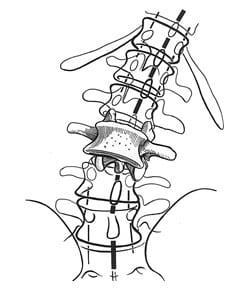
    In 1990, Dr. Wolfe and colleagues published the Criteria for the Classification of Fibromyalgia for the American College of Rheumatology (3). To complete this task, Dr. Wolfe and colleagues studied 558 consecutive patients: 293 patients with fibromyalgia and 265 control patients. Trained, blinded assessors performed interviews and examinations. The authors concluded that the clinical diagnosis of fibromyalgia required both of the following:

    • Widespread pain (axial plus upper and lower segment plus left and right-sided pain).
    • Excessive tenderness at 11 or more of 18 specific tender point sites.

    These fibromyalgia diagnostic criteria became the standard for the next 20 years, from 1990 to 2010. The location of the 18 specific tender point sites is included below: 

    18 specific tender point sites

    In 2010, Dr. Wolfe and colleagues updated the Criteria for the Classification of Fibromyalgia for the American College of Rheumatology (4). There had been a growing concern in the original diagnostic criteria for fibromyalgia that relied significantly on the presence of tender points. This is because of the fact that women in general are more sensitive to pressure point tenderness than are men. Relying on pressure point tenderness could cause a sex bias, which might account for the finding that about 90% of diagnosed fibromyalgia sufferers are women. It had become apparent that the “focus on tender points was not justified.” (5) Consequently, Dr. Wolfe and colleagues developed simple, practical criteria for clinical diagnosis of fibromyalgia that did not require a tender point examination, and to provide a severity scale for characteristic fibromyalgia symptoms (4). Specifically, they used an amalgamation of two prior developed assessment protocols:

    1) The Widespread Pain Index (WPI)

    This is a measure of the number of painful body regions (a total of 19 locations).

    2) The Symptom Severity (SS) Scale.

    This scale is designed to grade adjunct fibromyalgia symptoms, specifically cognitive symptoms (trouble remembering or thinking), un-refreshed sleep, fatigue, and number of somatic symptoms.

    A sample of the questionnaire is included below.

    The authors concluded that this simple form correctly classified 88.1% of fibromyalgia cases, and that classifying fibromyalgia  “does not require a physical or tender point examination.” This approach to the diagnosis of Fibromyalgia Syndrome continues to be supported (6). To diagnose fibromyalgia, patients should:

    • Have pain in a number of areas of the body, widespread pain (upper body, lower body, left side, right side).
    • Be substantially bothered by fatigue.
    • Have difficulty in getting restful sleep.
    • Often have problems with memory or thinking clearly.

    New Fibromyalgia Diagnostics 2010
    All Three Are Needed For the Diagnosis of Fibromyalgia Syndrome

    1)     Either WPI ≥ 7 and SS ≥ 5   OR    WPI 3 – 6 and SS ≥ 9

    2)     Symptoms are present and essentially unchanged for ≥ 3 months

    3)     There is no alternative explanation for the pain


    Widespread Pain Index

    Score: 1 For Each Spot

    Shoulder Girdle L
    Shoulder Girdle R
    Upper Arm L
    Upper Arm R
    Lower Arm L
    Lower Arm R
    Hip (buttock/trochanter) L
    Hip (buttock/trochanter) R
    Upper Leg R
    Upper Leg L
    Lower Leg L
    Lower Leg R
    Jaw L
    Jaw R
    Chest
    Abdomen
    Upper Back
    Lower Back
    Neck
    Total WPI (0-19)


    Symptom Severity (SS)

    0

    1

    2

    3

    Fatigue

    No Problem

    Mild

    Moderate

    Severe

    Waking up Un-refreshed

    No Problem

    Mild

    Moderate

    Severe

    Cognitive Symptoms(trouble remembering or thinking)

    No Problem

    Mild

    Moderate

    Severe

    Number of Somatic Symptoms

    None

    Few

    Moderate

    Many

    Total Symptom Severity (SS): (0-12)                                        

    Recent studies have assessed the influence of spinal mobilization and/or manipulation on the clinical status of patients with fibromyalgia.

    In 2014, Michel Reis and colleagues from the School of Medicine, Federal University of Rio de Janeiro, Brazil, published a study in the journal

    Rehabilitation Research and Practice titled (7):

    Effects of Posteroanterior Thoracic Mobilization on Heart Rate Variability and Pain in Women with Fibromyalgia

    These authors note that fibromyalgia is classically characterized by chronic pain, fatigue, depression, insomnia, and reduced cognitive performance. In addition, fibromyalgia is also associated with cardiac autonomic abnormalities.

    Heart rate variability is used to investigate cardiovascular autonomic abnormalities. It is a simple, sensitive, and noninvasive tool. Heart rate variability is reduced in fibromyalgia with increased sympathetic tone and activity. It is thought that there is a relationship between increased sustained sympathetic activity and tone and the symptoms of fibromyalgia. Studies suggest autonomic imbalance mechanistically contributes to the symptoms of fibromyalgia. The autonomic imbalance for fibromyalgia is characterized by sympathetic hyperactivity at rest.

    Sympathetic hyperactivity may also be responsible for frequent complaints of cold extremities in fibromyalgia patients. Studies have shown that fibromyalgia may be related to changes in autonomic tone, shifting toward an increase in sympathetic activity.

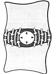
    The purpose of this study was to evaluate the effects of one session of a posteroanterior (P-A) glide technique on both autonomic modulation and pain in woman with fibromyalgia. The study used 20 women, half with diagnosed fibromyalgia. This is the first study to demonstrate the effect of a posteroanterior glide mobilization to the thoracic spine on autonomic modulation in patients with fibromyalgia. The mobilization technique used in this study was passive P-A push, sustained for 60 seconds at the T1-T2 spinal level, “corresponding to the thoracic sympathetic preganglionic neurons.”

    The upper thoracic mobilization was able to improve heart rate variability and improve autonomic profile through increased vagal activity. In women with fibromyalgia and impaired cardiac autonomic modulation, one session of spinal mobilization was able to acutely improve heart rate variability. In addition, the authors note that, in agreement with other studies, manual therapy protocols are effective in improving pain intensity in fibromyalgia patients. Their conclusions include:

    This study shows that “patients with fibromyalgia have increased sympathetic activity and decreased activity in the vagal control of heart rate.” “This sympathetic excitation could contribute to the diffuse pain and tenderness at specific points experienced by patients with fibromyalgia.”


    “The potentially significant impact of our findings is the demonstration that only one session of this manual intervention to the thoracic spine was able to modify heart rate variability in women with fibromyalgia.”

    “It is plausible to hypothesize that the posteroanterior glide technique utilized in the current study may significantly contribute to reducing the debilitating signs and symptoms of fibromyalgia, improve quality of life, and reduce cardiovascular risk when applied for more than one session.”

    There is an optimum balance of activity between the sympathetic and the parasympathetic nervous systems. When the balance is disrupted, it adversely influences the entire body, including pain thresholds. Dysfunctional spinal joints increase sympathetic tone, creating an imbalance with parasympathetic tone. Applied mechanical forces (manual therapy, mobilization/manipulation) to the dysfunctional spinal joints inhibits sympathetic tone, restoring autonomic balance, improving homeostasis and reducing pain thresholds.

    ••••

    David Vallez Garcia (from The Department of Nuclear Medicine and Molecular Imaging, University Medical Center Groningen, The Netherlands) and colleagues contributed a chapter in a 2014 book titled PET and SPECT in Neurology, pertaining to the physiology of chronic pain (8). In their review, the authors note:

    • Pain, anywhere in the body, is controlled by the periaqueductal gray (PAG) matter of the mesencephalon (the midbrain, at the top of the brainstem).
    • A major contributor to the periaqueductal gray matter is the vestibular nucleus (VN) of the medulla (the bottom of the brainstem).
    • The vestibular nucleus is also critically involved in posture control.
    • A major contributor to the vestibular nucleus is the proprioceptive neurology from the upper cervical spine, specifically from C1-C2-C3. This proprioception arises form neck muscle spindles, ligaments, and joint capsules.

    link of proprioception to pain control

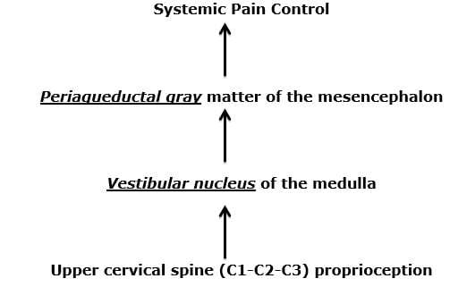
    Consequently, these authors propose this mechanism for widespread somatic pain syndromes: Mechanical dysfunction of the joints of the upper cervical spine alters the quality of the proprioceptive input from the upper cervical spine muscles, ligaments, and capsules to the vestibular nucleus. The vestibular nucleus becomes less efficient in activating the pain control neurons of the periaqueductal gray matter, resulting in increased chronic widespread body pain syndromes.

    These authors note:

    “More than 30% of all the spinal-periaqueductal gray fibers originate from the C1-C3 spinal segments.”

    Chronic pain symptoms are the “result of a mismatch between aberrant information from the cervical spinal cord and the information from the vestibular and visual systems, all of which are integrated in the mesencephalic periaqueductal gray and adjoining regions.”

    Chronic pain symptoms are due to a “mismatch in the midbrain and other structures via the upper cervical cord to the mesencephalon on the one hand and the intact information from the vestibular and visual systems to the mesencephalon on the other hand.”

    This chapter supports the perspective that chronic pain syndromes occur as a consequence of poor quality proprioceptive (mechanoreception) input from soft tissues into the central neural axis, and that management should be directed towards improving mechanical function of the dysfunctional soft tissues. This very much supports the chiropractic approach to management of chronic pain patients, especially chiropractic care that targets the function of the joints of the upper cervical spine.

    Are there any studies evaluating manipulation of the upper cervical spine in the treatment of patients suffering from fibromyalgia? See below:

    •••••

    In March 2015, Drs. Ibrahim M. Moustafa and Aliaa A. Diab, from the Basic Science Department, Cairo University, Giza, Egypt, published a study in the journal Rheumatology International, titled (9):

    The addition of upper cervical manipulative therapy in the treatment of patients with fibromyalgia: A randomized controlled trial

    The aim of this study was to investigate the immediate and long-term effects of a one-year multimodal program, with the addition of upper cervical manipulative therapy, on fibromyalgia management outcomes in addition to three-dimensional (3D) postural measures. It is a prospective, randomized, clinical trial.

    These authors note that fibromyalgia syndrome is a common and chronic disorder manifested by increased pain sensitivity and a number of other symptoms such as fatigue, stiffness, non-restorative sleep patterns, memory and cognitive difficulties, and reduced quality of life. Long-term treatment outcomes on fibromyalgia patients “are typically poor.” A survey of 1200 primary care physicians in the USA found that only 14% of respondents indicated very good or excellent satisfaction with the management of patients with fibromyalgia (10).

    Published theories as to the cause of fibromyalgia include:

    • Poor nutrition
    • Stress
    • Alterations in sleep patterns
    • Changes in neuroendocrine transmitters
    • Poor posture
    • Cervical spine dysfunction
    • Abnormal afferent processing and/or abnormal sensorimotor integration

    This is a randomized clinical trial with a one-year follow-up, assessing 120 patients with fibromyalgia syndrome and definite C1-2 joint dysfunction. Subjects were assessed at 12 weeks (at the end of the treatment program) and again at a 1-year follow-up period. The subjects were randomly assigned to the experimental [upper cervical manipulation] group (n = 60) or the control group (n = 60). Both groups completed a 12-week multimodal program consisting of an education program, cognitive behavior therapy, and an exercise program.

    The education program consisted of one 2-hour session per week for 12 weeks, and included:

    • Information about typical symptoms
    • The usual course for fibromyalgia
    • Potential causes of fibromyalgia
    • The influence of psychosocial factors on pain
    • Current pharmacologic and non-pharmacological treatments for fibromyalgia
    • The benefits of regular exercise on fibromyalgia

    The cognitive behavior therapy consisted of one 2-hour session per week for 12 weeks, and included educational, physical, cognitive, and behavioral elements.

    The exercise program was conducted for 1 hour three times per week for 12 weeks. The participants were instructed to perform the relaxation exercises at home twice daily as their home routine.

    The upper cervical spinal manipulation group consisted of both:

    • Low-velocity cervical joint mobilization techniques
    • High-velocity manipulation techniques for the treatment of cervical joint disorders
    • Upper cervical manipulative therapy was conducted in 12 treatments (three times per week) over a one-month period in addition to maintenance spinal manipulations in one session per week for the following 8 weeks. [3X/week for 4 weeks, then 1X/week for 8 weeks]

    The group receiving upper cervical spinal manipulation showed significant improvements in spinal posture. After 12 weeks of treatment, the two treatment arms were roughly equally successful in improving the fibromyalgia management outcomes. However, at the 1-year follow-up, the upper cervical spinal manipulation group showed greater improvements in all the fibromyalgia measurement outcomes. These authors made the following points:

            “Increasing evidence suggests that spinal dysfunction, particularly in the upper cervical region (which has more mechanoreceptors per unit surface area than any other region of the spinal column), might affect central neural processing and potentially lead to maladaptive central plastic changes.”

    “Cervical manipulation might help to modulate disordered sensorimotor integration and thus counteract changes in the processing of sensory information in the brain and spinal cord.”

    “Upper cervical manipulation might be required to achieve optimal full-spine postural correction because the rest of the spine orients itself in a top-down fashion.”

    “At the one-year follow-up after the end of the treatment, there were statistically significant changes that indicated that the fibromyalgia syndrome management outcomes of the experimental [upper cervical manipulation] group exhibited continued improvement and that the control subjects’ scores regressed back toward the baseline values (i.e., the scores worsened).”

    “The one-year improvements in the FMS management outcome measures observed in the experimental [upper cervical manipulation] group are the most significant findings of our investigation.”

    “Sustained postural imbalances can result in the establishment of a state of continuous asymmetric loading. Once this state is established and maintained beyond critical weight and time threshold, degenerative changes in the muscles, ligaments, bony structures, and neural elements increase.” When postural asymmetry is reversed, and the unbalanced loading is thereby corrected the “reversible of these degenerative changes or even their improvement requires some time.”

    “The continuous asymmetrical loading and muscle imbalance that results from biomechanical dysfunction due to abnormal spinal posture in the sagittal, transverse, and coronal planes elicits abnormal stress and strain in many structures, including the bones, intervertebral disks, facet joints, musculotendinous tissues, and neural elements and causes a barrage of nociceptive afferent input that results in dysafferentation.”

    “Importantly, our results from the one-year follow-up revealed statistically significant changes that favored the experimental [upper cervical manipulation] group’s outcomes in terms of all of the fibromyalgis management outcome variables.”

    “The addition of the upper cervical manipulative therapy to a multimodal program is beneficial in treating patients with fibromyalgia syndrome.”

    This is an important study, and the data suggests this model:

    Fibromyalgia is a multifaceted problem that tends to be linked to poor posture. This is because the postural system controls a large quantity of the afferent neurology into the central neural axis.

    The upper cervical spine has more afferent neurons that enter the central neural axis than any other spinal region. In addition, upper cervical afferents communicate mono-synaptically in the vestibular nucleus.

    The vestibular nucleus controls whole body posture.

    Upper cervical spinal manipulation makes a significant and lasting improvement on whole body posture by influencing the vestibular nucleus.

    This study also indicates that the benefits of chiropractic postural corrections take time to manifest, but once these benefits are obtained, they tend to be long lasting.

    These authors imply that new guidelines for the treatment of fibromyalgia syndrome should be established and they should include upper cervical manipulation.

    SUMMARY

    Six to 25 million Americans suffer from fibromyalgia syndrome. Their symptoms are chronic, treatment resistant, and often debilitating. Only 14% of traditionally managed fibromyalgia patients achieve an acceptable clinical outcome.

    Components of the proposed pathophysiology of fibromyalgia suggest that chiropractic mechanical care, especially of the upper cervical spine, makes biologically plausible sense. The evidence suggests that chiropractic care, along with other traditional approaches, result in significant clinical improvement for these patients, and that the improvement is long lasting. All chronic fibromyalgia patients should engage in chiropractic segmental and postural spinal corrections.

    REFERENCES

    • Clauw DJ; Fibromyalgia: a clinical review; Journal of the American Medical Association; April 16, 2014; 311(15):1547-55.
    • Clauw DJ; Fibromyalgia and Related Conditions; Mayo Clin Proc; May 2015;90(5):680-692.
    • Wolfe F, Smythe HA, Yunus MB, Bennett RM, Bombardier C, Goldenberg DL, Tugwell P, Campbell SM, Abeles M, Clark P et al.; The American College of Rheumatology 1990 Criteria for the Classification of Fibromyalgia. Report of the Multicenter Criteria Committee; Arthritis Rheum. 1990 Feb;33(2):160-72.
    • Wolfe F, Clauw DJ, Fitzcharles MA, Goldenberg DL, Katz RS, Mease P, Russell AS, Russell IJ, Winfield JB, Yunus MB; The American College of Rheumatology preliminary diagnostic criteria for fibromyalgia and measurement of symptom severity; Arthritis Care Res (Hoboken); 2010 May;62(5):600-10.
    • Smith HS, Harris R, Clauw DJ; Fibromyalgia: An Afferent Processing Disorder Leading to a Complex Pain Generalized Syndrome; Pain Physician; 2011; 14:E217-E245.
    • Häuser W, Wolfe F; Diagnosis and diagnostic tests for fibromyalgia (syndrome); Reumatismo; Sep 28, 2012;64(4):194-205.
    • Reis MS, Durigan JL, Arena R, Rossi BR, Mendes RG, Borghi-Silva A; Effects of Posteroanterior Thoracic Mobilization on Heart Rate Variability and Pain in Women with Fibromyalgia; Rehabilitation Research and Practice; May 29, 2014 [epub].
    • Garcia DV, Dierckx R, Otte a, Holstege G; PET and SPECT in Neurology, Chapter 46 “Whiplash: Real or Not Real: A Review and New Concept”; Springer-Verlag; 2014, pp. 947-963.
    • Moustafa IM, Diab AA; The addition of upper cervical manipulative therapy in the treatment of patients with fibromyalgia: A randomized controlled trial; Rheumatology International; March 18, 2015 [epub].
    • Hartz AJ, Noyes R, Bentler SE et al; (2000) Unexplained symptoms in primary care: perspectives of doctors and patients. Gen Hosp Psychiatry 22:144–152.
  • Lumbar Spine Degenerative Disc Disease and Chiropractic Spinal Adjusting Pathophysiology, Safety, Effectiveness

    Lumbar Spine Degenerative Disc Disease and Chiropractic Spinal Adjusting Pathophysiology, Safety, Effectiveness

    Introduction

    A pioneering study was published in The New England Journal of Medicine in 1994 (1). Researchers from Hoag Memorial Hospital in Newport Beach, California, performed magnetic resonance imaging (MRI) examinations on 98 people who did not have back pain. Sixty-four percent of the 98 asymptomatic subjects were found to have abnormal lumber spine discs. The pathologies included bulging, protrusions, an extrusion and other annular defects (disruption of the outer fibrous ring of the disk); all secondary to disc degenerative disease. The age of the afflicted subjects was from 20 to 80 years (mean age, 42.3 years), and the pathologies increased with age. The authors concluded:

    “These findings support the contentions that annular defects are generally associated with disk degeneration and that such defects are frequently asymptomatic.”

    This study indicates that the majority of chiropractic patients, including patients who do not have back pain, have abnormalities of the intervertebral disc. Chiropractors are routinely adjusting the low backs of patients who have intervertebral disc pathology.

    Anatomy

    The spinal column is made up of 24 segments (vertebrae). The low back (lumbar spine) is made up of 5 segments (L1, L2, L3, L4, L5). Total spinal column motion is dependent upon the additive motion between the individual segments. Individual segmental motion is largely dependent upon the integrity of the intervertebral disc. Picture from Kapandji (2):

    Total spinal column motion is dependent upon the additive motion between the individual segments

    The unique anatomy of the intervertebral disc allows for both cushioning (like a shock absorber) and motion. The disc has two distinct regions:

    •       The nucleus pulposus:

    The nucleus is mostly water and functions as a ball bearing.

    •       The annulus fibrosis:

    The annulus is composed of tough protein fibers and cells.

    Above and below each intervertebral disc is the vertebral body. The vertebral body is quite vascular (it has a well developed blood supply). Between the disc and the vertebral body is a porous cartilaginous end plate.

    The intervertebral disc has no inherent blood supply. Nutrients for the cells of the intervertebral disc must travel from the vertebral body through the porous cartilaginous end plate (see Physiology below) (from reference #2).

    The intervertebral disc has no inherent blood supply. Nutrients for the cells of the intervertebral disc must travel from the vertebral body through the porous cartilaginous end plate (see Physiology below) (from reference #2).

    Physiology

    The adult human has approximately 75 trillion cells (3). The health and function of each cell depends upon a steady supply of nutrients (glucose, oxygen, vitamins, minerals, amino acids, etc.). For most of the cells of the human body, the delivery of these nutrients is via the blood supply. The blood is pumped to the cells by the heart, beginning with the larger arteries and eventually to the minute capillaries. The capillaries are adjacent to most of the cells of the body.

    Unfortunately, the intervertebral disc is void of both arteries and capillaries. This makes the cells of the disc vulnerable to dysfunction, disease, and death. This lack of inherent vascular nutrition is the reason the intervertebral disc has increased risk for disease and degeneration (disc degenerative disease); it is also why disc injuries are notoriously slow to recover and heal.

    So how do the cells of the intervertebral disc obtain the nutrients required to maintain health and function?

    The answer is through motion. Anything that impairs the movement of the vertebral motor unit (two adjacent vertebrae with an intervertebral disc in between), impairs the supply of nutrients to the avascular disc.

    As Vert Mooney, MD, noted in his Presidential Address to the International Society for the Study of the Lumbar Spine (4):

    “Mechanical activity has a great deal to do with the exchange of water and oxygen concentration” in the disc. 

    An important aspect of disc nutrition and health is the mechanical aspects of the disc related to the fluid mechanics. The pumping action maintains the nutrition and biomechanical function of the intervertebral disc.

    Thus, “research substantiates the view that unchanging posture, as a result of constant pressure such as standing, sitting or lying, leads to an interruption of pressure-dependent transfer of liquid. Actually the human intervertebral disc lives because of movement.”

    Pathophysiology

    Aging is a multifactorial phenomenon involving many variables, including time, genetics, injuries, chronic stress, nutrition, habits, etc. The aging of joints is classically called arthritis or osteoarthritis or degenerative joint disease. Degenerative joint disease of the joints of our spinal column is known as spondylosis or spondyloarthritis. The aging of the intervertebral disc is classically called degenerative disc disease.

    In 2006, Susan Garstag, MD, and Todd Stitik, MD, wrote an informative article on joint degeneration (5). They attribute joint degeneration (osteoarthritis) to a number of factors, categorizing their list to local factors and systemic factors:

    Local Factors include:

    • Altered joint biomechanics
    • Malalignment
    • Muscle weakness
    • Impaired proprioception neurology

    Systemic Factors include:

    • Age
    • Genetic Factors
    • Nutritional Factors, especially inflammatory profile and free radical burden (generators of oxidative stress)

    Drs. Garstag’s and Stitik’s comments linking osteoarthritis to malalignment, muscle weakness, impaired proprioception, and altered joint biomechanics is particularly interesting to chiropractors, as these are all components of what chiropractors call the subluxation. Traditional chiropractic teaches that untreated joint subluxations lead to osteoarthritic changes.

    Recent concepts pertaining to aging in general indicate that the aging of all of our cells is the life-long process of glycation (also known as glycosylation). This concept is nicely described by Nora Gedgaudas in 2011 (6):

    “Aging is now being understood by people researching longevity as essentially a gradual process of glycation of all tissues, including the brain.” p. 126

    Technically, glycation is the abnormal attachment of sugars to proteins. When a sugar attaches to a protein, the function of the protein is altered. As noted above, the proteins of the intervertebral disc are suppose to allow for the necessary motion to pump the nutritious fluids from the vertebral bodies through the cartilaginous end plates into the avascular disc. When the disc proteins are glycated with sugars, the disc becomes stiff, reducing movement, and impairing the pumping of fluid into the disc. The consequence is that the disc becomes dry (desiccated), sick, and degenerated (degenerative disc disease). This process of intervertebral disc glycation was nicely documented in an article published earlier this year by Michael Adams and colleagues from the University of Bristol, UK (7).

    This process of glycation is directly linked to one’s consumption of sugars, especially fructose (6). Sadly, sugar consumption is addicting, and food scientists both know this and exploit it (8, 9). As a result, sugar has become the most successful food additive in human history. When sugar is added to food or drink, net sales (and profits) increase. If sugar is removed, sales decline. Consequently, essentially everything possible now contains added sugar (10). Sadly, this trend increases both glycation and cellular aging, including that of the intervertebral disc (6, 7).

    When the proteins of the intervertebral disc become glycated, the disc itself becomes stiffer. This increased stiffness reduces motion, impairing the pumping of nutritious fluid to the cells of the disc, contributing to disc degenerative disease (8).

    Another problem with glycation, is that glycated proteins increase the production of free radicals by a factor of about 50X. This is relevant in light of the osteoarthritis factors described by Drs. Garstag and Stitik above. Recall, a driving factor of osteoarthritis is one’s free radical burden and production of oxidative stress. Glycated intervertebral disc proteins and their substantially increased production of free radicals accelerate disc degenerative disease (7).

    Thus, glycated disc proteins drive disc degeneration through two mechanisms, both of which were described by Drs. Garstag and Stitik above (5):

    • Reducing disc motion, impairing disc nutrition.
    • Increase production of free radicals, accelerating oxidative stress.

    As the cells of one’s disc (or any other tissue) degenerate, the aging can be quantified. This is done by measuring the length of the telomeres on the end of the chromosomes of the disc cells. This process was awarded the 2009 Nobel Prize in Medicine/Physiology, and is nicely described by Michael Fossel, MD, PhD, and colleagues, in 2011 (11).

    Essentially, the ends of our chromosomes have a repeating sequence of nucleotide bases that are collectively referred to as the telomere. Each time the cell divides, the telomere shortens a little. When most of the telomere disappears, the cell dies. Consequently, telomere length has been proposed as a marker of biological aging. As noted, this concept was awarded the 2009 Nobel Prize in Medicine/Physiology to Elizabeth Blackburn, PhD, from the University of California, San Francisco, and colleagues.

    This concepts of telomere shortening is true when applied to disc degenerative disease (7). As noted by Dr. Fossell and colleagues, the main drivers of cellular aging are oxidation, inflammation, and glycation (11).

    The point of this discussion, and as noted with MRIs in 1994, the majority of people, and consequently the majority of chiropractic patients, have disc degenerative disease of varying degrees.

    Safety

            Is it safe to manipulate a low back spinal segment if that segment has disc degenerative disease?

    For six decades, studies have shown that lumbar spinal manipulation is both safe and effective in the treatment of patients with low back pain, with or without leg pain, that is secondary to pathology of the intervertebral disc (12, 13, 14, 15, 16, 17, 18). Importantly, there is primary research detailing the biomechanical reason for the safety of spinal manipulation for both normal and degenerated lumbar spine intervertebral discs, noted below. It is important to understand that “torsion” is a common component of lumbar spinal manipulation.

            In 1981, MA Adams, PhD, and WC Hutton, MSc, published an important study in the journal Spine, titled (19):

    The Relevance of Torsion to the Mechanical Derangement of the Lumbar Spine

            In their cadaver experiments, these authors noted that the limit of spinal segmental rotation was not created by the disc, but rather by the facet joint. During rotational stress, the compression facet is the first structure to yield at the limit of torsion, and this occurs after about 1-2° of rotation. “Much greater angles are required to damage the intervertebral disc, so torsion seems unimportant in the etiology of disc degeneration and prolapse.” Important statements in this article include:

    “Because of the protection offered by the compression facet, the intervertebral disc is subjected to relatively small stresses and strains in the physiologic range of torsion. By the time the facets are damaged, the disc is rotated only about one-third to one-tenth of its maximum angle and is bearing a small fraction of the torque required to rupture it.”

    “Except in cases of extreme trauma and as a sequel to crushing of the apophyseal joints, axial rotation can play no major part in the mechanical derangement of the intervertebral disc in life.”

    Two years later, in 1983, Adams and Hutton publish an additional cadaver studies in journal Spine, titled (20):

    The Mechanical Function of the Lumbar Apophyseal Joints

    Based upon their experiments, they conclude that the facet joints “prevent excessive movement from damaging the discs: the posterior annulus is protected in torsion by the facet surfaces and in flexion by the capsular ligaments.” They note that the facets only allow at most 2° of rotation, and also note that the disc will completely recover from all rotational stresses that are less then 3°. These authors state:

    “In flexion, as in torsion, the apophyseal joints protect the intervertebral disc.”

    “The function of the lumbar apophyseal joints is to allow limited movement between vertebrae and to protect the discs from shear forces, excessive flexion, and axial rotation.”

    In another follow-up article in 1995, Adams and Dolan published a study in the journal Clinical Biomechanics titled (21):

    Recent Advances in Lumbar Spinal Mechanics and their Clinical Significance

    Once again, these authors note that rotational loading of a spinal motor unit will always damage the facet joints “long before the disc.” If the facet joints are removed, rotational forces will damage the disc if subjected to rotational loads between 10-20 degrees.

    These authors also note “severely degenerated discs cannot be made to prolapse, presumably because the nucleus is too fibrous to exert a hydrostatic pressure on the annulus.”

    The information presented by Adams, Hutton, and Dolan, indicates that for most patients suffering from a lumbar disc disease, including intervertebral disc prolapse, manipulation of the lumbar spine is both safe and often effective. These authors also specifically applied their experiments to discs that were already degenerated. Even in the presence of disc degenerative disease, these authors were able to show that rotational and torsional spinal motions did not injure these discs. They found that even in the presence of disc degeneration that the facet joints protected the disc from injury.

    Outcomes

    Is the manipulation of a spinal segment with disc degenerative disease helpful to the patient with low back pain?

    A preliminary review of the literature to address this question was published in 2005 in the Journal of Manipulative and Physiological Therapeutics, and titled (22):

    High-velocity low-amplitude spinal manipulation for symptomatic lumbar disk disease: A systematic review of the literature

    The authors of this study performed a systematic review of the literature, including the Cochrane Central Register of Controlled Trials, Medline, Cumulative Index to Nursing and Allied Health Literature, and Mantis. Sixteen studies met the inclusion criteria, representing 203 total subjects. The authors concluded:

    “The reviewed evidence supports the hypothesis that high velocity low amplitude spinal manipulation may be effective in the treatment of symptomatic lumbar disc disease and does not support the hypothesis that high velocity low amplitude spinal manipulation is inherently unsafe in symptomatic lumbar disc disease cases. It appears that patients with lumbar disk pathology do undergo manipulative treatment in practice. Consequently, this should be an area of research importance.”

    A more recent study looking at the benefits of spinal manipulation for subjects suffering from degenerative disc disease was recently published (September 2014). The article was published in the journal Archives of Physical Medicine and Rehabilitation, and titled (23):

    Short-Term Effect of Spinal Manipulation on Pain Perception,

     Spinal Mobility, and Full Height Recovery in Male Subjects With Degenerative Disk Disease: A Randomized Controlled Trial

    The authors point out that lumbar intervertebral disk disease is one of the main causes for low back pain. Disk degeneration is the most common pathology in the adult spine and accounting for approximately 90% of surgery cases. Subjects with symptomatic disk disorders incur the highest health care expenditure among those with other low back pain diagnoses. In agreement with above studies, degenerative disc disease is an extremely common pathology.

    Disc degeneration results in structural disruption and loss of the hydrostatic capacity of the nucleus.  Thus, disc degeneration eventually ends up leading to a loss of disc height with a reduction of spinal range of motion.

    The purpose of the study was to evaluate, in subjects with lumbar disc degeneration, the immediate effect of a lumbosacral high-velocity low-amplitude spinal manipulation on four established variables that assess spinal function:

    A) The subjects’ height via stadiometer:

    The cumulative effect of the intervertebral disc loss of fluid in response to mechanical stress may reduce the subject’s measured height, spinal shrinkage. The stadiometer measures height variations and the amount of intervertebral disc compression caused by pressure on the spinal column. The stadiometer is a noninvasive method that has proven validity and is easier to use and less costly than MRI.

    B) Self perceived LBP (using a visual analog scale [VAS]):

    The VAS is an effective, sensitive, and appropriate tool to measure acute and chronic pain.

    C) Neural mechanosensitivity:

    Neural mechanosensitivity was assessed by means of the passive straight-leg raise (SLR) test. The point of initial appearance of pain or discomfort was the test endpoint, and was measured with a goniometer. The test is considered an easy-to use tool, with a reliability of 87%.

    D) Spinal mobility in flexion:

    Spine mobility in flexion was measured as finger-to-floor distance (FFD). This test is considered easy to conduct and has a high degree of inter-examiner reliability (96-98%).

    The study was a randomized, double-blind, controlled clinical trial that evaluated 40 men with an average age of about 40 years. All subjects had degenerative lumbar disc disease at L5-S1. Twenty of the men were randomally assigned to receive a side-posture L5-S1 manipulation. The spinal manipulation was a standard side-posture very fast thrust with a short range of motion. The other 20 men were given a single placebo intervention. The placebo maneuver (sham) was also side-posture set up but involved no manipulative thrust.

    Measured Outcome Improvements in Men with Degenerative Disc Disease Following a Single Side-Posture Spinal Adjustment

    (rounded)

    Measurement

    Outcome Improvement

    Height (stadiometer)

    4 ± 1 mm

    Pain (VAS)

    Reduced by 46%

    Neural Tension (SLR)

    Increased by 14° ± 9°

    Spinal Mobility (FFD)

    Increased by 4 ± 2 cm

    Results

    A)     Height Change Stadiometry:

    • “A significant height change was found in the TG:” (3.98 ± 1.46 mm).
    • The increase in paravertebral muscle activity in chronic low back pain is associated with greater compressive loads. The impact of spinal manipulation on diminishing paravertebral hyperactivity may produce changes in stadiometry (improved height).

    B)     Visual Analog Scale (VAS):

    “The lumbosacral spinal manipulation achieved an immediate reduction in self perceived low back pain.” Pain perception decreased by 46% in in the manipulation group in relation to baseline.

    C)     Neural Tension Straight Leg Raise (SLR):

    The passive straight-leg raising test increased with spinal manipulation by 14° ± 9° (rounded).

    D)    Spinal Mobility Finger-to-Floor Distance (FFD):

    Spinal mobility during the finger-to-floor distance test was increased in the manipulation group by 4 ± 2 cm (rounded). It appeared that spinal manipulation modulates the somatosensory system, which inhibits the paravertebral muscle hyperactivity and improves spine function.

    The authors of this study concluded:

    “Ahigh-velocity, low-amplitude spinal manipulation in the lumbosacral joint performed on men with degenerative disk disease immediately improves self-perceived pain, spinal mobility in flexion, hip flexion during the passive SLR test, and subjects’ full height.”

    “A side-lying spinal manipulation technique in the lumbosacral region decreases self-perceived low back pain in the short-term and produces an immediate improvement in spinal mobility in flexion, the subject’s height, and hip flexion mobility during the passive straight-leg raising test in men with disc degeneration.”

    The intragroup comparison indicated a significant improvement in all variables in the spinal manipulation group compared to the control group. “In the between-group comparison of the mean differences from pre- to post intervention, there was statistical significance for all cases.”

    “Only the mechanical effect from the thrust appears to be a key element to the effectiveness of the spinal manipulation.”

    “Spinal Manipulation has been demonstrated to decrease pain and improve function in symptomatic lumbar disc degeneration.”


    Summary

    Spinal mechanical neurophysiological function is dependent upon the integrity of the intervertebral disc. The adult intervertebral disc is avascular; adult disc health depends upon motion that pumps nutrients and fluid from the adjacent vertebral bodies through the cartilaginous end plates into the disc. Any impairment in spinal segmental motion negatively impacts disc health.

    The universal mechanism of cellular aging is glycation, the process of attaching sugars to proteins. The rate of glycation is linked to sugar consumption, inflammation, and oxidative stress. When the intervertebral disc is glycated it becomes stiff, reducing motion, impairing the nutritious fluid pump, accelerating degenerative disc disease. Consequently, disc degeneration is a ubiquitous adult finding. Disc degeneration is found even in asymptomatic subjects. Disc degeneration makes such individuals more susceptible to back injury and pain, and impairs recovery rates. More that half of adults have some form of low back disc degeneration.

    Spinal adjusting (manipulation) at segmental levels of disc degeneration is safe. Not only it is safe, it is beneficial. The improved neuro-biomechanics that occurs as a consequence of spinal adjusting (manipulation), increases height, improves range of motion, relieves stress in the nervous system, and significantly improves pain syndrome. 

    REFERENCES 

    • Jensen MC, Brant-Zawadzki MN, Obuchowski N, Modic MT, Malkasian D, Ross JS; Magnetic resonance imaging of the lumbar spine in people without back pain; New England Journal of Medicine; 1994 Jul 14;331(2):69-73.
    • Kapamdji IA; The Physiology of the Joints; Volume 3; The Trunk and the Vertebral Column; Churchill Livingstone; 1974.
    • Guyton A; Textbook of Medical Physiology; Saunders; 1981; p. 2.
    • Mooney V; Where Is the Pain Coming From?; Spine; Vol. 12; No. 8; 1987’ pp. 754-759.
    • Garstang SV, Stitik TP; Osteoarthritis: Epidemiology, Risk Factors, and Pathophysiology; American Journal of Physical Medicine and Rehabilitation November 2006, Vol. 85, No. 11, pp. S2-S11.
    • Gedgaudas N; Primal Body, Primal Mind: Beyond the Paleo Diet for Total Health and a Longer Life; Healing Arts Press; 2011.
    • Adams MA, Lama P, Zehra U, Dolan P;Why Do Some Intervertebral Discs Degenerate, When Others (in the Same Spine) Do Not?; Clinical Anatomy; April 19, 2014 [epub].
    • Kessler DA; The End of Overeating: Taking Control of the Insatiable American Appetite; Rodale; 2009.
    • Moss M; Salt, Sugar, Fat: How the Food Giants Hooked Us; Random House; 2013.
    • Lustig R; Fat Chance: Beating the Odds Against Sugar, Processed Food, Obesity, and Disease; Hudson Street Press; 2013.
    • Fossel M, Blackburn G, Woynarowski; The Immortality Edge: Realize the Secrets of Your Telomeres for a Longer, Healthier Life; Wiley; 2011.
    • Ramsey RH; Conservative Treatment of Intervertebral Disk Lesions; American Academy of Orthopedic Surgeons, Instructional Course Lectures; Volume 11, 1954, pp.118-120.
    • Mathews JA and Yates DAH; Reduction of Lumbar Disc Prolapse by Manipulation; British Medical Journal September 20, 1969, No. 3, 696-697.
    • Edwards BC; Low back pain and pain resulting from lumbar spine conditions: a comparison of treatment results; Australian Journal of Physiotherapy; 15:104, 1969.
    • White AA, Panjabi MM; Clinical Biomechanics of the Spine; Second edition, JB Lippincott Company, 1990.
    • Turek S; Orthopaedics, Principles and Their Applications; JB Lippincott Company; 1977; page 1335.
    • Kuo PP and Loh ZC; Treatment of Lumbar Intervertebral Disc Protrusions by Manipulation; Clinical Orthopedics and Related Research. No. 215, February 1987, pp. 47-55.
    • Cassidy JD, Thiel HW, Kirkaldy-Willis WH; Side posture manipulation for lumbar intervertebral disk herniation; Journal of Manipulative and Physiological Therapeutics; February 1993;16(2):96-103.
    • Adams MA and Hutton WC, MSc; The Relevance of Torsion to the Mechanical Derangement of the Lumbar Spine; Spine; Volume 6, Number 3, May/June 1981, pp. 241-248.
    • Adams MA and Hutton WC, MSc; The Mechanical Function of the Lumbar Apophyseal Joints; Spine; Volume 8, Number 3, April 1983, pp. 327-330.
    • Adams MA and Dolan P; Recent advances in lumbar spinal mechanics and their clinical significance; Clinical Biomechanics; Volume 10, Number 1, 1995, pp. 3-19.
    • Lisi AJ, Holmes EJ, Ammendolia C; High-velocity low-amplitude spinal manipulation for symptomatic lumbar disk disease: a systematic review of the literature; J Manipulative Physiol Ther. 2005 Jul-Aug;28(6):429-42.
    • Vieira-Pellenz F, Oliva-Pascual-Vaca A, Rodriguez-Blanco C, Heredia-Rizo AM, Ricard F, Almazán-Campos G; Short-term effect of spinal manipulation on pain perception, spinal mobility, and full height recovery in male subjects with degenerative disk disease: a randomized controlled trial; Arch Phys Med Rehabil; 2014 Sep;95(9):1613-9.
  • Fibromyalgia Syndrome

    Fibromyalgia Syndrome

    Fibromyalgia Syndrome is the third most commonly diagnosed rheumatologic disorder, following osteoarthritis and rheumatoid arthritis. Fibromyalgia is characterized by widespread pain and tenderness, fatigue, morning stiffness, and sleep disturbance. Fibromyalgia is often disabling.  Fibromyalgia is often treatment resistant. Fibromyalgia can be triggered by trauma (Greenfield, Waylonis, Buskila, Neumann). Studies report that between 25% and 50% of subjects with fibromyalgia recall physical trauma immediately prior to the onset of their symptoms (Al-Allaf).

    Fibromyalgia Syndrome is diagnosed by identifying widespread pain and tenderness upon palpation at multiple anatomically defined soft tissue body sites. Fibromyalgia Syndrome overwhelmingly affects women more than men. “FM is nine times more common in middle-aged women (between the ages of 30 and 50 years) than in men (Abraham).”

    THE GATE THEORY OF PAIN

              In 1965, Ronald Melzack and Patrick Wall introduced the GATE THEORY OF PAIN. In 1985, orthopedic surgeon Kirkaldy-Willis published a pilot study of chiropractic spinal adjusting in the management of 283 patients suffering from chronic, treatment-resistant, low back pain. The results were excellent, essentially fixing 81% of the referred pain syndrome patients. Dr. Kirkaldy-Willis’ explanation for the positive clinical outcomes was based upon the fact that the improved motion would close the PAIN GATE.

    Chiropractic management of patients with fibromyalgia syndrome traditionally involves the practical application of Melzack’s and Wall’s GATE THEORY OF PAIN. Chiropractic management of these patients involves improvement of the quality of mechanical neurological afferentation into the central nervous system in an effort to “close” the Pain Gate. Improving the sagittal and horizontal planes of spinal posture and motion through chiropractic is a proven method to “close” the pain gate, and has been so recognized at least since 1985 (Kirkalady-Willis). However, these mechanical efforts on those suffering from fibromyalgia syndrome may themselves worsen patient symptoms, at least temporarily, discouraging both patient and practitioner.

    Consequently, chiropractors often utilize a number of adjunct approaches in the management of patients with fibromyalgia syndrome. Several of the adjunct approaches are reviewed below.

    MALIC ACID and MAGNESIUM

    In 1992, Abraham and Flechas propose that fibromyalgia is caused by a deficiency of substances needed for ATP synthesis. The nociceptive nervous system requires a steady flow of ATP to remain at sub-action potential threshold. The nociceptive action potential is achieved primarily by an influx of positively charged Na+ ions. Keeping the potential across the membrane far away from excitation threshold is the job of the membrane-bound sodium pump, a mechanism that is ATP energy dependent. Reductions in ATP supplies could allow more Na+ ions to cross the membrane, bringing the nociceptive neuron much closer to excitation threshold. Trivial environmental stresses could now be sufficient to achieve nociceptive excitation and action potential, accounting for the widespread pain fibromyalgia patients experience.

    anaerobic glycolysis

    During anaerobic glycolysis, from glucose to acetyl-Co A, there are 11 distinct steps; 9 of 11 steps (82%) require magnesium.

    Central to ATP generation is the Krebs Cycle (Citric Acid Cycle). The Krebs Cycle has nine steps, and 3 of them require magnesium. The rate limiting Krebs Cycle step in the genesis of ATP is malic acid (malate). In humans, when there is increased demand for ATP, there is also an increased demand and utilization of malate and magnesium. Malate deficiency is the cause of the ATP deficiency seen with exhaustive physical activity.

     In humans, when there is increased demand for ATP, there is also an increased demand and utilization of malate and magnesium. Malate deficiency is the cause of the ATP deficiency seen with exhaustive physical activity.

    Without adequate levels of malate and magnesium, there is increased anaerobic glycolysis, resulting in increased pyruvate production and increased lactic acid, both of which increase pain perception. Magnesium and malate have a critical role in ATP production, and therefore fibromyalgia symptoms may be caused by magnesium and malate deficiency.

    Abraham and Flechas explain the synergistic role of magnesium and malic acid in the genesis of ATP. They detail the biochemistry of how reductions in magnesium and malic acid would result in ATP deficiency.

    Abraham and Flechas then treat 15 fibromyalgia patients with daily 300-600 mg of magnesium plus 1200-2400 mg malic acid. “All patients reported significant subjective improvement of pain within 48 hours of starting” supplementation.

    In 1995, Russell and colleagues, in a randomized, double blind, placebo controlled, crossover study, also used magnesium and malic acid to treat 29 patients with fibromyalgia, noting “significant reductions in the severity of all 3 primary pain/tenderness measures were observed.”  Better results were observed in those taking 600 mg of magnesium and 2400 mg of malic acid, as compared to those who took lower doses. The authors note that this supplementation should continue for al least 2 months, and often for as long as 6 months.

    THE SEROTONIN PATHWAY

    In 2004, Borut Banic and colleagues, writing in the journal Neurology, presented extensive evidence suggesting that fibromyalgia is the consequence of reduced levels of the brain neurotransmitter serotonin.

    In 1998, osteopath John H Juhl also proposed that fibromyalgia could be related to reduced serotonin. He notes that researchers have found low serum levels of serotonin in fibromyalgia patients. Low serum serotonin levels have been found to have an inverse correlation with clinical measures of pain.

    The serotonin pathway begins with the essential amino acid tryptophan. Tryptophan is the least common of the 8 essential amino acids, accounting for about 1% of protein content.

    After absorption, about 90% of tryptophan is used at the peripheral tissues for protein synthesis.

    About 9% of absorbed tryptophan is used to produce niacin. The RDA for niacin is 15 mg. It takes 60 mg of tryptophan to produce 1 mg of niacin. This is important, because if niacin levels are adequate in the diet, the body will not need to use this 9% to make niacin. In fact, the higher the dietary levels of niacin, the less tryptophan is converted to this pathway. This increases the tryptophan available to be converted to serotonin.

    About 1% of absorbed tryptophan is converted to serotonin.

    In the body, tryptophan is converted to 5-hydroxy-tryptophan

    (5-HTP). 5-HTP easily crosses the blood-brain barrier for conversion to serotonin in the central nervous system. The conversion of 5-HTP to serotonin requires vitamin B6. Consequently, inadequate levels of B6 impair the conversion of tryptophan to serotonin.

    Currently, tryptophan is available by prescription only in the United States. However, 5-HTP is sold, and as noted above, still crosses the blood brain barrier for conversion to serotonin, as does tryptophan. Commercially, 5-HTP is extracted from the seeds of Grifonia simplicifolia, a plant grown in West Africa.

    Dr. Juhl notes 2 published studies where supplementation of 5-HTP in the dose of 100 mg 3 times per day in patients with fibromyalgia resulted in significant improvement of clinical symptoms after 30-90 days. The effective daily dose range appears to be 200-1000 mg per day, and that it should be taken with meals. These patients should also be given vitamin B6 to increase conversion of 5-HTP to serotonin, and niacinamide to inhibit the need for tryptophan to convert to niacin.

    These patients should also be given vitamin B6 to increase conversion of 5-HTP to serotonin, and niacinamide to inhibit the need for tryptophan to convert to niacin.

    Dr. Rodger Murphree supports the tryptophan to 5HTP to serotonin pathway noted by Dr. Juhl. In his 2003 book titled Treating and beating Fibromyalgia and Chronic Fatigue Syndrome, Dr. Murphree suggests supplementing with 100-300 mg of 5HTP on an empty stomach 30 minutes before bed. He notes that it can take several nights to two weeks before 5HTP starts to work.

    LOW LEVEL LASER THERAPY

    Below are 3 studies that show significant benefit to management of chronic pain and fibromyalgia using low-level laser therapy. The first article is by Green, et al in 2000.  The authors claim excellent positive therapeutic results in treating patients with chronic painful diabetic neuropathy, chronic myofascial pain, or complex regional pain syndrome.

    Green et al conclude, “It appears that photon stimulation carries with it a significant potential for amelioration of chronic pain in which autonomic and neurovascular abnormalities are, in fact, present.”

    The second article is a randomized controlled clinical trial done in 2002 by Gur et al on patients with fibromyalgia.  The laser group of patients were treated for 3 minutes at each tender point daily for 2 weeks.  The authors note “Significant improvements were indicated in all clinical parameters in the laser group,” and that “laser therapy can be used as a monotherapy or as a supplementary treatment to other therapeutic procedures in fibromyalgia.”

    Gur and colleagues also published the third article in 2002 in the journal Lasers in Surgery and Medicine.  It is a single-blinded placebo-controlled trial of low power laser therapy in 40 female patients with fibromyalgia.  The authors note that there was a “significant difference was in parameters as pain, muscle spasm, morning stiffness and tender point numbers in favour of laser group.”  These authors conclude “Our study suggests that laser therapy is effective on pain, muscle spasm, morning stiffness, and total tender point number in fibromyalgia and suggests that this therapy method is a safe and effective way of treatment in the cases with fibromyalgia.”

    According to a book chapter titled “Low-Power Laser Therapy” by Tina Karu, low-level laser therapy physiologically increases the mitochondrial production on cellular energy ATP. This is similar to the proposed mechanism of supplementing with malic acid and magnesium, as noted above.

    EXCITOTOXINS

    Below are listed 5 books that deal extensively with dietary excitotoxins and their deleterious effects on human physiology. These deleterious effects include chronic fibromyalgia pain because dietary excitotoxins also function as excitatory neurotransmitters for chronic pain (Dickenson).

    In short, dietary excitotoxins are added to food because they function as excitatory neurotransmitters, enhancing the flavor of food.  The two main dietary excitotoxins are glutamate (often labeled monosodium glutamate or MSG, and aspartame because it is metabolized to the excitotoxin aspartate).

    In excess, these substances can literally excite neurons to death, and therefore have been associated with neurodegenerative diseases such as Alzheimer and Parkinson diseases, as well as a plethora of other symptoms, including fibromyalgia chronic pain. Unfortunately, excitotoxins such as glutamate can have dozens of names on food labels.

    In 2001, Smith reports on 4 cases of chronic pain fibromyalgia patients who where successfully treated after avoiding all products that contain the excitotoxins glutamate and aspartame.  Some of these patients had suffered for as long as 17 years, and were taking as many as 13 different drugs for their symptoms.

    Smith notes the following:

    “Excitotoxins are molecules, such as MSG and aspartate that act as excitatory neurotransmitters, and can lead to neurotoxicity when used in excess.”

    “MSG, the sodium salt of the amino acid glutamic acid or glutamate, is an additive used to enhance the flavor of certain foods.”

    The US Food and Drug Administration (FDA) grandfathered MSG, like salt and baking powder, as harmless food substances in 1959.

    Aspartame was first marketed in 1981, and is a dipeptide of aspartate and phenylalanine used in foods, beverages, and drugs.

    “Anecdotally, aspartame use in humans has been linked with head aches, seizures, dizziness, movement disorders, urticaria, angioedema, and anaphylaxis.”

    “Much of the research performed proving that glutamate was safe for human consumption may have been flawed.”

    Glutamate has a role in chronic pain sensitization:

    “MSG is nearly ubiquitous in processed food, appearing under many names, including gelatin, hydrolyzed vegetable protein, textured protein, and yeast extract.”

    Aspartame is the dominant artificial sweetener on the market since 1981.

    Fibromyalgia can be caused by exposure to dietary excitotoxins in susceptible individuals.

    Aspartate and glutamate taken together have additive neurotoxic effects.

    The elimination of MSG and other excitotoxins from the diets of patients with fibromyalgia offers a benign treatment option that has the potential for dramatic results in a subset of patients.

            There are dozens of names for glutamate as it is added to foods. A partial list of names seen on food packaging are listed below, from the website www.truthinlabeling.org

    HIDDEN SOURCES OF PROCESSED FREE GLUTAMIC ACID (MSG)

    NAMES OF INGREDIENTS THAT CONTAIN ENOUGH MSG TO SERVE                                                           AS COMMON MSG-REACTION TRIGGERS

    These ALWAYS contain MSG


    Glutamate                          Glutamic acid                      Gelatin

    Monosodium glutamate        Calcium caseinate                Textured protein

    Monopotassium glutamate    Sodium caseinate                        Yeast nutrient

    Yeast extract                      Yeast food                          Autolyzed yeast

    Hydrolyzed protein (any protein that is hydrolyzed)          Hydrolyzed corn gluten


    These OFTEN contain MSG or                                              create MSG during processing


    Carrageenan                       Maltodextrin                       Malt extract

    Natural pork flavoring          Soy protein isolate               Malt flavoring

    Bouillon and Broth               Natural chicken flavoring      Citric acid

    Natural beef flavoring          Ultra-pasteurized Soy sauce  Stock Barley malt

    Soy sauce extract                       Whey protein concentrate     Pectin

    Anything fermented             Whey protein                      Protease

    Soy protein concentrate       Whey protein isolate            Protease enzyme Anything protein fortified   Flavors(s) & Flavoring(s)                       Soy protein

    Anything enzyme modified    Enzymes anything               Seasonings

    Natural flavor & flavoring                                             

            The website further notes that “The new game is to label hydrolyzed proteins as pea protein, whey protein, corn protein, etc. If a pea, for example, were whole, it would be identified as a pea. Calling an ingredient pea protein indicates that the pea has been hydrolyzed, at least in part, and that processed free glutamic acid (MSG) is present.”

    “Low fat and no fat milk products often include milk solids that contain MSG. Drinks, candy, and chewing gum are potential sources of hidden MSG and of aspartame and neotame.”

    “Aspartic acid, found in neotame and aspartame (NutraSweet), ordinarily causes MSG type reactions in MSG sensitive people. Aspartame is found in some medications, including children’s medications.”

    EXERCISE

            The majority of fibromyalgia patients are aerobically unfit and have poor muscle strength and limited flexibility. A number of studies have shown that exercise improves the clinical status of fibromyalgia patients. As an example, in 2007, Bircan and colleagues published a study in which fibromyalgia patients were assigned to either treadmill (aerobic) exercise or weight lifting exercise. In both groups, the exercises were initially at a lower level of intensity; the intensity was increased in subsequent exercise sessions based on patient tolerance. No patient experienced musculoskeletal injury or exacerbation of fibromyalgia-related symptoms during either exercise intervention. The authors documented that both exercise groups were “effective at improving symptoms, tender point count, fitness, psychological status and quality of life in fibromyalgia patients.” Interestingly, they also found that muscle strengthening exercise was superior to aerobic training in improving the fitness in fibromyalgia patients.

    This month (November 2010), Carson and colleagues published a study in the journal Pain evaluating the effects of an 8-week yoga program on 53 women diagnosed with Fibromyalgia Syndrome. The program showed these women experienced significant improvement in all aspects of their symptoms and function.

    OMEGA-3 FATTY ACIDS

            A number of studies have shown that the omega-3 fatty acid eicosapentaenoic acid (EPA) is effective in the management of pain syndromes. Probably the article most referenced in this regard is the one authored by the neurosurgeon for the Pittsburgh Steeler football team, Dr. Joseph Maroon, in the journal Surgical Neurology, 2006. Dr. Maroon explains how EPA inhibits the cascade of the omega-6 fatty acid arachidonic acid into the pro-inflammatory eicosanoid hormone prostaglandin E2.

    In his 2002 book The Omega Zone, biochemist Barry Sears reports that he is able to completely eliminate fibromyalgia symptoms by administering 18 grams (18,000 mg) of fish oil. He notes that his fish oil formula contained twice as much eicosapentaenoic acid (EPA) as docosahexaenoic acid (DHA).

    This year (2010), Ko and colleagues published a study on patients with neuropathic pain syndrome, of which they included Fibromyalgia Syndrome. They reported excellent clinical outcomes with the administration of 2,400-7,500 mg of fish oil supplementation (EPA + DHA) per day.

    FIBROMYALGIA SYNDROME AND WHIPLASH TRAUMA

            As noted above, a number of studies have linked Fibromyalgia Syndrome to physical trauma, including whiplash injury.

    In their 1992 book, Painful Cervical Trauma, Diagnosis and Rehabilitative Treatment of Neuromusculoskeletal Injuries, C. David Tollison and John Satterthwaite state:

    “A particularly frustrating group of patients are those with a typical whiplash injury who, rather than gradually improving, actually seem to progressively develop a generalized chronic pain state identical to the fibromyalgia syndrome.”

    Tollison and Satterthwaite state that fibromyalgia follows trauma approximately 22% of fibromyalgia patients.

    In 1992, Greenfield and colleagues reviewed 127 cases of fibromyalgia and determined that 23% were triggered by a traumatic event. They also noted that patients suffering from trauma fibromyalgia were more disabled than those suffering from primary (non-traumatic) fibromyalgia.

    In 1994, Waylonis and Perkins evaluated 176 patients who had been suffering from post-traumatic fibromyalgia. The traumatic cause was determined to be whiplash injury in 61% of the subjects. Years after the initial diagnosis, “eighty-five percent of the patients continued to have significant symptoms and clinical evidence of fibromyalgia.

    In 1997, Buskila and colleagues studied the relationship between cervical spine injury and the development of fibromyalgia syndrome. They assessed 102 patients with neck injury and a control group of 59 patients with leg fracture. Twenty-two percent of the neck injury patients developed fibromyalgia, while only 1.7% of those with leg fracture developed fibromyalgia. The authors concluded “fibromyalgia syndrome was 15 times more frequent following neck injury than following lower extremity injury.”

    In 2002, Al-Allaf and colleagues stated that 25% to 50% of those with Fibromyalgia Syndrome have physical trauma immediately prior the onset.

    In 2003, Neumann and colleagues evaluated the outcomes of 78 post-traumatic neck injury fibromyalgia cases. They determined that 60% were still suffering from their fibromyalgia symptoms at the three-year follow-up. They also determined that nearly all of the persistently symptomatic patients were women, indicating that whiplash fibromyalgia recovery is worse in women than men.

    In 2005, Samuel McLean and colleagues from the University of Michigan Medical Center established the criteria to assign fibromyalgia to whiplash trauma. They state:

    “To summarize, there are abundant data suggesting that it is biologically plausible that physical trauma, acting as a stressor, could lead to the development of chronic widespread pain, as well as a number of other somatic symptoms.”

    “Using these above attribution elements, the association between fibromyalgia and motor vehicle collision meets criteria one (temporal association), two (lack of alternative explanations), three (biological plausibility), six (analogy), and possibly five (re-challenge). This meets or exceeds the recommended threshold for suspecting a causal relationship between an exposure and subsequent illness. To put the relationship between fibromyalgia and trauma in context, there are at least as much data supporting this relationship as there are for many other accepted environmentally associated rheumatic diseases.”

    “Thus, trauma may be only one of many types of stressors capable of producing symptoms characteristic of fibromyalgia.”

    SUMMARY

            Fibromyalgia patients are often quite resistant to traditional treatment approaches. Today’s chiropractors use all or some of the above adjuncts, along with traditional joint adjusting, tissue work, postural improvement and exercise. Most patients so treated experience substantial benefit and achieve acceptable clinical improvement. Whiplash-injured patients are treated identically to fibromyalgia cases that are of a non-traumatic origin. It should be understood that whiplash-injured patients who develop fibromyalgia would require more treatment and a longer course of treatment than other whiplash-injured patients. Because fibromyalgia syndrome patients are often treatment resistant, the prognosis for complete recovery is guarded.

    REFERENCES

    Abraham GE, Flechas JD. Management of Fibromyalgia:  Rationale for the Use of Magnesium and Malic Acid.  J of Nutritional Med. 1992 (3) 49-59.

    Al-Allaf AW, Dunbar KL, Hallum NS, Nosratzadeh B, Templeton KD, Pullar T. A case–control study examining the role of physical trauma in the onset of fibromyalgia syndrome; Rheumatology 2002; 41: 450-453.

    Bircan C, SKarasel SA, Akgün B, El O, Alper S; Effects of muscle strengthening versus aerobic exercise program in fibromyalgia; Rheumatology International; November 3, 2007.

    Borut Banic, Steen Petersen-Felix, Ole K. Andersen, Bogdan P. Radanov, P. M. Villiger, Lars Arendt-Nielse and Michele Curatolo. Evidence for spinal cord hypersensitivity in chronic pain after whiplash injury and in fibromyalgia. Pain. January 2004, Pages 7-15.

    Buskila D, Neumann L, Vaisberg G, Alkalay D, Wolfe F. Increased rates of fibromyalgia following cervical spine injury. A controlled study of 161 cases of traumatic injury. Arthritis Rheum. 1997 Mar;40(3):446-52.

    Carson JW, Carson KM, Jones KD, Bennett RM, Wright CL, Mist SD. A pilot randomized controlled trial of the Yoga of Awareness program in the management of fibromyalgia. Pain. 2010 Nov;151(2):530-9.

    Dickenson AH. Gate Control Theory of pain stands the test of time British Journal of Anaesthesia, Vol. 88, No. 6, June 2002, Pgs. 755-757.

    Green J, Fralicker D, Clewell W, Horowitz E, Lucey T, Yannacone V, Haber C. INFRARED PHOTON STIMULATION: A NEW FORM OF CHRONIC PAIN THERAPY. American Journal of Pain Management, Vol. 10, No. 3 July 2000,113-120;

    Greenfield S, Fitzcharles MA, Esdaile JM. Reactive fibromyalgia syndrome. Arthritis Rheum. 1992 Jun;35(6):678-81.

    Gur A, Karakoc M, Nas K, Cevik R, Sarac J, Ataoglu S. Effects of low power laser and low dose amitriptyline therapy on clinical symptoms and quality of life in fibromyalgia: a single-blind, placebo-controlled trial. Rheumatol Int. 2002 Sep;22(5):188-93.

    Gur A, Karakoc M, Nas K, Cevik R, Sarac J, Demir E. Efficacy of low power laser therapy in fibromyalgia: a single-blind, placebo-controlled trial. Lasers Med Sci. 2002;17(1):57-61.

    Juhl JH, (1998-10-01). “Fibromyalgia and the serotonin pathway”, Altern Med Rev;3(5):367-75.

    Karu, Tina, “Low-Power Laser Therapy”, Chapter 48 in Biomedical Photonics Handbook, Tuan Vo-Dinh, CRS Press, 2003.

    Kirkaldy-Willis WH, Cassidy JD; Spinal Manipulation in the Treatment of Low Back Pain; Canadian Family Physician; March 1985, Vol. 31, pp. 535-540.

    Ko GD, Nowacki NB, Arseneau L, Eitel M, Hum A; Omega-3 Fatty Acids for Neuropathic Pain: Case Series; The Clinical Journal of Pain; February 2010, Vol. 26, No, 2, pp 168-172.

    Maroon JC, Bost JW; Omega-3 Fatty acids (fish oil) as an anti-inflammatory: an alternative to nonsteroidal anti-inflammatory drugs for discogenic pain; Surgical Neurology; 65 (April 2006) 326– 331.

    Melzack R, Wall PD; Pain Mechanisms: A New Theory; Science; November 19, 1965’ 150(699): pp. 971-9.

    McLean SA, Williams DA Clauw DJ; Fibromyalgia After Motor Vehicle Collision: Evidence and Implications; Traffic Injury Prevention, 2005; No. 6, pp. 97–104.

    Murphree, Rodger, Treating and beating Fibromyalgia and Chronic Fatigue Syndrome, The Definitive Guide for Patients and Physicians, Harrison and Hampton Publishing, 2003.

    Neumann L, Zeldets V, Bolotin A, Buskila D. Outcome of posttraumatic fibromyalgia: A 3-year follow-up of 78 cases of cervical spine injuries.

    Semin Arthritis Rheum. 2003 Apr;32(5):320-5.

    Russell IJ, Michalek JE, Flechas JD, Abraham GE. Treatment of fibromyalgia syndrome with Super Malic: a randomized, double blind, placebo controlled, crossover pilot study. J Rheumatol. 1995 May;22(5):953-8.

    Sears B; The Omega Zone, Regan Books, 2002.

    Smith JD, Terpening CM, Schmidt SOF, Gums JG. Relief of Fibromyalgia Symptoms Following Discontinuation of Dietary Excitotoxins The Annals of Pharmacotherapy: Vol. 35, No. 6, pp. 702–706. June 2001

    Tollison CD, Satterthwaite JR; Painful Cervical Trauma, Diagnosis and Rehabilitative Treatment of

    Neuromusculoskeletal Injuries, Williams & Wilkins, 1992. Waylonis GW, Perkins RH. Post-traumatic fibromyalgia. A long-term follow-up. Am J Phys Med Rehabil. 1994 Nov-Dec;73(6)