Category: Whiplash

  • Whiplash Injury, Neck Pain AND Spinal Manipulation

    Whiplash Injury, Neck Pain AND Spinal Manipulation

    The understanding of whiplash biomechanics was forever changed in 1997. Researchers from Yale University School of Medicine performed a series of rear-end collisions on human cadavers while imaging their biomechanics with cineradiography (1). The results established that during the earliest phase of collision mechanics, the cervical spine forms an “S” shaped configuration, with flexion of the upper cervical spine and simultaneous significant hyperextension of the lower cervical spine. The tissue distortion noted during this “S” configuration of the cervical spine was of a magnitude that is injurious, especially in the lower cervical spine. As the subjects used in this initial assessment were cadavers, skepticism as to the relevance to live humans in real life collisions remained. This skepticism was mitigated within a few years with follow-up studies.

    In 1999, similar cineradiography studies were performed on live human volunteers (2). The results were the same as those of the cadaver studies, solidifying the concept that the cervical spine undergoes an “S” configuration during a rear-end motor vehicle collision, primarily injuring structures of the lower cervical spine. Essentially all articles published between 1997–1999 regarding whiplash biomechanics cite these studies, noting that the pathology of whiplash primarily occurs during this “S” configuration. A representative article from 2007 states (3):

    “The forward acceleration of the torso deforms the cervical spine into a non-physiologic S-shaped curve, with extension developing between the lower segments and flexion developing between the uppermost segments. Most of the whiplash injury occurs during this deformation phase.”

    “The cervical facet joint is the most common source of chronic neck pain after whiplash injury.”

    “The facet joints are the most common source of chronic neck pain after whiplash injury.”

    Injury to the facet joints and their capsular ligaments as a consequence of the “S” configuration should be emphasized. Findings include (2):

    “The zygapophysial joint is the suspected origin of neck pain after rear-end car collision.”

    “Most whiplash injuries occur during low-speed rear-end collisions and rarely produce morphologic changes such as fracture of the joint. The zygapophysial joint is a synovial joint and has a synovial fold (meniscus), between the articular facets that is innervated with nociceptive receptors. Thus, we hypothesize that facet collisions are likely to impinge on and inflame the synovial folds in the zygapophysial joints, causing neck pain (facet synovial fold impingement syndrome).”

    Many other studies support the premise of whiplash-mechanism facet joint injury and pain (4, 5, 6, 7, 8, 9, 10).

    •••••••••

    The entire December 1, 2011 supplement of the journal Spine is dedicated to whiplash trauma. The issue contains 27 articles by the world’s foremost authorities on whiplash biomechanics, pathology and outcomes, including the physician/clinical anatomist, Nikoli Bogduk, from Australia. Dr. Bogduk’s article in this issue of Spine is titled, “On Cervical Zygapophysial Joint Pain After Whiplash” (9).

    Dr. Bogduk cites 72 references while summarizing the evidence that implicates the cervical zygapophysial joints (facets) as the leading source of chronic neck pain after whiplash trauma. He states that the patho-anatomic basis for neck pain after whiplash is not elusive, but rather well documented and well known. Dr. Bogduk notes that there is convergent validity from

    • whiplash postmortem studies
    • whiplash biomechanics studies
    • whiplash clinical studies

    All of these studies and diverse methods of investigation indicate that the primary source of chronic whiplash pain is injury to the cervical zygapophysial (facet) joints. Dr. Bogduk states:

    “Collectively, these various biomechanics studies, in normal volunteers and in cadavers, predict or produce the same spectrum of lesions as that identified in postmortem studies. In particular, they indicate that the zygapophysial joints can be injured.”

    “The zygapophysial joints are the single, most common source of pain in patients with chronic neck pain after whiplash.”

    There is an extensive amount of evidence indicating that post-whiplash pain syndrome is attributed to injury to the cervical facet joints; no other explanation for whiplash pain has more evidence.

    •••••••••

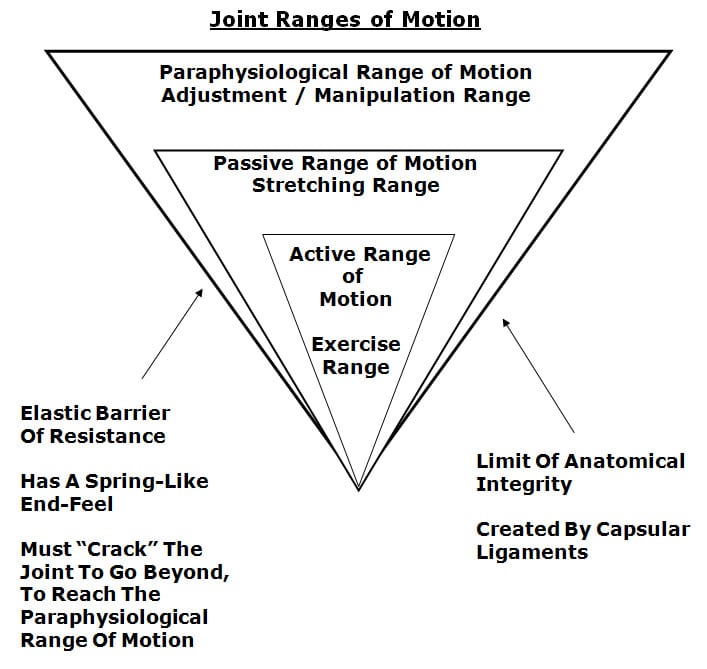
    Mechanically, spinal manipulation primarily affects the spinal facet joints. This is well accepted and not controversial. In 1985, Canadian Orthopedic Surgeon, WH Kirkaldy-Willis, MD, stated (11):

    “Spinal manipulation is essentially an assisted passive motion applied to the spinal apophyseal and sacroiliac joints.”

    Biological Plausibility

    1. The primary injury from whiplash biomechanics is to the facet joints.
    2. The primary source of both acute and chronic whiplash injury pain is the facet joints.
    3. Spinal adjusting (specific joint manipulation) primarily affects the facet joints.
    4. It would appear to be biologically plausible that spinal adjusting could improve the pain status and recovery time of individuals injured in a whiplash type mechanism.
    5. Studies assessing such biological plausibility should show positive clinical outcomes.

    A few pertinent studies are presented below:

    In 1996, clinicians from the University Department of Orthopaedic Surgery, Bristol, United Kingdom, published a study in the journal Injury, titled (12):

    Chiropractic Treatment of Chronic ‘Whiplash’ Injuries

    The authors note, “The whiplash syndrome is a cause of long-term symptoms for which conventional medicine has failed to discover an effective treatment.” They note that 43% of patients will suffer long-term symptoms following ‘whiplash’ injury, for which no conventional treatment has proven to be effective. Consequently, they performed a retrospective study to determine the effects of chiropractic spinal manipulation [a nonconventional treatment] in a group of 28 patients who were suffering with chronic ‘whiplash’ syndrome. The authors defined “spinal manipulation” as:

    “Spinal manipulation is a high-velocity low-amplitude thrust to a specific vertebral segment aimed at increasing the range of movement in the individual facet joint, breaking down adhesions and stimulating production of synovial fluid.”

    The severity of patient’s symptoms was assessed before and after treatment using the Gargan and Bannister (1990) classification protocol, as follows:

    The Gargan and Bannister Whiplash Classification

    GROUP SYMPTOMS
    A None
    B Nuisance
    C Intrusive
    D Disabling

    The 28 patients in this study had initially been treated with anti-inflammatories, soft collars and physiotherapy. These patients had all become chronic, and were referred for chiropractic at an average of 15.5 months (range was 3–44 months) after their initial injury. At the initial evaluation and prior to chiropractic treatment, 27/28 (96%) of the patients were classified as category C (intrusive) or D (disabling) symptoms.

    Following the chiropractic treatment, 93% of the patients had improved: 16/28 (57%) improved by one symptom group, and 10/28 (36%) improved by two symptom groups. The authors concluded:

    “The encouraging results from this retrospective study merit the instigation of a prospective randomized controlled trial to compare conventional with chiropractic treatment in chronic ‘whiplash’ injury.”

    “The results of this retrospective study would suggest that benefits can occur in over 90% of patients undergoing chiropractic treatment for chronic whiplash injury.”

    •••••••••

    In 1999, the same group of clinicians from the University Department of Orthopaedic Surgery, Bristol, United Kingdom, published a study in the Journal of Orthopaedic Medicine, titled (13):

    A Symptomatic Classification of Whiplash Injury and the Implications for Treatment

    In this study, the author’s objective was to determine which patients with chronic whiplash would benefit from chiropractic treatment. The study was a retrospective review involving 93 consecutive whiplash-injured patients. Once again, the patients were assessed using the Gargan and Bannister classification protocol. The authors note:

    “Conventional treatment of patients with whiplash symptoms is disappointing.”

    “In chronic cases, no conventional treatment has proved successful.”

    All patients underwent spinal manipulation, defined as a “high velocity, low amplitude thrust to a specific vertebral segment.” Patients underwent a mean of 19.3 treatments (range 1-53), over a period of 4.1 months.

    The authors note that their results “confirm the efficacy of chiropractic, with 69 of our 93 patients (74%) improving following treatment.” Importantly, 94% of whiplash patients with neurological signs and/or symptoms in association with neck pain and a restricted range of neck movement, including tingling, numbness, pins and needles in a dermatomal distribution in the arm or hand as well as both hypo- and hyperaesthesia, responded positively to chiropractic spinal manipulation. The authors concluded:

    “The results from this study provide further evidence that chiropractic is an effective treatment for chronic whiplash symptoms.”

    “Chiropractic is the only proven effective treatment in chronic [whiplash] cases.”

    •••••••••

    In 2004, a group of physiotherapists, physicians, and professors from the Rey Juan Carlos University, Spain, published a study in the Journal of Whiplash & Related Disorders, titled (14):

    Manipulative Treatment vs. Conventional Physiotherapy Treatment in Whiplash Injury: A Randomized Controlled Trial

    The authors note that the clinical syndrome of whiplash includes neck pain, restriction of neck motion, dizziness, headaches, tinnitus and blurred vision. Spinal joint dysfunction and myofascial pain syndrome are thought to be one of the greatest causes of musculoskeletal disorders and symptoms in patients diagnosed with whiplash injury.

    The goal of joint manipulation is to restore maximal, pain-free movement of the musculoskeletal system. “Only joints that are found to be hypomobile should be considered as candidates for high velocity low amplitude [manipulative] techniques.”

    The objective of this clinical trial was to compare the results obtained from a manipulative protocol with the results obtained from a conventional physiotherapy treatment in patients suffering from whiplash injury. This is the first controlled experimental trial documenting the effects of the manipulative protocol used in this study. It was a randomized controlled trial using 380 acute whiplash injury (less than 3 months duration) subjects. All subjects were Quebec Task Force classified as grades II and III.

    • Quebec GRADE II = neck complaint and musculoskeletal signs
    • Quebec GRADE III = neck complaint, musculoskeletal signs, and neurologic signs

    The injured subjects were randomly divided into 2 groups:

    Group A (experimental, manipulation group)

    This group was treated with high velocity-low amplitude spinal manipulation and soft tissue manipulation. The manipulation was delivered to the upper cervical spine, the cervical-thoracic junction, thoracic spine, thoracolumbar junction and pelvic girdle. Importantly, although the patients were suffering from neck pain and related symptoms, the manipulation was applies “full spine.” The treatment was weekly.

    Group B (control, physical therapy group)

    This group was treated with a conventional physiotherapy including active exercises, electrotherapy, ultrasound therapy and diathermy. The treatment was daily.

    The outcome measures were the visual analogue scale (VAS), the cervical range of motion (CROM) in flexion and rotation, and number of sessions needed to complete the treatment.

    Analysis of the change in visual analogue scale for pain showed:

    • The manipulation group obtained an average decrease of 40% in head and neck pain in 4 visits. (maximum improvement was 9 ± 1.5 visits)
    • The physical therapy group, obtained a decrease of 19% after 10 visits. (maximum improvement was 23 ± 3.2 visits)

    Analysis of the change in cervical range of motion showed:

    • An improvement of 20° in cervical rotation, and 17.5° in cervical flexion after 4 sessions of spinal manipulation.
    • An improvement of 2.5° in cervical rotation, and in cervical flexion after 10 sessions of physical therapy.
    Spinal Manipulation Physiotherapy
    Number of Visits 4 10
    Decrease in
    Neck/Head Pain
    40% 19%
    Increase ROM
    Rotation
    20° 2.5°
    Increase ROM
    Flexion
    17.5°
    Number of Visits to Maximum Improvement 9 ± 1.5 23 ± 3.2

    The authors note:

    Patients who had received manipulative treatment needed fewer sessions to complete the treatment than patients who had received physiotherapy treatment.”

    Patients of manipulative group needed an average of 9 sessions to complete the treatment, whereas physiotherapy group needed an average of 23 sessions.”

    “Results showed that the manipulative group had more benefits than the physiotherapy group in the VAS and CROM.”

    “Our clinical experience with these [whiplash-injured] patients has demonstrated that manipulative treatment gives better results than conventional physiotherapy treatment.”

    Interestingly and importantly, these authors propose that it is “necessary to manipulate a number of spinal levels” to obtain the best outcomes for the whiplash-injured patient. Thoracic spine joint dysfunction may cause secondary adaptive or maladaptive changes in cervical and sacroiliac joints. Such adaptative changes in the cervical spine may contribute to the resulting headaches and neck pain in these patients. They note that whiplash injury symptoms (head, neck and upper thoracic pain) decrease in response to a thoracic spinal manipulation.

    Also, in a rear-end impact, as the struck vehicle is accelerated forward, the seatback contacts the lumbo-pelvic region, causing compression and extension. “This compression of the lumbo-pelvic region produces a hypomobility in the pelvic girdle that is necessary to manipulate.”

    This study emphasizes the need to use spinal manipulation to multiple spinal regions, including the pelvis, to obtain the best outcomes in the whiplash-injured patient. This is consistent with the perspective of rheumatologist John Bland, MD, from the University of Vermont College of Medicine. Note the following quote from his his book Disorders of the Cervical Spine (15):

    “We tend to divide the examination of the spine into regions: cervical, thoracic, and lumbar spine clinical studies. This is a mistake. The three units are closely interrelated structurally and functionally – a whole person with a whole spine. The cervical spine may be symptomatic because of a thoracic or lumbar spine abnormality, and vice versa! Sometimes treating a lumbar spine will relieve a cervical spine syndrome, or proper management of cervical spine will relieve low backache.”

    This is the first study showing the effectiveness of spinal manipulation in the management of acute whiplash injuries (the prior studies pertained to chronic whiplash injuries). In this study, spinal manipulation was significantly more effective both subjectively and objectively in treating Quebec Task Force whiplash injuries Grades II and III than conventional physiotherapy. These authors concluded:

    “This clinical trial has demonstrated that head and neck pain decrease with fewer treatment sessions in response to a manipulative treatment protocol as compared to a physiotherapy treatment protocol among patients diagnosed with acute whiplash injury.”

    Manipulation is “effective in the management of whiplash injury.”

    “Manipulative treatment is more effective in the management of whiplash injury than conventional physiotherapy treatment.”

    •••••••••

    Recently (September 2015), researchers and clinicians from the Orthopedic University Hospital Balgrist, University of Zurich, Switzerland, published a study in the Journal of Manipulative and Physiological Therapeutics, titled (16):

    Prognostic Factors for Recurrences in Neck Pain Patients

    Up to 1 Year After Chiropractic Care

    This is a prospective cohort study assessing 545 neck pain patients. After a course of chiropractic spinal manipulation, they were followed up for one year regarding recurrence of their neck pain. Nine independent prognostic variables were assessed:

    • Age
    • Use of pain medication
    • Sex
    • Work status
    • Duration of complaint
    • Previous episodes of neck pain
    • Trauma onset
    • Numeric Rating Scale (NRS) was used to quantify their pain
    • Bournemouth Questionnaire was used for neck pain

    These authors note that most people will see a health care provider at least once in their lifetime due to neck pain. In patient populations that do not include chiropractic care, those who have experienced an episode of neck pain are likely to have another episode within the next 1 to 5 years. Overall, for these individuals, the prognosis for a complete recovery from their neck pain is “quite poor.” Those who do not recover from their neck pain within the 3 months after treatment begins tend to suffer from residual neck pain and disability.

    There are known prognostic factors for the onset of neck pain. They include:

    • Computer work
    • Heavy physical work
    • Longer duration of complaint
    • Older age
    • Female sex
    • Previous neck injury

    The results of this study are impressive:

    • Fifty-four (54) participants (11%) were identified as “recurrent.”
    • Four hundred ninety one (491) participants (89%) were not recurrent.

    The authors state:

    “89% of neck pain patients had recovered from their neck pain episode up to 1 year after receiving chiropractic care.”

    This study also found that older age and a previous episode of neck pain were useful predictors of neck pain recurrence within 1 year. This is because an increase in age was associated with recurrence. Patients older than 45 years are twice as likely to experience a neck pain recurrence within 1 year after the start of chiropractic treatment. They state:

    “Having had a previous episode as well as increasing age are increased risk factors for predicting a subsequent new episode of neck pain within a year.”

    These authors concluded:

    “The results of this study suggest that recurrence of neck pain within 1 year after chiropractic intervention is low.”

    This study indicates that chiropractic is both effective in the treatment of neck pain and that its benefits are stable and long lasting.

    •••••••••

    SUMMARY

    Both acute and chronic neck pain is primarily attributed to the facet joints. This is especially true of those injured in a whiplash-mechanism accident. Chiropractic spinal adjusting (specific joint manipulation) affects many tissues, but primarily the facet joints. Evidence continues to support that spinal manipulation is both effective and safe for patients suffering with neck pain, including patients with neck pain initiated by a whiplash injury. The evidence supports spinal manipulation for both acute and chronic neck pain/. The evidence also shows that spinal manipulation is not only effective, but the benefits are also long-lasting with only a small incidence of recurrence.


    REFERENCES

    • Grauer JN, Panjabi MM, Cholewicki J, Nibu K, Dvorak J; Whiplash produces an S-shaped curvature of the neck with hyperextension at lower levels; Spine; November 1, 1997; Vol. 22; No. 21; pp. 2489-94.
    • Kaneoka K, Ono K, Inami S, Hayashi K; Motion analysis of cervical vertebrae during whiplash loading; Spine; April 15, 1999; Vol. 24; No. 8; pp. 763-9.
    • Schofferman J, Bogduk N, Slosar P; Chronic whiplash and whiplash-associated disorders: An evidence-based approach; Journal of the American Academy of Orthopedic Surgeons; October 2007; Vol. 15; No. 10; pp. 596-606.
    • Uhrenholt L, Grunnet-Nilsson N; Hartvigsen J; Spine; Cervical spine lesions after road traffic accidents: a systematic review; September 1, 2002; Vol. 27; No. 17; pp. 1934-1941.
    • Pearson AM, Ivancic PC, Ito S, Panjabi MM; Facet joint kinematics and injury mechanisms during simulated whiplash; Spine; February 15, 2004; Vol. 29; No. 4; pp. 390-397.
    • Bogduk N, Aprill C; On the nature of neck pain, discography and cervical zygapophysial joint blocks; Pain; August 1993; Vol. 54; No. 2; pp. 213-217.
    • Barnsley L, Lord SM, Wallis BJ; Bogduk N; The prevalence of chronic cervical zygapophysial joint pain after whiplash; Spine; January 1, 1995; Vol. 20; No. 1; pp. 20-25.
    • Lord SM, Barnsley L, Wallis BJ; Bogduk N; Chronic cervical zygapophysial joint pain after whiplash. A placebo-controlled prevalence study; Spine; August 1, 1996; Vol. 21; No. 15; pp. 1737-1744.
    • Bogduk N; On Cervical Zygapophysial Joint Pain After Whiplash; Spine December 1, 2011; Vol. 36; No. 25S; pp. S194–S199.
    • Quinn KP, Winkelstein BA; Detection of Altered Collagen Fiber Alignment in the Cervical Facet Capsule After Whiplash-Like Joint Retraction; Annals of Biomedical Engineering; August 2011; Vol. 39; No. 8; pp. 2163–2173.
    • Kirkaldy-Willis WH, Cassidy JD; Spinal Manipulation in the Treatment of Low back Pain; Canadian Family Physician; March 1985; Vol. 31; pp. 535-540.
    • Woodward MN, Cook JCH, Gargan MF, and Bannister GC; Chiropractic treatment of chronic ‘whiplash’ injuries; Injury; Vol. 27; No. 9; November 1996; pp. 643-645.
    • Khan S, Cook J, Gargan M, Bannister G; A symptomatic classification of whiplash injury and the implications for treatment; The Journal of Orthopaedic Medicine; Vol. 21; No. 1; 1999; pp. 22-25.
    • Fernández-de-las-Peñas C, Fernández-Carnero J, Palomeque del Cerro L, Miangolarra-Page JC; Manipulative Treatment vs. Conventional Physiotherapy Treatment in Whiplash Injury: A Randomized Controlled Trial; Journal of Whiplash & Related Disorders; 2004; Vol. 3; No. 2.
    • Bland J; Disorders of the Cervical Spine; WB Saunders Company; 1987; p. 84. [Professor of Medicine, University of Vermont College of Medicine].
    • Langenfeld A, Humphreys K, Swanenburg J, Cynthia K. Peterson CK; Prognostic Factors for Recurrences in Neck Pain Patients Up to 1 Year After Chiropractic Care; Journal of Manipulative and Physiological Therapeutics; September 2015; Vol. 38; No. 7; pp. 458-464.
  • Whiplash Injury and Cervicogenic Headache

    Whiplash Injury and Cervicogenic Headache

    nervous system, side view of head

    Barbara is a 45-year old woman with two adult children. She is employed full-time as a sales clerk at the local mall. Her job is not physically demanding nor is it ergonomically challenging. Her job allows her to assume multiple physical positions throughout the day while she is assisting a variety of customers with a variety of needs. There is no required heavy lifting or prolonged postures.

    Barbara is fit, with good muscle tone and posture. She stands 5 feet 4 inches tall and weight 120 pounds. Her exercise regime consists of walking several miles per day, nearly every day of the week, with a group of her friends.

    Barbara has suffered with chronic headaches for 24 years. In addition, her headaches seemed to make her right shoulder ache.

    Barbara’s headaches began when she was involved in a motor vehicle collision that occurred at 21 years of age. She did not recall many of the details of the collision other than that she was the driver of a vehicle that was struck from the rear. The collision caught her by surprise and she remembers her head being thrown backwards. There was no loss of consciousness, and she did not experience being dazed, confusion, disorientation, or loss of any memory. The damage to her vehicle was minor, and she was able to drive away from the accident scene after exchanging insurance information with the man who was driving the striking vehicle.

    Barbara did not experience pain or any other complaints at the accident scene. However, as the day progressed, she became aware of some minor neck stiffness. The next day was a different story. Barbara recalls that the next morning she was unable to pick her head up off her pillow without using her hands to assist her. Her neck was painful and stiff. And, she had a headache.

    Barbara attributed her neck and head signs and symptoms to a “strain” injury caused by the vehicle collision she was involved in. She took some over-the-counter pain pills, and within a few days she was much improved.

    However, about a week after the collision, Barbara became more aware that she still had a headache, and that it did not appear to be improving. Rather it seemed to be becoming more pronounced. The headache was located at the right upper posterior area of her neck and also around and behind her right eye.

    Since being injured 24 years ago, Barbara has had to constantly deal with her headaches. They occur frequently and range in severity from annoying to debilitating. When she is suffering from a bad headache, she also notices an abnormal sensitivity to bright lights (photophobia). She notes that apparent triggers for her headaches range from certain neck movements to prolonged neck postures. Her headaches are always only on her right side.

    Barbara’s examination shows significantly reduced lateral flexion and rotation of the upper cervical spine on the right side. She is very sensitive to mild/moderate digital pressure applied to the suboccipital region and muscles. Importantly, her right-sided frontal (around her eye) headache can be triggered by sustained deeper pressure at the inferior margin of the right inferior oblique muscle. Recall, the inferior oblique muscle exists between the spinous process of the axis (C2) and the transverse process of the atlas (C1). (Two easily identifiable landmarks for a practicing chiropractor; see drawing page 10).

    Barbara reports that she has consulted a number of medical doctors (general practitioners, not specialists) about these headaches, resulting in her taking a variety of over-the-counter and/or prescription medications. She reports that these drugs definitely help her, especially when her headache is severe. She states that she takes pain medicines for her headache 10-15 days per month. But, after developing some gastrointestinal bleeding from taking over-the-counter drugs, her primary care physician suggested she try the COX-2 inhibitor drug Celebrex. She has now been consuming Celebrex 10-15 days per month, reporting that it is quite helpful when she has a bad headache.

    However, Barbara became concerned after hearing media reports of Celebrex and other pain medicines being associated with an increased risk of heart attacks. In addition, she reported that she was weary of having to consume pain medicines 10-15 days per month to function appropriately in her life. Barbara acknowledges that medicines she had been taking for her headaches were helpful, but that they had not cured her headaches, and her suffering had been going on for 24 years.

    Barbara self referred herself to our office as it was on her way home from work. She had seen no other chiropractors or physical therapists for her headaches. Our office was the first.

    ••••••••••

    It has been written in top, respected journals, since at least the 1940s, that whiplash injury to the neck can cause chronic headaches. Whiplash injury pioneer Ruth Jackson, MD, wrote about this phenomenon as early as 1947.

    Ruth Jackson, MD (1902-1994), was the world’s first female admitted into the American Academy of Orthopedic Surgeons (1937). She began her orthopedic private practice in Dallas in 1932. From 1936 to 1941, Dr. Jackson was Chief of Orthopaedics at Parkland Hospital in Fairmont, Texas. In 1945, she had her own private clinic built in Dallas. In 1956 she published her acclaimed, authoritative book entitled TheCervicalSyndrome. The fourth and final edition of her book was published in 1978 (1). Dr. Jackson retired from clinical practice in 1989 at the age of 87 years.

    In 1947, Dr. Jackson published a study in the Journal of the American Medical Women’s Association titled (2):

     The Cervical Syndrome As a Cause of Migraine

    In this article, Dr. Jackson notes that at least half of patients suffering from cervical syndrome will also complain of headaches as one of their principle symptoms. The cervical syndrome is caused by “cervical nerve root irritation,” and this nerve root irritation can occur as a consequence of whiplash trauma.

    Dr. Jackson noted that irritation of the upper cervical spine nerve roots, C1-C2-C3, are most likely to cause headache, and that it is these upper cervical spine nerve roots that are most vulnerable to whiplash trauma. In addition, these post-traumatic headaches may still be present decades later. (Recall, Barbara’s headaches were triggered by a motor vehicle collision, and she had been suffering with headaches for 24 years).

    •••••

    About a decade later, in the late 1950s, the concept of chronic whiplash-generated headache was supported by the writings of Emil Seletz, MD.

    Dr. Emil Seletz (1907-1999) was a neurosurgeon in Beverly Hills, California. Dr. Seletz worked at the Los Angeles General Hospital, and he was faculty at the University of California, Los Angeles, medical school. He was also chief of neurosurgery at Cedar’s Hospital (now called Cedars-Sinai Medical Hospital) in Los Angeles, and Professor of Neurological Surgery at the University of Southern California School of Medicine. By 1957, Dr. Seletz had treated more than 20,000 injury patients, and he began publishing a series of articles pertaining to whiplash trauma and headaches. These include (3, 4, 5):

    Craniocerebral Injuries and the Postconcussion Syndrome

    Journal of the International College of Surgeons

    January 1957, Vol. 27, No. 1, pp. 46-53

    Headache of Extracranial Origin

    California Medicine

    November 1958, Vol. 89, No. 5, pp. 314-17

    Whiplash Injuries

    Neurophysiological Basis for Pain and Methods Used for Rehabilitation

    Journal of the American Medical Association

    November 29, 1958, pp. 1750-1755


    In these articles, Dr. Seletz stressed that the cervical spine causes headaches because of a neuroanatomical relationship between the 2nd cervical nerve root and the trigeminal nerve (cranial nerve V). Dr. Seletz notes that many patients involved in whiplash trauma will develop incapacitating severe headaches that may persist for months or even years following the injury. These headaches are often severe and begin in the suboccipital area and radiate to the vertex or to behind one eye; or they may be frontal or temporal.

    Dr. Seletz believes that the 2nd cervical nerve root is most often involved in the generation of headaches, stating:

    “The 2nd cervical nerve root is more vulnerable to trauma than other nerve roots because it is not protected by pedicles and facets.”


    Dr. Seletz emphasizes that a major portion of the headaches associated with the whiplash syndrome are derived from a traction injury to the second cervical nerve root. The second cervical nerve root is particularly vulnerable to injury because it is not protected by pedicles and facets, as are the other cervical nerve roots. Also, the second cervical nerve root exits between the atlas and axis, “the point of greatest rotation of the head on the neck.”

    Dr. Seletz explains that sensory changes in any of the three sensory branches (ophthalmic, maxillary, mandibular) of the trigeminal nerve (cranial nerve V) are capable of producing headaches. He also explains that the three sensory branches of the trigeminal nerve communicate (synapse with) with the 2nd and 3rd cervical nerve roots in the upper neck in a nucleus he calls the spinal fifth tract of the medulla.

    cervicogenic headache pathway

    Dr. Seletz’s model of post-whiplash headache is quite simple:

    1) Whiplash trauma injures the vulnerable 2nd cervical nerve root.

    2) The sensory input change derived from the injured 2nd nerve root synapses in the spinal fifth tract of the medulla where it synaptically communicates with the branches of the trigeminal nerve.

    3) The signal in the spinal fifth tract of the medulla is interpreted as a headache in one or more of the branches of the trigeminal nerve.

    Dr. Seletz explains that the ophthalmic fibers of cranial nerve V descend the deepest into the cervical spine. Consequently, traumatized patients with an irritated 2nd nerve root often perceive their headache in the distribution of the ophthalmic branch, which is around and behind the eye.

    Adding to the mechanism of chronic post-traumatic headache, Dr. Seletz notes that trauma causes hemorrhage, leading to the development of adhesions forming about the upper cervical nerve roots. He states that these nerve root adhesions are visible during surgical exposure. These nerve root adhesions chronically irritate the nerve roots, sending a signal into the spinal fifth tract of the medulla and ultimately causing chronic headache.

    In summary, Dr. Seletz states:

    “The physiological communication between the second cervical and the trigeminal nerves in the spinal fifth tract of the medulla [trigeminal-cervical nucleus] involves the first division of the trigeminal nerve [opthalamic] and thereby gives attacks of hemicrania with pain radiating behind the corresponding eye. This is the mechanism whereby a great many chronic and persistent headaches have their true origin in injury to the second cervical nerve.”

    “Many headaches are not headaches at all, but really a pain in the neck.”

    •••••

    Although Drs. Jackson and Seletz described the neuroanatomical basis of headaches arising from the cervical spine in the 1940s and 1950s, “Cervicogenic Headache” was not officially recognized until 1983 by Sjaastad (6). In his 1983 article titled “Cervicogenic Headache” An Hypothesis, Sjaastad listed the diagnostic criteria for cervicogenic headache as:

    • Precipitation of head pain by neck movement and/or sustained awkward head positioning.
    • Precipitation of head pain by external pressure over the upper cervical or occipital region on the symptomatic side.
    • Restriction of neck range of motion.
    • Ipsilateral neck, shoulder, or arm pain of a rather vague nonradicular nature, or, occasionally, arm pain of a radicular nature.
    • Unilaterality of the head pain, without sideshift.
    • Head pain is moderate-severe, nonthrobbing, and nonlancinating, usually starting in the neck.
    • Occasionally there is nausea, phonophobia, photophobia, dizziness, ipsilateral blurred vision, difficulties on swallowing, ipsilateral edema (mostly in the periocular area).
    • The pain typically starts at the back of the head, spreading to frontal areas.

    A recent (June 2011) PubMed search of the National Library of Medicine database using the key words “cervicogenic headache” listed 744 articles, with publication dates ranging from September 1942 to June 2011.

    •••••

    Perhaps the most detailed anatomical description for the physiological basis for cervicogenic headache was written by Australian physician and clinical anatomist Nikoli Bogduk, MD, PhD, in 1995. Dr. Bogduk published an article in the journal Biomedicine and Pharmacotherapy titled (7):

    Anatomy and Physiology of Headache

    In this article, Dr. Bogduk notes that all headaches have a common anatomy and physiology in that they are all mediated by the trigeminocervical nucleus, and are initiated by noxious stimulation of the endings of the nerves that synapse in this nucleus. “Trigeminocervical nucleus” is contemporary terminology for what Dr. Emil Seletz termed spinal fifth tract of the medulla. The trigeminocervical nucleus is a region of grey matter in the medulla of the brainstem that descends into the upper cervical spinal cord.

    In agreement with Dr. Seletz above, Dr. Bogduk notes that the trigeminocervical nucleus receives afferents from all three branches (ophthalmic, maxillary, mandibular) of the trigeminal nerve (cranial nerve V). In slight variance with Dr. Seletz, Dr. Bogduk’s detailed anatomical sections indicate that the trigeminocervical nucleus receives afferents from nerve roots C1, C2, and C3.cervicogenic headache pathway


    Consequently, irritation of any of the upper three cervical nerve roots can cause headaches. In addition, Dr. Bogduk stresses that irritation or injury to any tissue innervated by the upper cervical nerve roots can cause headaches.

    Both Dr. Seletz and Dr. Bogduk indicate that the ophthalmic branch of the trigeminal nerve extends the farthest into the trigeminocervical nucleus, and consequently cervical afferent nerve irritation is most likely to refer pain to the frontal-orbital region of the head.

    Both Dr. Seletz and Dr. Bogduk agree that the C1 and C2 nerve roots are particularly likely to be involved in the genesis of cervicogenic headache because the C1 and C2 spinal nerve roots “do not emerge through intervertebral foramina.” This make these nerve roots more vulnerable to stretch or compressive stresses.

    Dr. Seletz commented that in his surgically managed cervicogenic headache patients he would find scar tissue or adhesions that were responsible for chronic C2 nerve root irritation. Dr. Bodguk’s anatomical sections further isolate these C2 nerve root post-traumatic adhesions at two locations:

    • At the C2 dorsal root ganglion as it crosses the C1-C2 joint capsule.
    • At the under belly of the inferior oblique muscle.

    adhesions interfering with nerve function

    •••••

    In 2005, Dr. David Biondi, an instructor in Neurology at Harvard Medical School, published an article titled (8):

    Cervicogenic Headache:

    A Review of Diagnostic and Treatment Strategies

    Dr. Biondi notes that cervicogenic headache is a relatively common cause of chronic headaches with a prevalence as high as 20%. He notes that post-whiplash cervicogenic headache is particularly porne to chronicity.

    Dr. Biondi notes that in the management of cervicogenic headache, drugs alone are often ineffective. He states, “Many patients with cervicogenic headache overuse or become dependent on analgesics.” He also notes that COX-2 inhibitors cause both gastrointestinal and renal toxicity after long-term use, and they cause an increased risk of cardiovascular and cerebrovascular events.

    Dr. Biondi is an osteopathic physician, and therefore has an understanding of manual and manipulative techniques. He states:

    “All patients with cervicogenic headache could benefit from manual modes of therapy and physical conditioning.”

    Dr. Biondi notes that the treatment of cervicogenic headache usually requires manipulation of the upper cervical facet joints, and that manipulative techniques are particularly well suited for the management of cervicogenic headache, including high velocity, low amplitude manipulation. These techniques are commonly used by chiropractors in the management of cervicogenic headaches.

    In April of this year (2011), Dr. Maurice Vincent published a detailed review article pertaining to the relationship between the cervical spine and headache (9). In the article, Dr. Vincent lists five requirements for the diagnosis of cervicogenic headache. They are:

    1) unilateral pain preponderance

    2) reduction of cervical range of motion

    3) pain in the ipsilateral shoulder or arm

    4) attacks precipitation from triggering spots in the neck

    5) precipitation from awkward neck positions.

    These five requirements are all present in my patient Barbara:

    Barbara was suffering from post-traumatic chronic cervicogenic headache. Recall that her “headache was triggered by sustained deep pressure at the inferior margin of the right inferior oblique muscle.” Consequently, my assessment included that Barbara was suffering from an ectopic depolarization of the C2 nerve root at the inferior margin of the right inferior oblique muscle; the electrical signal communicated with the ophthalmic branch of the trigeminal nerve in the trigeminocervical nucleus, creating a cortical brain perception of a headache around her right eye. As there is a history of trauma and 24 years of chronicity, it seems plausible that the primary nidus of C2 irritation was scar tissue or adhesions at the inferior oblique muscle, consistent with the writings of both Drs. Seletz and Bogduk. The irritation of the C2 nerve root is most likely aggravated by alignment and motion dysfunctions of the upper cervical spinal segments.

    My clinical protocols included the following:

    1) Analysis and chiropractic management of upper cervical spinal segmental alignment.

    2) Analysis and chiropractic management of asymmetry of upper cervical spinal segmental movement.

    3) Manual friction myotherapy at the inferior margin of the inferior oblique muscle. This is done in a effort to reduce the adverseness of adhesions and/or scar tissue that appeared to be irritating the C2 nerve root.

    4) Low level laser therapy (in the office) and cryotherapy (homecare) to reduce inflammation subsequent to tissue work.

    Barbara was so treated three times per week for four weeks, a total of twelve visits. The one-month re-evaluation showed a 100% resolution of both signs and symptoms. Twenty-four years chronicity and suffering resolved within one moth of manual therapy.

    REFERENCES:

    1) Jackson R, The Cervical Syndrome, Thomas, 1978.

    2) Jackson R; The Cervical Syndrome As a Cause of Migraine. Journal of the American Medical Women’s Association. December 1947, Vol. 2, No. 12, pp. 529-534.

    3) Seletz E; Craniocerebral Injuries and the Postconcussion Syndrome; Journal of the International College of Surgeons; January, 1957; 27(1):46-53.

    4) Seletz E; Headache of Extracranial Origin; California Medicine; November 1958, Vol. 89, No. 5, pp. 314-17.

    5) Seletz E; Whiplash Injuries, Neurophysiological Basis for Pain and Methods Used for Rehabilitation; Journal of the American Medical Association; November 29, 1958, pp. 1750 – 1755.

    6) Sjaastad O; “Cervicogenic” Headache: An Hypothesis; Cephalagia; December 1983; 3(4):249-256.

    7) Bogduk N; Anatomy and Physiology of Headache; Biomedicine and Pharmacotherapy; 1995, Vol. 49, No. 10, pp. 435-445.

    8) Biondi DM; Cervicogenic Headache: A Review of Diagnostic and Treatment Strategies; Journal of the American Osteopathic Association; April 2005, Vol. 105, No. 4 supplement, pp. 16-22.

    9) Vincent MB; Headache and Neck; Current Pain Headache Report; April 5, 2011 [Epub].

  • Car Accidents, Chiropractic And Children

    Car Accidents, Chiropractic And Children

    In the legal cases I have consulted in, often it is claimed that children cannot be injured in motor vehicle collisions, and therefore they do not require any treatment. To escalate this perspective, I have consulted in cases where the chiropractor treating such a child is accused of committing fraud, a crime. Occasionally, these cases will even progress to courtroom trial.

    Pertaining to the causes of death of our children, the following statistics were compiled from the United States Centers for Disease Control (CDC) National Center for Health Statistics (NCHS).

    Age Group

    Most Prevalent Cause of Death

    Most Prevalent Cause of Accidental Death

    <1

    Birth Defects

    1 – 4

    Accidental

    Motor Vehicle Accident

    5 – 9

    Accidental

    Motor Vehicle Accident

    10 – 14

    Accidental

    Motor Vehicle Accident

    15 – 24

    Accidental

    Motor Vehicle Accident


    In addition, the second leading cause for accidental death in children < 1 year of age was motor vehicle accident.

    Based upon these statistics, it seems ludicrous to claim that children cannot be injured in motor vehicle crashes.

    •••••

    Recently, a new-graduate chiropractor asked my advice regarding the management of an infant who had been injured in a motor vehicle collision. The insurance adjuster controlling the case stated: “our chiropractic consultant informs us that it is unlikely that an infant can be injured in a motor vehicle collision and therefore treatment of an infant after a motor vehicle collision is not likely to be reasonable or necessary.” Chiropractors that treat motor vehicle collision injuries, including those to children, are probably familiar with this attitude.

    More than a decade ago, I had the opportunity to testify in a case in which a 7-year-old child and a 22-month-old toddler were injured in a motor vehicle collision. The children were treated successfully by a chiropractor. The mother of the children was adamant that the chiropractic care her children received was necessary for the improvement of their condition caused by the motor vehicle collision. Yet the case went to trial because of the attitude by the insurance company and their chiropractic paper reviewer that the children did not need the amount of care they received; or that the treating chiropractor’s records could not justify the care that he gave to the children.

    One of the consequences of this trial was my generation of a chapter in a book, Pediatric Chiropractic, edited by Claudia Anrig and Greg Plaugher, Williams and Wilkins, 1998. The second edition of this book is due out later this year (2011). I did an extensive review of the literature pertaining to injuries to children from motor vehicle collisions, using more than 200 references. This article is a summary of some of the main principles of child injuries from motor vehicle collisions.

    •••••

    Many of the concepts that pertain to adults in motor vehicle collisions also apply to children, including the basic principles of inertial acceleration/deceleration injuries, patient preparedness prior to impact, and rotation of the head or trunk prior to impact. Overall, studies indicate that the pattern of injury among children in motor vehicle collisions is similar to those of the general population.

    However, injuries to children in motor vehicle collisions can be unique as a consequence of the following reasons:

    1) Child safety seats.

    2) The increased size of the child’s head as a proportion of the overall body mass.

    3) The child’s ability to be restrained while facing rearward.

    4) The use of seat belts that are designed for adults.

    5) The use of lap belts without shoulder harnesses.

    6) The reduced height of the developing pediatric pelvis.

    7) The underdevelopment of the pediatric anterior superior iliac spine.

    8) The higher center of gravity for the pediatric body.

    9) The diminished development and strength of various spinal musculoskeletal components.

    10) The ability to sit on the lap of adults when traveling in a vehicle.

    11) The probability that a child injured in a motor vehicle collision is unprepared for the collision, or caught by surprise.

    12) The more unfavorable head diameter to neck diameter ratio, as compared to adults.

    I believe that each aspect (above) of this uniqueness regarding children injury during motor vehicle collisions should be understood by the health care provider so that he/she can better explain the appropriateness of treatment given to these injured children. Specifically, I believe that the health care provider should:

    1) Understand the biomechanical uniqueness of injury for each age group of children involved in a motor vehicle collision.

    2) Learn how to examine and document pediatric trauma, including daily charting.

    3) Become proficient at the treatment management of injuries in such small bodies.

    I will briefly review these concepts below. A more detailed explanation

    with graphics and references is available in the next edition of Chiropractic Pediatrics, edited by Anrig and Plaugher, 2011.

    •••••

    Anthropometric Variables For Children

    Head Size

    The increased size of the pediatric head as a proportion of the overall body mass influences the location and type of injuries sustained by children involved in a motor vehicle collision. At birth the head is proportionately larger and accounts for approximately 25% of the body length as compared with 15% in the adult. Consequently in motor vehicle collisions it is the head and cervical spine of the newborn that is most likely to be injured in a motor vehicle collision.

    Toddlers up to 3 years of age continue to have disproportionately large head size and higher centers of gravity, and, therefore, also tend to sustain head injuries. Rear facing child safety seats tend to restrict forward head movement and prevent young heads from striking the interior of the vehicle.

    Pelvic Height

    The reduced height of the developing pediatric pelvis predisposes children to unique injury. Every anatomical part of children is reduced in size as compared to the adult, including the height of their pelvis. This reduced height increases the probability for a lap belt to slip over the top of the brim of the pelvis during a motor vehicle collision, resulting in more serious abdominal visceral and lumbar spine fulcrum injuries.

    Anterior Superior Iliac Spine

    The underdevelopment of the pediatric anterior superior iliac spine increases the probability for unique injury for young patients. Children younger than 10 years of age have less development of the anterior superior iliac spine as compared to the adult. This increases the probability for a lap belt to slip over the top of the brim of the pelvis during a motor vehicle collision, resulting in more serious abdominal visceral and lumbar spine fulcrum injuries.

    Center of Gravity

    The higher center of gravity for the pediatric body changes the nature and location of injury. Children have a relatively higher center of gravity and a greater tendency for the lap belt to ride cephalad to across the abdomen as compared to adults. This elevated position allows the child to submarine forward under the belt, increasing injury to the abdomen and/or the spine.

    4-9 year olds have a relatively lower center of gravity in contrast to infants and toddlers, closer to the umbilicus but still above the lap belt. Yet the iliac crests are underdeveloped in this age group and the lap belt tends to slip up over the bony pelvis and onto the abdomen. With a rapid deceleration event, with a greater proportion of body mass above the lap belt and with the lap belt already in contact with the abdomen, “jackknifing” occurs with compression and injury of abdominal viscera. The hallmark indicator of abdominal viscera and mid-lumbar spine injury is abdominal or flank ecchymosis.

    Tissue Strength

    The diminished development and strength of various spinal musculoskeletal components increases the probability of significant tissue injury in children. Children have less well developed muscle and connective tissue, which increases probabilities for spinal joint and neurological injury.

    Submarining:

    Primarily because of the shortness of their pelvis and under development of their anterior superior iliac spine, children, especially those between ages 4-9, have a higher probability of having their torso slip under the lap belt during a motor vehicle collision thus sustaining associated injuries. This is termed submarining. 10-14 year olds have a better developed anterior superior iliac spine, a “taller” pelvis, and consequently experience submarining less often.

    Child on Adult Lap

    A parent should never hold an infant or child on their lap while riding in a motor vehicle. In a front-end collision at 25 miles per hour at impact, the forces on the baby may reach 20 G. If the weight of the baby is 7.5 pounds its effective weight raises to 150 pounds (7.5 lb X 20 G = 150 lb). If the weight of the child is 25 pounds its effective weight raises to 500 pounds (25 lb X 20 G = 500 lb). It is impossible for the adult to hold the baby under those circumstances. To hold a 10-pound infant at 30 mph the adult strength required would be roughly that needed to lift 300 pounds one foot off the ground.

    If the adult holder is also unrestrained, their body may crush the baby against the dashboard or the back of the front seat. When the adult is not restrained, the infant is crushed by a force equal to the mass of the adult multiplied by the square of the speed and divided by two. When the child is held in the arms of an adult and both are not using belt restraints, the weight of the adult is added to the child’s weight as they are thrown forward. The adult will crush the child with an incredible force.

    Studies indicate that many infants under the age of one travel in cars while being carried on adult laps.

    Unrestrained Children

    Careful observation of anthropomorphic video graphically shows that even though the principles of inertia apply to children, they are different, especially when the child is less than 40 lbs. When young children are unrestrained, their entire body functions as a single piece of inertial mass, and will fly through the air during motor vehicle collisions, becoming “human projectiles.” Injuries include crashing through the glass and being thrown from the vehicle, as well as colliding with the inside of the vehicle. In a moving vehicle that is stopped suddenly by an impact, an unrestrained smaller child will continue to move at the original vehicle speed until stopped by the interior of the vehicle. Even in low speed collisions an unrestrained child becomes a human projectile.

    Studies indicate that children run more risk of injury or death traveling unrestrained in a vehicle than by being hit by a vehicle as a pedestrian. It is estimated that disabling to fatal injuries to these children would decrease by 78-91% if the child was using a restraint system during motor vehicle collisions. It is estimated that 49% of child passenger deaths from motor vehicle collisions could have been prevented with appropriate child restraint use. Children not in safety restraint devices are 11 times more likely to die in a motor vehicle collision than children placed in restraints. Unrestrained children are three times more likely to sustain a brain injury than restrained children.

    Children in Restraints

    Reduction In Injuries:

    The April 2011 edition of the journal Pediatrics published the policy recommendations of the Committee on Injury, Violence, and Poison Prevention pertaining to Child Passenger Safety in motor vehicle collisions. This project used twenty-two expert collaborators. The abstract of their work project includes:

    Child passenger safety has dramatically evolved over the past decade; however, motor vehicle crashes continue to be the leading cause of death of children 4 years and older.

    This policy statement provides 4 evidence-based recommendations for best practices in the choice of a child restraint system to optimize safety in passenger vehicles for children from birth through adolescence:

    1) Rear-facing car safety seats for most infants up to 2 years of age.

    2) Forward-facing car safety seats for most children through 4 years of age.

    3) Belt-positioning booster seats for most children through 8 years of age.

    4) Lap-and-shoulder seat belts for all who have outgrown booster seats.

    In addition, a fifth evidence-based recommendation is for all children younger than 13 years to ride in the rear seats of vehicles.

    It is important to note that every transition is associated with some decrease in protection; therefore, parents should be encouraged to delay these transitions for as long as possible.

    The American Academy of Pediatrics urges all pediatricians to know and promote these recommendations as part of child passenger safety anticipatory guidance at every health-supervision visit.

    Injuries from Restraints

    The leading cause of morbidity and mortality in children is trauma and the most frequent mechanism is motor vehicle collisions. Restraining children decreases their chance of injury or death. Seat belts prevent ejections and reduce impact between the child and the interior of the vehicle. Yet serious injury can still occur even when restraining belts are used because the belts themselves can cause harm and injury. The belt systems have their own unique pattern of injury as they change the distribution of forces, especially to the abdominal viscera in a deceleration event. Violent hyperflexion of the child’s torso over the lap belt applies flexion-distraction forces to the spine. Submarining, or slipping of the child underneath the lap belt can occur and predispose the child to additional abdominal trauma. Children at maximum risk are those too large to be in a safety seat yet too small for the available restraint belt system which are designed for adults (transition age from above). In spite of the drawbacks, adult seat belts are recommended over no restraint at all as they reduce injury and death.

    Seat belts may cause injuries from the neck to the pelvis. The probability of seat belt induced injuries increases when the restraint device is not used properly. Common errors in restraint use include:

    • The child is placed in a restraint not designed for his/her size or weight.
    • The child restraint is not properly anchored to the vehicle.
    • The restraint is not properly applied to the child.

    Children and Lap Belt Injuries

    Lap belt injuries are usually associated with children between ages 4-9, as these children are too large to use restraint seats and are too small to safely use adult lap belts. Children in this age group have special and unique anatomical characteristics that increase their vulnerability to lap belt injuries. Children have relatively larger heads and less well developed spinal musculature than adults, putting children at greater risk of hyperflexion injuries. The immature pelvis is more likely to slip below the seat belt creating fulcrum load injuries to the abdomen.

    Conventional lap restraints do not properly restrain or protect children because the anterior superior iliac spine is under developed in this population. The belt rides up onto the abdomen and chest and may itself cause significant injury. If the vehicle rapidly decelerates the child may whip forward with increased force than an adult because of the child’s higher center of gravity and greater body mass above the waist. Children have greater probability of lap belt induced abdominal and spinal injuries because of their greater percentage of body mass above the umbilicus, the poorly developed anterior superior iliac spine, and the frequent lack or misuse of the shoulder harness for children. Children lap belt syndrome injuries typically have an abrasion or contusion across the abdomen, created by the lap belt. These children may suffer from fracture, dislocations, neurologic damage, and significant intra-abdominal injuries.

    A 1986 report from the National Transportation Safety Board suggested that the use of rear seat lap belts may be more harmful than no seat belt use at all for children, stating: “In many cases, the lap belts induced severe to fatal injuries that probably would not have occurred if the lap belts had not been worn.” Although rear seat lap belts do not meet the special needs of children, most agree that restraining a child with a lap belt is preferable to having no restraint at all.

    Children and Shoulder Harness Injury

    Children between ages 4-9 are generally too large to use a restraint seat and yet are too small to safely use an adult shoulder harness restraint. If such children use an improperly fitting adult shoulder harness across their neck or face, serious and fatal injuries have been reported. As the neck / face position for the shoulder harness is uncomfortable for these children, they often will modify its placement by putting the shoulder harness behind their back or under their arm.

    Infants and Air Bags

    The deployment of air bags occurs at high velocity and creates a serious hazard for children as a result of “bag slap.” The air bag mushrooms out in a fraction of a second, reaching speeds up to 200 mph. Because the rear of the infant child restraint seat is close to the air bag compartment, it will receive a tremendous force from air bag deployment, resulting in serious head injuries to the child. Therefore, rear-facing infant seats should be used only in the back seat of vehicles that have front passenger air bags.

    Types of Injuries to Children

    As noted, both injury and death are frequently reported in children who are involved in motor vehicle crashes. Injury risk and seriousness is greatest when the child is unrestrained or improperly restrained. However, serious injury and death routinely occurs in properly restrained children. The injuries best documented in the literature include:

    Brain Injury

    Facial Fractures

    Cervical Spine Injuries

    Upper Cervical Injury

    Cervical Disc Injury

    Apophysis (growth center)Injury

    Cervical Spinal Cord Injury

    The Gamete of Soft Tissue Injuries

    Seat Belt Syndrome Abdominal Injuries

    Psychological Injury

    •••••

    Summary

    Children are injured in motor vehicle collisions, and it is not a small problem; rather, it is a huge problem. Motor vehicle collisions have consistently proven to be the number one reason for both mortality and morbidity in children younger than 25 years of age.

    The bodies of children younger than one year of age function as a single piece of inertial mass during motor vehicle collisions. Children in this age group have a proportionately larger head size as compared to overall body mass. Consequently, when they are unrestrained during a motor vehicle collision, they tend to “lead” with their head. Their heads and bodies will collide with the interior of the vehicle, and ejections of their bodies are known to occur. Such unrestrained children sustain serious head, brain, and cervical spinal cord injuries, leading to death and significant lifelong disabilities.

    When children younger than one year of age are restrained in a child restraint seat and that child restraint seat is not securely attached to the vehicle seat with the appropriate adult restraint belt, the child’s body and the restraint seat together will function as a single piece of inertial mass. Once again the child will sustain serious head and brain injuries. Not securing the child safety seat to the vehicle is considered to be misuse of the safety device.

    When children younger than one year of age are properly restrained in a forward facing child safety seat, and that child safety seat is properly secured to the vehicle with the adult restraint belt, serious head, brain, and cervical spinal cord injuries are largely avoided. Yet, in this forward facing position the properly restrained child has increased vulnerability to cervical spine injury, especially in frontal impacts. This is because the restraints immobilize the child’s body, yet their head remains moveable. With this young child’s proportionately larger head size as compared to overall body mass, and with the child’s poorly developed strength of the cervical spine musculoskeletal tissues, significant cervical spine soft tissues occur.

    Most serious injuries to restrained children younger than one year of age occur during a frontal impact collision. These serious injuries can be reduced by placing the child restraint seat in a rearward facing direction, and then properly securing this child restraint seat to the vehicle using the adult restraint belts. Serious injuries are reduced as the forces of the frontal impact are dispersed over a broader surface area of the child; over the back of the skull, the thoracic cage, and the pelvis. There is no doubt that a child of this age group who is properly restrained in the rearward facing position has the best chance of avoiding injury in a motor vehicle collision, and especially in serious frontal impact collisions.

    When children younger than one year of age are restrained in a child safety seat that is properly secured to the vehicle by the adult belt, but the crotch strap of the child safety seat is not properly attached, the child’s body will “submarine” under the waist strap, catching the child under the chin. The results are serious cervical spine injury, including fracture of the odontoid process or a bipedicular (hangman’s) fracture of C2. An adult must always properly secure the crotch strap portion of the child restraint seat for children in this age group.

    Children younger than one year of age should not be restrained in a child safety seat in the rearward facing position in the front seat of a vehicle that has a passenger side air bag. In this position, the closeness of the child to a rapidly outwardly exploding air bag can launch the safety seat and child at an extremely high velocity, resulting in serious head and brain injury.

    Children between 1 – 4 years of age are similar to children younger than age one in that their heads are proportionately larger as compared to overall body mass, the strength of their musculoskeletal spinal tissues are not as developed as those of the adult. When they are unrestrained they tend to “lead” with their heads sustaining serious head, brain, and cervical spinal cord injuries after colliding with the interior of the vehicle, and are at risk of ejection. Recent retrospective statistical studies show that the children in this age group are least injured when they are properly restrained in a child safety seat facing the rearward direction. When children in this age group are properly restrained and facing the forward direction, they sustain significant cervical spine soft tissue injury during frontal collisions. Contrary to common practice, it is recommended that children remain in child restraint seats facing the rearward position for a long as possible as they age, ideally to approximately age 4.

    Caution should be used when restraining children between 1- 4 years of age in an adult lap belt. The pelvis of children in this age group is much shorter in height, and the anterior superior iliac spine is grossly underdeveloped as compared to that of the adult, increasing the tendency for the lap belt to slip up over the top of the pelvis rim and to be in contact with the abdomen and its contents. Because of the shorter stature of these children, in a frontal impact their face or chest will not collide with the dashboard or with the seat in front of them. This results in a serious rapid flexion of the child’s torso around the adult lap belt, or “jackknifing.” Serious and fatal abdominal viscera and mid lumbar spinal injuries result.

    Children between ages 4 – 9 years have the greatest difficulty with motor vehicle collision safety. Children in this age group face forward nearly always and are restrained in the adult seat belt. Unfortunately, adult seat belts do not meet the special needs of this group of children. Often they are riding in the rear seat of the vehicle, and there are still many vehicles that do not have shoulder harness restraints available for rear seat passengers. As the developing pelvis remains short in height with an underdeveloped anterior superior iliac spine in this age group, it is once again common for the adult lap belt restraint to slip over the rim of the pelvis and to come into contact with the abdomen and its contents. The center of gravity for these children is higher as compared to that of the adult, superior to the lap belt. This proportionately increases the fulcrum stress above the lap belt in a frontal impact or during a rebound flexion following a rear impact. Again, this results in serious injuries to the abdominal viscera and mid lumbar spine, including Chance fractures. Depending on the stature of these children, their face/head may impact the dashboard or the seat in front of them, resulting in significant face, head, brain, and cervical spinal cord injuries. It is, therefore, recommended that whenever possible, children of this age group should be restrained in a lap belt shoulder belt combination.

    Children between ages 4 – 9 years also have unique problems when using the recommended adult lap belt with shoulder harness combination. Because of their short stature, the shoulder harness does not fit their body adequately. For many children in this age group, the shoulder harness will cut across their cervical spine or face rather than their chest. When left in this position, the shoulder harness can cause serious and fatal cervical spine and facial injuries. Also, because of the uncomfortable annoyance of the shoulder belt crossing the neck or face, many children of this age group will simply place the shoulder strap behind their back, rendering them susceptible to the lap belt injuries noted above. Other children will place the shoulder harness under the arm. This position is also quite dangerous, as the thoracic cage is not capable of handling the forces of a frontal collision during this age of skeletal maturation that are generated by the shoulder harness. The stresses imparted to the child can seriously injure the thoracic cage, including imparting cardiac and pulmonary trauma. The proper shoulder harness placement for this age is across the chest, over the clavicle, but remaining off the cervical spine and face. This is best accomplished by using a booster seat that effectively increases the height of the child, or by using a device that lowers the shoulder harness away from the face and neck and into the proper position and secures it in place by attaching to the lap belt.

    Very young children cannot communicate to their parents or health care providers the location or nature of their injuries. Even non-life threatening injuries in children should be documented and properly managed.


    REFERENCES

    Boyd, William, M.D., Pathology, Lea & Febiger, (1952).

    Buckwalter J, Effects of Early Motion on Healing of Musculoskeletal Tissues, Hand Clinics, Volume 12, Number 1, February 1996.

    Cohen, I. Kelman; Diegelmann, Robert F; Lindbald, William J; Wound Healing, Biochemical & Clinical Aspects, WB Saunders, 1992.

    Cyriax, James, M.D., Orthopaedic Medicine, Diagnosis of Soft Tissue Lesions, Bailliere Tindall, Vol. 1, (1982).

    Fischgrund, Jeffrey S, Neck Pain, monograph 27, American Academy of Orthopaedic Surgeons, 2004.

    Gargan, MF, Bannister, GC, Long-Term Prognosis of Soft-Tissue Injuries of the Neck, Journal of Bone and Joint Surgery, September, 1990.

    Gunn, C. Chan, Pain, Acupuncture & Related Subjects, C. Chan Gunn,

    (1985).

    Gunn, C. Chan, Treating Myofascial Pain: Intramuscular Stimulation (IMS) for Myofascial Pain Syndromes of Neuropathic Origin, University of Washington, 1989.

    Hodgson, S.P. and Grundy, M., Whiplash Injuries: Their Long-term Prognosis and Its Relationship to Compensation, Neuro-Orthopedics, (1989), 7.88-91.

    Jonsson H, Cesarini K, Sahlstedt B, Rauschning W, Findings and Outcome in Whiplash-Type Neck Distortions; Spine, Vol. 19, No. 24, December 15, 1994, pp. 2733-2743.

    Kannus P, Immobilization or Early Mobilization After an Acute Soft-Tissue Injury?; The Physician And Sports Medicine; March, 2000; Vol. 26 No 3, pp. 55-63.

    Kellett J; Acute soft tissue injuries-a review of the literature; Medicine and Science of Sports and Exercise, American College of Sports Medicine, Vol. 18 No.5, (1986), pp. 489-500.

    Kirkaldy-Willis, W.H., M.D., Managing Low Back Pain, Churchill Livingston, (1983 & 1988).

    Kirkaldy-Willis, W.H., M.D., & Cassidy, J.D.,”Spinal Manipulation in the Treatment of Low-Back Pain,” Can Fam Physician, (1985), 31:535-40.

    Majno, Guido and Joris, Isabelle, Cells, Tissues, and Disease: Principles of General Pathology, Oxford University Press, 2004.

    Mealy K, Brennan H, Fenelon GCC; Early Mobilization of Acute Whiplash Injuries; British Medical Journal, March 8, 1986, 292(6521): 656-657.

    Oakes BW. Acute soft tissue injuries. Australian Family Physician. 1982; 10 (7): 3-16.

    Omoigui S; The biochemical origin of pain: The origin of all pain is inflammation and the inflammatory response: Inflammatory profile of pain syndromes; Medical Hypothesis; 2007, Vol. 69, pp. 1169-1178.

    Oschman, James L, Energy Medicine: The Scientific Basis, Churchill Livingstone, 2000.

    Rogier M. van Rijn, Anton G. van Os, Roos M.D. Bernsen, Pim A. Luijsterburg, Bart W. Koes, Professor, Sita M.A. Bierma-Zeinstra; What Is the Clinical Course of Acute Ankle Sprains? A Systematic Literature Review; The American Journal of Medicine; April 2008, Vol. 121, No. 4, pp. 324-331.

    Rosenfeld M, Gunnarsson R, Borenstein P, Early Intervention in Whiplash-Associated Disorders, A Comparison of Two Treatment Protocols; Spine, 2000;25:1782-1787.

    Roy, Steven, M.D., and Irvin, Richard, Sports Medicine: Prevention, Evaluation, Management, and Rehabilitation, Prentice-Hall, Inc. (1983).

    Salter R, Continuous Passive Motion, A Biological Concept for the Healing and Regeneration of Articular Cartilage, Ligaments, and Tendons; From Origination to Research to Clinical Applications, Williams and Wilkins, 1993.

    Seletz E; Whiplash Injuries; Neurophysiological Basis for Pain and Methods Used for Rehabilitation; Journal of the American Medical Association; November 29, 1958, pp. 1750-1755.

    Schofferman J, Bogduk N, Slosar P; Chronic whiplash and whiplash-associated disorders: An evidence-based approach; Journal of the American Academy of Orthopedic Surgeons; October 2007;15(10):596-606.

    Stonebrink, R.D., D.C., “Physiotherapy Guidelines for the Chiropractic Profession,” ACA Journal of Chiropractic, (June1975), Vol. IX, p.65-75.

    Stearns, ML, Studies on development of connective tissue in transparent chambers in rabbit’s ear; American Journal of Anatomy, vol. 67, 1940, p. 55.

    Sturzenegger M, DiStefano G, Radanov BP, Schnidrig A. Presenting symptoms and signs after whiplash injury: the influence of accident mechanisms. Neurology. April 1994;44(4):688-93.

    Sturzenegger M, Radanov BP, Di Stefano G. The effect of accident mechanisms and initial findings on the long-term course of whiplash injury. Journal of Neurology. July 1995;242(7):443-9.

    Wyke, B.D., Articular neurology and manipulative therapy, Aspects of Manipulative Therapy, Churchill Livingstone, 1980, pp.72-77.

    Woo, Savio L.-Y.,(ed.), Injury and Repair of the Musculoskeletal Soft Tissues, American Academy of Orthopaedic Surgeons,(1988), p.18-21; 106-117; 151-7; 199-200; 245-6; 300-19; 436-7; 451-2; 474-6.

  • Whiplash Update Two Important Studies And Their Clinical Applications

    Whiplash Update Two Important Studies And Their Clinical Applications

    BACKGROUND INFORMATION FROM DAN MURPHY


    • All pain has an inflammatory component. In his 2007 article, Omoigui, concludes:

    “The origin of all pain is inflammation and the inflammatory response.”

    “Irrespective of the type of pain, whether it is acute or chronic pain, peripheral or central pain, nociceptive or neuropathic pain, the underlying origin is inflammation and the inflammatory response.”

    “Activation of pain receptors, transmission and modulation of pain signals, neuroplasticity and central sensitization are all one continuum of inflammation and the inflammatory response.”

    “Irrespective of the characteristic of the pain, whether it is sharp, dull, aching, burning, stabbing, numbing or tingling, all pain arises from inflammation and the inflammatory response.”

    • Post-traumatic inflammation is often the consequence of the membrane release of arachidonic acid fat cascading into the pro-inflammatory hormone prostaglandin E2 (PGE2). In his 2010 article, Maroon states:

    “A major component of the inflammatory pathway is called the arachidonic acid pathway because arachidonic acid is immediately released from traumatized cellular membranes.”

    Cell membrane trauma releases arachidonic acid. Arachidonic acid is then transformed into the pro-inflammatory hormones prostaglandins and thromboxanes through the enzymatic action of cyclooxygenase.

    This is why omega-6/omega-3 fatty acid balancing is an important clinical strategy in the management of patients suffering from pain syndromes (Boswell, 2006).

    • Inflammation alters the pain threshold and increases pain perception (Omoigui, 2007; Boswell, 2006; Maroon, 2006; Cleland, 2006; Goldberg 2007; Maroon, 2010). In 2007, Omoigui states:

    The unifying Law of Pain indicates that there is an inflammatory soup of biochemical mediators that are present in all pain syndromes.

    • The resolution of inflammation is fibrosis or scar tissue (Manjo, 2004). In 2004, Manjo states:

    “After a day or two of acute inflammation, the connective tissue—in which the inflammatory reaction is unfolding—begins to react, producing more fibroblasts, more capillaries, more cells—more tissue. In other words, granulation tissue arises from normal connective tissue, but it cannot be mistaken for normal connective tissue, because its fibroblasts are plump and activated.”

    “Fibrosis means an excess of fibrous connective tissue. It implies an excess of collagen fibers, with a varying mixture of other matrix components. It can be a local phenomenon, as an end result to chronic inflammation and of wound healing.”

    “When fibrosis develops in the course of inflammation it may contribute to the healing process.” “By contrast, an excessive or inappropriate stimulus can produce severe fibrosis and impair function.”

    “Why does fibrosis develop? In most cases the beginning clearly involves chronic inflammation. Fibrosis is largely secondary to inflammation.”

    • Fibrotic granulation tissue is capable of maintaining an inflammatory response long after the completion of the healing process, a component of chronic pain (Cyriax, 1982). In 1982, Cyriax states:

    “Fibrous tissue appears capable of maintaining an inflammation, originally traumatic, as the result of a habit continuing long after the cause has ceased to operate.”

    “It seems that the inflammatory reaction at the injured fibers continues, not nearly during the period of healing, but for an indefinite period of time afterwards, maintained by the normal stresses to which such tissues are subject.”

    • Tension within the scar granulation tissue initiates remodeling, reducing inflammation. [Supports the need for early persistent mobilization and chiropractic adjustments]. Once again, in 1982 Cyriax states:

    “Tension within the granulation tissue lines the cells up along the direction of stress. Hence, during the healing of mobile tissues, excessive immobilization is harmful. It prevents the formation of a scar strong in the important direction by avoiding the strains leading to due orientation of fibrous tissue and also allows the scar to become unduly adherent, e.g. to bone.”

    ••••••••••

     Americans eat a lot of fat. This is important in a trauma clinical practice because one particular type of fat is linked to both acute and chronic pain. In fact, this fat was the central theme of the 1982 Nobel Prize in Medicine/Physiology, which pertained to pain.

    Our bodies have somewhere around 75 trillion cells. The cell membranes are composed primarily of fat, and it is the fat that we habitually eat. Trauma/injury to tissues disrupts the cell membranes, releasing the fat and activating enzymes that metabolize those fats (Maroon, 2010).

    There is a type of dietary fat that is linked to inflammation and pain. If people eat this pro-inflammatory pain producing fat, then it is that fat that is released as a consequence of trauma/injury. This fat is not a saturated fat. It is a poly-unsaturated fatty acid called arachidonic acid. Arachidonic acid is an omega-6 fat. Our bodies have enzymes that convert arachidonic acid into pro-inflammatory hormones (leukotrienes, thromboxanes, prostaglandins); and these pro-inflammatory hormones are linked to pain (Omoigui, 2007; Boswell, 2006; Maroon, 2006; Cleland, 2006; Goldberg 2007; Maroon, 2010).

    inflammation

    The primary American source of dietary arachidonic acid is eating meat. Meat is not bad per se. Meat becomes bad when the animal is fed junk food that makes it fat and sick. Economically, our food animals are fed the food that is most fattening. That’s because they are sold by the pound. Fatter animals are worth more in the marketplace. Our food animals are proven to become really fat on a diet of corn and/or soybeans.

    Of course, fattening animal feed is a poor economic choice unless it is also cheap feed. In today’s political environment, the cheapest feed is the food that is subsidized by the taxpayers; and it makes so much sense: lobbying our politicians to use taxpayer dollars to grow corn and soybeans creates a win-win situation for all, cheap meat (this is sarcasm, as noted below).

    Meat, a source of complete proteins, historically was an expensive and therefore rare commodity (at least since the Agricultural Revolution, beginning about 10,000 years ago). Animals become big and fat on a corn/soybean diet, and if the taxpayers subsidize these crops, the corn and soybeans also become much cheaper. By extension, the taxpayers (and the Chinese, or whoever is buying our debt) are subsidizing the cost of meat, making it so that nearly all Americans can afford to eat meat daily (if they choose to do so).

    Recent evidence suggests that nearly 100% of our chickens and 93% of our cows are exclusively fed corn (USA Today, 2008). A major source of feed for our farmed fish is soybeans (Greenberg, 2010). Sadly, when these food animals are fed corn and/or soybeans, they have enzymes that convert the fat found in these crops (linoleic acid) into the pro-inflammatory hormone precursor fat, arachidonic acid.

    These pro-inflammatory fats are in the omega-6 family. One hundred years ago, the amount of omega-6 fats consumed by Americans was about 2 pounds per year. Today, as a consequence of politics and economics, consumption of omega-6 fats has increased to about 25 pounds (Boswell, 2006). In contrast, the quantity of anti-inflammatory omega-3 fats in our diets had decreased substantially.

    Paleolithic humans evolved with a ratio of omega-6/omega-3 fats of about 1/1; the average modern ratio is about 25/1 (Boswell, 2006). This means that the average American is prone to pain syndromes as a consequence of dietary choices and habits. At any given moment, 28% of Americans are suffering from pain (Krueger, 2008); the omega-6/omega-3 ratio is critical. The sarcastic downside from the win-win of cheap fat meat is that it predisposes the consumers, Americans, to pain syndromes. This has resulted in Americans consuming more than 70 million nonsteroidal anti-inflammatory drug (NSAID) prescriptions every year; and 30 billion over-the-counter NSAID tablets are sold annually (Maroon, 2006). The cost is $17 billion per year (Krueger, 2008). Michael Pollan states in his 2008 book In Defense of Food “The billions we spend on anti-inflammatory drugs such as aspirin, ibuprofen, and acetaminophen is money spent to undo the effects of too much omega-6 in the diet.”

    Dietary strategies to rebalance the omega-6/omega-3 ratio have proven to prevent an/or reverse many of these pathological syndromes. Such strategies have proven to be more effective than pain medications about 88% of the time (Maroon, 2006).

    ••••••••••

    This month (August 2011), primary research from the Department of Bioengineering and the Department of Neurosurgery, University of Pennsylvania, provides some of the most important insights into the patho-biomechanics of chronic whiplash injury to date. The study was published in the journal Annals of Biomedical Engineering, and titled (Quinn, 2011):

    Detection of Altered Collagen Fiber Alignment in the Cervical Facet Capsule After Whiplash-Like Joint Retraction

    The authors review that the cervical facet joint is the primary source of pain in patients with whiplash-associated disorders; yet, most clinical studies show no radiographic or MRI evidence of tissue injury. To evaluate this puzzle, these authors used quantitative polarized light imaging to assess the potential for altered collagen fiber alignment in human cadaveric cervical facet capsule specimens during and after a joint retraction simulating whiplash exposure.

    The authors document that the whiplash mechanism involves a retraction event to the facet joint capsular ligaments. Although no evidence of ligament damage was detected during whiplash-like retraction, mechanical and microstructural changes of the facet joint capsular ligaments were identified following these whiplash loadings. The retraction experience produced significant decreases in ligament stiffness and increases in ligament laxity. The strained capsule regions showed altered fiber alignment, “suggesting the altered mechanical function may relate to a change in the tissue’s fiber organization.” The altered capsular ligament fiber alignment occurred without any tears that would classically be identified with diagnostic imaging, including radiographs and/or MRI. Consequently, the authors indicate that whiplash kinematics is a potential cause of microstructural damage that is not detectable using standard clinical imaging techniques.

    The authors make these key points:

    1) This is the first study that has assessed changes in tissue microstructural organization of the facet capsule following whiplash-like loading.

    2) “Whiplash is a common cause of chronic neck pain, and the cervical facet joint has been identified as the site of pain in the majority of these cases.”

    3) “Up to 62% of people affected by whiplash injuries report pain lasting 2 years or more after injury.”

    4) Facet joint injuries cannot be imaged in most whiplash patients with x-rays or magnetic resonance imaging (MRI).

    5) “The lack of any definitive evidence of facet capsular ligament damage following whiplash, despite the high incidence of facet-mediated pain, suggests radiographic and MRI techniques may lack the resolution or contrast to identify these subtle injuries.”

    6) Low-speed rear-end impact collision causes the lower cervical spine to undergo a combination of compression, posterior shear, and extension. “This combination of forces and moments primarily induces a retraction of each vertebra in the posterior direction relative to its adjacent inferior vertebra in the lower cervical spine prior to head-headrest contact.” The facet capsular ligaments are at risk for excessive motion during this vertebral retraction, creating subfailure injuries to the facet capsule. “The facet capsular ligament may sustain partial failures and/or unrecovered deformation during whiplash.”

    7) Facet joint injury causes altered collagen fiber organization and facet capsular ligament laxity that may produce persistent pain. “Neither partial failure nor capsule rupture is required to initiate facet-mediated pain, suggesting painful facet joint injuries cannot be identified through traditional load-based or medical imaging techniques.”

    8) Prior to ligament visible rupture or mechanical failure, there is an anomalous fiber realignment, which may be used as a marker for subfailure capsule injury.

    9) The retraction caused permanent deformation of ground substance materials of the ligament, leading to altered collagen fiber organization. This tissue damage may be sufficient to induce an inflammatory response or nociceptor firing in the ligament.

    10) “These findings would suggest that radiographic or MRI diagnostic approaches may lack the resolution to detect the microstructural changes that can occur in the facet capsule without overt capsule rupture after a whiplash exposure.”

    11) “Facet joint displacements that produce persistent pain symptoms also induce laxity in the capsular ligament and collagen fiber disorganization.”

    12) “The detection of altered fiber alignment and unrecovered strain observed after facet retraction in the current study would suggest that whiplash-like loading may be sufficient to generate facet-mediated pain.”

    This study indicates that whiplash injury causes microstructural changes, anomalous fiber realignment and laxity of the facet capsular ligaments. These injuries may cause permanent deformation of ground substance of the ligament, leading to altered collagen fiber organization. These injuries are subfailure in magnitude, but are capable of causing pain and permanent alterations in capsular mechanics. These injuries are not identifiable clinically, with x-ray, or MRI imaging. The tissue damage may be sufficient to induce an inflammatory response and/or nociceptor firing.

    The anomalous fiber realignment noted in this study is probably

    analogous to the writings of Cyriax when he stated that fibrotic granulation tissue is capable of maintaining an inflammatory response long after the completion of the healing process. This inflammatory granulation tissue becomes a factor in the initiation of chronic pain perception. Consequently, Cyriax also states “…that the scar tissue remains painful whenever tension is put upon it, perhaps for decades.”

    This is an important study advancing the understanding of whiplash injury pathoanatomy, yet I believe there is still a missing piece. The authors document post-traumatic anomalous fiber realignment, but they only speculate that it is associated with pain producing inflammation. They offer no evidence for the existence of an actual inflammatory process. Fortunately, the next study does just that.

    ••••••••••

    Clas Linnman (from Harvard Medical School) and an international team of colleagues published a study in April of this year (2011) titled:

    Elevated [11C]-D-Deprenyl Uptake in Chronic Whiplash Associated Disorder Suggests Persistent Musculoskeletal Inflammation

    These authors note that there are few diagnostic tools for chronic musculoskeletal pain, and especially for whiplash injury. In agreement with Quinn above, they note that structural imaging methods seldom reveal pathological alterations that can account for a patient’s ongoing pain. Therefore, they sought to visualize inflammatory processes in the neck region by means of Positron Emission Tomography (PET) using an inflammatory marker, 11C-D-deprenyl, or DDE. They evaluated 22 patients with chronic pain after a rear impact car accident and 14 healthy controls. The whiplash-injured subjects had pain and reduced motion but no neurological signs.

    The whiplash-injured patients displayed significantly elevated inflammatory tracer uptake in the neck, suggesting that whiplash patients have signs of local persistent peripheral tissue inflammation. The authors concluded that inflammation and its associated pain in the periphery could be objectively visualized and quantified with PET using the inflammatory tracer DDE. Key points from this study include:

    1) “Chronic musculoskeletal pain syndromes are common, cause extensive individual suffering and place a large burden on health care in society. Yet, pain remains notoriously difficult to visualize and diagnose objectively.”

    2) “The pathophysiology of persistent pain is elusive and there is a great need for ways to visualize and quantify pain mechanisms.”

    3) In a sub-portion of the population, “whiplash injuries proceed to chronic debilitating pain.”

    4) “Structural imaging does not capture on-going biological processes; where as molecular imaging with positron emission tomography (PET) has the potential to visualize such mechanisms.”

    5) The authors present evidence that shows “DDE can be used to visualize chronic inflammatory processes.”

    6) The site of inflammation “appeared to be localized to adipose tissue surrounding deep cervical muscles.” “The tracer retention observed in fatty regions surrounding deep cervical muscle may indicate that adipose tissue is actively involved in the inflammatory process.”

    7) Patients displayed elevated DDE retention in cervical soft tissue, suggesting that localized chronic inflammation is apparent in many chronic pain whiplash patients.

    8) “A large subset of patients with chronic pain after a whiplash injury displayed elevated DDE retention, suggestive of persistent peripheral tissue inflammation.”

    9) “The possibility to visualize and quantify sites of inflammation in chronic pain may be very useful in diagnosis and treatment monitoring.”

    SUMMARY POINTS:

    • All pain has an inflammatory component.

    • Post-traumatic inflammation is often the consequence of the membrane release of the arachidonic acid fat cascading into pro-inflammatory hormones, including prostaglandin E2 (PGE2). [Therefore omega-6/-3 balancing is an important clinical strategy].

    • Inflammation alters the pain threshold and increases pain perception.

    • The resolution of inflammation is granulation, fibrosis, or scar tissue.

    • Fibrotic granulation tissue is capable of maintaining an inflammatory response long after the completion of the healing process, a component of chronic pain.

    • Whiplash trauma can create anomalous fiber alignment and granulation tissue.

    • From whiplash, granulation tissue and inflammation occurs as a consequence of subfailure injuries. Therefore, these injuries cannot be visualized with either x-rays or MRI.

    • Persistent post-traumatic inflammation has been linked to chronic pain syndrome. This inflammation can be documented with PET using the inflammatory tracer DDE.

    • Tension within the scar granulation tissue initiates remodeling, reducing inflammation. This supports the need for early persistent mobilization, exercise, and chiropractic adjustments.

    • I believe that anti-inflammatory omega-6/omega-3 balancing is critical in chronic pain management.

    Dan Murphy, DC, DABCO

    REFERENCES

    Omoigui S; The biochemical origin of pain: The origin of all pain is inflammation and the inflammatory response: Inflammatory profile of pain syndromes; Medical Hypothesis; 2007, Vol. 69, pp. 1169 – 1178.

    Maroon J, Bost JW, Maroon A; Natural anti-inflammatory agents for pain relief; Surgical Neurological International; December 2010.

    Boswell M, Cole EB; American Academy of Pain Management; Weiner’s Pain Management: A Practical Guide for Clinicians; Seventh Edition, 2006, pp.584-585.

    Maroon JC, Bost JW; Omega-3 Fatty acids (fish oil) as an anti-inflammatory: an alternative to nonsteroidal anti-inflammatory drugs for discogenic pain; Surgical Neurology; 65 (April 2006) 326– 331.

    Cleland LG, James MJ, Proudman SM; Fish oil: what the prescriber needs to know; Arthritis Research & Therapy; Volume 8, Issue 1, 2006, pp. 402.

    Goldberg RJ, Katz J; A meta-analysis of the analgesic effects of omega-3 polyunsaturated fatty acid supplementation for inflammatory joint pain; Pain; May 2007, 129(1-2), pp. 210-223.

    Manjo G, Joris I; Cells, Tissues, and Disease, Principles of General Pathology; Second Edition; Chapter 13: “Chronic Inflammation: Defense at a Price”; Oxford University Press; 2004.

    Cyriax, James, M.D., Orthopaedic Medicine, Diagnosis of Soft Tissue Lesions, Bailliere Tindall, Vol. 1, (1982).

    USA Today, November 12, 2008, quoting Proceedings of the National Academy of Sciences.

    Greenberg P; Four Fish, The Future of the Last Wild Food; The Penguin Press, New York, 2010.

    Krueger AB, Stone AA; Assessment of pain: a community-based diary survey in the USA; Lancet; 2008 May 3;371(9623):1519-25.

    Pollan, M; In Defense of Food; 2008, pg. 131.

    Quinn KP, Winkelstein BA; Detection of Altered Collagen Fiber Alignment in the Cervical Facet Capsule After Whiplash-Like Joint Retraction; Annals of Biomedical Engineering; August 2011, Vol. 39, No. 8, pp. 2163–2173.

    Linnman C, Appel L, Fredrikson M, Gordh T, Soderlund A, Langstrom B, Engler H; Elevated [11C]-D-Deprenyl Uptake in Chronic Whiplash Associated Disorder Suggests Persistent Musculoskeletal Inflammation; Public Library of Medicine (PLoS) ONE; April 6, 2011, Vol. 6 No. 4, pp. e19182.

  • Whiplash Injury, Chronic Pain, Non-Pharmacologic Managements Adjuncts

    Whiplash Injury, Chronic Pain, Non-Pharmacologic Managements Adjuncts

    Not everyone injured in a motor vehicle collision recovers completely. A percentage of those injured will suffer for years or sometimes even for decades. Documented examples of this chronic pain syndrome include:

    • In 1964, the Journal of Bone and Joint Surgery (American) published a study where the author followed 145 whiplash-injured patients for more than two years. The author reported that after a minimum of two years, between 45% to 83% of the injured patients continued to suffer from pain.* (1)

    *The author’s study initially included 266 injured patients, but at the follow-up assessment (more then 2 years later) only 145 were evaluated (121 of the original group were not evaluated at the two plus year follow-up). Of the 145 followed patients, 83% were still suffering pain symptoms. The author noted that if he assumed that 100% of the 121 subjects who were not evaluated were completely symptom free, then the incidence of chronic pain in the entire initial 266 patient set fell to 43%.

    • In 1989, the journal Neuro-Orthopedics published a 12.5-year (mean duration) study on whiplash-injured patients. The authors reported that 62% continued to suffer from significant pain symptoms attributed to the motor vehicle collision 12.5 years later. (2)
    • In 2000, the Journal of Clinical Epidemiology published a 7-year study on whiplash-injured patients. The authors reported that 39.6% continued to suffer from neck-shoulder pain 7 years after injury. This 39.6% chronic pain rate was three times greater than the pain noted in the matched control populations. (3)
    • In 2005, the journal Injury published a 7.5 year prospective study on whiplash-injured patients. The authors reported that 21% of these patients continued to suffer from clinically relevant pain 7.5 years after injury. An additional 48% continued to suffer from nuisance pain at the 7.5-year analysis. (4)
    • In 1990, the Journal of Bone and Joint Surgery (British) published a 10.8 year study on whiplash-injured patients. The authors reported that 40% of these patients continued to suffer from clinically significant pain 10.8 years after injury. An additional 40% continued to suffer from nuisance pain at the 10.8-year analysis. (5)
    • In 1996, the Journal of Bone and Joint Surgery (British) published a 15.5-year study on whiplash-injured patients. The authors reported that 43% of these patients continued to suffer from clinically significant pain 15.5 years after injury. An additional 28% continued to suffer from nuisance pain at the 15.5-year analysis. (6)
    • In 2002, the European Spine Journal published a 17-year study on whiplash-injured patients. The authors reported that 55% of these patients continued to suffer from residual pain 17 years after injury. Of those with residual symptoms, 25% suffered from neck pain every day, and 23% had pain radiating into their arm daily. (7)
    • In 2006, the Journal of Bone and Joint Surgery (British) published a 30-year study on whiplash-injured patients. The authors reported that 15% of these patients continued to suffer from clinically significant pain 30 years after injury; their pain was such that they still required ongoing treatment. An additional 40% continued to suffer from nuisance pain at the 30-year analysis. (8)

    •••••

    In the vast majority of these chronic pain patients, secondary monetary gain does not appear to be the reason for their suffering. If secondary monetary gain were the motivation behind ongoing pain and suffering, such pain and suffering would resolve after receiving the monetary compensation. When an individual continues to complain of post-whiplash pain 2 plus, 7, 7.5, 10.8, 12.5, 15.5, 17, and even 30 years after the initial injury and after all possible monetary compensation has already been awarded, it is difficult to ascribe those chronic complaints to the desire to enhance monetary compensation. Several of the authors of the above studies made comments on this fact, including these:

    “If the symptoms resulting from an extension-acceleration injury of the neck are purely the result of litigation neurosis, it is difficult to explain why 45% [minimum, could be as high as 83%] of the patients should still have symptoms two years or more after settlement of their court action.” (1)

    “If symptoms were largely due to impending litigation it might be expected that symptoms would improve after settlement of the claim. Our results would seem to discount this theory, with the long-term outcome seeming to be determined before the settlement of compensation.” (2)

    The fact that symptoms do not resolve even after a mean 10 years supports the conclusion that litigation does not prolong symptoms. (5)

    Symptoms did not improve after settlement of litigation, which is consistent with previous published studies. (6)

    “It is not likely that the patients exposed to motor vehicle accidents would over-report or simulate their neck complaint at follow-up 17 years after the accident, as all compensation claims will have been settled.” (7)

    •••••

    In 1997, a study published in the journal Pain reported that chronic pain whiplash-injured patients have an abnormal psychological profile (9). However, the authors noted that in their review, they were unable to find any evidence that appropriate psychotherapy was able to effectively treat the patient’s pain. Rather, the psychotherapy helped the patient deal with their pain, but it did not remove their pain. In contrast, the authors were able to effectively eliminate the patient’s abnormal psychological profile, essentially 100% of the time, if and only if they were able to establish an organic lesion causing the patient’s pain and effectively treating it. The authors reported that the abnormal psychological profile was the consequence of the chronic pain.

    Other studies have also concluded that the whiplash-injured patient’s abnormal psychological profile is secondary to their chronic pain. As an example, in 1996, Squires and colleagues note (6):

    Studies have found that patients that were psychologically normal at the time of injury will develop abnormal psychological assessments if their symptoms persisted for three months.

    This study showed an “abnormal psychological profile in patients with symptoms after 15 years suggesting that this is both reactive to physical pain and persistent.”

    In 2010, Rooker and colleagues note (8):

    Whiplash-injured patients with a disability [including chronic pain symptoms] often develop an abnormal psychological profile.

    Other studies have also concluded that chronic whiplash pain is not, as a rule, psychometric, but rather it has an organic basis. In 1997, a study published in the Journal of Orthopedic Medicine followed whiplash-injured patients and a matched control population for a period of 10 years (10). Neck pain was 8 times more prevalent in the whiplash group than in the control group. Paraesthesia was 16 times more prevalent in the whiplash group than in the control group. Headaches were 11 times more prevalent in the whiplash group than in the control group. The combination of both back pain and neck pain was 32 times more prevalent in the whiplash group than in the control group. Importantly, objectively, the x-rays showed that radiographic degenerative changes in the cervical spine appeared 10 years earlier in the whiplash group than in the control group. The authors reported:

    “The prevalence of degenerative changes in the younger cervical spine [of the whiplash group] suggests that the condition has an organic basis.”

    “Degenerative change and its association with neck stiffness support an organic basis for the symptoms that follow soft tissue injuries of the neck.”

    •••••

    In 2007, a study was published summarizing the basis of all pain, including chronic pain, in the journal Medical Hypothesis (11). The author, from the Division of Inflammation and Pain Research, Los Angeles Pain Clinic, cites studies to support these conclusions:

    “The origin of all pain is inflammation and the inflammatory response.”

    “Irrespective of the type of pain, whether it is acute or chronic pain, peripheral or central pain, nociceptive or neuropathic pain, the underlying origin is inflammation and the inflammatory response.”

    “Activation of pain receptors, transmission and modulation of pain signals, neuroplasticity and central sensitization are all one continuum of inflammation and the inflammatory response.”

    “Irrespective of the characteristic of the pain, whether it is sharp, dull, aching, burning, stabbing, numbing or tingling, all pain arises from inflammation and the inflammatory response.”

    •••••

    In 1975, Stonebrink (12) addresses that the last phase of the pathophysiological response to trauma is tissue fibrosis. Boyd in 1953 (13), Cyriax in 1983 (14), and Majno/Joris in 2004 (15) note that there is tissue fibrosis subsequent to trauma. This fibrosis of repair subsequent to soft tissue trauma creates problems that can adversely affect the tissues and the patient for years, decades, or even forever.

    As an example, Cyriax (14):

    “Fibrous tissue is capable of maintaining an inflammation, originally traumatic, as a result of a habit continuing long after the initial [cause] has ceased to operate.”

    Connecting the dots, I propose the following model:

    Tissue trauma, including whiplash trauma,

    heals with varying degrees of fibrous tissue.

    Post-traumatic fibrous tissue is capable of maintaining an inflammatory response long after the initial cause has ceased to exist.

    This inflammatory fibrous tissue alters the threshold of the pain neurons, increasing the probability of chronic pain perception.

    •••••

    In 1971, biochemists Sune K. Bergström (Sweden; d.2004), Bengt I. Samuelsson (Sweden) and John R. Vane (United Kingdom; d. 2004) determined that nonsteroidal anti-inflammatory drugs (NSAIDs) could inhibit the synthesis of prostaglandins from the toxic fat arachidonic acid. They subsequently jointly received the 1982 Nobel Prize in Physiology or Medicine for their research on prostaglandins. The official Nobel Prize press release acknowledged:

    “Prostaglandins are continuously formed in the stomach, where they prevent the tissue from being damaged by the hydrochloric acid. If the formation of prostaglandins is blocked a peptic ulcer can rapidly be formed.”

    As a consequence of the 1982 Nobel Prize in Medicine or Physiology, scientists and healthcare providers have a much better understanding of the mechanisms of how aspirin and other nonsteroidal antiinflammatory drugs reduce pain, but also increase the risk for gastrointestinal bleeding and kidney damage. In an effort to reduce the gastrointestinal bleeding, a new class of NSAIDs, the COX-2 inhibitors, was developed. These drugs are also known as cyclo-oxygenase 2 inhibitors or ‘coxibs’, and the major brand names are Vioxx and Celebrex.

    Cox enzymes convert the omega-6 fatty acid arachidonic acid into the pro-inflammatory pain producer prostaglandin E2 (PGE2).

    Cox enzymes convert the omega-6 fatty acid arachidonic acid into the pro-inflammatory pain producer prostaglandin E2 (PGE2).

    In 2002, a study published in the European Spine Journal reported that of the 55% of whiplash-injured patients with pain 17 years after their injury, 53% of the patients were still using analgesics to manage their pain (7). Of these:

    • 29% used analgesics 2-6 times per week
    • 46% used analgesics 7-30 times per week
    • 17% used analgesics more than 30 times per week

    Although non-steroidal anti-inflammatory drugs inhibit the genesis of prostaglandin E2 (PGE2) and subsequently reduce pain, there are problems with habitual consumption of these products in the management of chronic pain syndromes, including the chronic pain that is often observed following whiplash injury (7). Importantly, habitual consumption of NSAIDs for chronic pain conditions has been associated with a number of deleterious health events, including:

    • End stage renal disease (16)
    • Gastrointestinal Bleeding (17, 18)
    • Myocardial infarction (18, 19, 20)
    • Stroke (18, 20)
    • Alzheimer’s and other dementias (21)
    • Hearing loss (22)
    • Erectile dysfunction (23)

    In 2003, the journal Spine published a study stating (24):

    “Adverse reactions to nonsteroidal antiinflammatory (NSAID) medication have been well documented.”

    “Gastrointestinal toxicity induced by NSAIDs is one of the most common serious adverse drug events in the industrialized world.”

    “The newer COX-2-selective NSAIDs are less than perfect, so it is imperative that contraindications be respected.”

    There is “insufficient evidence for the use of NSAIDs to manage chronic low back pain, although they may be somewhat effective for short-term symptomatic relief.”

    In 2006, a study published in Surgical Neurology stated (25):

    “The use of NSAID medications is a well-established effective therapy for both acute and chronic nonspecific neck and back pain.”

    “Extreme complications, including gastric ulcers, bleeding, myocardial infarction, and even deaths, are associated with their [NSAIDs] use.”

    Blockage of the COX enzyme [with NSAIDs] inhibits the conversion of arachidonic acid to the very pro-inflammatory prostaglandins that mediate the classic inflammatory response of pain (dolor), edema (tumor), elevated temperature (calor), and erythema (rubor).

    “More than 70 million NSAID prescriptions are written each year, and 30 billion over-the-counter NSAID tablets are sold annually.”

    “5% to 10% of the adult US population and approximately 14% of the elderly routinely use NSAIDs for pain control.”

    Almost all patients who take the long-term NSAIDs will have gastric hemorrhage, 50% will have dyspepsia, 8% to 20% will have gastric ulceration, 3% of patients develop serious gastrointestinal side effects, which results in more than 100,000 hospitalizations, an estimated 16,500 deaths, and an annual cost to treat the complications that exceeds 1.5 billion dollars.

    “NSAIDs are the most common cause of drug-related morbidity and mortality reported to the FDA and other regulatory agencies around the world.”

    One author referred to the “chronic systemic use of NSAIDs to ‘carpet-bombing,’ with attendant collateral end-stage damage to human organs.”

    COX 2 inhibitors [Celebrex], designed to alleviate the gastric side effects of COX 1 NSAIDs, are “not only associated with an increased incidence of myocardial infarction and stroke but also have no significant improvement in the prevention of gastric ulcers.”

    •••••

    There are effective nontoxic alternatives to NSAIDs in the management of chronic spinal pain. A well-respected physician who is an advocate of these alternative approaches to chronic pain management is Joseph Charles Maroon, MD. Dr. Maroon is a neurosurgeon from the University of Pittsburgh Medical Center. Dr. Maroon specializes in painful degenerative spinal diseases, and he is also the neurosurgeon for Pro Football’s Pittsburgh Steelers.

    Recently (2006 and 2010), Dr. Maroon has published two studies and one book on the efficacy of natural anti-inflammatory agents for pain relief (25, 26, 27). The effective products Dr. Maroon details include:

    Omega-3 Essential Fatty Acids (fish oil)

    White willow bark

    Curcumin (turmeric)

    Green tea

    Pycnogenol (maritime pine bark)

    Boswellia serrata resin (Frankincense)

    Resveratrol

    Uncaria tomentosa (cat’s claw)

    Capsaicin (chili pepper)

    In his writings, Dr. Maroon discusses the biological plausibility for the use of each of these products, as well as their therapeutic doses. He particularly emphasizes the viability of omega-3 essential fatty acids, noting that these oils powerfully inhibit the production of both pro-inflammatory prostaglandins and pro-inflammatory leukotrienes. In his 2006 study (25), Dr. Maroon found that he could eliminate pain medication in 59% of his study subjects.

    Other studies also support the utilization of omega-3 fatty acids (fish oil) in an effort to achieve an anti-inflammatory state:

    In 2006, the journal Arthritis Research & Therapy published a study noting that an anti-inflammatory dose of fish oil had to be a minimum of 2,700 g/d of EPA plus DHA (the active anti-inflammatory ingredients in fish oil) (28).

    In 2007, the journal Pain published a study also study noting that an anti-inflammatory dose of fish oil had to be a minimum of 2,700 mg/d of EPA plus DHA (29).

    Both studies (28, 29) indicated that it might take a period of 2-3 months before maximum benefit of fish oil supplementation to be observed.

    In 2010, The Clinical Journal of Pain presented a case series of patients suffering from chronic neuropathic pain, including patients injured in whiplash collisions (30). The authors noted that in these more difficult neuropathic pain patients, that more aggressive fish oil supplementation may be required to achieve a good clinical outcome. They suggest doses of EPA plus DHA between 2400-7500 mg/d. Their case series was very successful with these high doses of fish oil, stating:

    “These patients had clinically significant pain reduction, improved function as documented with both subjective and objective outcome measures up to as much as 19 months after treatment initiation.”

    “No serious adverse effects were reported.”

    “This first-ever reported case series suggests that omega-3 fatty acids may be of benefit in the management of patients with neuropathic pain.”

    •••••

    In summary, it is inevitable that some patients injured in motor vehicle collisions will develop chronic pain syndrome. The cause of their chronic pain is rarely psychometric. Rather their pain usually has an organic basis, which includes post-traumatic scarring with persistent inflammation. Inflammation predisposes tissues to pain generation and perception. Management with anti-inflammatory agents makes sense. However, NSAIDs taken for chronic pain syndromes are associated with a number of serious adverse events, including death. There is good evidence that there are a number of alternative natural products for pain management that are both safe and effective, especially omega-3 fish oils. Alternative health care practioners routinely uses these products in the management of chronic pain patients, including those injured in whiplash trauma. The results are improved outcomes, few if any side effects, and great patient satisfaction.

    •••••

    References:

    1) Macnab, I; Acceleration Injuries of the Cervical Spine; Journal of Bone and Joint Surgery (American); Vol. 46, No. 8, December 1964.

    2) Hodgson SP, Grundy M; Whiplash Injuries: Their Long-term Prognosis and its Relationship to Compensation; Neuro-Orthopedics; No.7, 1989, pp. 88-91.

    3) Berglund A, Alfredsson L, Cassidy JD, Jensen I, Nygren A; The association between exposure to a rear-end collision and future neck or shoulder pain; Journal of Clinical Epidemiology; 2000; 53:1089-1094.

    4) Tomlinson PJ, Gargan MF, Bannister GC. The fluctuation in recovery following whiplash injury: 7.5-year prospective review. Injury. Volume 36, Issue 6, June 2005, Pages 758-761.

    5) Gargan MF, Bannister GC. Long-Term Prognosis of Soft-Tissue Injuries of the Neck. Journal of Bone and Joint Surgery (British); Vol. 72-B, No. 5, September 1990, pp. 901-3.

    6) Squires B, Gargan MF, Bannister CG. Soft-tissue Injuries of the Cervical Spine: 15-year Follow-up. Journal of Bone and Joint Surgery (British). November 1996, Vol. 78-B, No. 6, pp. 955-7.

    7) Bunketorp L, Nordholm L Carlsson J; A descriptive analysis of disorders in patients 17 years following motor vehicle accidents; European Spine Journal, 11:227-234, June 2002.

    8) Rooker J, Bannister M, Amirfeyz R, B. Squires, M. Gargan, G. Bannister; Whiplash Injury 30-Year follow-up of a single series; Journal of Bone and Joint Surgery – British Volume; 2010; Volume 92-B, Issue 6, pp. 853-855.

    9) Wallis B, Lord S, Bogduk N; Resolution of Psychological Distress of Whiplash Patients Following Treatment by Radiofrequency Neurotomy: A randomized, double-blind, placebo controlled trial; Pain, October 1997; Vol. 73, No. 1; pp. 15-22.

    10) Gargan MF, Bannister GC. The Comparative Effects of Whiplash Injuries. The Journal of Orthopaedic Medicine, 19(1), 1997, pp. 15-17.

    11) Omoigui S; The biochemical origin of pain: The origin of all pain is inflammation and the inflammatory response: Inflammatory profile of pain syndromes; Medical Hypothesis; 2007, Vol. 69, pp. 1169 – 1178.

    12) Stonebrink, R.D., D.C., “Physiotherapy Guidelines for the Chiropractic Profession,” ACA Journal of Chiropractic, (June1975), Vol. IX, p.65-75.

    13) Boyd, William, M.D., Pathology, Lea & Febiger, (1953).

    14) Cyriax J, Orthopaedic Medicine, Diagnosis of Soft Tissue Lesions, Bailliere Tindall, Vol. 1, (1982).

    15) Majno, Guido and Joris, Isabelle, Cells, Tissues, and Disease: Principles of General Pathology, Oxford University Press, 2004.

    16) Perneger PV, Whelton PK, Klag MJ; Risk of Kidney Failure Associated with the Use of Acetaminophen, Aspirin, and Nonsteroidal Antiinflammatory Drugs; New Eng J Med, Number 25, Volume 331:1675-1679, December 22, 1994.

    17) Wolfe MM, Lichtenstein DR, Singh, G; Gastrointestinal Toxicity of Nonsteroidal Anti-inflammatory Drugs; The New England Journal of Medicine June 17, 1999.

    18) Vaithianathan R, Hockey PM, Moore TJ, Bates DW; Iatrogenic Effects of COX-2 Inhibitors in the US Population; Drug Safety 2009; 32 (4): 335-343.

    19) Helin-Salmivaara A, Virtanen A, Vesalainen R, Gronroos JM, Klaukka T, Idanpaan-Heikkila JE, Huupponen R; NSAID use and the risk of hospitalization for first myocardial infarction in the general population: a nationwide case-control study from Finland; European Heart Journal May 26, 2006.

    20) Trelle S, Reichenbach S, Wandel S, Hildebrand P, Tschannen B, Villiger PM, Egger M; Cardiovascular safety of non-steroidal anti-inflammatory drugs: Network meta-analysis; British Medical Journal; January 11, 2011; Vol. 342:c7086.

    21) Breitner JC, Haneuse SJPA, Walker R, Dublin S, Crane PK, Gray SL, Larson EB, Risk of dementia and AD with prior exposure to NSAIDs in an elderly community-based cohort; Neurology; June 2, 2009; Vol. 72, No. 22; pp. 1899-905.

    22) Curhan SG, Eavey R, Shargorodsky J, Curhan GC; Analgesic Use and the Risk of Hearing Loss in Men; The American Journal of Medicine; March 2010; Vol. 123; No. 3; pp. 231-237.

    23) Gleason JM, Slezak JM, Jung H, Reynolds K, Van Den Eeden SK, Haque R, Quinn VP, Loo RK, Jacobsen SJ; Regular nonsteroidal anti-inflammatory drug use and erectile dysfunction; Journal of Urology; April 11, 2011; Vol. 185; No. 4; pp. 1388-93.

    24) Giles LGF, Muller R; Chronic Spinal Pain: A Randomized Clinical Trial Comparing Medication, Acupuncture, and Spinal Manipulation; Spine; July 15, 2003; 28(14):1490-1502.

    25) Maroon JC, Bost JW; Omega-3 Fatty acids (fish oil) as an anti-inflammatory: an alternative to nonsteroidal anti-inflammatory drugs for discogenic pain; Surgical Neurology; 65 (April 2006) 326– 331.

    26) Maroon JC, Bost JW, Maroon A; Natural anti-inflammatory agents for pain relief; Surgical Neurological International; December 2010.

    27) Maroon JC; Fish Oil, The Natural Anti-Inflammatory, Basic Health, 2006.

    28) Cleland CG, James MJ, Proudman SM; Fish oil: what the prescriber needs to know; Arthritis Research & Therapy; Volume 8, Issue 1, 2006, p. 402.

    29) Goldberg RJ Katz J; A meta-analysis of the analgesic effects of omega-3 polyunsaturated fatty acid supplementation for inflammatory joint pain; Pain; May 2007, 129(1-2), pp. 210-223.

    30) Ko GD, Nowacki NB, Arseneau L, Eitel M, Hum A; Omega-3 Fatty Acids for Neuropathic Pain: Case Series; The Clinical Journal of Pain; February 2010, Vol. 26, No, 2, pp 168-172.

  • The Healing of Injured Soft Tissues

    The Healing of Injured Soft Tissues

    In this month’s issue we’re going to touch on area of patient treatment that has undergone enormous leaps and bounds in our understanding over the last decade. An area I will refer to as “Post-Traumatic Soft Tissue Injury”.

    Even with recent breakthroughs in understanding the physiology of repair (and possibly because of these RECENT breakthroughs) there is a considerable amount of misunderstanding regarding soft tissue injury and its repair.

    The most common (almost knee-jerk) misconception is that injured soft tissue will heal in a period of time between four and eight weeks.

    Frequently it is claimed that injured soft tissues will heal spontaneously, leaving no long-term residual damage, and that treatment is not required. This type of information is extremely misleading and confusing to both doctor and patient alike.

    Published articles and books concerning the healing of injured soft tissues (Oakes 1982; Roy and Irving 1983; Kellett 1986; Buckwalter/Woo 1988, Majno 2004) indicate that the time frame for such healing is approximately one year.

    Needless to say the difference between a recovery time of 4-8 weeks and 12 months dramatically impacts both clinical practice and expected outcomes.

    Healing Takes Place In Three Specific Phases. Soft Tissue Healing Phase #1 Acute Inflammatory Phase.

    This phase will last approximately 72 hours. During this phase, after the initial injury, an electrical current is generated at the wound, called the “current of injury.”

    This “current of injury” attracts fibroblasts to the wound (Oschman, 2000).

    During this phase there is also initial bleeding and continual associated inflammation of the injured tissues. Because of the increasing inflammatory cascade during this period of time, it is not uncommon for the patient to feel worse for each of the first three days following injury.

    Because there is disruption of local vascular supplies, there is insufficient availability of substrate (glucose, oxygen, etc.) to produce large enough quantities of ATP energy to initiate collagen protein synthesis to repair the wound.

    After 72 hours following injury, the damaged blood vessels have mended. The resulting increased availability of glucose and oxygen elevates local ATP levels and collagen repair begins by the fibroblasts that accumulated during the acute inflammatory phase.

    Soft Tissue Healing Phase #2 Phase Of Regeneration

    During the regeneration phase the disruption in the injured muscles and ligaments is bridged. Some references call the regeneration phase the phase of repair, which creates confusion about the timing of healing (Jackson, 1977).

    “Repair” connotation is that the process has completed, which, as we well see, is not the case. The fibroblasts manufacture and secrete collagen protein glues that bridge the gap in the torn tissues. This phase will last approximately 6-8 weeks (Jackson, 1977).

    At the end of 6-8 weeks, the gap in the torn tissues is more than 90% bridged. Many will erroneously claim this to be the end of healing. However, it clearly is not. There is a third and final phase of healing. This phase is called the phase of remodeling.

    Soft Tissue Healing Phase #2 Phase Of Remodeling

    The phase of remodeling starts near the end of the phase of regeneration. During the phase of remodeling the collagen protein glues that have been laid down for repair are remodeled in the direction of stress and strain.

    This means that the fibers in the tissue will become stronger, and will change their orientation from an irregular pattern to a more regular pattern, a pattern more like the original undamaged tissues.

    Proper treatment during this remodeling phase is very necessary if the tissues are to get the best end product of healing. It is during this remodeling phase that the tissues regain strength and alignment. Remodeling takes approximately one year after the date of injury.

    It is established that remodeling takes place as a direct byproduct of motion. Chiropractic healthcare puts motion into the tissues in an effort at getting them to line up along the directions of stress and strain, thereby giving a stronger, more elastic end product of healing.

    stages of healing following soft tissue injury

    Traditional chiropractic joint manipulation healthcare is directed towards putting motion into the periarticular paraphysiological space.

    The concept of paraphysiological joint motion was first described by Sandoz in 1976, and is explained well by Kirkalady-Willis 1983 and 1988, by Kirkalady-Willis/Cassidy 1985, and in the 2004 monograph on Neck Pain (edited by Fischgrund) published by the American Academy of Orthopedic Surgeons (see picture).

    These discussions clearly show that there is a component of motion that cannot be properly addressed by exercise, massage, etc, and that this component of motion can be properly addressed by osseous joint manipulation.

    Therefore, traditional chiropractic osseous joint manipulation adds a unique aspect to the treatment and the remodeling of periarticular soft tissues that have sustained an injury.

    There are some problems associated with the healing of injured soft tissues. Microscopic histological studies show that the repaired tissue is different than the original, adjacent, undamaged tissues.

    During the initial acute inflammatory phase there is bleeding from the damaged tissues and consequent local inflammation. This progressive bleeding releases increased numbers of fibroblasts into the surrounding tissues.

    Chemicals that are released trigger the inflammation response that is noted in cases of trauma. Subsequent to the inflammatory response and to the number of fibrocytes that are released into the tissues, the healing process is really a process of fibrosis.

    Fibrosis

    In 1975, Stonebrink addresses that the last phase of the pathophysiological response to trauma is tissue fibrosis. Boyd in 1953, Cyriax in 1983, and Majno/Joris in 2004 note that there is tissue fibrosis subsequent to trauma.

    This fibrosis of repair subsequent to soft tissue trauma creates problems that can adversely affect the tissues and the patient for years, decades, or even forever.

    Fibrosed tissues are functionally different from the adjacent normal tissues. The differences fall into two main categories:

    Fibrosis Category 1:

    The repaired tissue is weaker and less strong than the undamaged tissues. This is because the diameter of the healing collagen fibers is smaller, and the end product of healing is deficient in the number of crossed linkages within the collagen repair.

    Fibrosis Category 2:

    The repaired tissue is stiffer or less elastic than the original, undamaged tissues. This is because the healing fibers are not aligned identically to that of the original. Examination range of motion studies will indicate that there are areas of decrease of the normal joint ranges of motion.

    In addition, Cyriax notes “fibrous tissue is capable of maintaining an inflammatory response long after the initial cause has ceased to operate.”

    Since inflammation alters the thresholds of the nociceptive afferent system, physical examinations in these cases will show these fibrotic areas display increased sensitivity, and digital pressure may show hypertonicity and spasm.

    This increased sensitivity can be documented with the use of an algometer, which is a device that uses pressure to determine the initiating threshold of pain.

    Because the fibrotic residuals have rendered the tissues weaker, less elastic, and more sensitive, the patient will have a history of flare-ups of pain and/or spasm at times of increased use or stress.

    These episodes of pain and/or spasm at times of increased use or stress of the once damaged soft tissues is the rule rather than the exception, and a problem that the patient will have to learn to live with.

    It is likely that the patient will continue to have episodes of pain and/or spasm for an indefinite period of time in the future. It is probable that the patient will have a need for continuing care subsequent to these episodes of pain and/or spasm.

    Consistent with these concepts, a study by Hodgson in 1989 indicated that…

    62% of those injured in automobile accidents still have significant symptoms caused by the accident 12 1/2 years after being injured; and that of the symptomatic 62%, 62.5% had to permanently alter their work activities and 44% had to permanently alter their leisure activities in order to avoid exacerbation of symptoms.

    One of the conclusions of the article is that these long-term residuals were most likely the result of post-traumatic alterations in the once damaged tissues.

    A study by Gargan in 1990 indicated that…

    Only 12% of those sustaining a soft tissue neck injury had achieved a complete recovery more than ten years after the date of the accident.

    One of the conclusions of this study is that the patient’s symptoms would not improve after a period of two years following the injury.

    It is established neurologically (Wyke 1985, Kirkalady-Willis and Cassidy 1985) that when a chiropractor adjusts (specific directional spinal manipulation) the joints of the region of pain and/or spasm, that there is a depolarization of the mechanoreceptors that are located in the facet joint capsular ligaments, and that the cycle of pain and/or spasm can be neurologically aborted. This is why many patients feel better after they receive specific joint manipulation from a chiropractor following an episode of increased pain and/or spasm.

    What Is The Basis For The Chronic Post-Trauma Pain Syndromes So Many Patients Suffer From?

    A good explanation is found from Gunn (1978, 1980, 1989). He refers to this type of pain as supersensitivity.

    The supersensitivity type pain is a residual of the scarring or the fibrosis that was created by the injuries sustained in this accident.

    The treatment that we give to the patient for the injuries sustained in an accident is really not designed to heal the sprain or strain but rather, to change the fibrotic nature of the reparative process that has left the patient with residuals that are weaker, stiffer, and more sore.

    The actual diagnosis for this type of problem is initial sprain/strain injuries of the paraspinal soft tissues with fibrotic residuals subsequent to the fibrosis of repair of once damaged soft tissues that have left these tissues weaker, stiffer, and more sensitive as compared to the original tissues.

    The majority of our efforts in the treatment of post-traumatic chronic pain syndrome patients is in dealing with the residual fibrosis of repair and its associated mechanical and neurological consequences.

    These residuals to some degree are most probably permanent. The patient will have to learn to deal with the long-term residuals and the occasional episodes of pain and/or spasm.

    However, as noted above, occasional specific joint manipulation in the involved areas can neurologically inhibit muscle tone, improve ranges of motion, disperse accumulated inflammatory exudates, and the patient will have less pain and improved function.

    The concepts briefly discussed above are frequently not understood or appreciated. There is a tendency for healthcare providers to not properly examine the patient in order to document these regions of tissue fibrosis and its consequent mechanical and neurological consequences and, therefore, to quote Stonebrink, the real problem is missed.

    joint ranges of motion

    References:

    Boyd, William, M.D., Pathology, Lea & Febiger, (1952).

    Cyriax, James, M.D., Orthopaedic Medicine, Diagnosis of Soft Tissue Lesions, Bailliere Tindall, Vol. 1, (1982).

    Fischgrund, Jeffrey S, Neck Pain, monograph 27, American Academy of Orthopaedic Surgeons, 2004.

    Gargan, MF, Bannister, GC, Long-Term Prognosis of Soft-Tissue Injuries of the Neck, Journal of Bone and Joint Surgery, September, 1990.

    Gunn, C. Chan, Pain, Acupuncture & Related Subjects, C. Chan Gunn,

    (1985).

    Gunn, C. Chan, Treating Myofascial Pain: Intramuscular Stimulation (IMS) for Myofascial Pain Syndromes of Neuropathic Origin, University of Washington, 1989.

    Hodgson, S.P. and Grundy, M., Whiplash Injuries: Their Long-term Prognosis and Its Relationship to Compensation, Neuro-Orthopedics, (1989), 7.88-91.

    Kellett, John, “Acute soft tissue injuries-a review of the literature,” Medicine and Science of Sports and Exercise, American College of Sports Medicine, Vol. 18 No.5, (1986), pp 489-500.

    Kirkaldy-Willis, W.H., M.D., Managing Low Back Pain, Churchill Livingston, (1983 & 1988).

    Kirkaldy-Willis, W.H., M.D., & Cassidy, J.D.,”Spinal Manipulation in the Treatment of Low-Back Pain,” Can Fam Physician, (1985), 31:535-40.

    Majno, Guido and Joris, Isabelle, Cells, Tissues, and Disease: Principles of General Pathology, Oxford University Press, 2004.

    Oakes BW. Acute soft tissue injuries. Australian Family Physician. 1982; 10 (7): 3-16.

    Oschman, James L, Energy Medicine: The Scientific Basis, Churchill Livingstone, 2000.

    Roy, Steven, M.D., and Irvin, Richard, Sports Medicine: Prevention, Evaluation, Management, and Rehabilitation, Prentice-Hall, Inc. (1983).

    Stonebrink, R.D., D.C., “Physiotherapy Guidelines for the Chiropractic Profession,” ACA Journal of Chiropractic, (June1975), Vol. IX, p.65-75.

    Wyke, B.D., Articular neurology and manipulative therapy, Aspects of Manipulative Therapy, Churchill Livingstone, 1980, pp.72-77.

    Woo, Savio L.-Y.,(ed.), Injury and Repair of the Musculoskeletal Soft Tissues, American Academy of Orthopaedic Surgeons,(1988), p.18-21; 106-117; 151-7; 199-200; 245-6; 300-19; 436-7; 451-2; 474-6.

  • Whiplash Trauma, Alar Ligament Injury, and Chronic Neck Pain

    Whiplash Trauma, Alar Ligament Injury, and Chronic Neck Pain

    The primary region of the human body to be injured in a whiplash accident is the neck. The whiplash injury is an inertial injury to the neck. This means that there is no direct impact, blow, or contact to the neck. Rather, the injury is indirect, and there is no contact. Another well-known example of a neck inertial injury is “shaken baby syndrome.” The injury to the baby’s neck is indirect, or inertial.

    During the whiplash mechanism, two large pieces of inertial mass, the head and the trunk, tend to move in opposite directions. As an example, in a rear end collision, the trunk moves forward with the struck vehicle, while the inertial mass of the head leaves the head behind. The neck, existing between these two large inertial masses, is subjected to mechanical stresses, and may become injured. The injury occurs because there are mechanical stresses to the structures of the neck that occur as a consequence of inertial loading.

    The cranial-cervical junction and the upper cervical spine are a mechanically unique region of the spinal column. Their mechanically unique characteristics increase the vulnerability of the upper cervical spine tissues to inertial injury. Four relevant unique mechanical characteristics to this discussion include:

    1) The center of mass of the head exists at the location of the sella turcica, the bony location of the pituitary gland. A mechanical lever arm exists between the sella turcica and the joints of the upper cervical spine, especially the occiput, first cervical vertebrae (C1, or atlas), and the second cervical vertebrae (C2 or axis).

    When the head becomes inertially involved in the mechanism of a trauma, this unique lever arm increases the inertial injury to the cranial-upper cervical spine region.

    2) A general biomechanical principle includes the understanding that there is a trade-off between mobility and stability. Joints that have greater mobility have reduced stability. Joints that have great stability have reduced mobility. Joints that have great mobility have increased vulnerability to injury (including inertial injury).

    Although it is not commonly understood, 55% of cervical spine (neck) rotation (turning to the left or right) occurs at a single joint. This joint possesses great mobility, but at a price of reduced stability and increased vulnerability to inertial injury. The joint is the atlas-axis joint (C1-C2).

    3) Very little motion occurs between the skull (occiput bone, CO) and the atlas vertebrae (C1). Consequently, during inertial loading, the occiput bone and the atlas vertebrae often function together. This increases the mechanical stresses between the atlas (C1) and the axis (C2). Once again, the atlas-axis joint (C1-C2) has increased vulnerability to inertial injury.

    4) A main stabilizing ligament of the cranial-cervical region is called the alar ligament. The alar ligaments exist between the odontoid process of the axis (C2) and the lateral masses of the occiput bone.

    alar ligament

    The alar ligaments connect the odontoid process (dens) of the axis vertebrae (C2) to the occipital condyles of the occiput bone of the skull.

    •••••

    There is no doubt that a percentage of whiplash injured patients will develop chronic pain that does not improve or go away after all possible monetary compensation has been obtained. Representative examples include:

    • A 1990 study reviewed the long-term status of whiplash-injured patients. They reviewed 43 patients who had sustained soft-tissue injuries of the neck after a mean 10.8 years. Of these, only 12% had recovered completely and 88% suffered from residual symptoms. Of these residual symptoms, 28% were intrusive and 12% were severe. After two years, symptoms did not alter with further passage of time, remaining chronic. This indicates that 40% of whiplash-injured patients continued to suffer from significant residual symptoms more than a decade after being injured.

    (Gargan, Journal of Bone and Joint Surgery (British), 1990)

    • A 1996 study reviewed the long-term status of whiplash-injured patients 15.5 years after injury. The authors documented that 70% of the patients continued to complain of symptoms referable to the original accident. In addition, 33% complained of intrusive symptoms and 10% were unable to work and relied heavily on analgesics or alternative therapy. This means that 43% had significant problems caused by whiplash injury more than 15 years after being injured. Surprisingly, these authors also documented that 60% of symptomatic patients had not seen a doctor in the previous five years because the doctors were unable to help them, and that 18% of these patients had taken early retirement due to their health problems, which they related to the whiplash injury. They also documented that whiplash symptoms do not improve after settlement of litigation.

    • A 2002 study looked at the health status of whiplash-injured patients 17 years after injury. At the time, this was the longest follow-up study on whiplash-injured patients published. The authors documented that 55% of the patients still suffered from pain caused by the original trauma 17 years later.

    (Bunketorp, European Spine Journal, 2002)

    • A 2007 review article documents that between 15-40% of those who are injured in a motor vehicle collision will suffer from ongoing chronic pain.

    (Schofferman, Journal of the American Academy of Orthopedic Surgeons, 2007)

    • A 2005, 7.5-year prospective study on whiplash-injured patients found that 21% had intrusive symptoms that interfered with work and leisure, and required continued treatment and drugs. In addition, 2% of these whiplash-injured patients had severe pain and problems that required ongoing medical investigations and drugs. This means that 23% of whiplash-injured patients had significant problems more than 7 years after being injured.

    (Tomlinson, Injury, 2005)

    • A 2009 review article pertaining to whiplash injury and including 100 citations, thoroughly reviewed 15 studies pertaining to whiplash-injury outcomes. The authors document that fewer than 50% of all patients made a full recovery and that 4.5% are permanently disabled. In addition, they document that whiplash-injured patients are 5 times more likely to suffer from chronic neck pain than control populations. The view that a whiplash-injured patient’s symptoms will improve once litigation has finished “is unsupported by the literature.”

    (Bannister, Journal of Bone and Joint Surgery, British, 2009)

    • A very recent study (June 2010), published the assessment of whiplash-injured patients 30 years after injury, making this the longest follow-up of whiplash-injured patients to date. Once again, this study shows that a significant number of those injured in whiplash trauma will suffer with chronic symptoms. Thirty years after being injured, 40% of patients retain nuisance symptoms and 15% have significant symptoms and impairments, requiring ongoing treatment.

    (Rooker, Journal of Bone and Joint Surgery, British, 2010)

    Again, with there being no doubt to the chronicity of symptoms for some patients following whiplash trauma, numerous clinical investigations have been performed in the assessment of the tissue origin of these symptoms. These investigations have included the careful fluoroscopic insertions of anesthetic needles using gold-standard protocols and techniques. The majority of these studies have focused on the tissues of the lower cervical spine. Since 1993, it has been firmly established the primary tissue source for chronic whiplash injury symptoms are the facet joints of the lower cervical spine, with the annulus of the disc being a close second source.

    (Bogduk, Pain, 1993)

    However, recently, researchers have turned their attention to the tissues of the upper cervical spine as a source of chronic symptoms following whiplash trauma, especially if the symptom complex includes headaches. Specifically, these researchers have focused on the alar ligaments. As noted above, the alar ligaments are particularly vulnerable to inertial loading injury.

    Historically, the most important whiplash injury physician was Ruth Jackson, MD. Ruth Jackson was born in 1902 and graduated from Baylor University College of Medicine in Dallas in 1928. In 1937, she became the first woman to be certified by the American Board of Orthopaedic Surgery. From 1936 to 1941, Dr. Jackson was Chief of Orthopedics at Parkland Hospital in Fairmont, Texas. In 1945, she had her own private clinic built in Dallas, retiring in 1989 at the age of 87.

    In her career, Dr. Jackson published more than twenty-five articles, and she lectured extensively in the United States and throughout the world. Dr. Jackson had a special interest in injuries of the cervical spine. Her interest arose after a neck injury she sustained in a motor-vehicle accident. In 1956 she published her acclaimed, authoritative book entitled The Cervical Syndrome. The fourth and final edition of her book was published in 1978. Dr. Jackson personally treated more than 20,000 whiplash-injured patients.

    (Jackson, The Cervical Syndrome, 1978)

    In her 1978 book, Dr. Jackson discusses the mechanics of the alar ligament injury from whiplash trauma. She also discussed documentation of these injuries using stress radiographs of the upper cervical spine.

    After Computed Tomography (CT) scanning became more available, the documentation of alar ligament injury from whiplash trauma became more precise and definitive than the stress radiography methods of Dr. Ruth Jackson. The CT scanning procedure recommended consists of using high-resolution images of the ligaments of the occiput-atlas-axis complex while the patient is in a position of maximum upper cervical spine and head rotation.

    (Panjabi, Journal of Spinal Disorders, 1991)

    (Panjabi, Journal of Orthopedic Research, 1991)

    (Dvorak, Spine, 1987)

    Recent advances in MR imaging have further enhanced the assessment of the health of the alar ligaments, and have eliminated the concerns of excessive exposure to ionizing radiation coupled with CT technology. These studies have specifically compared the status of alar ligament health in chronic whiplash patients and compared them to asymptomatic control populations. A pioneering such study appeared in the journal Neurology in 2002, and was titled:

    MRI assessment of the alar ligaments in the late stage of whiplash injury:

    A study of structural abnormalities and observer agreement

    These authors were able to characterize and classify structural changes in the alar ligaments in the late stage of whiplash injuries by using proton density weighted MRI technology, and evaluate the reliability and the validity of their procedures. They studied 92 whiplash-injured and 30 uninjured individuals who underwent proton density-weighted MRI of the cranial-cervical junction in three orthogonal planes. They concluded:

    “Whiplash trauma can cause permanent damage to the alar ligaments, which can be shown by high-resolution proton density-weighted MRI.”

    The authors of this study made the following important points:

    • Alar ligaments consist primarily of collagen proteins with a few elastic fibers. In contrast to elastic fibers, which can tolerate elongation up to 200% before failure, collagen ligaments will fail at only 8% elongation. Consequently, the alar ligaments are particularly vulnerable to traumatic stretching loads.

    • The cranial-cervical ligaments are very vulnerable to sudden acceleration and/or deceleration of the head.

    • Several studies have documented traumatic alar ligament ruptures or injuries from whiplash trauma mechanisms.

    • Plain cervical radiographs are usually normal following whiplash injury.

    • The strength of the MRI magnet is important. They suggest that the strength be at least 1.5 tesla.

    • The thickness of the section slices is important. They suggest that the slices not be less than 2 mm thick from the foramen magnum to the base of the dens. A slice thickness of 2 mm gives excellent spatial resolution of injured alar ligaments.

    • T2-weighted images give inadequate discrimination between ligament, bone and soft tissue due to a low signal-to-noise ratio.

    • T1-weighted images give poor contrast resolution and thus less ability to differentiate small variations in signal and therefore to assess injury.

    • “A proton-density weighted sequence is the technique of choice for assessment of [alar] ligamentous abnormalities.”

    • This study confirms that the alar ligaments are vulnerable to whiplash trauma, “and that the severity of the lesions can be graded using high-resolution MRI.”

    • Whiplash trauma can cause permanent damage to the alar ligaments, and this damage can be shown by high-resolution proton density-weighted MRI.


    • Alar ligament damage can take up to 2 years for complete healing.

    • Cranial cervical junction ligament injury may prove to be the structural substrate for the chronic whiplash syndrome.

    •••••

    In 2005, a follow-up study was published in the Journal of Neurotrauma, titled:

    Whiplash-Associated Disorders Impairment Rating:

    Neck Disability Index Score According to Severity of MRI Findings of Ligaments and Membranes in the Upper Cervical Spine


    These authors evaluated the ligaments of the upper cervical spine with proton-density weighted MR imaging, comparing it to pain and functional disability in whiplash-injured patients. The authors found:

    “Symptoms and complaints among [whiplash-injured] patients can be linked with structural abnormalities in ligaments and membranes in the upper cervical spine, in particular the alar ligaments.”

    The authors of this study made the following important points:

    • Whiplash can injure the ligaments of the upper cervical spine.

    • Injury to the ligaments of the upper cervical spine can be imaged with proton-density weighted MRI.

    • “Post-traumatic changes of the alar ligaments have been proposed to be the cause of chronic pain in patients after whiplash.”

    • The alar and transverse ligaments, and membranes in the upper cervical spine, as well as the lesions of these structures, can be visualized by high resolution MRI. “Thus, it now seems possible to demonstrate physical evidence of a neck injury in WAD patients.”

    • Documented lesions to the alar ligaments were associated with more severe chronic whiplash symptoms.

    • Chronic whiplash subjective symptoms and complaints can be a consequence of injuries to ligaments and membranes in the cranial-cervical junction.

    • “The alar ligaments appeared to be the most important structure in a whiplash trauma, as it was the structure with the most frequent high-grade MRI abnormalities.”

    • Alar ligament abnormalities also show the most consistency with disability scores.

    • “Lesions of the alar ligaments can be a common denominator in explaining the pain and functional disability in the neck after a WAD trauma.”

    • Women appeared to be more injured, explained by the “fact that the neck muscles are weaker in females, thus making their neck structures more vulnerable when under the influence of abrupt external forces.”

    • “In summary, the present study shows that increasing severity of MRI findings of soft tissue structures in the upper cervical spine is related to increasing levels of neck pain and functional disability, as experienced by persons with a diagnosis of [whiplash injury].”


    •••••

    Later in 2005, the Journal of Neurotrauma published another study titled:

    Head Position and Impact Direction in Whiplash Injuries:

    Associations with MRI-Verified Lesions of Ligaments and

    Membranes in the Upper Cervical Spine

    These authors compared magnetic resonance imaging (MRI) findings of soft tissue structures in the upper cervical spine of whiplash-injured patients, a control population, and specific facts regarding the mechanism of injury. They were able to determine that the alar ligaments of the upper cervical spine were most vulnerable to injury when the patient’s “head/neck was turned to one side at the moment of collision.” The authors also made the following important points:

    • Since the late 1980s it has been known that the alar ligaments could be injured from neck trauma, especially if the head is rotated at time of accident.

    • The alar ligaments could be irreversibly overstretched or ruptured when the head is rotated and bent by the impact of whiplash trauma.

    • There is growing evidence that whiplash injury is linked to soft tissue lesions in the cranial-cervical junction.

    • The patients who had their head rotated at the instant of collision had more often injuries of the alar ligaments than those with their head in a neutral position.

    • In rear end collisions, the alar ligament most likely to be injured was on the side opposite of head rotation. [If the head was turned to the left, the usual alar ligament injury was on the right].

    • The alar ligaments are the most injured from neck trauma, especially if the head is rotated at time of accident.

    • Alar ligaments can be irreversibly overstretched or ruptured when the head is rotated and bent by impact trauma.

    • An abnormal alar ligament is the strongest predictor for severity of subjective symptoms and functional disability in whiplash-injured patients.

    • The severity of injury to the alar and transverse ligaments depends on head rotation at the moment of collision.

    • The best diagnostic tool to assess injury to the upper cervical ligaments and membranes is the proton density-weighted MRI examination.

    • The alar ligaments are particularly vulnerable when the head is rotated and bent by impact trauma, “especially in unexpected rear-end collision.”

    • Head rotation and awareness are more important than speed, type of headrest, sitting position, and fitness level when considering injury from whiplash trauma.

    • Upper cervical ligament injuries represent “chronic lesions” that are responsible for chronic WAD pain.

    •••••

    In 2006, the journal Spine published an article titled:

    Magnetic Resonance Imaging Assessment of Craniovertebral Ligaments and Membranes After Whiplash Trauma

    These authors presented a review the literature on soft tissue lesions of the upper cervical spine in whiplash trauma with focus on neuroimaging. They concluded:

    “Whiplash trauma can damage soft tissue structures of the upper cervical spine, particularly the alar ligaments”

    The authors of this study also made the following important points:

    • “Most investigators who have studied the natural history of whiplash patients have found long-term symptoms in 24% to 70%, among whom 12% to 16% are severely impaired many years after the accident interfering with their job and everyday activities.”

    • Whiplash trauma can damage soft tissue structures of the upper cervical spine, particularly the alar ligaments.

    • The soft tissues of the cranial-cervical joints can be imaged by use of high-resolution proton-density weighted MR imaging.

    • “The alar ligaments are particularly vulnerable to neck trauma when the head is rotated at the moment of impact.”

    “When the head rotates, the alar ligaments twist around the dens. Reaching 90° rotation, these ligaments are maximally tightened and obtain an anteroposterior orientation. Not unexpected, such tightened anteroposteriorly oriented alar ligaments are more vulnerable to hyperextension-hyperflexion trauma than relaxed, transversely oriented ligaments.”

    • “Our findings add support to the hypothesis that injured soft tissue structures in the upper cervical spine, particularly the alar ligaments, play an important role in the understanding of the chronic whiplash syndrome.”

    • To best examine the alar ligaments with MRI, proton-density weighted formatting should be used (not T1- and T2-weighted sequences), the magnet should have a least 1.5 Tesla strength, and the slice thickness should not exceed 2 mm.

    • “Injured soft tissue structures in the upper cervical spine, particularly the alar ligaments, play an important role in the understanding of the chronic whiplash syndrome.”

    •••••

    In 2009, the journal Pain Research and Management published an article titled:

    Dynamic kinemagnetic resonance imaging in whiplash patients

    These authors presented a study comparing the motion of the upper cervical spine in chronic whiplash-injured patients with that of normal controls, and comparing both groups with findings from proton-density weighted MR imaging. They concluded:

    Whiplash patients with longstanding symptoms had both more abnormal signals from the alar ligaments and more abnormal movements on dMRI at the Occiput-C2 level than controls.

    The authors of this study also made the following important points:

    • On average, 30% (range 11% to 42%) of people with acute whiplash develop chronic whiplash symptoms.

    • “Injury to the alar ligaments associated with neck sprain could be a cause of pain and disability among these [chronic whiplash] patients.”

    • Whiplash injury to the upper cervical spine can cause balance disturbance, dizziness, visual problems and jaw problems.

    • The stability of the cranial-cervical junction is primarily provided by the alar and transverse ligaments.

    • “The alar ligaments restrain rotation of the upper cervical spine.”

    • “The alar ligaments may be irreversibly overstretched or even ruptured in unexpected rear-end collisions.”

    • Alar ligament integrity can be assessed using high-resolution proton density-weighted dynamic MRI.

    • Chronic whiplash patient symptoms attributable to Occiput-C1-C2, include:

    Neck pain

    Headache

    Upper limb symptoms

    Lower limb symptoms

    Loss of balance

    Some tongue numbness

    • “Because of the lack of a disc and the horizontal nature of the facet joints, the stability of the atlanto-axial complex depends mainly on the ligaments and muscles.”

    • 55% of the rotation of the cervical spine occurs at the C1-C2 joint.

    5% of the rotation of the cervical spine occurs at the Occiput-C1 joint.

    40% of the rotation of the cervical spine occurs at C2-C7.

    • “Symptoms and complaints among WAD patients can be linked with structural abnormalities of the ligaments and membranes of the upper cervical spine, particularly the alar ligaments.”

    •••••

    ENDING REMARKS:

    The discussion presented here indicates the following pertaining to the alar ligaments of the upper cervical spine:

    1) They can sustain inertial injuries during whiplash trauma.

    2) The injuries to the alar ligaments can be responsible for chronic whiplash symptoms.

    3) Alar ligament injury can cause neck pain, but also headache, tinnitus, vertigo, light-headedness, unsteadiness, etc.

    4) Alar ligament injuries are often permanent.

    5) Alar ligament injuries are best diagnosed using proton density MR imaging.

    Alar ligament problems pose both mechanical and proprioceptive problems for the patient. Experience indicates that carefully applied chiropractic adjustments to this sensitive spinal region can significantly improve and help manage these otherwise very difficult chronic injuries.

    REFERENCES

    Gargan MF, Bannister GC. Long-Term Prognosis of Soft-Tissue Injuries of the Neck. Journal of Bone and Joint Surgery (British); Vol. 72-B, No. 5, September 1990, pp. 901-3.

    Squires B, Gargan MF, Bannister CG. Soft-tissue Injuries of the Cervical Spine: 15-year Follow-up. Journal of Bone and Joint Surgery (British). November 1996, Vol. 78-B, No. 6, pp. 955-7.

    Schofferman J, Bogduk N, Slosar P.; Chronic whiplash and whiplash-associated disorders: An evidence-based approach; Journal of the American Academy of Orthopedic Surgeons; October 2007;15(10):596-606.

    Bunketorp L, Nordholm L, Carlsson J; A descriptive analysis of disorders in patients 17 years following motor vehicle accidents; European Spine Journal, June 2002; 11:227-234.

    Tomlinson PJ, Gargan MF, Bannister GC. The fluctuation in recovery following whiplash injury: 7.5-year prospective review. Injury. Volume 36, Issue 6, June 2005, Pages 758-761.

    Bannister GC, Amirfeyz R, Kelley S, Gargan MF. Whiplash Injury. Journal of Bone and Joint Surgery (British). July 2009, Vol. 91B, no. 7, pp. 845-850.

    Rooker J, Bannister M, Amirfeyz R, Squires B, Gargan M, Bannister G; Whiplash Injury: 30-Year follow-up of a single series; Journal of Bone and Joint Surgery – British Volume, Volume 92-B, Issue 6, pp. 853-855.

    Bogduk N, Aprill C; On the nature of neck pain, discography and cervical zygapophysial joint blocks; Pain. August 1993;54(2):213-7.

    Jackson R, The Cervical Syndrome, Thomas, 1978.

    Panjabi M, Dvorak J, Crisco J, Oda T, Hilibrand A, Grob D; Journal of Spinal Disorders; June 1991;4(2):157-67.

    Panjabi M, Dvorak J, Crisco JJ, Oda T, Wang P, Grob D; Effects of alar ligament transection on upper cervical spine rotation. Journal of Orthopedic Research. July 1991;9(4):584-93.

    Dvorak J, Panjabi M, Gerber M, Wichmann W; CT-functional diagnostics of the rotatory instability of upper cervical spine: An experimental study on cadavers. Spine. April 1987;12(3):197-205.

    Krakenes J, Kaale BR, Moen G, Nordli H, Gilhus NE, Rorvik J. MRI assessment of the alar ligaments in the late stage of whiplash injury:

    A study of structural abnormalities and observer agreement; Neuroradiology 2002 Jul;44(7):617-24.

    Kaale BR, Krakenes J, Albreksten G, Wester K; Whiplash-Associated Disorders Impairment Rating: Neck Disability Index Score According to Severity of MRI Findings of Ligaments and Membranes in the Upper Cervical Spine Journal of Neurortrauma; Volume 22, Number 4, April 2005, pp. 466–475.

    Kaale BR, Krakenes J, Albreksen G, Wester K; Head Position and Impact Direction in Whiplash Injuries: Associations with MRI-Verified Lesions of Ligaments and Membranes in the Upper Cervical Spine; Journal of Neurotrauma; Volume 22, Number 11, November 2005, pp. 1294–1302.

    Krakenes J, Kaale BR; Magnetic Resonance Imaging Assessment of Craniovertebral Ligaments and Membranes After Whiplash Trauma; Spine;

    November 15, 2006, Volume 31, Number 24, pp 2820-2826.

    Lindgren KA, Kettunen JA, Paatelma M, Mikkonen RH. Dynamic kine magnetic resonance imaging in whiplash patients; Pain Research and Management; Nov-Dec 2009;Vol. 14, No. 6; pp. 427-32.

  • Whiplash Injury, Chronic Pain, Non-Pharmacologic Managements Adjuncts

    Whiplash Injury, Chronic Pain, Non-Pharmacologic Managements Adjuncts

    Not everyone injured in a motor vehicle collision recovers completely. A percentage of those injured will suffer for years or sometimes even for decades. Documented examples of this chronic pain syndrome include:

    • In 1964, the Journal of Bone and Joint Surgery (American) published a study where the author followed 145 whiplash-injured patients for more than two years. The author reported that after a minimum of two years, between 45% to 83% of the injured patients continued to suffer from pain.* (1)

    *The author’s study initially included 266 injured patients, but at the follow-up assessment (more then 2 years later) only 145 were evaluated (121 of the original group were not evaluated at the two plus year follow-up). Of the 145 followed patients, 83% were still suffering pain symptoms. The author noted that if he assumed that 100% of the 121 subjects who were not evaluated were completely symptom free, then the incidence of chronic pain in the entire initial 266 patient set fell to 43%.

    • In 1989, the journal Neuro-Orthopedics published a 12.5-year (mean duration) study on whiplash-injured patients. The authors reported that 62% continued to suffer from significant pain symptoms attributed to the motor vehicle collision 12.5 years later. (2)
    • In 2000, the Journal of Clinical Epidemiology published a 7-year study on whiplash-injured patients. The authors reported that 39.6% continued to suffer from neck-shoulder pain 7 years after injury. This 39.6% chronic pain rate was three times greater than the pain noted in the matched control populations. (3)
    • In 2005, the journal Injury published a 7.5 year prospective study on whiplash-injured patients. The authors reported that 21% of these patients continued to suffer from clinically relevant pain 7.5 years after injury. An additional 48% continued to suffer from nuisance pain at the 7.5-year analysis. (4)
    • In 1990, the Journal of Bone and Joint Surgery (British) published a 10.8 year study on whiplash-injured patients. The authors reported that 40% of these patients continued to suffer from clinically significant pain 10.8 years after injury. An additional 40% continued to suffer from nuisance pain at the 10.8-year analysis. (5)
    • In 1996, the Journal of Bone and Joint Surgery (British) published a 15.5-year study on whiplash-injured patients. The authors reported that 43% of these patients continued to suffer from clinically significant pain 15.5 years after injury. An additional 28% continued to suffer from nuisance pain at the 15.5-year analysis. (6)
    • In 2002, the European Spine Journal published a 17-year study on whiplash-injured patients. The authors reported that 55% of these patients continued to suffer from residual pain 17 years after injury. Of those with residual symptoms, 25% suffered from neck pain every day, and 23% had pain radiating into their arm daily. (7)
    • In 2006, the Journal of Bone and Joint Surgery (British) published a 30-year study on whiplash-injured patients. The authors reported that 15% of these patients continued to suffer from clinically significant pain 30 years after injury; their pain was such that they still required ongoing treatment. An additional 40% continued to suffer from nuisance pain at the 30-year analysis. (8)

    •••••

    In the vast majority of these chronic pain patients, secondary monetary gain does not appear to be the reason for their suffering. If secondary monetary gain were the motivation behind ongoing pain and suffering, such pain and suffering would resolve after receiving the monetary compensation. When an individual continues to complain of post-whiplash pain 2 plus, 7, 7.5, 10.8, 12.5, 15.5, 17, and even 30 years after the initial injury and after all possible monetary compensation has already been awarded, it is difficult to ascribe those chronic complaints to the desire to enhance monetary compensation. Several of the authors of the above studies made comments on this fact, including these:

    “If the symptoms resulting from an extension-acceleration injury of the neck are purely the result of litigation neurosis, it is difficult to explain why 45% [minimum, could be as high as 83%] of the patients should still have symptoms two years or more after settlement of their court action.” (1)

    “If symptoms were largely due to impending litigation it might be expected that symptoms would improve after settlement of the claim. Our results would seem to discount this theory, with the long-term outcome seeming to be determined before the settlement of compensation.” (2)

    The fact that symptoms do not resolve even after a mean 10 years supports the conclusion that litigation does not prolong symptoms. (5)

    Symptoms did not improve after settlement of litigation, which is consistent with previous published studies. (6)

    “It is not likely that the patients exposed to motor vehicle accidents would over-report or simulate their neck complaint at follow-up 17 years after the accident, as all compensation claims will have been settled.” (7)

    •••••

    In 1997, a study published in the journal Pain reported that chronic pain whiplash-injured patients have an abnormal psychological profile (9). However, the authors noted that in their review, they were unable to find any evidence that appropriate psychotherapy was able to effectively treat the patient’s pain. Rather, the psychotherapy helped the patient deal with their pain, but it did not remove their pain. In contrast, the authors were able to effectively eliminate the patient’s abnormal psychological profile, essentially 100% of the time, if and only if they were able to establish an organic lesion causing the patient’s pain and effectively treating it. The authors reported that the abnormal psychological profile was the consequence of the chronic pain.

    Other studies have also concluded that the whiplash-injured patient’s abnormal psychological profile is secondary to their chronic pain. As an example, in 1996, Squires and colleagues note (6):

    Studies have found that patients that were psychologically normal at the time of injury will develop abnormal psychological assessments if their symptoms persisted for three months.

    This study showed an “abnormal psychological profile in patients with symptoms after 15 years suggesting that this is both reactive to physical pain and persistent.”

    In 2010, Rooker and colleagues note (8):

    Whiplash-injured patients with a disability [including chronic pain symptoms] often develop an abnormal psychological profile.

    Other studies have also concluded that chronic whiplash pain is not, as a rule, psychometric, but rather it has an organic basis. In 1997, a study published in the Journal of Orthopedic Medicine followed whiplash-injured patients and a matched control population for a period of 10 years (10). Neck pain was 8 times more prevalent in the whiplash group than in the control group. Paraesthesia was 16 times more prevalent in the whiplash group than in the control group. Headaches were 11 times more prevalent in the whiplash group than in the control group. The combination of both back pain and neck pain was 32 times more prevalent in the whiplash group than in the control group. Importantly, objectively, the x-rays showed that radiographic degenerative changes in the cervical spine appeared 10 years earlier in the whiplash group than in the control group. The authors reported:

    “The prevalence of degenerative changes in the younger cervical spine [of the whiplash group] suggests that the condition has an organic basis.”

    “Degenerative change and its association with neck stiffness support an organic basis for the symptoms that follow soft tissue injuries of the neck.”

    •••••

    In 2007, a study was published summarizing the basis of all pain, including chronic pain, in the journal Medical Hypothesis (11). The author, from the Division of Inflammation and Pain Research, Los Angeles Pain Clinic, cites studies to support these conclusions:

    “The origin of all pain is inflammation and the inflammatory response.”

    “Irrespective of the type of pain, whether it is acute or chronic pain, peripheral or central pain, nociceptive or neuropathic pain, the underlying origin is inflammation and the inflammatory response.”

    “Activation of pain receptors, transmission and modulation of pain signals, neuroplasticity and central sensitization are all one continuum of inflammation and the inflammatory response.”

    “Irrespective of the characteristic of the pain, whether it is sharp, dull, aching, burning, stabbing, numbing or tingling, all pain arises from inflammation and the inflammatory response.”

    •••••

    In 1975, Stonebrink (12) addresses that the last phase of the pathophysiological response to trauma is tissue fibrosis. Boyd in 1953 (13), Cyriax in 1983 (14), and Majno/Joris in 2004 (15) note that there is tissue fibrosis subsequent to trauma. This fibrosis of repair subsequent to soft tissue trauma creates problems that can adversely affect the tissues and the patient for years, decades, or even forever.

    As an example, Cyriax (14):

    “Fibrous tissue is capable of maintaining an inflammation, originally traumatic, as a result of a habit continuing long after the initial [cause] has ceased to operate.”

    Connecting the dots, I propose the following model:

    Tissue trauma, including whiplash trauma,

    heals with varying degrees of fibrous tissue.

     Post-traumatic fibrous tissue is capable of maintaining an inflammatory response long after the initial cause has ceased to exist.

     This inflammatory fibrous tissue alters the threshold of the pain neurons, increasing the probability of chronic pain perception.

    •••••

    In 1971, biochemists Sune K. Bergström (Sweden; d.2004), Bengt I. Samuelsson (Sweden) and John R. Vane (United Kingdom; d. 2004) determined that nonsteroidal anti-inflammatory drugs (NSAIDs) could inhibit the synthesis of prostaglandins from the toxic fat arachidonic acid. They subsequently jointly received the 1982 Nobel Prize in Physiology or Medicine for their research on prostaglandins. The official Nobel Prize press release acknowledged:

    “Prostaglandins are continuously formed in the stomach, where they prevent the tissue from being damaged by the hydrochloric acid. If the formation of prostaglandins is blocked a peptic ulcer can rapidly be formed.”

    As a consequence of the 1982 Nobel Prize in Medicine or Physiology, scientists and healthcare providers have a much better understanding of the mechanisms of how aspirin and other nonsteroidal antiinflammatory drugs reduce pain, but also increase the risk for gastrointestinal bleeding and kidney damage. In an effort to reduce the gastrointestinal bleeding, a new class of NSAIDs, the COX-2 inhibitors, was developed. These drugs are also known as cyclo-oxygenase 2 inhibitors or ‘coxibs’, and the major brand names are Vioxx and Celebrex.

    Cox enzymes convert the omega-6 fatty acid arachidonic acid into the pro-inflammatory pain producer prostaglandin E2 (PGE2).

    Cox enzymes convert the omega-6 fatty acid arachidonic acid into the pro-inflammatory pain producer prostaglandin E2 (PGE2).

    In 2002, a study published in the European Spine Journal reported that of the 55% of whiplash-injured patients with pain 17 years after their injury, 53% of the patients were still using analgesics to manage their pain (7). Of these:

    • 29% used analgesics 2-6 times per week
    • 46% used analgesics 7-30 times per week
    • 17% used analgesics more than 30 times per week

    Although non-steroidal anti-inflammatory drugs inhibit the genesis of prostaglandin E2 (PGE2) and subsequently reduce pain, there are problems with habitual consumption of these products in the management of chronic pain syndromes, including the chronic pain that is often observed following whiplash injury (7). Importantly, habitual consumption of NSAIDs for chronic pain conditions has been associated with a number of deleterious health events, including:

    • End stage renal disease (16)
    • Gastrointestinal Bleeding (17, 18)
    • Myocardial infarction (18, 19, 20)
    • Stroke (18, 20)
    • Alzheimer’s and other dementias (21)
    • Hearing loss (22)
    • Erectile dysfunction (23)

    In 2003, the journal Spine published a study stating (24):

    “Adverse reactions to nonsteroidal antiinflammatory (NSAID) medication have been well documented.”

    “Gastrointestinal toxicity induced by NSAIDs is one of the most common serious adverse drug events in the industrialized world.”

    “The newer COX-2-selective NSAIDs are less than perfect, so it is imperative that contraindications be respected.”

    There is “insufficient evidence for the use of NSAIDs to manage chronic low back pain, although they may be somewhat effective for short-term symptomatic relief.”

    In 2006, a study published in Surgical Neurology stated (25):

    “The use of NSAID medications is a well-established effective therapy for both acute and chronic nonspecific neck and back pain.”

    “Extreme complications, including gastric ulcers, bleeding, myocardial infarction, and even deaths, are associated with their [NSAIDs] use.”

    Blockage of the COX enzyme [with NSAIDs] inhibits the conversion of arachidonic acid to the very pro-inflammatory prostaglandins that mediate the classic inflammatory response of pain (dolor), edema (tumor), elevated temperature (calor), and erythema (rubor).

    “More than 70 million NSAID prescriptions are written each year, and 30 billion over-the-counter NSAID tablets are sold annually.”

    “5% to 10% of the adult US population and approximately 14% of the elderly routinely use NSAIDs for pain control.”

    Almost all patients who take the long-term NSAIDs will have gastric hemorrhage, 50% will have dyspepsia, 8% to 20% will have gastric ulceration, 3% of patients develop serious gastrointestinal side effects, which results in more than 100,000 hospitalizations, an estimated 16,500 deaths, and an annual cost to treat the complications that exceeds 1.5 billion dollars.

    “NSAIDs are the most common cause of drug-related morbidity and mortality reported to the FDA and other regulatory agencies around the world.”

    One author referred to the “chronic systemic use of NSAIDs to ‘carpet-bombing,’ with attendant collateral end-stage damage to human organs.”

    COX 2 inhibitors [Celebrex], designed to alleviate the gastric side effects of COX 1 NSAIDs, are “not only associated with an increased incidence of myocardial infarction and stroke but also have no significant improvement in the prevention of gastric ulcers.”

    •••••

    There are effective nontoxic alternatives to NSAIDs in the management of chronic spinal pain. A well-respected physician who is an advocate of these alternative approaches to chronic pain management is Joseph Charles Maroon, MD. Dr. Maroon is a neurosurgeon from the University of Pittsburgh Medical Center. Dr. Maroon specializes in painful degenerative spinal diseases, and he is also the neurosurgeon for Pro Football’s Pittsburgh Steelers.

    Recently (2006 and 2010), Dr. Maroon has published two studies and one book on the efficacy of natural anti-inflammatory agents for pain relief (25, 26, 27). The effective products Dr. Maroon details include:

    Omega-3 Essential Fatty Acids (fish oil)

    White willow bark

    Curcumin (turmeric)

    Green tea

    Pycnogenol (maritime pine bark)

    Boswellia serrata resin (Frankincense)

    Resveratrol

    Uncaria tomentosa (cat’s claw)

    Capsaicin (chili pepper)

     In his writings, Dr. Maroon discusses the biological plausibility for the use of each of these products, as well as their therapeutic doses. He particularly emphasizes the viability of omega-3 essential fatty acids, noting that these oils powerfully inhibit the production of both pro-inflammatory prostaglandins and pro-inflammatory leukotrienes. In his 2006 study (25), Dr. Maroon found that he could eliminate pain medication in 59% of his study subjects.

    Other studies also support the utilization of omega-3 fatty acids (fish oil) in an effort to achieve an anti-inflammatory state:

    In 2006, the journal Arthritis Research & Therapy published a study noting that an anti-inflammatory dose of fish oil had to be a minimum of 2,700 g/d of EPA plus DHA (the active anti-inflammatory ingredients in fish oil) (28).

    In 2007, the journal Pain published a study also study noting that an anti-inflammatory dose of fish oil had to be a minimum of 2,700 mg/d of EPA plus DHA (29).

    Both studies (28, 29) indicated that it might take a period of 2-3 months before maximum benefit of fish oil supplementation to be observed.

    In 2010, The Clinical Journal of Pain presented a case series of patients suffering from chronic neuropathic pain, including patients injured in whiplash collisions (30). The authors noted that in these more difficult neuropathic pain patients, that more aggressive fish oil supplementation may be required to achieve a good clinical outcome. They suggest doses of EPA plus DHA between 2400-7500 mg/d. Their case series was very successful with these high doses of fish oil, stating:

    “These patients had clinically significant pain reduction, improved function as documented with both subjective and objective outcome measures up to as much as 19 months after treatment initiation.”

    “No serious adverse effects were reported.”

    “This first-ever reported case series suggests that omega-3 fatty acids may be of benefit in the management of patients with neuropathic pain.”

    •••••

    In summary, it is inevitable that some patients injured in motor vehicle collisions will develop chronic pain syndrome. The cause of their chronic pain is rarely psychometric. Rather their pain usually has an organic basis, which includes post-traumatic scarring with persistent inflammation. Inflammation predisposes tissues to pain generation and perception. Management with anti-inflammatory agents makes sense. However, NSAIDs taken for chronic pain syndromes are associated with a number of serious adverse events, including death. There is good evidence that there are a number of alternative natural products for pain management that are both safe and effective, especially omega-3 fish oils. Alternative health care practioners routinely uses these products in the management of chronic pain patients, including those injured in whiplash trauma. The results are improved outcomes, few if any side effects, and great patient satisfaction.

    •••••

    References:

    1) Macnab, I; Acceleration Injuries of the Cervical Spine; Journal of Bone and Joint Surgery (American); Vol. 46, No. 8, December 1964.

    2) Hodgson SP, Grundy M; Whiplash Injuries: Their Long-term Prognosis and its Relationship to Compensation; Neuro-Orthopedics; No.7, 1989, pp. 88-91.

    3) Berglund A, Alfredsson L, Cassidy JD, Jensen I, Nygren A; The association between exposure to a rear-end collision and future neck or shoulder pain; Journal of Clinical Epidemiology; 2000; 53:1089-1094.

    4) Tomlinson PJ, Gargan MF, Bannister GC. The fluctuation in recovery following whiplash injury: 7.5-year prospective review. Injury. Volume 36, Issue 6, June 2005, Pages 758-761.

    5) Gargan MF, Bannister GC. Long-Term Prognosis of Soft-Tissue Injuries of the Neck. Journal of Bone and Joint Surgery (British); Vol. 72-B, No. 5, September 1990, pp. 901-3.

    6) Squires B, Gargan MF, Bannister CG. Soft-tissue Injuries of the Cervical Spine: 15-year Follow-up. Journal of Bone and Joint Surgery (British). November 1996, Vol. 78-B, No. 6, pp. 955-7.

    7) Bunketorp L, Nordholm L Carlsson J; A descriptive analysis of disorders in patients 17 years following motor vehicle accidents; European Spine Journal, 11:227-234, June 2002.

    8) Rooker J, Bannister M, Amirfeyz R, B. Squires, M. Gargan, G. Bannister; Whiplash Injury 30-Year follow-up of a single series; Journal of Bone and Joint Surgery – British Volume; 2010; Volume 92-B, Issue 6, pp. 853-855.

    9) Wallis B, Lord S, Bogduk N; Resolution of Psychological Distress of Whiplash Patients Following Treatment by Radiofrequency Neurotomy: A randomized, double-blind, placebo controlled trial; Pain, October 1997; Vol. 73, No. 1; pp. 15-22.

    10) Gargan MF, Bannister GC. The Comparative Effects of Whiplash Injuries. The Journal of Orthopaedic Medicine, 19(1), 1997, pp. 15-17.

    11) Omoigui S; The biochemical origin of pain: The origin of all pain is inflammation and the inflammatory response: Inflammatory profile of pain syndromes; Medical Hypothesis; 2007, Vol. 69, pp. 1169 – 1178.

    12) Stonebrink, R.D., D.C., “Physiotherapy Guidelines for the Chiropractic Profession,” ACA Journal of Chiropractic, (June1975), Vol. IX, p.65-75.

    13) Boyd, William, M.D., Pathology, Lea & Febiger, (1953).

    14) Cyriax J, Orthopaedic Medicine, Diagnosis of Soft Tissue Lesions, Bailliere Tindall, Vol. 1, (1982).

    15) Majno, Guido and Joris, Isabelle, Cells, Tissues, and Disease: Principles of General Pathology, Oxford University Press, 2004.

    16) Perneger PV, Whelton PK, Klag MJ; Risk of Kidney Failure Associated with the Use of Acetaminophen, Aspirin, and Nonsteroidal Antiinflammatory Drugs; New Eng J Med, Number 25, Volume 331:1675-1679, December 22, 1994.

    17) Wolfe MM, Lichtenstein DR, Singh, G; Gastrointestinal Toxicity of Nonsteroidal Anti-inflammatory Drugs; The New England Journal of Medicine June 17, 1999.

    18) Vaithianathan R, Hockey PM, Moore TJ, Bates DW; Iatrogenic Effects of COX-2 Inhibitors in the US Population; Drug Safety 2009; 32 (4): 335-343.

    19) Helin-Salmivaara A, Virtanen A, Vesalainen R, Gronroos JM, Klaukka T, Idanpaan-Heikkila JE, Huupponen R; NSAID use and the risk of hospitalization for first myocardial infarction in the general population: a nationwide case-control study from Finland; European Heart Journal May 26, 2006.

    20) Trelle S, Reichenbach S, Wandel S, Hildebrand P, Tschannen B, Villiger PM, Egger M; Cardiovascular safety of non-steroidal anti-inflammatory drugs: Network meta-analysis; British Medical Journal; January 11, 2011; Vol. 342:c7086.

    21) Breitner JC, Haneuse SJPA, Walker R, Dublin S, Crane PK, Gray SL, Larson EB, Risk of dementia and AD with prior exposure to NSAIDs in an elderly community-based cohort; Neurology; June 2, 2009; Vol. 72, No. 22; pp. 1899-905.

    22) Curhan SG, Eavey R, Shargorodsky J, Curhan GC; Analgesic Use and the Risk of Hearing Loss in Men; The American Journal of Medicine; March 2010; Vol. 123; No. 3; pp. 231-237.

    23) Gleason JM, Slezak JM, Jung H, Reynolds K, Van Den Eeden SK, Haque R, Quinn VP, Loo RK, Jacobsen SJ; Regular nonsteroidal anti-inflammatory drug use and erectile dysfunction; Journal of Urology; April 11, 2011; Vol. 185; No. 4; pp. 1388-93.

    24) Giles LGF, Muller R; Chronic Spinal Pain: A Randomized Clinical Trial Comparing Medication, Acupuncture, and Spinal Manipulation; Spine; July 15, 2003; 28(14):1490-1502.

    25) Maroon JC, Bost JW; Omega-3 Fatty acids (fish oil) as an anti-inflammatory: an alternative to nonsteroidal anti-inflammatory drugs for discogenic pain; Surgical Neurology; 65 (April 2006) 326– 331.

    26) Maroon JC, Bost JW, Maroon A; Natural anti-inflammatory agents for pain relief; Surgical Neurological International; December 2010.

    27) Maroon JC; Fish Oil, The Natural Anti-Inflammatory, Basic Health, 2006.

    28) Cleland CG, James MJ, Proudman SM; Fish oil: what the prescriber needs to know; Arthritis Research & Therapy; Volume 8, Issue 1, 2006, p. 402.

    29) Goldberg RJ Katz J; A meta-analysis of the analgesic effects of omega-3 polyunsaturated fatty acid supplementation for inflammatory joint pain; Pain; May 2007, 129(1-2), pp. 210-223.

    30) Ko GD, Nowacki NB, Arseneau L, Eitel M, Hum A; Omega-3 Fatty Acids for Neuropathic Pain: Case Series; The Clinical Journal of Pain; February 2010, Vol. 26, No, 2, pp 168-172.

  • Whiplash Outcomes And Compensation

    Whiplash Outcomes And Compensation

    In 1961, Henry Miller published an article in the British Medical Journal titled (1):

    Accident Neurosis

    In this article, Dr. Miller stated that whiplash-injured patients are:

    “… likely to improve with cessation of litigation”

    The concept that chronic symptoms following whiplash injury exist as a consequence of an attempt to gain monetarily is often encountered. Insurance adjusters and defense medical experts often express this concept, referring to it as “litigation neurosis” or “secondary gain.” The concept remains the most frequently expressed defense opinion on the prognosis for recovery from whiplash injury.

    However, Dr. Miller’s article has significant methodological problems which damage his conclusions. Dr. Miller saw a group of litigants who were pre-selected for referral by the insurers and their legal advisors; this constitutes selection bias. In Dr. Miller’s final follow-up, he selected fifty patients who showed “gross neurotic symptoms;” once again, this constitutes selection bias.

    In 1982, Dr. George Mendelson published an article in the Medical Journal of Australia, titled (2):

    Not cured by a verdict:
    Effect of legal settlement on compensation claimants

    Dr. Mendelson is from the Department of Psychological Medicine at Monash University (Australia) and an Assistant Psychiatrist at Prince Henry’s Hospital, Melbourne. In this article he reviews 20 years of literature on compensation neurosis, and notes the following:

    “There appears to be a widely held belief that litigants claiming compensation after industrial and road-traffic accidents improve and return to work within a short time of the finalisation of the claim…”

    “There is a body of evidence that, in a significant proportion of patients with acceleration/deceleration injuries of the cervical spine, and in those after relatively slight closed head injury, organic disorder is responsible for prolonged symptoms and incapacity. Therefore, to label these patients as suffering from ‘compensation neurosis’ in the expectation that their symptoms and disability will be cured by financial settlement is clearly erroneous.”
    “That the simplistic notion of financial gain as the all-important motive is not borne out by follow-up studies of patients when there is no further prospect of monetary reward from continuing disability.”

    Dr. Mendelson notes that the literature does not support the view that patients invariably become symptom-free and resume work within months of the finalization of their injury claims. In contrast, he states that up to 75% of those injured in compensable accidents fail to return to gainful employment two years after legal settlement.

    Additionally, Dr. Mendelson specifically comments on the conclusions of Dr. Miller from 1961, indicating that Dr. Miller’s study stands alone in showing that patients nearly always recover after the conclusion of litigation. He specifically states:

    “To the best of my knowledge, all studies published in the past 20 years have shown Miller’s conclusions to be incorrect…” and “myths.”

    Dr. Mendelson’s conclusion is: “At present, there is no justification for a medical practitioner to stand up in court and state that it is well known that litigants lose their symptoms and return to work shortly after their claim has been settled.”

    ••••

    In 1993, Parmar and Raymakers published an article in the journal Injury: The British Journal of Accident Surgery, titled (3):

    Neck Injuries from Rear Impact Road Traffic Accidents:
    Prognosis in Persons Seeking Compensation

    In this study, the authors retrospectively studied the natural history and prognostic factors in 100 patients (60 women, 40 men, mean age at injury 47 years) who sustained neck sprains from rear impact road traffic accidents for eight years, with the following results:

    50% had significant pain at 8 months

    44% had significant pain at 1 year

    22% had significant pain at 2 years

    18% had significant pain at 3 years

    14% had significant pain at 8 years

    42% were pain free at 8 years

    58% had continued pain at 8 years

    These authors noted that between years 3 to 8 (a 5 year period of time), only 4% of the patients continued to symptomatically improve. The authors therefore stated:

    “After 3 years there is unlikely to be any improvement”

    “We conclude that for those still in pain, 3 years from injury is a reasonable time to make final medicolegal assessment.”

    Interestingly, the author found 5 factors that were associated with a longer duration of significant pain. They were:

    1) Front seat position during collision

    2) Pain onset within 12 hours of injury

    3) Past history of neck pain: this factor had the greatest influence on the duration of significant pain

    4) Pre-injury degenerative changes on radiographs

    5) Being older than 45 years at the time of collision

    Additionally, these authors found:

    Hyperextension strains cause prolonged symptoms because they are “clearly mechanically different from other neck strains.”

    “There was no relationship between the prognosis and the type of car or the severity of damage it sustained.”

    The “use of seat belts and head restraints did not alter the outcome.”

    “Some factors bore no relationship to the prognosis, and they included root pain, mechanical influences such as use of seat belts and head rests, and the amount of damage sustained by the vehicle.”

    Pertaining to compensation, these authors made the following comments:

    “The timing of compensation was not associated with improvement in symptoms.”

    “The majority of our patients were free of significant pain before the settlement of their claims, and only four improved soon after receiving compensation. The belief that compensation neurosis is likely to develop after this type of neck injury is not borne out by our study.”

    ••••

    In 1993, clinical anatomist Nikolai Bogduk MD, PhD, and physician Charles Aprill, MD, were able to show that in 23% of individuals who were suffering from chronic post-traumatic neck pain, the tissue source for the pain was a single cervical zygapophyseal joint (4). This finding has allowed the Australian research team of Leslie Barnsley, Susan Lord, Barbara Wallis, and Nikolai Bogduk to investigate the relationship between whiplash pain psychology and organic whiplash pain (5, 6, 7).

    In 1996, Barbara Wallis and colleagues published an article in the journal Spine, titled (5):

    Pain and Psychologic Symptoms of Australian Patients with Whiplash

    In this study, the authors evaluated 140 consecutive patients with chronic neck pain (defined as more than 3 months duration) following a motor vehicle accident with the SCL-90-R psychological profile and the McGill Pain Questionnaire to assess if their pain was due to organic or psychological causes. The study group of 137 patients (52 males and 85 females, aged 21-69) with a median illness length of 37.5 years (range 6 months to 530 months), presented with the following symptoms:

    100% neck pain

    78% headache

    76% shoulder pain

    70% irritability

    69% disturbance of concentration or memory

    65% sleep disturbance

    54% dizziness / light headedness

    40% tiredness

    37% visual disturbance

    25% arm pain

    In their review of the literature, these authors make the following comments:

    • The late or chronic whiplash syndrome is characterized by pain persisting for months or years after an accident.
    • 10-25% of those injured in whiplash develop chronic symptoms, mostly neck pain.
    • Chronic whiplash symptoms have been ascribed to:
      • secondary gain
      • pain-prone disorder
      • abnormal illness behavior
      • hysteria
      • compensation neurosis
    • The argument for compensation neurosis is based only on single cases or anecdotal evidence and is unsupported by any valid epidemiological or sociological studies.
    • Formal studies and reviews have shown that financial compensation does not effect a cure and that despite settlement a substantial proportion of patients suffer persistent pain and distress.
    • Recent reviews failed to identify any substantive data implicating psychological factors as the primary cause for persisting whiplash pain.

    In their study, these authors found that the psychological profiles and pain intensity ratings of these chronic pain whiplash patients was similar to the psychological profiles obtained from patients suffering from rheumatoid arthritis and organic low back pain. Because both rheumatic arthritis and organic back pain are considered to be organic and not psychological, they concluded that chronic neck pain following whiplash injury is organic. The authors state the psychological profile: “… leads itself readily to the interpretation that psychological distress exhibited by patients with whiplash is secondary to chronic pain.”

    ••••

    In 1996 Bogdan Radanov and colleagues published an article in the journal Pain, titled (8):

    Course of psychological variables in whiplash injury: A 2-year follow-up with age, gender and education pair-matched patients

    These authors evaluated the course of psychological variables during a 2-year follow-up in patients after common whiplash of the cervical spine. Patients with head impact or traumatic loss of consciousness were excluded from their study. 21 of 117 (18%) patients suffered trauma related symptoms 2 years following their initial injury. These symptomatic patients were psychologically compared to twenty-one age, gender and education matched patients who had completely recovered during the 2-year follow-up period from their symptoms following whiplash injury (asymptomatic group).

    The authors concluded:

    “These results highlight that patients’ psychological problems are rather a consequence than a cause of somatic symptoms in whiplash.”

    ••••

    In 1996, Squires and colleagues published an article in the Journal of Bone and Joint Surgery (British) titled (9):

    Soft-tissue injuries of the cervical spine: 15-year follow-up

    The authors reported on the status of 40 patients who sustained whiplash injury 14-17 years prior (mean of 15.5 years), by physical examination (cervical range of motion and neurological testing), pain (visual analogue scale, pain map, and McGill), and psychometric testing. Two of the authors of this study had evaluated this same group of patients after a mean of 10.8 years. Consequently, the aim of this study was to establish whether there was improvement in symptoms between 10 and 15 years after injury, and whether psychological abnormalities were seen in the long term. The author’s findings include:

    • 70% of the patients continued to complain of symptoms referable to the original accident
    • 30% of the patients were asymptomatic
    • Between 10 and 15 years after the accident symptoms remained static in 54%, improved in 18%, and deteriorated in 28%

    Pertaining to the influence of psychological influence in continued symptoms, the authors concluded:

    “Our study shows an abnormal psychological profile in patients with symptoms after 15 years suggesting that this is both reactive to physical pain and persistent.”

    ••••

    Again in 1996, Barbara Wallis and colleagues published a study in the journal Pain, titled (6):

    Faking a profile:
    Can naive subjects simulate whiplash responses?

    The authors evaluated 132 whiplash patients and compared them to 40 pain-free university students who were asked to simulate chronic pain 6 months after a motor vehicle accident in order to ensure compensation. The evaluation included the SCL-90-R psychological profile, the McGill Pain Questionnaire, and the visual analogue pain scale.

    The authors note that two factors bedevil the field of whiplash:

    1) The belief that patients suffering with neck pain after whiplash are not suffering as a result of an organic lesion, but have pain as a function of psychological disturbance.

    2) The fear that patients with whiplash may be malingering because of the financial gain associated with insurance claims.

    The authors review the evidence for why the SCL-90-R psychological assessment is a suitable device to assess psychological distress as well as to screen the ingenuine patient. They conclude:

    • Students would be expected to be more intelligent than the average population. Patients are less likely to be as skillful at acting an ingenuine role than these students. Accordingly, the results are equivalent to ‘worse case’ possibility.
    • The students were not able to reproduce the true whiplash patient psychological profile.
    • “The results indicates that the SCL-90-R is robust against deliberate faking. Hence, it is very difficult for an ingenuine individual to fake a psychological profile typical of a whiplash patient.”
    • Since psychological profiles of genuine distress in whiplash patients cannot be faked, “there are no legitimate grounds for dismissing such profiles as those of a malingerer.”

    ••••

    In 1996, Swartzman and colleagues published a study in the journal Spine titled (10):

    The effect of litigation status on adjustment to whiplash injury

    The authors retrospectively evaluated 82 whiplash patients to determine the effect of litigation on adjustment to chronic pain. Of the 82 patients, 41 of them were currently in the process of litigation and 21 had already completed the litigation process. The author’s conclusions include:

    “That litigation status does not predict employment status suggests that secondary gain does not figure prominently in influencing the functionality of these patients.”

    “…current litigants were not more psychologically distressed than postlitigants, nor did they report more sleep problems.”

    “Current litigants for the most part were not more functionally impaired than postlitigants.”

    “The results of this study suggests that litigation does not affect pain related disability nor psychological distress.”

    “Our data does not suggest that chronic pain completely resolves and functionality is restored after litigation is concluded.”

    ••••

    In 1997, Barbara Wallis once again published a study in the journal Pain, titled (7):

    Resolution of psychological distress of whiplash patients following treatment by radiofrequency neurotomy:
    A randomized, double-blind, placebo-controlled trial

    The author’s goal was to determine between:

    1) The psychological model of chronic neck pain following whiplash: whether psychological distress precedes and causes the chronic pain, or

    2) The medical model: whether the psychological distress is a consequence of chronic pain.

    The authors used the SCL-90-R psychological profile, the McGill Pain Questionnaire, and the visual analogue pain scale to evaluate 17 randomized, double-blind, placebo-controlled patients with a single painful cervical zygapophyseal joint, using percutaneous radiofrequency neurotomy. These 17 patients were found to have a single painful zygapophyseal joint diagnosed by double-blind, placebo-controlled cervical medial branch blocks. The placebo group received the same invasive procedure, but no radiofrequency current was delivered.

    These authors note:

    • There is little evidence of useful clinical improvement following psychological treatment in these patients. “Even when psychological improvement has been demonstrated, it has not been associated with clinically useful degree of pain reduction, let alone complete relief of pain. At best, psychological interventions enable patients to return to work in spite of their pain.”
    • Percutaneous radiofrequency neurotomy is a 3 hour, local anesthetic, operative neuroablative procedure which provides long-term, complete pain relief by coagulating the nerves that innervate the painful zygapophyseal joint. This neurosurgical procedure has been validated in a randomized, double-blind, placebo-controlled study.
    • Radiofrequency neurotomy does not effect a permanent cure. It provides long-term analgesia (months to years). Recurrence of the pain is natural as the coagulated nerve heals.

    The results of this study were:

    • At 3-months post-operative assessment, all patients who were pain free exhibited resolution of psychological distress. In contrast, only one patient whose pain was present at 3-month assessment exhibited improvement in her level of psychological distress. “The association between complete relief of pain and resolution of psychological distress was very strong.”
    • “As their original pain recurred, so did their psychological distress, but when successful active neurosurgical treatment again achieved pain relief, the psychological distress was again resolved.”
    • “None of the patients received any formal psychological therapy. The only intervention was the operative procedure. Therefore, such changes in the psychological profile as were observed can only be ascribed to the neurosurgical intervention.”
    • The results of this study clearly refute the psychological model, which would have predicted that because no psychological intervention was administered, no patient should have exhibited improvement in either their pain or psychological status. “Yet, ten patients exhibited complete resolution of psychological distress.”

    These authors concluded:

    “This result calls into question the present nihilism about chronic pain, that proclaims medical therapy alone to be ineffectual, and psychological co-therapy to be imperative.”

    “All patients who obtained complete pain relief exhibited resolution of their pre-operative psychological distress. In contrast, all but one of the patients whose pain remained unrelieved continued to suffer from psychological distress. Because psychological distress resolved following a neurosurgical treatment which completely relieved pain, without psychological co-therapy, it is concluded that the psychological distress exhibited by these patients was a consequence of the chronic somatic pain.”

    ••••

    In 1997, Martin Gargan and colleagues published a study in the Journal of Bone and Joint Surgery (British), titled 11):

    The Behavioural Response To Whiplash Injury

    These authors prospectively evaluated 50 consecutive patients after a rear-end vehicle collision and recorded symptoms and psychological profile within 1 week of injury, at 3 months, and 2 years. Cervical range of motion was noted at 3 months. All patients had plain cervical spine radiographs and initial treatment with a soft cervical collar, non-steroidal anti-inflammatory drugs and a self-help advice sheet. The authors noted:

    • Two years following injury, 40% of whiplash patients report continuing discomfort and 10% are unable to work.
    • Whiplash symptoms which are still present after 2 years, tend to persist.
    • If litigation had been consciously adopted for financial gain, it is curious that it should persist for so long after compensation had been paid.

    These authors concluded:

    “Our findings suggest that the symptoms of whiplash injury have both physical and psychological components, and that the psychological response develops after the physical damage.”

    ••••

    In another study in 1997, Mayou and colleagues published a study in the journal Psychosomatic Medicine, titled (12):

    Long-term outcome of motor vehicle injury

    These authors assessed the psychological outcome of 111 consecutive non-head injured motor vehicle accident victims at 3 months, 1 year, and 5 years. Their conclusion were:

    “Although most subjects reported a good outcome, a substantial minority described continuing social, physical, and psychological difficulties and a quarter of those studied suffered phobic anxiety about travel as a driver or passenger.”

    Psychological complications are important and persistent after injury in a motor vehicle accident and are associated with adverse effects on everyday activities.

    Trends for a poor outcome may be due to having more serious physical problems.

    Compensation proceedings were often a cause of distress, but were not significantly associated with outcomes.

    ••••

    In 2001, Sapir and colleagues published a study in the journal Spine, titled (13):

    Radiofrequency Medial Branch Neurotomy in Litigant and Nonlitigant Patients With Cervical Whiplash: A Prospective Study

    These authors state:

    “The influence of litigation on treatment [of whiplash injury] outcome is a subject of controversy in both the medical and legal professions.”

    This is the first study to examine this issue in a prospective manner using a previously proven diagnostic and therapeutic method, radiofrequency neurotomy. Sixty patients with cervical whiplash who remained symptomatic after 20 weeks of conservative management were referred for radiofrequency cervical medial neurotomy. The patients were classified as litigant or nonlitigant based on whether the potential for monetary gain via litigation existed. Each group underwent identical evaluation and treatment. Patients were observed for 1 year, examined and evaluated.

    These authors make the following conclusions:

     “These results demonstrate that the potential for secondary gain in patients who have cervical facet arthropathy as a result of a whiplash injury does not influence response to treatment.”

    “These data contradict the common notion that litigation promotes malingering.”

    “To consider whiplash injury only as a secondary gain syndrome and deny treatment based on a presumption of malingering is a grave injustice to patients who have this syndrome.”

    “The fact that litigants and nonlitigants both experienced significant and equivalent reductions in pain after radiofrequency neurotomy refutes the contention that litigation exacerbates symptoms of whiplash injury.”

    “An inevitable consequence that some physicians have drawn is that patients with whiplash syndrome suffer only from a ‘litigation neurosis’ rather than an organically based disorder. Our data do not support this conclusion.”

    “Another bias has been that treatment resistance in whiplash syndrome is caused by psychological factors.” However, the “uniform response to treatment supports the contention that psychological problems were not a major factor either in producing symptoms or in modulating the response to treatment.”

    “Litigation is not an etiologic factor in the genesis of pain in cervical whiplash injury and that treatment is not likely to be more or less effective in patients with pending or potential litigation.”

    “There is no statistical difference in medical outcome between litigant and nonlitigant whiplash patients.”

    ••••

    In 2003, Scholten Peeters and colleagues published a study in the journal Pain, titled (14):

    Prognostic factors of whiplash-associated disorders:
    A systematic review of prospective cohort studies

    These authors presented a systematic review of prospective cohort studies to assess prognostic factors associated with functional recovery of patients with whiplash injuries. This study is considered to be the best-done study on the topic to date (2010, (15)) because it is judged to have the best methodological quality.

    In their conclusions, these authors found:

    “There was strong evidence that compensation is not associated with an adverse prognosis.”

    ••••

    This year (2010), Spearing and colleague published a study in the journal Injury, titled (15):

    Is compensation “bad for health”? A systematic meta-review

    These authors performed a systematic meta-review (a “review of reviews”) on this topic, and constitutes the most comprehensive review pertaining to compensation and health outcomes to date. Their conclusions include:

    “There is a common perception that injury compensation has a negative impact on health status among those with verifiable and non-verifiable injuries, and systematic reviews supporting this thesis have been used to influence policy and practice. However, such reviews are of varying quality and present conflicting conclusions.”

    “Systematic reviews that have sought to examine the link between compensation and health outcomes are subject to the inherent methodological weaknesses of observational studies and many do not evaluate the quality of the studies that comprise the dataset for their analysis. Moreover, the extant approaches to health outcomes measurement in this literature may bear a dubious relation to the latent health state of interest, and their use is not validated.”

    “There is evidence from one well-conducted systematic review (focusing on one legal process and on health outcome measures) of no association between litigation and poor health outcomes among people with whiplash, contradicting the hypothesis that such an approach contributes to poorer health status.” (14)

    The contention that “compensation is ‘bad for health’, should be viewed with caution.”

    The study that these authors judged to be the best quality (14) found no association between compensation and whiplash recovery.

    SUMMARY

    The studies presented in this review were published in the best journals over a period of decades. Based upon these studies, it can be said:

    • Studies that claim that those suffering from chronic problems following whiplash injury do so in hope of gaining financial compensation have methodological flaws.
    • The best methodologically done studies show there is no association between litigation/compensation and recovery from whiplash injury.
    • Individuals suffering from chronic whiplash injuries do exhibit an abnormal psychological profile. However, their abnormal psychological profile is consistent with the abnormal psychological profile of those who are suffering from other types of organically based chronic pain syndromes.
    • Smart individuals attempting to obtain financial compensation are unable to fake the psychological profile of a true chronic pain whiplash sufferer.
    • Psychotherapy has not been shown to be effective in treating chronic whiplash pain. This does not undervalue psychotherapy for the treatment of other aspects of whiplash trauma, such as post-traumatic stress disorder, etc.
    • Successful treatment of a whiplash patient’s chronic pain normalizes their psychological profile.
    • The abnormal psychological profile of chronic whiplash patients is secondary to the chronic pain.
    • It is wrong to claim that chronic whiplash symptoms are primarily the consequence of litigation and desire for monetary gain.

    REFERENCES:

    1) Miller H, Accident Neurosis; British Medical Journal; April 8, 1961; 1(5231):pp. 992-8.

    2) Mendelson G, Not cured by a verdict: Effect of legal settlement on compensation claimants; Medical Journal of Australia; August 7, 1982; pp. 132-134

    3) Parmar HV, Raymakers, R; Neck injuries from rear impact road traffic accidents: prognosis in persons seeking compensation; Injury: The British Journal of Accident Surgery; 1993, Vol. 24, No. 2, pp.75-78.

    4) Bogduk N, Aprill C; On the nature of neck pain, discography and cervical zygapophysial joint blocks; Pain, 54, 1993, 213-217.

    5) Wallis, BJ, Lord, SM, Barnsley, L and Bogduk, N (1996). “Pain and psychologic symptoms of Australian patients with whiplash.” Spine 21(7): 804-810.

    6) Wallis, BJ and Bogduk, N (1996). “Faking a profile: can naive subjects simulate whiplash responses?” Pain 66: 223-227.

    7) Wallis, BJ, Lord, SM and Bogduk, N (1997). “Resolution of psychological distress of whiplash patients following treatment by radiofrequency neurotomy: a randomized, double-blind, placebo-controlled trial.” Pain; 73: 15-22.

    8) Radanov, BP, Begre, S, Sturzenegger, M and Augustiny, KF (1996). “Course of psychological variables in whiplash injury: A 2 year follow-up with age, gender and education pair-matched patients.” Pain 64: 429-434.

    9) Squires, B, Gargan, MF and Bannister, GC (1996). “Soft-tissue injuries of the cervical spine: 15-year follow-up.” J Bone Joint Surg [Br] 78 B(6): 955-7.

    10) Swartzman LC, Teasell RW, Shapiro AP, McDermid AJ; The effect of litigation status on adjustment to whiplash injury; Spine, Vol. 21, No 1, pp. 53-58.

    11) Gargan, M, Bannister, G, Main, C and Hollis, S (1997). “The behavioural response to whiplash injury.” J Bone Joint Surg [Br], 79-B(4): 523-6.

    12) Mayou, R, Tyndel, S and Bryant, B (1997). “Long-term outcome of motor vehicle injury.” Psychosomatic Medicine; 59: 578-584.

    13) Sapir DA, Gorup JM; Radiofrequency Medial Branch Neurotomy in Litigant and Nonlitigant Patients With Cervical Whiplash’ A Prospective Study; Spine; June 15, 2001;26:e268-e273.

    14) Scholten-Peeters GGM, Verhagen AP, Bekkering GE, van der Windt DAWM, Barnsley L, Oostendorp RAB, Hendriks EJM; Prognostic factors of whiplash-associated disorders: A systematic review of prospective cohort studies; Pain ; July 2003, Vol. 104, pp. 303–322.

    15) Spearing NM, Connelly LB; Is compensation “bad for health”? A systematic meta-review; Injury January 8, 2010.

  • The 3 Critical Components Of A Whiplash Injury

    The 3 Critical Components Of A Whiplash Injury

    When considering whiplash injuries, three questions are important:

    1. What are the primary tissues that are injured during whiplash trauma?
    2. Which injured tissues are responsible for chronic whiplash injury pain?
    3. Which conservative treatments are best at preventing and treating chronic whiplash injury pain?

    Discussions to help answer these questions are to come in the following pages. However, I believe first a brief review of fundamental yet often misunderstood whiplash biomechanics is warranted.

    Historically, it had been thought and extensively taught that whiplash injury was the consequence of hyperextension of the cervical spine. The usual explanation was based upon Sir Isaac Newton’s Law of Inertia, first published in 1687 (1).

    However, in 1995, Whitman E. McConnell and colleagues from Biodynamic Research Corporation performed a series of 28 low speed automobile collisions (velocity changes up to 6.8 mph) on 7 male volunteers (2).

    All 7 of the volunteer test subjects initially sustained typical whiplash symptoms such as neck discomfort and headaches. Prior to being exposed to the collision, each subject had his sitting range of cervical spine extension measured.

    The video assessment of cervical extension during the collision clearly showed that none of the volunteer subject’s cervical spines actually hyperextended. In fact, in some cases the extension of the cervical spine during the collision was up to 40° less extension that the subject could perform while seated prior to the collision. The authors concluded that…

    Hyperextension Of The Cervical Spine
    Was Not The Cause Of The Subject’s Symptoms.

    These findings by McConnell and colleagues initiated a series of experiments to determine the mechanism by which whiplash trauma could cause injury and symptoms without causing cervical spine hyperextension.

    The first widely published experiment appeared in the journal Spine two years later, in November 1997 (3).

    In this study, Grauer and associates from the Department of Orthopaedics and Rehabilitation at Yale University School of Medicine performed a series of rear-end collisions on human cadavers. Because the subjects in this series were cadavers, exposure to ionizing radiation was moot, allowing the team to view cervical spine dynamics during the collision using cineradiography.

    This unique method of assessment brought forth the following conclusions and opinions:

    1. In the earliest phase of the cervical spine dynamics following a rear-end collision, the cervical spine forms an “S” shaped configuration, with flexion of the upper cervical spine and simultaneous significant hyperextension of the lower cervical spine.
    2. The tissue distortion noted during this “S” configuration of the cervical spine was of a magnitude that is injurious.
    3. This injurious “S” configuration of the cervical spine occurs very quickly, between 50 – 75 milliseconds following impact.
    4. The quickness of this “S” configuration of the cervical spine is shorter than the time required by the stretched muscles to react and to afford meaningful protection of the cervical spine joints. Therefore, the injury is primarily imparted to the joints of the cervical spine.
    5. In most cases, this quick injurious “S” configuration of the cervical spine occurs before the head contacts the head restraint, meaning the head restraint often does not offer adequate protection.

    Several other cadaver studies confirmed this “S” configuration of the cervical spine in the initial phase of whiplash injury.

    In 1999, similar cineradiography studies were performed on live human volunteers by Kaneoka and colleagues (4), and the results were the same as those of the cadaver studies.

    This 1999 live human volunteer study generated this following official Point of View, published in Spine:

    POINT OF VIEW
    Nikolai Bogduk, MD, PhD, DSc, FAFRM
    Department of Anatomy and Musculoskeletal Medicine
    University of Newcastle
    Newcastle Bone and Joint Institute
    Royal Newcastle Hospital
    Newcastle, New South Wales, Australia

    “The study of Kaneoka et al now fills a critical gap in the story of cervical facet pain. It provides the missing biomechanical link. Their’s is the most significant advance in the biomechanics of whiplash since the pioneering studies of Severy et al in 1955.”

    “As a result of this study, we no longer rely on inference or speculation; we have a direct demonstration of the mechanism of injury in whiplash.”

    Essentially all articles published regarding whiplash biomechanics since 1997 – 1999 cite these studies that agree the pathology of whiplash primarily occurs during this “S” configuration very early on (50–75 ms) following the collision. As an example, last fall (October 2007), a review article by Schofferman and colleagues (5) titled:

    Chronic whiplash and whiplash-associated disorders: An evidence-based approach
    Journal of the American Academy of Orthopedic Surgeons
    October 2007;15(10):596-606

    makes the following comments:

    “In a typical rear-end motor vehicle collision, the injury is caused by the abnormal biomechanics of neck motion resulting from the forward and upward motion of the torso while the head lags behind as the result of inertia.” 

    “Whiplash injury is any structural damage sustained because of the whiplash forces.”

    “The forward acceleration of the torso deforms the cervical spine into a nonphysiologic S-shaped curve, with extension developing between the lower segments and flexion developing between the uppermost segments. Most of the whiplash injury occurs during this deformation phase.”

    With this basic and fundamental review complete now we’ve laid the groundwork for this month’s discussion of “The 3 Critical Components Of A Whiplash Injury…

    Critical Whiplash Component #1
    What are the primary tissues that are injured during whiplash trauma?

    Importantly, the 1999 live human cineradiography cervical spine biomechanical study by Kaneoka and colleagues (4) showed that the primary injury from whiplash trauma was to the facet joints and to the intervertebral disc. Their article makes the following points:

    “The zygapophysial joint is the suspected origin of neck pain after rear-end car collision.”

    “The facet joint collision that occurs during the first phase of whiplash trauma creates a bending moment. “If this bending moment is large enough, this motion is likely to cause the disruption of the disc from the vertebral rim (rim lesion) or to cause a zygapophysial joint injury.”

    “Most whiplash injuries occur during low-speed rear-end collisions and rarely produce morphologic changes such as fracture of the joint. The zygapophysial joint is a synovial joint and has a synovial fold (meniscus), between the articular facets that is innervated with nociceptive receptors. Thus, we hypothesize that facet collisions are likely to impinge on and inflame the synovial folds in the zygapophysial joints, causing neck pain (facet synovial fold impingement syndrome).”

    Once again, in the POINT OF VIEW by Dr. Nikolai Bogduk noted previously following the Kaneoka study, the following comments are found:

    “The critical observation is that in whiplash the lower cervical segments undergo sagittal rotation about an abnormally high instantaneous axis of rotation. As a result, there is no translation; there is only rotation. As the vertebra spins, its anterior elements separate from, while the posterior elements crunch into, the vertebra below. This mechanism predicts that the resultant lesions should be tears of the anterior annulus and fractures of the zygapophysial joints or contusions of their meniscoids. These are the very lesions seen at postmortem.”

    In 2002, additional evidence for whiplash trauma causing injury to the facet joints and intervertebral disc of the lower cervical spine was presented by Lars Uhrenholt and colleagues from the Institute of Sports Science and Clinical Biomechanics, University of Southern Denmark, Odense, Denmark (6). Their study, published in Spine, was titled:

    Cervical spine lesions after road traffic accidents: a systematic review

    The authors concluded that occult pathoanatomical lesions in the cervical intervertebral disc and zygapophysial joints were possible in survivors of motor vehicle collisions. This article was also well summarized in the Point Of View from Dr. Nikoli Bogduk, as follows:

    Point of View
    Nikolai Bogduk, MD

    This study has “harvested the best available evidence concerning the possible pathology of whiplash.”

    The injuries documented include:

    (1) Articular fractures
    (2) Intra-articular contusions
    (3) Tears of the anterior annulus

    “The credibility of these injuries is enhanced because different lines of investigation, using totally independent methods, point to the same conclusion. “This constitutes convergent validity.”  

    “In the case of whiplash, postmortem studies, biomechanics studies, and clinical studies converge.”

    “Postmortem studies point to lesions in the zygapophysial joints.”

    “Biomechanical studies show how these joints can be injured to produce the lesions seen at mortem.”

     “Clinical studies have shown that zygapophysial joint pain is common in patients with chronic neck pain after whiplash.”

    “All three lines of investigation point to the same culprit,” the facet joint.”

    Two years later, in 2004, Pearson and colleagues from the Biomechanics Research Laboratory, Department of Orthopaedics and Rehabilitation, Yale University School of Medicine, published in the journal Spine the most detailed evidence to date showing the biomechanics of facet joint injury during whiplash mechanism (7). Their article is titled:

    Facet joint kinematics and injury mechanisms during simulated whiplash

    The same year, 2004, the same group from Yale, lead by Panjabi, published in the journal Spine the most detailed experimental evidence on the biomechanics of intervertebral disc injury during the whiplash mechanism (8). Their article is titled:

    Injury mechanisms of the cervical intervertebral disc during simulated whiplash

    Critical whiplash component #1 answers the question… “what are the primary tissues that are injured during whiplash trauma?”

    The answer appears to be the facet and the intervertebral disc. These studies conclusively show that the primary soft-tissue injury caused by whiplash collisions is to the facets and intervertebral discs of the lower cervical spine.

    Critical Whiplash Component #2
    Which injured tissues are responsible for chronic whiplash injury pain?

    The tissue sources for chronic post-traumatic neck pain were thoroughly evaluated by Drs. Nikoli Bogduk and Charles Aprill in 1993 and published in the journal Pain (9). In this study, the authors evaluated the sources of chronic neck pain by using both provocation discography and cervical zygapophysial joint blocks. Comments found in their study include:

    “Both a symptomatic disc and a symptomatic zygapophysial joint were identified in the same segment in 41% of the patients.”  

    “Discs alone were symptomatic in only 20% of the sample.”

    “Zygapophysial joints were symptomatic but discs were asymptomatic in 23%.”

    “Only 17% of the patients had neither a symptomatic disc nor a symptomatic zygapophysial joint at the segments studied.”

    Neck muscle injury “does not provide a satisfying model for persistent or chronic neck pain” because extremity muscle injuries heal rapidly, “in a matter of days or weeks.”

    “Persistent neck pain suggests injury to tissues that heal poorly or slowly, such as the intervertebral disc and the facet joints. “However, painful disorders of these structures are not demonstrable by plain radiography, computed tomography or magnetic resonance images.”

    No findings on plain radiography, computed tomography or magnetic resonance images are correlated with pain.

    The most frequent finding was “both a symptomatic disc and a symptomatic zygapophysial joint at the same segment,” seen in 41%.

    The second most frequent finding was a symptomatic zygapophysial joint, alone, with no disc involvement, which was found in 23%.

    “This indicated that 64% of the sample had a symptomatic zygapophysial joint.” [41% + 23% = 64%]

    The third most frequent finding was a symptomatic disc alone, with no zygapophysial joint involvement, found at 20%.

    “This indicated that 61% of the sample had a symptomatic disc.”[41% + 20% = 61%]

    “If cervical segments are fully investigated, it emerges that cervical discs are not the most common, primary source of neck pain.”

    “A large proportion, if not the majority, of patients with post-traumatic neck pain have symptomatic zygapophysial joints.”

    In 1995, members of the Cervical Spine Research Unit from the Faculty of Medicine, University of Newcastle, Callaghan, Australia, published a study in Spine (10) to determine the prevalence of cervical zygapophysial joint pain in patients with chronic neck pain after whiplash. In this study, these authors note and conclude:

    “In a significant proportion of patients with whiplash, chronic, refractory neck pain develops.”

    “Painful joints were identified in 54% of the patients.” 

    “In this population, cervical zygapophysial joint pain was the most common source of chronic neck pain after whiplash.”

    The following year, in 1996, the same Australian group repeated their study, this time including a placebo control. Once again they published their findings in Spine (11). Their findings and conclusions include:

    “Overall, the prevalence of cervical zygapophysial joint pain (C2-C3 or below) was 60%.”

    “Cervical zygapophysial joint pain is common among patients with chronic neck pain after whiplash.”

    The evidenced based review of chronic whiplash and whiplash-associated disorders, published in the Journal of the American Academy of Orthopedic Surgeons, October 2007 (5), makes the following statements:

    “The cervical facet joint is the most common source of chronic neck pain after whiplash injury, followed by disk pain. Some patients experience pain from both structures.”

    “The facet joints are the most common source [more than half of the cases] of chronic neck pain after whiplash injury.”

    “Some patients have pain that arises from a disk, and some have a combination of facet joint pain and discogenic pain.”

    Critical whiplash component #2 answers the question, “Which injured tissues are responsible for chronic whiplash injury pain?”

    The answer appears to be the facets and the intervertebral disc. Note, the same tissues responsible for chronic whiplash injury pain (Critical Whiplash Component #2) are the same tissues primarily injured during whiplash trauma (Critical Whiplash Component #1): the facet joints and the intervertebral disc.

    Critical Whiplash Component #2
    Which conservative treatments are best at preventing and treating chronic whiplash injury pain?

    Less is known about the successful conservative treatment of whiplash injuries than is known about the biomechanics of whiplash injuries. However, with respects to the healing of injured soft tissues, studies indicate that early persistent mobilization is significantly superior to immobilization. Two such studies include the 1986 article by physician John Kellett (12), published in the journal Medicine and Science in Sports and Exercise and titled:

    Acute Soft Tissue Injuries
    A Review of the Literature

    The second article is by Pekka Kannus, MD, PhD (13). Dr. Kannus is chief physician and head of the Accident and Trauma Research Center and a sports medicine specialist at the Tampere Research Center of Sports Medicine at the UKK Institute in Tampere, Finland. His article was published in the journal The Physician And Sports Medicine in 2000, and titled:

    Immobilization or Early Mobilization After an Acute Soft-Tissue Injury?

    Specifically pertaining to whiplash injury, Mark Rosenfeld and colleagues (14) compared the six-month outcome of whiplash-injured patients who were treated either with a cervical collar or no collar and in contrast with early mobilization. Their article was published in Spine in 2000, and titled:

    Early Intervention in Whiplash-Associated Disorders
    A Comparison of Two Treatment Protocols

    Clearly, early mobilization was superior in clinical improvement as compared to the use of a cervical collar. Sadly, the patients who were immobilized earliest following their injury reported a 90% incidence of chronic pain at the six-month follow-up evaluation.

    In 2002, physical therapist Jan Hoving and colleagues published a randomized clinical trial in the treatment of acute neck pain involving physician care v. exercise v. manual manipulative therapy (15). The article was published in the Annals of Internal Medicine and titled:

    Manual Therapy, Physical Therapy, or Continued Care by a General Practitioner for Patients with Neck Pain A Randomized, Controlled Trial

     In this study, “Manual Therapy” was defined as:

    “Orthopedic manipulative (manual) therapy is a specialization within physical therapy and provides comprehensive conservative management for pain and other symptoms of neuro-musculo-articular dysfunction in the spine and extremities.”

    These authors also made the following points and conclusions:

    “At 7 weeks, the success rates were 68.3% for manual therapy, 50.8% for physical therapy, and 35.9% for continued [physician] care.”

    “Manual therapy scored consistently better than the other two interventions on most outcome measures.”

    “In daily practice, manual therapy is a favorable treatment option for patients with neck pain compared with physical therapy or continued care by a general practitioner.”

    “Primary care physicians should consider manual therapy when treating patients with neck pain.”

    “The success rates for manual therapy were statistically significantly higher than those for physical therapy.”

    “Patients receiving manual therapy had fewer absences from work than patients receiving physical therapy or continued [physician] care.”

    “In our study, mobilization, the passive component of the manual therapy strategy, formed the main contrast with physical therapy or continued care and was considered to be the most effective component.”

    There are two studies evaluating the chiropractic management of chronic whiplash injuries. The first was published in the journal Injury in 1996 (16), and titled:

    Chiropractic treatment of chronic ‘whiplash’ injuries

    The authors of this study are from the University Department of Orthopaedic Surgery, Bristol, UK. The authors retrospectively evaluated the effects of chiropractic in a group of 28 patients who had been referred with chronic ‘whiplash’ syndrome.

    The 28 chronic whiplash patients in this study were treated by a chiropractor using “specific spinal manipulation, proprioceptive neuromuscular facilitation, and cryotherapy.”

    The treatment was evaluated by an independent orthopedic surgeon, M. Woodward, who was blinded as to the treatment. The results showed that following chiropractic treatment, 93% of the patients had improved. The authors stated:

    “The results of this retrospective study would suggest that benefits can occur in over 90% of patients undergoing chiropractic treatment for chronic whiplash injury.”

    The second article pertaining to the chiropractic management of chronic whiplash appeared in the Journal of Orthopedic Medicine in 1999 (17), and is titled:

    A symptomatic classification of whiplash injury and the implications for treatment

    In this study, the authors retrospectively evaluated 93 consecutive patients seen in chiropractic clinics for chronic whiplash symptoms. All patients underwent spinal manipulation, a high velocity, low amplitude thrust to a specific vertebral segment by a licensed chiropractor. These authors made the following points and conclusions:

    “Conventional treatment of patients with whiplash symptoms is disappointing.”

    “In chronic cases, no conventional treatment has proved successful.”

    “Chiropractic is the only proven effective treatment in chronic [whiplash] cases.”

    “Our results confirm the efficacy of chiropractic, with 69 of our 93 patients (74%) improving following treatment.”

    “The results from this study provide further evidence that chiropractic is an effective treatment for chronic whiplash symptoms.”

    Lastly, in the evidenced-based review article on whiplash from the October 2007 Journal of the American Academy of Orthopedic Surgeons noted above (5), Dr. Schofferman and colleagues note:

    Treatments for acute neck pain include remaining active despite ongoing pain, performance of prescribed exercises, and possible inclusion of spinal manipulation, which can improve outcomes over exercise alone.

    Critical whiplash component #3 answers the question, “Which conservative treatments are best at preventing and treating chronic whiplash injury pain?” And it clearly appears to be early mobilization, including manual therapy and manipulation, along with exercise.

    Summary and Conclusion

    In summary and conclusion, this article supports the following:

    1. Whiplash biomechanically injures the facets and intervertebral discs of the lower cervical spine.
    2. The facets and intervertebral discs are proven to be the most probable source of chronic post-traumatic whiplash pain.
    3. Early persistent mobilization is the most effect treatment for post-traumatic whiplash pain. Superior clinical outcomes are achieved if the early mobilization includes a combination of passive motions, manipulation, and exercise. This protocol has the best chance to avoid post-traumatic chronic pain. If post-traumatic chronic pain does occur in a whiplash-injured patient, specific joint manipulation is appropriate treatment with a high percentage of clinical improvement.

    References:

    1)         Newton I. Principia Mathematica; July 5, 1687.

    2)         McConnell, Whitman E; Howard, Richard P; Van Poppel, Jon; Krause, Robin; Guzman, Herbert M; Bomar, John B; Raddin, James H; Benedict, James V; Human Head and Neck Kinematics After Low Velocity Rear-End Impacts–Understanding “Whiplash”; Society of Automobile Engineers Document Number: 952724, November 1995.

    3)         Grauer JN, Panjabi MM, Cholewicki J, Nibu K, Dvorak J. Whiplash produces an S-shaped curvature of the neck with hyperextension at lower levels. Spine. 1997 Nov 1;22(21):2489-94.

    4)         Kaneoka K, Ono K, Inami S, Hayashi K. Motion analysis of cervical vertebrae during whiplash loading. Spine. 1999 Apr 15;24(8):763-9.

    5)         Schofferman J, Bogduk N, Slosar P. Chronic whiplash and whiplash-associated disorders: An evidence-based approach; Journal of the American Academy of Orthopedic Surgeons; October 2007;15(10):596-606.

    6)         Uhrenholt L, Grunnet-Nilsson N, Hartvigsen J. Spine. Cervical spine lesions after road traffic accidents: a systematic review; 2002 Sep 1;27(17):1934-41.

    7)         Pearson AM, Ivancic PC, Ito S, Panjabi MM. Facet joint kinematics and injury mechanisms during simulated whiplash; Spine; 2004 Feb. 15; 29(4):390-7.

    8)         Panjabi MM, Ito S, Pearson AM, Ivancic PC. Injury mechanisms of the cervical intervertebral disc during simulated whiplash; Spine; 2004 Jun 1; 29(11):1217-25.

    9)         Bogduk N, Aprill C. On the nature of neck pain, discography and cervical zygapophysial joint blocks; Pain; August 1993;54(2):213-7.

    10)       Barnsley L, Lord SM, Wallis BJ, Bogduk N. The prevalence of chronic cervical zygapophysial joint pain after whiplash. Spine. 1995 Jan 1;20(1):20-5.

    11)       Lord SM, Barnsley L, Wallis BJ, Bogduk N. Chronic cervical zygapophysial joint pain after whiplash. A placebo-controlled prevalence study. Spine. 1996 Aug 1;21(15):1737-44.

    12)       Kellett J. Acute soft tissue injuries–a review of the literature; Medicine and Science in Sports and Exercise. Oct. 1986;18(5):489-500.

    13)       Kannus P. Immobilization or Early Mobilization After an Acute Soft-Tissue Injury?; The Physician And Sports Medicine; March, 2000; Vol. 26 No. 3, pp. 55-63.

    14)       Rosenfeld M, Gunnarsson R, Borenstein P. Early Intervention in Whiplash-Associated Disorders: A Comparison of Two Treatment Protocols; Spine July 15, 2000;25:1782-1787.

    15)       Hoving JL, Koes BW, de Vet HCW, van der Windt DAWM, Assendelft WJJ, van Mameren H, Devillé WLJM, Pool JJM, Scholten RJPM, Bouter LM. PhD. Manual Therapy, Physical Therapy, or Continued Care by a General Practitioner for Patients with Neck Pain A Randomized, Controlled Trial; Annals of Internal Medicine, May 21, 2002, Vol. 136 No. 10, pp. 713-722.

    16)       M. N. Woodward MN, Cook JCH, Gargan MF, and Bannister GC. Chiropractic treatment of chronic ‘whiplash’ injuries; Injury; Volume 27, Issue 9, November 1996, pp 643-645.

    17)       Khan S, Cook J, Gargan M, Bannister G. A symptomatic classification of whiplash injury and the implications for treatment; The Journal of Orthopaedic Medicine 21(1) 1999, 22-25.