Category: Whole Body Health

  • Tissue Healing and Low Level Laser Therapy

    Tissue Healing and Low Level Laser Therapy

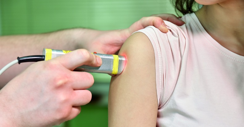
    The 2010 Olympic gold-medal downhill skier Lindsey Vonn was recently (March 8, 2010) interviewed in the magazine Time. Lindsey Vonn was the first American woman to win gold in an Olympic downhill, and she did so while being injured. One of the questions posed to her was:

    “How did you find the strength to ski with an injury?”

    Her response was, in part:

    “It’s been a real challenge for me to be able to ski well despite this injury. I’ve been doing as much therapy as I can.”

    “And I do laser therapy and massage.”

    •••••

    The healing of injured musculoskeletal tissue is primarily done by the formation of a protein patch. The primary repair protein is the connective tissue collagen. The primary cell that creates the collagen repair is the connective tissue fibrocyte cell.

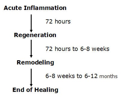
    Healing of injured musculoskeletal tissues takes place in three specific phases. A review of these phases is as follows (Kellett 1986):

    The first phase is called the acute inflammatory phase. This phase will last approximately 72 hours. After the initial injury, an electrical current is generated at the wound, called the “current of injury.” This “current of injury” attracts fibroblasts to the wound (Oschman, 2000). During this phase there is also initial bleeding and continual associated inflammation of the injured tissues. Because of the increasing inflammatory cascade during this period of time, it is not uncommon for the patient to feel worse for each of the first three days following injury. Because there is disruption of local vascular supplies, there is insufficient availability of substrate (glucose, oxygen, etc.) to produce large enough quantities of ATP energy to initiate collagen protein synthesis to repair the wound.

    After about 72 hours following injury, the damaged blood vessels have repaired. The resulting increased availability of glucose and oxygen elevates local ATP levels and collagen repair begins by the fibroblasts that accumulated during the acute inflammatory phase. This second phase of healing is called the phase of regeneration. During the regeneration phase the disruption in the injured muscles and ligaments is bridged. This phase will last approximately 6-8 weeks (Jackson, 1977). At the end of 6-8 weeks, the gap in the torn tissues is more than 90% bridged.

    There is a third and final phase of healing. This phase is called the phase of remodeling. The phase of remodeling starts near the end of the phase of regeneration. During the phase of remodeling the collagen protein glues that have been laid down for repair are remodeled in the direction of stress and strain. This means that the fibers in the tissue will become stronger, and will change their orientation from an irregular pattern to a more regular pattern, a pattern more like the original undamaged tissues.

    stages of healing following soft tissue injury

    It is established that remodeling takes place as a direct byproduct of motion. Traditional chiropractic management of injured musculoskeletal tissues involves the application of controlled motion into those tissues and joints. Consequently, chiropractic management has its greatest influence on the third phase of healing, the phase of remodeling. This is reflected in the studies that show that chiropractic spinal adjusting (specific directional manipulation) achieves its greatest clinical improvement in patients that are suffering from chronic problems and who have failed to achieve an acceptable clinical outcome from other approaches to management (Kirkaldy-Willis 1985, Meade 1990, Woodward 1996, Khan 1999, Giles 2003). The important question for this discussion is this:

    Is there a safe, effective approach to enhance the timing and quality of musculoskeletal healing that targets the second phase of the response, the phase of regeneration?

    Key to this discussion, and it is important to restate, is that ATP energy is required to synthesize repair proteins. In 1997, Douglas Wallace wrote an article for Scientific American titled “Mitochondrial DNA In Aging and Disease.” In this article, he notes that an intracellular organelle, the mitochondria, is the power plant of cells because it produces ATP energy. “Mitochondria provide about 90% of the energy that cells, and thus tissues, organs and the body as a whole, need to function.” Every cell in the body contains hundreds of mitochondria that produce the energy that the body requires.

    Synthesizing repair proteins occurs primarily in the phase of regeneration. Attachment of a single amino acid onto the string of amino acids that will become a protein requires the availability and expenditure of 4 ATP molecules (Champe 1994). Also, additional ATP molecules are required for both initiation and termination of the amino acid chain synthesis. The development of repair proteins is limited by the availability of ATP energy.

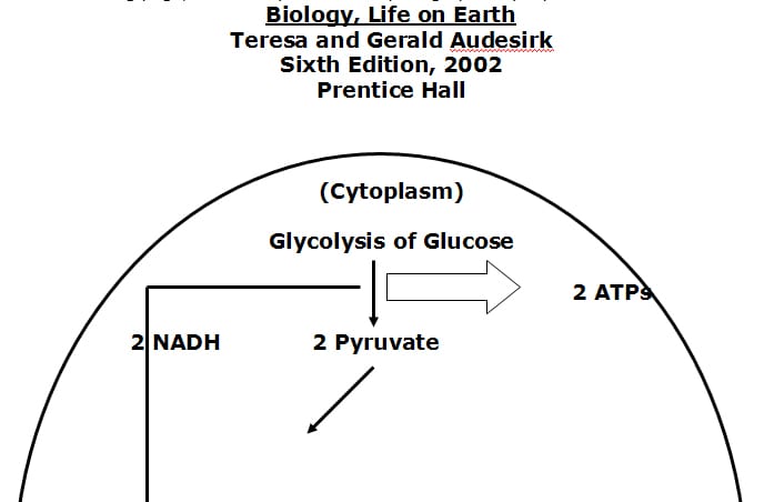
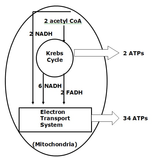
    A summary of the genesis of ATP energy is as follows:

    • The two variable but absolute substances required for ATP genesis are glucose and oxygen.

    • Without adequate oxygen the cytoplasm of the cell can produce 2 ATP molecules per glucose molecule. This is called anaerobic glycolosis. Aerobic life cannot be sustained through the anaerobically derived quantity of ATP energy.

    • With oxygen, additional ATP energy is formed in an intracellular organelle called the mitochondria.

    • With adequate oxygen, the mitochondrial Kreb’s Cycle can produce 2 more ATP molecules per original glucose molecule. Simultaneously, the Kreb’s Cycle is generating the all important electron transport proteins, which are shunted to the inner membrane of the mitochondria, know as the electron transport system (chain).

    • The electron transport chain of the mitochondria produces the majority of ATP molecules, an astonishing 34 per glucose molecule. Function and efficiency of the electron transport chain is also oxygen dependent.

    On the following page, a summary of these steps is graphically represented:

    image explained above

    image explained above

    Things that compromise the synthesis of ATP energy will impair healing. As an example, smoking cigarettes. The carbon monoxide in cigarette smoke has a greater affinity for hemoglobin than does oxygen. Smoking reduces the delivery of oxygen to the cells, impairing both the Kreb’s Cycle and electron transport chain genesis of ATP energy. Consequently, smokers heal more slowly and less completely. They suffer from accelerated tissue degradation and degeneration as a consequence of reduced ability to replicate proteins.

    In contrast, things that enhance tissue oxygenation will enhance the genesis of ATP energy, in turn enhancing the genesis of repair proteins. Some examples of this include:

    • Yogi breathing exercises (Weil 1996).

    Improved breathing results in improved tissue oxygenation, improving the genesis of mitochondrial (Kreb’s Cycle and electron transport chain) ATP, enhancing the timing and the quality of the genesis of repair proteins, accelerating healing.

    • Being aerobically fit.

    The number of mitochondria one has per cell is not a constant, it is a variable. The more aerobically fit one is the more mitochondria one will have per cell. The more mitochondria one has, the more efficient one is at utilizing glucose and oxygen in generating mitochondrial (Kreb’s Cycle and electron transport chain) ATP energy. More ATP enhances the timing and the quality of the genesis of repair proteins, accelerating healing.

    There is another variable controlling the mitochondrial genesis of ATP energy that is becoming increasingly noticed and documented in the National Library of Medicine literature database: it is an enzyme called:

    cytochrome c oxidase

    The cytochrome c oxidase enzyme is a terminal enzyme of the mitochondrial electron transport chain. In fact, it is the rate-limiting enzyme in the production of ATP energy by the electron transport system. Upregulating or increasing the activity of the cytochrome c oxidase will proportionally increase the genesis of ATP energy. How can the treating doctor upregulate or increase the activity of the cytochrome c oxidase enzyme? A portion of the answer is the understanding that “chrome” pertains to color. The remainder of the answer is Low Level Laser Therapy.

    Laser light is different than background environmental light. In the electromagnetic spectrum, visible background environmental light exists between wavelengths of about 400 – 800 nanometers (nm). Different wavelengths will produce different colors. As examples, 410 nm is a blue color, 535 nm is a green color, and 635 nm is a red color. Background environmental light is a combination of all wavelengths, and therefore of all colors. Laser light is monochromatic. This means laser light is of a single wavelength and therefore a single color. Laser eliminates all wavelengths but one.

    Background environmental light is also scattered, meaning the light is spread in all directions. In contrast, laser light is coherent. This means that the light is not scattered. All of the light waves are brought to a single point.

    As a consequence of laser light’s monochromatism and coherency, laser light can elicit interesting biological and physiological responses.

    In the understanding of low level laser physiology, it is important to restate that the primary producer of ATP energy is the electron transport system of the mitochondria. The inner membrane of the mitochondria contains 4 protein complexes called the respiratory chain. Electrons from food pass through these protein complexes with the help of Coenzyme Q10, interacting with oxygen and hydrogen to produce water and ATP energy.

    As the respiratory chain participates in ATP energy production, toxic by-products known as oxygen free radicals are generated. These free radicals can attack all components of cells, including respiratory chain proteins and mitochondrial DNA. Anything that impedes the flow of electrons through the respiratory chain can increase their transfer to oxygen molecules and promote the generation of free radicals. Importantly, anything that improves the flow of electrons through the respiratory chain will increases the production of ATP while reducing the generation of free radicals.

    Tiina Karu, PhD, is the world’s leading authority on low level laser therapy. Dr. Karu is Head of the Laboratory of Laser Biology and Medicine, Institute on Laser and Informatic Technologies of the Russian Academy of Science. Dr. Karu holds two PhD’s; one in Science and Biophysics, and another in Photochemistry. Pertaining to laser biophysics and laser physiology, Dr. Karu has authored 3 books, 16 book chapters, and 146 journal articles.

    Dr. Karu wrote “Low-Power Laser Therapy” in Biomedical Photonics Handbook, in 2003. She notes that low-level laser therapy probably works because the laser light is absorbed by the mitochondria photoreceptors, which enhances cellular metabolism. This means the mitochondria produce more ATP as a result of exposure to laser light. She notes that the primary reaction of laser light is in the mitochondria, which results in increased ATP energy. Dr. Karu states:

    “The mechanism of low-power laser therapy at the cellular level is based on the increase of oxidative metabolism of mitochondria, which is caused by electronic excitation of components of the respiratory chain.”

    “It is known that even small changes in ATP level can significantly alter cellular metabolism.”

    “Our results also provide evidence that various wavelengths (670, 632.8, and 820 nm) can be used for increasing respiratory activity.”

    “The photobiological action mechanism via activation of the respiratory chain is a universal mechanism” [for laser light effects].

    In 2007, Dr. Karu wrote a book titled:

    Ten Lectures on Basic Science of Laser Phototherapy

    This book is perhaps the most detailed accounting of the biological and physiological basis for low-level laser therapy published to date. Dr. Karu reviews how laser radiation can modulate cell metabolism through the mediation of a universal photoreceptor, the cytochrome c oxidase enzyme. Once again, the cytochrome c oxidase enzyme is the enzyme that catalyzes the final step in the mitochondrial respiratory chain, thereby becoming the rate-limiting step in the genesis of ATP energy. In this book, Dr. Karu makes the following points:

    Cytochrome c oxidase plays a central role in the bioenergetics of the cell.”

    “It is well known that it is only in the ideal case where the supply of electrons from cytochrome c oxidase is unlimited that electrons are always present to reduce oxygen,” thus maximizing production of ATP and minimizing production of oxygen free radicals.

    “One of the most important biological mechanisms [for low level laser therapy is] based on activation/upgrading of the terminal enzyme of the mitochondrial respiratory chain, cytochrome c oxidase, is shown to be a universal mechanism controlling many aspects of the metabolism in different types of [laser] irradiated cells.”

    “The activation of cells via the mitochondria is suggested to be a universal photobiological mechanism.”

    “Laser phototherapy is used by physiotherapists (to treat a wide variety of acute and chronic musculoskeletal aches and pains), by dentists (to treat inflamed oral tissues and to heal diverse ulceration), by dermatologists (to treat edema, indolent ulcers, burns, and dermatitis), by rheumatologists (to relieve pain and treat chronic inflammation and autoimmune diseases), and by other specialists, as well as by general practitioners. Laser phototherapy is also widely used in veterinary medicine (especially in racehorse-training centers) and in sports medicine and rehabilitation clinics (to reduce acute swelling and hematoma, relieve pain, improve mobility, and treat acute soft-tissue injuries.”

    •••••

    Another reference text pertaining to low-level laser therapy was published in 2002 by Jan Turner and Lars Hode, titled:

    Laser Therapy Clinical Practice and Scientific Background

    This book contains 1,281 references. These authors note:

    1) “Today, we can safely say that therapeutic lasers have an important biological effect, and a very positive one at that.”

    2) “We believe that lasers have a tremendous and as yet untapped potential in the field of healthcare.”

    3) “Therapeutic lasers have no undesirable side effects in the hands of a reasonably qualified therapist.”

    4) Lasers are “sterile, painless, and often less expensive than methods already in use,” and does not have side effects as does pharmacotherapy.

    5) “Laser therapy of wounds is ideal, since it promotes healing and reduces pain at the same time.”

    6) Laser light increases the cell’s ATP energy.

    In their text, Turner and Hode note that the first company to receive a 510(k) market clearance from the Federal Drug Administration (FDA) was a US company, Erchonia Laser. This occurred in 2002. There are now (as of March 28, 2010) 7 FDA market clearances for low-level laser therapy, Erchonia obtaining 5 of the 7. Erchonia is located in McKinney, TX, and they can be contacted at phone number (214) 544-2227 or web address www.erchonia.com.

    •••••

    A recent (March 28, 2010) search of the National Library of Medicine database using the PubMed search engine and the words “low-level laser therapy” brought up 2,204 titles. Two representative articles pertaining to low level laser therapy and tissue healing are reviewed below:

    Effects of Helium-Neon Laser on Levels of Stress Protein and Arthritic Histopathology in Experimental Osteoarthritis

    American Journal of Physical Medicine & Rehabilitation

    October 2004, 83(10):758-765

    Lin, Yueh-Shuang MS; Huang, Mao-Hsiung MD, PhD; Chai, Chee-Yin MD, PhD; Yang, Rei-Cheng MD, PhD

    In this study, researchers injured the knees of 42 rats giving them arthritis. Twenty-one of the rats were given 632 nm low-level laser (red color), applied over the arthritic knee for 15 minutes, three times per week, for 8 weeks; the other 21 rats were not similarly exposed. The results showed a marked repair of arthritic cartilage in the lased rats, but not in the non-lased group. The authors concluded that the 632 nm low-power laser enhances protein production in arthritic joints and repairs the arthritic cartilage.

    These authors also note: Laser is “thought to cause electronic excitation of the photoacceptor molecules, which are thought to be various cytochrome enzymes that are terminal electron carriers in the respiratory chain.” This is thought to accelerate electron transfer. “Electron transport in the mitochondrial membrane is one of the main fueling mechanisms underpinning metabolism and proliferation of cells, including generation of adenosine triphosphate (ATP).” “Low-level laser mediated increase in efficiency of the electron carriers in the respiratory chain would increase generation of adenosine triphosphate, which could manifest itself as increased DNA and protein synthesis and result in cell proliferation, as shown in present study.”

    In summary, and consistent with the explanations above, these researchers credited the outcomes of this animal study to laser driven excitation of the cytochrome enzyme terminal electron carriers of the mitochondrial respiratory chain, increasing ATP synthesis and enhancing repair protein production.

    •••••

    Therapeutic low powered lasers are commercially available in many different wavelengths and power outputs. It appears clear that there is no one wavelength that is ideal for all appropriately treated clinical syndromes. Few studies compare the outcome of different wavelengths and exposure fluences on measured outcomes. However, the second reviewed article did just that:

    Comparative Study Using 685-nm and 830-nm Lasers in the Tissue Repair of Tenotomized Tendons in the Mouse

    Photomedicine and Laser Surgery

    December 2006, Volume 24, Number 6, pp. 754–758

    Carrinho PM, Renno ACM, Koeke P, Salate ACB, Parizotto NA, Vidal BC

    In this study, the authors compared the tissue repair of injured mouse tendons when treated with either a 685 nm laser or an 830 nm laser, each at fluences of both 3 J/cm2 and 10 J/cm2. This study used 48 mice that were divided into six experimental groups:

    Group A, tenomized animals, treated with 685 nm laser, at the dosage of

    3 J/cm2.

    Group B, tenomized animals, treated with 685-nm laser, at the dosage of

    10 J/cm2.

    Group C, tenomized animals, treated with 830-nm laser, at dosage of 3 J/cm2.

    Group D, tenomized animals, treated with 830-nm laser, at dosage of 10 J/cm2.

    Group E, injured control (placebo treatment).

    Group F, non-injured standard control.

    Laser irradiation started 24 hours after the tenotomy of the Achilles tendon. A total of 12 laser sessions were performed on consecutive days. The rats were killed on day 13, and the injured tendons were surgically removed and analyzed with polarized light microscopy to analyze the organization and molecular order of the collagen fibers. All laser treated groups showed improved healing when compared to injured control group. The best organization and aggregation of the collagen bundles was shown by the animals of group A (685 nm, 3 J/cm2), followed by the animals of group C (830 nm, 3 J/cm2), and B (685 nm, 10 J/cm2), and finally, the animals of group D (830 nm, 10 J/cm2). The authors concluded:

    “All wavelengths and fluences used in this study were efficient at accelerating the healing process of Achilles tendon post-tenotomy, particularly after the 685-nm laser irradiation, at 3 J/cm2. It suggests the existence of wavelength tissue specificity and dose dependency.”

    Interestingly, in this study, the shorter wavelength was associated with the better healing outcome. Counterintuitively, lesser exposure to laser irradiation resulted in an improved healing outcome than higher doses of exposure. These authors note:

    “The better tissue response was observed after the irradiation with the 685-nm laser, at the dosage of 3 J/ cm2.”

    “The animals irradiated with the 830-nm laser, at the dosage of 10 J/ cm2 presented the weaker response to laser irradiation.”

    “The best tissue response was obtained after the 685-nm laser irradiation, at the dosage of 3 J/cm2.”

    Specifically, the 685-nm laser irradiation at 3 J/cm2 showed (the shortest of the compared wavelengths and the lowest amount of irriadence):

    16% improved tendon healing over the 830-nm laser at 3 J/cm2.

    33% improved tendon healing over the 685-nm laser at 10 J/cm2.

    54% improved tendon healing over the 830-nm laser at 10 J/cm2.

    Compared to the control tendons, the improved tissue response was as follows:

    101% improved tissue response with the 830-nm laser at 10 J/cm2.

    114% improved tissue response with 685-nm laser at 10 J/cm2

    167% improved tissue response with the 830-nm laser at 3 J/cm2.

    208% improved tissue response with 685-nm laser irradiation at 3 J/cm2

    These authors concluded:

    “Our results suggest that laser irradiation produced an increase of cell proliferation through changes in mitochondrial physiology, subsequently affecting RNA synthesis, which, in turn, alters the expression of various cell regulatory proteins.”

    •••••

    In summary, the evidence suggests that low level laser therapy targets the mitochondrial enzyme that is the rate-limiting factor in the production of ATP energy, the cytochrome c oxidase enzyme. The increased production of ATP energy allows the DNA to enhance the replication of repair proteins, accelerating healing and improving symptoms. Since lasers can achieve this without side effects or risks, low-level laser therapy is here to stay. As a consequence, many chiropractors now incorporate low level laser therapy as a component of their patient clinical management, especially of injured patients.

    References:

    Audesirk T, Audesirk G. Biology, Life on Earth Sixth Edition, Prentice Hall 2002.

    Carrinho PM, Renno ACM, Koeke P, Salate ACB, Parizotto NA, Vidal BC; Comparative Study Using 685-nm and 830-nm Lasers in the Tissue Repair of Tenotomized Tendons in the Mouse; Photomedicine and Laser Surgery; December 2006, Volume 24, Number 6, pp. 754–758.

    Champe PC, Havey RA. Lippincott’s Illustrated Reviews: Biochemistry, Second Edition, Lippincoll Eilliams & Wilkins, 1994.

    Giles LGF, Muller R. Chronic Spinal Pain: A Randomized Clinical Trial Comparing Medication, Acupuncture, and Spinal Manipulation. Spine. July 15, 2003; 28(14):1490-1502.

    Jackson, R. The Cervical Syndrome, Fourth Edition, Thomas, 1977.

    Karu T. “Low-Power Laser Therapy” Chapter 48 in Biomedical Photonics Handbook, Tuan Vo-Dinh, CRS Press, 2003.

    Karu T. Ten Lectures on Basic Science of Laser Phototherapy. Prima Books

    2007.

    Kellett, John, “Acute soft tissue injuries-a review of the literature,” Medicine and Science of Sports and Exercise, American College of Sports Medicine, Vol. 18 No.5, (1986), pp 489-500.

    Khan S, Cook J, Gargan MF, Bannister GC. A symptomatic classification of whiplash injury and the implications for treatment. The Journal of Orthopaedic Medicine 21(l) 1999, 22-25.

    Kirkaldy-Willis, W.H., M.D., & Cassidy, J.D., “Spinal Manipulation in the Treatment of Low-Back Pain,” Canadian Family Physician, (1985), 31:535-40.

    Lin, Yueh-Shuang MS; Huang, Mao-Hsiung MD, PhD; Chai, Chee-Yin MD, PhD; Yang, Rei-Cheng MD, PhD, Effects of Helium-Neon Laser on Levels of Stress Protein and Arthritic Histopathology in Experimental Osteoarthritis. American Journal of Physical Medicine & Rehabilitation. 83(10):758-765, October 2004.

    Meade TW, Dyer S, Browne W, Townsend J, Frank AO. Low back pain of mechanical origin: Randomized comparison of chiropractic and hospital outpatient treatment. British Medical Journal. Volume 300, JUNE 2, 1990, pp. 1431-7.

    Oschman, James L, Energy Medicine: The Scientific Basis, Churchill Livingstone, 2000.

    Turner J, Hode L, Laser Therapy Clinical Practice and Scientific Background, Prima Books, 2002.

    Wallace D, Scientific American, Mitochondrial DNA In Aging and Disease, Scientific American, August 1997.

    Weil A. Spontaneous Healing. Ballantine Books, 1996.

    Woodward NM, Cook JHC, Gargan MF, Bannister GC. Chiropractic treatment of chronic ‘whiplash’ injuries. Injury. Volume 27, Issue 9, November 1996, Pages 643-645.

  • Osteoarthritis

    Osteoarthritis

    November 2, 2010, researchers from the Veterans Affairs Medical Center at the University of California, San Francisco, published a study in the Annals of Internal Medicine titled (1):

    The Epidemiology of Pain During the Last 2 Years of Life

    The study included 4,703 older adults with mean age of 75.7 years at the time of their deaths. These subjects were evaluated for the presence of clinically significant pain, as indicated by a report that the participant was “often troubled” by pain of at least moderate severity. Their records showed that the prevalence of such pain 24 months before death was 26%. The pain prevalence remained flat until 4 months before death, then the pain increased, reaching 46% in the last month of life.

    Importantly, the prevalence of pain in the last month of life was 60% among patients with arthritis versus 26% among patients without arthritis and did not differ by terminal diagnosis category (cancer, heart disease, frailty, sudden death, or other causes).

    The authors concluded:

    “Although the prevalence of pain increases in the last 4 months of life, pain is present in more than one quarter of elderly persons during the last 2 years of life.”

    “Arthritis is strongly associated with pain at the end of life.”

    This study indicates that a non-fatal condition, arthritis, significantly affects the quality of life in the elderly.

    •••••

    A number of well-respected reference texts indicate that the incidence of osteoarthritis is strongly associated with single event macro-trauma, repeated micro-trauma, alignment problems, and age. Such texts include:

    • The Cervical Syndrome by Ruth Jackson (3).
    • Anatomico-Roentgenographic Studies of the Spine by Lee Hadley (4).
    • Managing Low Back Pain by Harry Kirkaldy-Willis (5).
    • Clinical Implications of Normal Biomechanical Stresses on Spinal Function by Herbert Junghanns (6).

    A 1993 study published in the journal Injury (7) showed strong evidence that a single motor vehicle collision accelerates the process of disc degenerative disease of the cervical spine.

    A 1997 study published in the Journal of Orthopedic Medicine (8) showed that a single motor vehicle collision accelerated degenerative changes in the cervical spine by about 10 years earlier compared to the control group.

    A 2004 study published in the European Spine Journal (9) showed that the repetitive heading of soccer balls accelerates degenerative changes in the cervical spine by 10–20 years earlier than that of the normal population.

    A 2009 study published in the Journal of Physical Activity and Health (10) showed that knee and hip arthritis is over 3 times more prevalent in retired NFL players than in the general U.S. population of the same age.

    PATHOPHYSIOLOGY

    An excellent discussion of the pathophysiological process leading to spinal osteoarthritis is presented by the late (d. 2006) William H Kirkaldy-Willis, MD. Dr. Kirkaldy-Willis was Emeritus Professor, Department of Orthopaedic Surgery, University Hospital, University of Saskatchewan College of Medicine. In his 1988 (second edition) book Managing Low Back Pain, there are 19 international multidisciplinary distinguished contributing authors (5). Dr. Kirkaldy-Willis authored a chapter in the book titled:

    “The Three Phases of the Spectrum of Degenerative Disease”

    Dr. Kirkaldy-Willis describes how spinal segments are comprised of a three-joint complex: the two posterior facet joints and the intervertebral disc. He notes that the three joints always work together. Consequently, injury or stress to any single component of the three-joint complex will mechanically affect the other two components. His breakdown of the three phases of spinal degenerative disease is as follows:

    First Phase of Spondylosis

    “Dysfunction”

    In the first phase, the normal function of the three-joint complex is interrupted as a consequence of injury or chronic stress. This causes the posterior musculature of the involved segment to go into a state of hypertonic contraction. This restricts normal movement. The hypertonic contraction of the muscles also causes muscle ischemia, causing pain. The muscle hypertonicity also causes a slight misalignment of the posterior facet joints, which is known as a “subluxation.” Eventually tissue fibrosis begins to appear.

    Second Phase of Spondylosis

    “Instability”

    If the first phase is allowed to persist, the second phase will eventually ensue. In the second phase, there is abnormal increased movement. Laxity of the posterior joint capsule and of the annulus fibrosus is seen in anatomical sections. Local fibrosis is problematic because “the collagen of scar tissue is not as strong as normal collagen.” Therefore, there is an increased propensity for additional injury, inflammation, pain, and muscle hypertonicity.

    Third Phase of Spondylosis

    “Stabilization”

    If the second phase is allowed to persist, the third phase will eventually ensue. In the third phase, degenerative changes begin to appear. As the degenerative changes advance, the unstable segment regains its stability because of fibrosis and osteophytes form around the posterior joints and within and around the disc. In this stabilization phase, the facet joints will progress through the following sequence:

    Synovitis

    Degeneration of Articular Hyaline Cartilage

     ↓

    Development of Intra-articular Adhesions

     ↓

     Increasing Capsular Laxity

    Subluxation of the Joint Surfaces

     ↓

    Formation of Subperiosteal Osteophytes

     ↓

    Enlargement of Both Inferior and Superior Facets

     ↓

    Ultimately Greatly Reduced Movement

    Simultaneous with this sequence of changes in the facet joints, there are parallel changes in the intervertebral disc. These changes follow this sequence:

    The Development of Small Circumferential Tears in the Annulus Fibrosis

     ↓

    The Circumferential Tears in the Annulus Become Larger and Coalesce to form Radial Tears that Pass From the Annulus to the Nucleus Pulposus

     ↓

    Eventual Internal Disruption of the Disc

     ↓

    Disc Degeneration with Disc Resorption

     ↓

    Peripheral Osteophytes Around the Circumference of the Disc

    Ultimately Greatly Reduced Movement

    Dr. Kirkaldy-Willis notes that the “greatly reduced motion” associated with spinal degenerative disease opens Melzack’s and Wall’s “pain gate,” accounting for the increased pain that is often associated with osteoarthritis (12).

    •••••

    A more recent comprehensive review of osteoarthritis appeared in the American Journal of Physical Medicine and Rehabilitation in 2006, titled (11):

    Osteoarthritis

    Epidemiology, Risk Factors, and Pathophysiology

    The authors, Susan V Garstand, MD and Todd P Stitik, MD, are from the University of Medicine and Dentistry of New Jersey. In this article, Drs. Garstand and Stitik note that osteoarthritis is “the clinical and pathologic outcome of a range of disorders that results in structural and functional failure of synovial joints. Osteoarthritis occurs when the dynamic equilibrium between the breakdown and repair of joint tissues is overwhelmed.”

    Drs. Garstand and Stitik note that osteoarthritis is the most prevalent form of arthritis and a major cause of disability in people aged 65 and older. Osteoarthritis affects the majority of adults over age 55.

    Garstand and Stitik note that the incidence of osteoarthritis is influenced by both systemic and local factors:

    Systemic Factors:


    • Ethnicity

    • Age: “The presence of radiographic osteoarthritis rises with age at all joint sites.”

    • Gender

    • Hormonal Status

    • Genetic Factors; osteoarthritis has a major genetic component

    • Bone Density

    • Nutritional Factors:

    There is evidence that osteoarthritis is linked to free radicals, and that high dietary antioxidants (especially vitamins C and D) are protective against the development of osteoarthritis. “Chondrocyte senescence is thought to be the result of chronic oxidative stress.”

    Local Factors: Local factors “result in abnormal biomechanical loading of

    affected joints.”

    • Obesity

    • “Altered joint biomechanics”

    • ligamentous laxity

    • malalignment

    • impaired proprioception. With aging, there is a decline in proprioception, causing

    decreased neurologic responses, impairing proprioceptive joint-protective

    mechanisms. Consequently, reduced proprioception advances osteoarthritis.

    • muscle weakness

    • Prior joint injuries

    • Occupational Factors

    • Effects of sports and physical activities

    • Developmental abnormalities

    Garstand and Stitik note that if systemic factors are present, the joints are vulnerable, and thus local biomechanical factors will have more of an impact on joint degeneration and osteoarthritis.

    •••••

    Essential Fatty Acid Connection

    In his 2008 book Toxic Fat, biochemist Barry Sears (13) describes how the omega-6 fatty acid arachidonic acid is biochemically converted into the pro-inflammatory eicosanoid hormone prostaglandin E2 (PGE2).

    COX LOX pathways

    Cyclo-oxygenase (COX)/Lipo-oxygenase (LOX) Pathways

    The conversion of the omega-6 fatty acid arachidonic acid is biochemically converted into the pro-inflammatory eicosanoid hormone prostaglandin E2 (PGE2) and is important in a discussion of osteoarthritis because a number of studies have implicated prostaglandin E2 in the accelerated degradation of cartilage. For example, in 2002 the journal Neurosurgery Focus published a study linking disc degeneration to prostaglandin E2 (14). Later in 2002, the journal Spine published a study linking intervertebral disc herniation to the enzyme cyclo-oxygenase-2, which converts the toxic fat arachidonic acid to prostaglandin E2 (15).

    Also in 2002, the journal Arthritis & Rheumatism (16) published a study showing that the pathologic indicators manifested in human osteoarthritic cartilage could be significantly abrogated by exposure of the cartilage to omega-3 fatty acids. This is important because omega-3 fatty acids, especially the omega-3 eicosapentaenoic acid (EPA), is known to inhibit the conversion of arachidonic acid into prostaglandin E2. Neither omega-6 nor omega-9 (such as olive oil) could stop the cartilage breakdown biomarkers. These authors concluded that omega-3 fatty acids play a role in halting or slowing degradative and inflammatory factors that contribute to the progression of osteoarthritis. Specifically, they state:

    “Dietary supplementation with n-3 PUFA may prove useful in both quiescent and active arthritis.”


    “Our findings support the results of epidemiologic and clinical studies that have demonstrated dietary supplementation with omega-3 fatty acids to be beneficial in reducing pain and inflammation in human arthritic diseases.”


    “It has long been recognized that dietary supplementation with fish oils that are enriched with n-3 PUFAs can provide benefit in the treatment of arthritis.”


    •••••

    Nutritional Management

    In 2006, Leslie Cleland and colleagues published a study pertaining to the utilization of omega-3 fish oil prescription for the purposes of achieving an anti-inflammatory state in arthritis sufferers (17). The pain of arthritis is primarily caused by prostaglandin E2 (PGE2), which is derived from the omega-6 fatty acid arachidonic acid through the activity of the enzyme COX. Nonsteroidal anti-inflammatory drugs (NSAIDs) inhibit the COX enzyme. Dr. Cleland restates that the omega-3 fatty acid found in fish oil, eicosapentaenoic acid (EPA), not only inhibits the conversion of arachidonic acid to the pro-inflammatory eicosanoid prostaglandin E2 (PGE2), but also inhibits the conversion of arachidonic acid into another pro-inflammatory eicosanoid, leukotriene B4 (LTB4). Cleland and colleagues suggest that as a therapeutic agent, EPA should be superior to NSAIDs because NSAIDs do not inhibit the lipoxygenase (LOX) pathway.

    Cleland and colleagues state that in order to achieve an anti-inflammatory state for arthritis patients, they would need to consume a minimum of 2,700 mg of long chain omega-three fatty acids per day, and they would have to do so daily for a minimum of 2-3 months.

    osteoarthritis process

    Also in 2006, Dr. Joseph Maroon, the neurosurgeon for the Pittsburgh Steelers, published a study in the journal Surgical Neurology titled (18):


    Omega-3 Fatty acids (fish oil) as an anti-inflammatory:

    An alternative to nonsteroidal anti-inflammatory drugs for discogenic pain

    This paper won first prize in the poster competition at the American Association of Neurological Surgeons Annual Meeting, New Orleans, LA, April 2005. Dr. Maroon is a specialist in the management of degenerative spinal disease in the Department of Neurological Surgery at the University of Pittsburgh Medical Center.

    In this study, Dr. Maroon notes that NSAIDs are the most common cause of drug-related morbidity and mortality reported to the FDA and other regulatory agencies around the world. The use of NSAIDs is associated with extreme complications, including gastric ulcers, bleeding, myocardial infarction, stroke, and even death. “More than 70 million NSAID prescriptions are written each year, and 30 billion over-the-counter NSAID tablets are sold annually.” “The agent best documented by hundreds of references in the literature for its anti-inflammatory effects is omega-3 EFAs found in fish and in pharmaceutical-grade fish oil supplements.”

    In this study, after 75 days on fish oil, 59% of patients who were taking NSAIDs for chronic spinal pain and who had spinal osteoarthritis, were able to discontinue their prescription NSAIDs, and 88% stated they were satisfied with their improvement and that they would continue to take the fish oil. Dr. Maroon concluded:

    “Omega-3 essential fatty acid fish oil supplements appear to be a safer alternative to NSAIDs for treatment of nonsurgical neck or back pain.”

    Mechanical Management

    Drs. Garstand and Stitik (11) indicate that there exists biomechanical factors in the development of osteoarthritis, including altered joint biomechanics, reduced motion, ligamentous laxity, malalignment, impaired proprioception, and muscle weakness. Chiropractic training and clinical practice emphasizes and specializes in the diagnosis and management of these problems.

    Additionally, Dr. Kirkaldy-Willis (5) indicates that it is during the First Phase or Dysfunction Phase of spinal degeneration when most patients experience their first episode of back pain. Importantly, Dr. Kirkaldy-Willis indicates that the pathological changes during the First Phase are minor and potentially reversible. Dr. Kirkaldy-Willis suggests that spinal manipulation is a viable approach for the management of this first phase of spinal degeneration, noting:

    “Manipulation is an art that requires much practice to acquire the necessary skill and competence. Few medical practitioners have the time or inclination to master it. This modality has much to offer to the patient with low back pain, especially in the earlier stages during the phase of dysfunction. The majority of patients are first seen while in this phase. Most practitioners of medicine, whether family physicians, or surgeons, will wish to refer their patients to a practitioner of manipulative therapy with whom they can cooperate, whose work they know, and whom they can trust. The physician who makes use of this resource will have many contented patients and save himself many headaches.”

    Although this quote suggests that joint manipulation is most beneficial during the earliest phases of joint degeneration, a number of studies document that joint manipulation is excellent or even superior in the management of patients suffering from chronic back pain (19, 20, 21). Long-term chronicity of spinal pain (up to 8.3 years, (21)), suggests that these patients are not in earlier phases of spinal degeneration, but are probably more in the phase of stabilization with osteoarthritic changes.

    Summary

    The eicosanoid hormone prostaglandin E2 is best known as a pain mediator, but it is also linked to the development of osteoarthritis. Studies indicate that an effective management for both the pain and the development of osteoarthritis is supplemental omega-3 fatty acid fish oil. Chiropractors are well trained in the biochemistry and application of such omega-3 supplements.

    Biomechanically, spinal osteoarthritis begins as an injury or stress that causes local muscular hypertonicity, segmental joint dysfunction and misalignment. This early stage of pathophysiology is ideally aborted with spinal manipulation. Later stages of the osteoarthritic pathophysiological response, involving altered proprioception, muscle weakness, and reduced mobility from stabilization are also improved with joint manipulation, exercise, and targeted tissue work. The modern chiropractor is well trained in each of these clinical approaches.

    Additionally, today’s modern chiropractor has management skills in several of the systemic factors in osteoarthritis, including obesity, free radical damage, repetitive strains, and ergonomics.

    REFERENCES

    1) Smith AK, Cenzer IS, Knight SJ, Puntillo KA, Widera E, Williams BA, Boscardin WJ, Covinsky KE; Annals of Internal Medicine; The epidemiology of pain during the last 2 years of life; November 2, 2010;153(9):563-9.

    2) Schmoral G, Junghanns H; Schmorl’s and Junghanns’ The Human Spine in Health and Disease; Grune & Stratton; 1971.

    3) Jackson R, The Cervical Syndrome, Thomas, 1978.

    4) Hadley LA, Anatomico-Roentgenographic Studies of the Spine, fourth printing, Charles Thomas, 1979.

    5) Kirkaldy-Willis H; Managing Low Back Pain, Second Edition, Churchill Livingstone, 1988.

    6) Junghanns H; Clinical Implications of Normal Biomechanical Stresses on Spinal Function; Aspen, 1990.

    7) Hamer AJ, Gargan MF, Bannister GC, Nelson RJ. Whiplash injury and surgically treated cervical disc disease. Injury. 1993 Sep;24(8):549-50.

    8) Gargan MF, Bannister GC. The Comparative Effects of Whiplash Injuries. The Journal of Orthopaedic Medicine, 19(1), 1997, pp. 15-17.

    9) Kartal A, Yldran B, Enköylü A, Korkusuz F; Soccer causes degenerative changes in the cervical spine; European Spine Journal, February 2004, 13(1):76-82.

    10) Golightly YM, Marshall SW, Callahan LF, Guskiewicz K; Early-Onset Arthritis in Retired National Football League Players; Journal of Physical Activity and Health, 2009, 6, 638-643.

    11) Garstand SV, Stitik, TP; Osteoarthritis: Epidemiology, Risk Factors, and Pathophysiology; American Journal of Physical Medicine and Rehabilitation; November 2006, Vol. 85, No. 11, pp. S2-S11.

    12) Melzack R, Wall PD. Pain mechanisms: a new theory. Science. 1965 Nov 19;150(699):971-9.

    13) Sears B; Toxic Fat; 2008.

    14) Martin MD, Boxell CM, Malone DG; Pathophysiology of lumbar disc degeneration: A review of the literature; Neurosurgery Focus; Vol. 13, No. 2, August 2002.

    15) Miyamoto H, Saura R, Doita M, Kurosaka M, Mizuno K; Role of Cyclo-oxygenase-2 in Lumbar Disc Herniation; Spine; Vol. 27, No. 22, November 15, 2002, pp. 2477-2483.

    16) Curtis CL, Rees SG, Little CB, Flannery CR, Hughes CE, Wilson C, Dent CM, Otterness IG, Harwood JL, Caterson B; Pathologic indicators of degradation and inflammation in human osteoarthritic cartilage are abrogated by exposure to n-3 fatty acids; Arthritis & Rheumatism; Volume 46, Issue 6, 2002, pp 1544-1553.

    17) Cleland LG, James MJ, Proudman SM; Fish oil: what the prescriber needs to know; Arthritis Research & Therapy; Volume 8, Issue 1, 2006.

    18) Maroon JC, Bost JW; Omega-3 Fatty acids (fish oil) as an anti-inflammatory: an alternative to nonsteroidal anti-inflammatory drugs for discogenic pain; Surgical Neurology; 65 (April 2006) 326– 331.

    19) Kirkaldy-Willis WH, Cassidy JD; Spinal Manipulation in the Treatment of Low back Pain; Canadian Family Physician; March 1985, Vol. 31, pp. 535-540.

    20) Meade TW, Dyer S, Browne W, Townsend J, Frank AO; Low back pain of mechanical origin: Randomized comparison of chiropractic and hospital outpatient treatment; British Medical Journal; Volume 300, JUNE 2, 1990, pp. 1431-7.

    21) Giles LGF, Muller R; Chronic Spinal Pain: A Randomized Clinical Trial Comparing Medication, Acupuncture, and Spinal Manipulation; Spine, July 15, 2003; 28(14):1490-1502.

  • Soft Tissue Injury and Repair

    Soft Tissue Injury and Repair

    HISTORY:

    Mandy, a 50-year-old female, was injured in a motor vehicle collision.

    Her stationary vehicle was struck from the rear by a vehicle of similar mass that was traveling at a speed of approximately 20 miles per hour at the time of collision. Damage to her vehicle was judged to be $6,600.

    The collision caught Mandy by surprise, and she was looking left at the moment of impact. This is important because there is good evidence (Sturzenegger 1994, Sturzenegger 1995) that factors associated with greater initial injury and a worse outcome one year after injury are (in descending order):

    1) Being involved in a collision in which the patient is caught by surprise.

    2) Being involved in a collision in which the patient’s head is rotated.

    3) Being involved in a collision in which the impact is from a rear-end direction.

    Although dazed, Mandy did not lose consciousness. An ambulance did take her to the emergency department of her own HMO, which was geographically close by.

    At the hospital, Mandy was evaluated and radiographs were taken. There were no broken bones, signs of instability, congenital anomalies, or degenerative disease. She was told she had suffered a soft tissue injury and that it would get better in a few days to a few weeks.

    For treatment, Mandy’s doctors put her into a soft cervical spine collar, she was prescribed a nonsteroidal anti-inflammatory drug, and given a heat pack for home use. She was advised to return for a follow-up visit if she was having continuing problems. Mandy was advised to continue to work in her usual occupation as a real estate agent.

    Mandy began wearing her cervical spine soft collar most of the time. Although the anti-inflammatory medicines and the hot pack seemed to help her (this seems

    paradoxical), she did not seem to be making overall relevant improvement. She continued to have significant neck pain and stiffness with an occasional headache.

    Mandy did continue to work and actually missed no work at all. However, her neck was sore and stiff making her grumpy, and she became fatigued easily.

    Mandy wore her cervical collar nearly constantly for about two weeks, then she reduced its usage to only when she was engaging in more strenuous activities, such as driving or shopping, and during certain activities at work. She liked her hot pack and she used it on average 3-4 times per day. She still needed her pain medicine, taking them a few times daily.

    After about two months of being essentially unchanged, Mandy returned to her HMO physician and asked him if there was anything else she could do for her neck pain with occasional headaches. Her doctor authorized for Mandy to see one of the HMO’s physical therapists. She was prescribed one physical therapy session per week for the next four weeks.

    Mandy’s physical therapist talked to her about her posture and gave her some exercises. He evaluated the ergonomics of her desk, primarily as related to when she was doing work on her computer.

    After her four physical therapy visits, Mandy had incorporated his advice and exercises into her routine. Yet, she continued to suffer from neck pain and stiffness with occasional headaches.

    Nine months after being injured, Mandy was still suffering and she could not go a full day without taking pain medications. Mandy needed to do something different.

    Mandy became my patient nine months after being injured.

    •••••

    There is a common misconception that injured soft tissues will heal in a period of time between four and eight weeks. It is frequently claimed that injured soft tissues will heal spontaneously, leaving no long-term residuals, and that treatment is not required. This type of information is misleading and confusing because it is not true. As an example, in 2008, The American Journal of Medicine published a systemic review of the literature evaluating the clinical course of acute ankle sprain (Rijn 2008). The authors conducted a database search in MEDLINE, CINAHL, PEDro, EMBASE, and the Cochrane Controlled trial register. They found 31 studies that met their inclusion criteria. Their findings include:

    • 5% to 33% of patients still experienced ankle pain after 1 year.
    • The studies reported an incidence of subjective instability of their injured ankle in up to 53% patients.
    • 15% to 64% had not fully recovered at 3 years.
    • The incidence of re-sprains ranged from 3% to 34% of the patients.

    Most of my med-legal experience is with whiplash injury. Often, insurance defense personnel and their chiropractic/medical experts make an analogy between the whiplash-injured neck and a sprained ankle. Their classic claim is that a sprained ankle will heal spontaneously (without any treatment) and quickly (weeks), and there are no long-term residuals.

    This article by Rijn presents a much different reality pertaining to the healing of the sprained ankle: at 3 years up to 64% have not fully recovered, up to 33% have residual pain, up to 53% suffer from residual instability, and up to 34% suffer from re-injury. It appears that 15% to 64% have some degree of permanent injury. Additionally, the severity of ankle injury is not a strong predictor for the ultimate clinical outcome. It appears that trauma from ankle sprain and whiplash have a number of shared characteristics: significant residual pain, instability, re-injury rates, permanent injury residuals, and the severity of injury not being a predictor for the ultimate clinical outcome.

    Consequently, I believe that the most important question is:

    Is there an approach to the management of injured soft tissues that improves the timing and quality of the healing outcome?

    •••••

    Published articles and books concerning the healing of injured soft tissues (Oakes 1982; Roy and Irving 1983; Kellett 1986; Buckwalter/Woo 1988, Majno 2004) indicate that the time frame for soft healing is approximately one year.

    The healing of injured soft tissues takes place in three specific phases. The first phase is called the acute inflammatory phase. This phase will last approximately 72 hours. During this phase, after the initial injury, an electrical current is generated at the wound, called the “current of injury.” This “current of injury” attracts fibroblasts to the wound (Oschman, 2000). During this phase there is also initial bleeding and continual associated inflammation of the injured tissues. Because of the increasing inflammatory cascade during this period of time, it is not uncommon for the patient to feel worse for each of the first three days following injury. Because there is disruption of local vascular supplies, there is insufficient availability of substrate (glucose,

    oxygen, etc.) to produce large enough quantities of ATP energy to initiate collagen protein synthesis to repair the wound.

    Experience and published studies (Kellett 1996) indicate that the best management of soft tissue injuries during the acute inflammatory phase is ice therapy. Ice therapy during the first 72 hours following injury reduces pain and swelling, and minimizes the formation of scar tissue that often causes prolonged disability (Seletz 1958). [Unfortunately for Mandy, during this phase of soft tissue healing, she was prescribed and used heat].

    After 72 hours following injury, the damaged blood vessels have mended. The resulting increased availability of glucose and oxygen elevates local ATP levels and collagen repair begins by the fibroblasts that accumulated during the acute inflammatory phase. This second phase of healing is called the phase of regeneration. During the regeneration phase the disruption in the injured muscles and ligaments is bridged. Some references call the regeneration phase the phase of repair, which creates confusion about the timing of healing (Jackson, 1977). “Repair” connotation is that the process has completed, which, as we will see, is not the case. The fibroblasts manufacture and secrete collagen protein glues that bridge the gap in the torn tissues. This phase will last approximately 6-8 weeks (Jackson, 1977). At the end of 6-8 weeks, the gap in the torn tissues is more than 90% bridged; more than 90% of the collagen that is laid down in the breach occurs during this second phase of healing. Consequently, many will erroneously claim this to be the end of healing. However, it clearly is not.

    Experience and published studies (Stearns 1940, Seletz 1958, Cyriax 1982, Roy 1983, Kellett 1986, Mealy 1986, Cohen 1992, Salter 1993, Jonsson 1994, Buckwalter 1996, Kannus 2000, Rosenfeld 2000) document that the best management of soft tissue injuries during the second phase of healing is early, persistent, controlled mobilization. In contrast, immobilization is harmful, leading to increased risk of slowed healing and chronicity (Stearns 1940, Mealy 1986, Cohen 1992, Salter 1993, Jonsson 1994, Kannus 2000, Rosenfeld 2000, Schofferman 2007). [Unfortunately for Mandy, during this second phase of soft tissue healing, she was prescribed and used a cervical collar].

    There is a third and final phase of healing. This phase is called the phase of remodeling.

    The phase of remodeling starts near the end of the phase of regeneration. During the phase of remodeling the collagen protein glues that have been laid down for repair are remodeled in the direction of stress and strain. This means that the fibers in the tissue will become stronger, and will change their orientation from an irregular pattern to a more regular pattern, a pattern more like the original undamaged tissues. Proper treatment during this remodeling phase is very necessary if the tissues are to get the best end product of healing. It is during this remodeling

    phase that the tissues regain strength and alignment. Remodeling takes approximately one year from the date of injury. It is established that remodeling takes place as a direct byproduct of motion. Chiropractic healthcare puts motion into the tissues in an effort at getting them to line up along the directions of stress and strain, thereby giving a stronger, more elastic end product of healing.

    Stages of Healing Following Soft Tissue Injury

    Stages of Healing Following Soft Tissue Injury

    Possible Residual Fibrotic Changes

    Traditional chiropractic joint manipulation healthcare is directed towards putting motion into the periarticular paraphysiological space. The concept of paraphysiological joint motion was first described by Sandoz in 1976, and is explained well by Kirkaldy-Willis 1983 and 1988, by Kirkaldy-Willis/Cassidy 1985, and in the 2004 monograph on Neck Pain (edited by Fischgrund) published by the American Academy of Orthopedic Surgeons (see picture). These discussions clearly show that there is a component of motion that cannot be properly addressed by exercise, massage, etc., and that this component of motion can be properly addressed by osseous joint manipulation. Therefore, traditional chiropractic osseous joint manipulation adds a unique aspect to the treatment and the remodeling of periarticular soft tissues that have sustained an injury.

    During this third phase of healing, the phase of remodeling, Mandy continued to wear a cervical collar, especially during high-demand activities. Although she did add some exercises to her management, which is helpful, she employed no management aspects that would have introduced motion into the periarticular paraphysiological

    space. As Schofferman and Bogduk state in their 2007 article titled: Chronic whiplash and whiplash-associated disorders: An evidence-based approach,

    “exercise alone is rarely curative”

    Additionally, Drs. Schofferman and Bogduk suggest there is value in spinal manipulation in the management of chronic whiplash patients (Schofferman, 2007).

    •••••

    Mandy’s whiplash soft tissue injury management that included heat, immobilization and limited exercises did not result in an acceptable clinical outcome. Nine months after being injured, she was suffering from chronic neck pain, weakness, and occasional headaches.

    My approach to her management included:

    • Regular and strenuous resistive effort exercises of the muscles of her cervical and thoracic spines.
    • No more use of a cervical collar.
    • Transverse friction myotherapy to reduce the adverseness of post-traumatic muscular adhesions and fibrosis (Cyriax, 1982).
    • Specific osseous joint manipulation to the joints that were reduced in the symmetry and/or magnitude of normal motion. Such manipulations will reduce articular adhesions, remodel periarticular fibrosis, reduce muscle hypertonicity and spasm, and close the “pain gate” (Kirkaldy-Willis, 1983, 1985, 1988).
    • Specific chiropractic postural correctional techniques. Improved posture reduces stresses in both soft tissues, muscles, and articulations.

    Mandy remained under my care for a period of 4 months, and she was seen a total of 32 visits at our clinic. Her progress was steady and progressive. When she was released from additional regularly scheduled treatment, she was instructed to continue to do her prescribed exercises. Mandy’s symptoms were not completely resolved, but she judged her clinical status to be 85% improved as compared to when she first entered our clinic.

    •••••

    There are some problems associated with the healing of injured soft tissues. Microscopic histological studies show that the repaired tissue is different than the original, adjacent, undamaged tissues. During the initial acute inflammatory phase there is bleeding from the damaged tissues and consequent local inflammation. This progressive bleeding releases increased numbers of fibroblasts into the surrounding tissues. Chemicals that are released trigger the inflammation response that is noted in cases of trauma. Subsequent to the inflammatory response and to the number of fibrocytes that are released into the tissues, the healing process is really a process of fibrosis. In 1975, Stonebrink addresses that the last phase of the pathophysiological response to trauma is tissue fibrosis. Boyd in 1953, Cyriax in 1983, and Majno/Joris in 2004 note that there is tissue fibrosis subsequent to trauma. This fibrosis of repair subsequent to soft tissue trauma creates problems that can adversely affect the tissues and the patient for years, decades, or even forever.

    Fibrosed tissues are functionally different from the adjacent normal tissues. The differences fall into two main categories:

    Category 1: The repaired tissue is weaker and less strong than the undamaged tissues. This is because the diameter of the healing collagen fibers are smaller, and the end product of healing is deficient in the number of crossed linkages within the collagen repair.

    Category 2: The repaired tissue is stiffer or less elastic than the original, undamaged tissues. This is because the healing fibers are not aligned identically to that of the original. Examination range of motion studies will indicate that there are areas of decrease of the normal joint ranges of motion.

    In addition, Cyriax notes “fibrous tissue is capable of maintaining an inflammatory response long after the initial cause has ceased to operate.” Since inflammation alters the thresholds of the nociceptive afferent system (Omoigui 2007), physical examinations in these cases will show these fibrotic areas display increased sensitivity, and digital pressure may show hypertonicity and spasm. This increased sensitivity can be documented with the use of an algometer, which is a device that uses pressure to determine the initiating threshold of pain.

    Because the fibrotic residuals have rendered the tissues weaker, less elastic, and more sensitive, the patient will have a history of flare-ups of pain and/or spasm at times of increased use or stress. These episodes of pain and/or spasm at times of increased use or stress of the once damaged soft tissues is the rule rather than the exception, and a problem that the patient will have to learn to live with. It is likely that the patient will continue to have episodes of pain and/or spasm for an indefinite period of time in the future. It is probable that the patient will have a need for continuing care subsequent to these episodes of pain and/or spasm.

    Consistent with these concepts, a study by Hodgson in 1989 indicated that 62% of those injured in automobile accidents still have significant symptoms caused by the accident 12 1/2 years after being injured; and that of the symptomatic 62%, 62.5% had to permanently alter their work activities and 44% had to permanently alter their

    leisure activities in order to avoid exacerbation of symptoms. One of the conclusions of the article is that these long-term residuals were most likely the result of post-traumatic alterations in the once damaged tissues.

    A study by Gargan in 1990 indicated that only 12% of those sustaining a soft tissue neck injury had achieved a complete recovery more than ten years after the date of the accident. One of the conclusions of this study is that the patient’s symptoms would not improve after a period of two years following the injury.

    It is established neurologically (Wyke 1985, Kirkaldy-Willis and Cassidy 1985) that when a chiropractor adjusts (specific directional spinal manipulation) the joints in the region of pain and/or spasm, that there is a depolarization of the mechanoreceptors that are located in the facet joint capsular ligaments, and that the cycle of pain and/or spasm can be neurologically aborted. This is why many patients feel better after they receive specific joint manipulation from a chiropractor following an episode of increased pain and/or spasm.

    What is the basis for the chronic post-trauma pain syndromes that so many patients suffer from? A good explanation is found from Gunn (1978, 1980, 1989). He refers to this type of pain as supersensitivity. The supersensitivity type pain is a residual of the scarring or the fibrosis that was created by the injuries sustained in this accident. The treatment that we give to the patient for the injuries sustained in an accident is really not designed to heal the sprain or strain but rather, to change the fibrotic nature of the reparative process that has left the patient with residuals that are weaker, stiffer, and more sore. The actual diagnosis for this type of problem is initial sprain/strain injuries of the paraspinal soft tissues with fibrotic residuals subsequent to the fibrosis of repair of once damaged soft tissues that have left these tissues weaker, stiffer, and more sensitive as compared to the original tissues. The majority of our efforts in the treatment of post-traumatic chronic pain syndrome patients is in dealing with the residual fibrosis of repair and its associated mechanical and neurological consequences. These residuals to some degree are most probably permanent. The patient will have to learn to deal with the long-term residuals and the occasional episodes of pain and/or spasm. However, as noted above, occasional specific joint manipulation in the involved areas can neurologically inhibit muscle tone, improve ranges of motion, disperse accumulated inflammatory exudates, and the patient will have less pain and improved function.

    The concepts briefly discussed above are frequently not understood or appreciated. There is a tendency for healthcare providers to not properly examine the patient in order to document these regions of tissue fibrosis and its consequent mechanical and neurological consequences and, therefore, to quote Stonebrink, the real problem is missed.

    •••••

    I believe that for Mandy, her poor early management resulted in excessive tissue fibrosis, and as noted above, that was the basis for her chronicity. Our management of her problems reduced the magnitude of her fibrotic residuals and their adverseness. Her cervical spine range of motion increased, her posture improved, her muscle strength increased and her musculoskeletal fatigue resolved. She no longer used her cervical collar and she did not need any pain medications. Her residual symptoms were manageable and tolerable with continuing cervical spine exercise. It is probable that some of her fibrotic residuals were not reducible, creating the pathoanatomical basis for her residual symptoms (Josson 1994).

    Addendum

    Three months after Mandy was released from treatment, she was involved in another similar motor vehicle collision. She sustained significant soft tissue injuries of her cervical and thoracic spines, essentially in the exact locations of the collision she had sustained approximately nineteen months prior.

    The day of this second injury, Mandy presented herself to our clinic for management.

    Our acute care protocol included recumbent traction (for 20 minutes) with a cervical pillow and ice pack. This was done four times per day, once in the office and three times at home. She was initially seen in our office daily for the first three weeks following her injury. Low-level laser therapy was applied for 20 minutes daily to her injured spinal regions in an effort to elevate ATP levels, accelerating the healing process.

    On the fourth day, we added to her management a passive motion protocol to the joints of the cervical and thoracic spines; each of her joints were carefully pushed into the passive range of motion (see picture below) while using the laser with an anti-inflammatory setting. This is done in an effort to disperse inflammation and thereby reduce long-term scarring (fibrosis). It also puts tension in the developing granulation tissue, improving alignment and strength. This is a benefit that cannot be achieved by exercise alone.

    At two weeks, we began to provide specific joint manipulation (adjustments) to the articulations that showed reduced and/or altered motion patterns. Simultaneously, postural corrections, transverse friction myotherapy and resistive effort exercises were initiated.

    Mandy’s entire trauma management program lasted eighteen weeks; she was seen in our office a total of 34 times. When she was released from additional regularly scheduled treatment, she reported to be 100% resolved of all signs and symptoms. This means that the residuals she had from her prior accident had completely resolved. I believe that the second accident had re-torn the fibrotic residuals she retained from her prior collision. The magnitude of the second collision was such that

    it reduced fibrotic residuals that I was unable to reduce therapeutically. But I now had the opportunity to manage her new acute injuries with a different, superior approach. The results were gratifying for both Mandy and myself.

    Joint Ranges of Motion

    Joint Ranges of Motion

    REFERENCES

    Boyd, William, M.D., Pathology, Lea & Febiger, (1952).

    Buckwalter J, Effects of Early Motion on Healing of Musculoskeletal Tissues, Hand Clinics, Volume 12, Number 1, February 1996.

    Cohen, I. Kelman; Diegelmann, Robert F; Lindbald, William J; Wound Healing, Biochemical & Clinical Aspects, WB Saunders, 1992.

    Cyriax, James, M.D., Orthopaedic Medicine, Diagnosis of Soft Tissue Lesions, Bailliere Tindall, Vol. 1, (1982).

    Fischgrund, Jeffrey S, Neck Pain, monograph 27, American Academy of Orthopaedic Surgeons, 2004.

    Gargan, MF, Bannister, GC, Long-Term Prognosis of Soft-Tissue Injuries of the Neck, Journal of Bone and Joint Surgery, September, 1990.

    Gunn, C. Chan, Pain, Acupuncture & Related Subjects, C. Chan Gunn,

    (1985).

    Gunn, C. Chan, Treating Myofascial Pain: Intramuscular Stimulation (IMS) for Myofascial Pain Syndromes of Neuropathic Origin, University of Washington, 1989.

    Hodgson, S.P. and Grundy, M., Whiplash Injuries: Their Long-term Prognosis and Its Relationship to Compensation, Neuro-Orthopedics, (1989), 7.88-91.

    Jonsson H, Cesarini K, Sahlstedt B, Rauschning W, Findings and Outcome in Whiplash-Type Neck Distortions; Spine, Vol. 19, No. 24, December 15, 1994, pp. 2733-2743.

    Kannus P, Immobilization or Early Mobilization After an Acute Soft-Tissue Injury?; The Physician And Sports Medicine; March, 2000; Vol. 26 No 3, pp. 55-63.

    Kellett J; Acute soft tissue injuries-a review of the literature; Medicine and Science of Sports and Exercise, American College of Sports Medicine, Vol. 18 No.5, (1986), pp. 489-500.

    Kirkaldy-Willis, W.H., M.D., Managing Low Back Pain, Churchill Livingston, (1983 & 1988).

    Kirkaldy-Willis, W.H., M.D., & Cassidy, J.D.,”Spinal Manipulation in the Treatment of Low-Back Pain,” Can Fam Physician, (1985), 31:535-40.

    Majno, Guido and Joris, Isabelle, Cells, Tissues, and Disease: Principles of General Pathology, Oxford University Press, 2004.

    Mealy K, Brennan H, Fenelon GCC; Early Mobilization of Acute Whiplash Injuries; British Medical Journal, March 8, 1986, 292(6521): 656-657.

    Oakes BW. Acute soft tissue injuries. Australian Family Physician. 1982; 10 (7): 3-16.

    Omoigui S; The biochemical origin of pain: The origin of all pain is inflammation and the inflammatory response: Inflammatory profile of pain syndromes; Medical Hypothesis; 2007, Vol. 69, pp. 1169-1178.

    Oschman, James L, Energy Medicine: The Scientific Basis, Churchill Livingstone, 2000.

    Rogier M. van Rijn, Anton G. van Os, Roos M.D. Bernsen, Pim A. Luijsterburg, Bart W. Koes, Professor, Sita M.A. Bierma-Zeinstra; What Is the Clinical Course of Acute Ankle Sprains? A Systematic Literature Review; The American Journal of Medicine; April 2008, Vol. 121, No. 4, pp. 324-331.

    Rosenfeld M, Gunnarsson R, Borenstein P, Early Intervention in Whiplash-Associated Disorders, A Comparison of Two Treatment Protocols; Spine, 2000;25:1782-1787.

    Roy, Steven, M.D., and Irvin, Richard, Sports Medicine: Prevention, Evaluation, Management, and Rehabilitation, Prentice-Hall, Inc. (1983).

    Salter R, Continuous Passive Motion, A Biological Concept for the Healing and Regeneration of Articular Cartilage, Ligaments, and Tendons; From Origination to Research to Clinical Applications, Williams and Wilkins, 1993.

    Seletz E; Whiplash Injuries; Neurophysiological Basis for Pain and Methods Used for Rehabilitation; Journal of the American Medical Association; November 29, 1958, pp. 1750-1755.

    Schofferman J, Bogduk N, Slosar P; Chronic whiplash and whiplash-associated disorders: An evidence-based approach; Journal of the American Academy of Orthopedic Surgeons; October 2007;15(10):596-606.

    Stonebrink, R.D., D.C., “Physiotherapy Guidelines for the Chiropractic Profession,” ACA Journal of Chiropractic, (June1975), Vol. IX, p.65-75.

    Stearns, ML, Studies on development of connective tissue in transparent chambers in rabbit’s ear; American Journal of Anatomy, vol. 67, 1940, p. 55.

    Sturzenegger M, DiStefano G, Radanov BP, Schnidrig A. Presenting symptoms and signs after whiplash injury: the influence of accident mechanisms. Neurology. April 1994;44(4):688-93.

    Sturzenegger M, Radanov BP, Di Stefano G. The effect of accident mechanisms and initial findings on the long-term course of whiplash injury. Journal of Neurology. July 1995;242(7):443-9.

    Wyke, B.D., Articular neurology and manipulative therapy, Aspects of Manipulative Therapy, Churchill Livingstone, 1980, pp.72-77.

    Woo, Savio L.-Y.,(ed.), Injury and Repair of the Musculoskeletal Soft Tissues, American Academy of Orthopaedic Surgeons,(1988), p.18-21; 106-117; 151-7; 199-200; 245-6; 300-19; 436-7; 451-2; 474-6.

  • The Vertebral Artery

    The Vertebral Artery

    The brain is supplied by blood from two arterial sources: the paired internal carotid arteries and the paired vertebral arteries. The blood supply to brain from the carotid arteries is referred to as the anterior circulation to the brain. The blood supply to brain from the vertebral arteries is referred to as the posterior circulation to the brain.

    The vertebral arteries are exceptionally unique: they ascend to the brain through an opening, a foramen, in the transverse process of the cervical vertebrae.

    This opening is called the foramen transversarium. The foramen transversarium exists in the cervical vertebrae C6-C1. The vertebral arteries ascend in the foramen transversarium before entering the skull through the foramen magnum.

    After entering the skull, the paired vertebral arteries merge to become the singular basilar artery (drawing from reference 1).

     singular basilar artery

            The singular basilar artery ascends along the anterior surface of the brain stem, supplying its vascular needs through the pontine arteries. The basilar artery ends when it bifurcates into the paired posterior cerebral arteries (drawing from reference 1).

      The singular basilar artery ascends along the anterior surface of the brain stem, supplying its vascular needs through the pontine arteries. The basilar artery ends when it bifurcates into the paired posterior cerebral arteries

            The posterior cerebral arteries form the posterior aspect of the Circle of Willis. The Circle of Willis is the unique anatomical location where the posterior circulation (originates with the vertebral arteries) and the anterior circulation (originates with the internal carotid arteries) to the brain amalgamate together.Circle of Willis

    The atlas-axis (C1-C2) vertebral articulation of the cervical spine is mechanically unique. It is designed for the function of rotational motion. When one maximally turns one’s head, approximately 55% of that motion occurs at the atlas-axis articulation. The vertebral artery in the foramen transversarium between the atlas and axis must accommodate this rotational motion. This places the vertebral artery at increased risk of tractional types of stress and potential injury as a consequence of a variety of upper cervical spine mechanical loads.

    The potential tractional injury to the vertebral artery is a dissection, and usually referred to as vertebral artery dissection, or VAD.

    Right cervical spine (head) rotation, showing the tension on the left vertebral artery between the atlas and axis (drawing from reference from 1).

     Right cervical spine (head) rotation, showing the tension on the left vertebral artery between the atlas and axis

    Cervical arterial dissection is one of the main causes of ischemic stroke in young adults. Cervical arterial dissections can be categorized as traumatic or spontaneous. Cervical artery dissections occur when a tear forms in the tunica intima and blood enters into the space between intima and media. This can lead to a complete occlusion of the vessel lumen, which is mostly followed by recanalization after several months (2).

    Approximately 2/3 of cervical artery dissections are spontaneous and approximately 1/3 of them are posttraumatic. The overall annual incidence of spontaneous and posttraumatic dissections of the carotid artery is 26 / 1 million. The incidence of vertebral arterial dissection (spontaneous and posttraumatic) is 15 / 1 million. As noted, spontaneous cervical artery dissections occur twice as often as posttraumatic cervical artery dissections (2).

    Signs and symptoms that would warn of a possible vertebral artery dissection with ischemia are often summarized as the 5 Ds And the 3 Ns (1):

    Dizziness (vertigo, light-headedness)

    Drop attacks

    Diplopia (or other visual problems)

    Dysarthria [Speech Disorder]

    Dysphagia [Difficult or Painful Swallowing]

    Ataxia of gait (Hemiparesis)

    Nausea (possibly with vomiting)

    Nystagmus

    Numbness (hemianesthesia)

    A history that would warn of a possible vertebral artery dissection with ischemia involves a sudden onset of severe head and/or neck pain, which is like no other pain the patient has previously suffered. This is especially important if the patient can isolate the pain to the suboccipital region (1).

    According to a review of the literature by Alan Terrett (1), a number of non-manipulative mechanical events have been linked to vertebral artery dissections. These mechanical events usually involve rotation and/or extension, and include:

    Childbirth

    By Surgeon or Anesthetist During Surgery

    Calisthenics, Athletics, Fitness Exercise

    Yoga

    Overhead Work, Painting a Wall

    Hanging Out the Washing

    Neck Extension during Radiography

    Neck Extension for a Bleeding Nose

    Turning the Head while Driving a Vehicle

    Tonic Clonic Convulsive Seizure

    Amusement Park Ride

    Protracted Dental Work

    Archery

    Sneezing/Nose Blowing/Coughing

    Wrestling

    Emergency Resuscitation

    Star Gazing, Watching Aircraft

    Sleeping Position

    Swimming

    Break Dancing

    Football

    Beauty Parlor Stroke, Sitting in a Barber’s Chair

    Tai Chi

    Sexual Intercourse

    With respects to risk of vertebral artery dissection associated with cervical manipulation, all chiropractors are well aware of the issue. Vertebral artery dissection is extensively discussed in both chiropractic undergraduate and post graduate continuing educational programs. Entire books are written on the subject and are a part of core curriculum at chiropractic colleges (1). Chiropractors are well schooled on the pertinent anatomy, signs/symptoms, clinical presentations, examination findings, and procedures that may possibly be associated with increased risk. Although the risk of vertebral artery dissection is quite rare (as infrequent as 1/ 3,800,000 cervical manipulations in one study (4)), it appears to have a higher risk with the coupling of rotation with extension of the atlas (C1) on the axis (C2).

    In an important article published in 1995 (3), Alan Terrett reviewed the published literature pertaining to the incidence of reported adverse events associated with chiropractic spinal adjusting (manipulation). Astonishingly, his results revealed that in many of the published adverse events ascribed to chiropractic manipulation were, in fact, not associated with chiropractic in any manner. Apparently, the authors of the articles assumed “chiropractic” and “manipulation” were synonyms. When untrained laypersons or physicians performed a manipulation resulting in a reported adverse event, authors would claim that the manipulation was performed by a chiropractor. The list of discovered manipulators included:

    A Blind Masseur

    An Indian Barber

    A Wife

    A Kung-Fu Practitioner

    Self Manipulation

    Often the manipulation was performed by a medical doctor, an osteopath, a naturopath, or a physical therapist.

    Dr. Terrett concluded:

    “This study reveals that the words chiropractic and chiropractor commonly appear in the literature to describe spinal manipulative therapy, or practitioner of spinal manipulative therapy, in association with iatrogenic complications, regardless of the presence or absence of professional training of the practitioner involved.”

     “The words chiropractic and chiropractor have been incorrectly used in numerous publications dealing with spinal manipulative therapy injury by medical authors, respected medical journals and medical organizations.”

     “In many cases, this is not accidental; the authors had access to original reports that identified the practitioner involved as a non-chiropractor. The true incidence of such reporting cannot be determined.”

     “Such reporting adversely affects the reader’s opinion of chiropractic and chiropractors.”

     “It has been clearly demonstrated that the literature of medical organizations, medical authors and respected, peer-reviewed, indexed journals have, on numerous occasions, misrepresented the facts regarding the identity of a practitioner of manual therapy associated with patient injury.”

     “Such biased reporting must influence the perception of chiropractic held by the reader, especially when cases of death, tetraplegia and       neurological deficit are incorrectly reported as having been caused by chiropractic.”

     “Because of the unwarranted negative opinion generated in medical readers and the lay public alike, erroneous reporting is likely to result in hesitancy to refer to and underutilization of a mode of health care delivery.”

    In 2002, Dr. Scott Haldeman from the Department of Neurology, University of California, Irvine, and colleagues, published a study titled (5):

    “Unpredictability of cerebrovascular ischemia associated with cervical spine manipulation therapy: a review of sixty-four cases after cervical spine manipulation”

            The study, published in Spine, was a retrospective review of 64 medicolegal records describing cerebrovascular ischemia after cervical spine manipulation. The authors note, that up to 2002, only about “117 cases of post-manipulation cerebrovascular ischemia have been reported in the English language literature.”

    The authors further indicate that proposed risk factors for cerebrovascular ischemia secondary to spinal manipulation include age, gender, migraine headaches, hypertension, diabetes, birth control pills, cervical spondylosis, and smoking, and that it is often assumed that these complications may be avoided by clinically screening patients and by pre-manipulation positioning of the head and neck to evaluate the patency of the vertebral arteries. However, after an extensive review, these authors conclude:

    “This study was unable to identify factors from the clinical history and physical examination of the patient that would assist a physician attempting to isolate the patient at risk of cerebral ischemia after cervical manipulation.”

    “Cerebrovascular accidents after manipulation appear to be unpredictable and should be considered an inherent, idiosyncratic, and rare complication of this treatment approach.”

    In 2008, Dr. David Cassidy and colleagues published the most comprehensive study to date pertaining to the risk of vertebral artery dissection as related to chiropractic cervical spine manipulation (6). The article was published in Spine, and titled:

    “Risk of Vertebrobasilar Stroke and Chiropractic Care:

    Results of a Population-Based Case-Control and Case-Crossover Study”

    Key points from this article include:

    1)     “Vertebrobasilar artery stroke is a rare event in the population.”

    2)     “We found no evidence of excess risk of vertebral artery stroke associated chiropractic care.”

    3)     “Neck pain and headache are common symptoms of vertebral artery dissection, which commonly precedes vertebral artery stroke.”

    4)     “The increased risks of vertebral artery stroke associated with chiropractic and primary care physicians visits is likely due to patients with headache and neck pain from vertebral artery dissection seeking care before their stroke.”

    5)     Most cases of vertebral arterial dissection occur spontaneously.

    6)     “Because patients with vertebrobasilar artery dissection commonly present with headache and neck pain, it is possible that patients seek chiropractic care for these symptoms and that the subsequent vertebral artery stroke occurs spontaneously, implying that the association between chiropractic care and vertebral artery stroke is not causal.”

    7)     “Since it is unlikely that primary care physicians cause stroke while caring for these patients, we can assume that the observed association between recent primary care physician care and vertebral artery stroke represents the background risk associated with patients seeking care for dissection-related symptoms leading to vertebral artery stroke. Because the association between chiropractic visits and vertebral artery stroke is not greater than the association between primary care physicians visits and vertebral artery stroke, there is no excess risk of vertebral artery stroke from chiropractic care.”

    8)     Neck manipulation “is unlikely to be a major cause” of these rare vertebral artery stroke events.

    9)     “Our results suggest that the association between chiropractic care and vertebral artery stroke found in previous studies is likely explained by presenting symptoms attributable to vertebral artery dissection.”

    10)    “There is no acceptable screening procedure to identify patients with neck pain at risk of vertebral artery stroke.”

    In 2004, the American Academy of Orthopedic Surgeons published a monograph titled Neck Pain (7). The second to last chapter in the monograph, chapter 7, is titled:

    “Manual Therapy Including Manipulation For Acute and Chronic Neck Pain”

            The editor of the monograph is Jeffery Fischgrund, MD, from the Department of Orthopaedic Surgery at William Beaumont Hospital in Royal Oaks, Michigan. This monograph has twelve respected contributors, including the authors of chapter 7, Scott Haldeman, Clinical Professor of Neurology at the University of California, Irvine, and Eric Hurwitz, Associate Professor of Epidemiology at the University of California, Los Angeles. With respect to the safety of spinal manipulation, the authors make the following comments:

    “Major complications from manual therapies are extremely rare but, nonetheless, have been a source of much discussion.”

    “Estimates of vertebral artery dissections or stroke rates associated with cervical manipulation have ranged form 1 per 400,000 to 1 per 10 million manipulations.”

    “An estimate of 1 per 5.85 million manipulations, based on 1988 to 1997 medical record and chiropractic malpractice data from Canada, reflects the experience of practitioners of manipulation.”

    “No serious complications from spinal manipulation or other chiropractic forms of manual treatment have been reported from any of the published clinical trials involving manipulation or mobilization for neck pain.”

    “It should be noted that complications rates from medications, surgery, and most other neck pain treatments for which data are available are estimated to be higher than those from manual and manipulative therapies.”

    Whiplash Trauma and Vertebral Artery Dissection

    On January 1, 2011, I performed a PubMed search of the National Library of Medicine database using the words “whiplash AND vertebral artery” and 60 titles were produced. The first published study was dated in 1961.

    A 1991 study reported on three cases of posttraumatic vertebral artery dissection (8). All patients were young or middle-aged (range 27 to 49 years). Pain preceded neurological symptoms from hours to six weeks.

    In 1995, a study from Jefferson Medical College assessed the occurrence rate for vertebral artery injury after acute cervical spine trauma using MR angiography (9). The authors found that 24% (9/37) of those suffering from acute nonpenetrating cervical spine trauma had sustained vertebral artery injuries. The authors concluded:

    “Vertebral artery injuries due to major cervical spine trauma as determined by MR angiography are common. Although these vascular abnormalities usually remain clinically occult, a small percentage of patients may suffer devastating neurologic complications of posterior fossa infarction. Noninvasive assessment of the vertebral arteries by means of MR imaging should be an integral part of the evaluation of the acutely injured cervical spine.”

    Also in 1995, a study published in the journal Stroke reports on a case of lethal basilar thrombotic embolus that occurred 2 months after the patient’s injury in a vehicle collision (10). His complaints were headache and episodic visual disturbances. Two months after the accident he suddenly lost consciousness and died. The autopsy revealed a lesion of the right vertebral artery was found at the level of the atlantoaxial joint. The authors concluded:

    “We suggest that in patients with disturbances of the vertebrobasilar circulation, attention should be paid to occurrence of [whiplash] neck trauma in the preceding 3 months. Further, anticoagulant therapy should particularly be considered in patients who after suffering neck injuries develop signs of transient ischemic attacks with origin from the posterior cerebral circulation.”

    In 1997, researchers from Yamaguchi University School of Medicine in Japan showed that cadaver vertebral arteries sustain significant stretch and elongation during the whiplash trauma (11). The vertebral artery was shown to exceed its physiological range at even low levels of acceleration.

    A year later, 1998, researchers from Yale University School of Medicine confirmed that whiplash mechanics persistently and quickly causes excessive elongation of the vertebral artery (12).

    In 2000, the European Journal of Emergency Medicine presented a case study of a patient who had headache and neck pain after whiplash injury and subsequently developed cerebellar infarction due to vertebral artery dissection (13). This patient’s pain was out of proportion to his apparent injury and it was a clue to the final diagnosis. The authors opined that gross motor examination for spinal cord injury may not be adequate for patients with minor neck trauma because of the risk of vertebral artery dissection, and therefore detailed cranial nerve and cerebellar examination should be performed for detection of circulatory insufficiency.

    In 2002, the journal Neurological Research (14) published an investigational study of the incidence of vertebral artery dissection following minor whiplash trauma. The authors found the incidence to be 24% (7/29).

    In 2006, researchers from Yale University School of Medicine exposed cadaver cervical spines to rear-end low acceleration mechanical events (15). They determined that “Elongation-induced vertebral artery injury is more likely to occur in those with rotated head posture at the time of rear impact, as compared to head-forward.”

    In 2007, researchers once again from Yale University School of Medicine exposed cadaver cervical spines to frontal and side impact low acceleration mechanical events (16). They determined that “Elongation-induced vertebral artery injury is more likely to occur during side impact as compared with frontal impact.”

    Last year (2010), a study published in the journal European Neurology retrospectively analyzed the data on 500 consecutive patients with whiplash injury and discovered that the incidence of cervical arterial dissections in patients with whiplash injury was much higher than the overall incidence of cervical arterial dissections in the general population (17). They conclude that there is a “causal relationship between arterial dissection and cervical spine distortion injury.”

    The authors noted that cervical arterial dissection can become symptomatic months after a whiplash injury. In this study, 37.5% occurred between 4 -12 months post whiplash injury.

    These authors make these comments:

    “Whiplash trauma in a road traffic accident can lead to cervical arterial dissection, which initially is asymptomatic.”

    “Most clinicians are not aware that patients with arterial dissections are still at risk of cerebrovascular events months after the dissection.”

    “Dissections of cervical arteries following car accidents are often not recognized by clinical examination.”

    “Many dissections of cervical arteries remain clinically asymptomatic, and the association with a car accident is not recognized.”

    “The clinical implementation of this finding should be that the patients with whiplash injury acquired in a car accident are screened for arterial dissections. In case of clinically suspected cervical arterial dissection, each patient should receive Doppler sonography.”

    “Initial MRI of the cervical spine and follow-up investigations after 1–3 months should be considered in patients with whiplash trauma in order to detect vascular, osseous, ligamentous and nerve injuries.”

    In this study, the authors found that head-on collisions and rear-end collisions were equally likely to produce a cervical artery dissection; and that low speed collisions were just as likely as higher speed collisions to create a post-traumatic cervical artery dissection. Most importantly, they found that there is an increased risk of posttraumatic cervical artery dissection within 12 months after whiplash injury by about 400 times compared to the uninjured population. Car accidents are an important risk factor for arterial dissections. The victims of car accidents should be screened for arterial dissections.

    Lastly, in 2005, Drs. Michael Haneline and John Triano published a review of the literature comparing the incidence of cervical artery dissection between cervical chiropractic manipulation versus whiplash motor vehicle collision. They conclude:

    “Long-lasting abnormalities of blood flow velocity within the vertebral artery have been reported in patients following common whiplash injuries, whereas no significant changes in vertebral artery peak flow velocity were observed following cervical chiropractic manipulative therapy.”

    “Perceived causation of reported cases of cervical artery dissection is more frequently attributed to chiropractic manipulative therapy procedures than to motor vehicle collision related injuries, even though the comparative biomechanical evidence makes such causation unlikely.”

    “The direct evidence suggests that the healthy vertebral artery is not at risk from properly performed chiropractic manipulative procedures.”

    SUMMARY

            No therapeutic intervention is without risk. The risk of vertebral artery dissection as a consequence of upper cervical spine manipulation is extremely rare. When untrained individuals attempt spinal manipulation, there is an increased incidence of adverse events. For decades, chiropractors have been taught about the potential for vertebral artery injury with certain manipulative maneuvers, and are extremely well trained in the effective delivery of safe spinal manipulations. Newer evidence has even questioned if there is any increased risk of cervical artery dissection as a consequence of chiropractic cervical spine manipulation, yet they remind the practitioner that the symptoms associated with spontaneous vertebral artery dissection may bring the patient into their offices (6).

    In contrast, there is significant evidence that a much greater risk for vertebral artery dissection (400 times higher) exists as a consequence of whiplash motor vehicle collisions (17). The evidence clearly shows that during whiplash mechanics, the vertebral artery sustains significant abnormal stretch and injurious elongation. Post-whiplash vertebral artery injury can be asymptomatic for months following the collision, yet it can result in catastrophic outcomes, including death. The evidence supports that all whiplash-injured patients should be observed for symptoms and/or signs of vertebral-basilar insufficiency for months following injury; if any such symptoms and/or signs present, additional diagnostics are warranted, and the statistical etiology should be understood.

    REFERENCES

    1)                 Terrett AGJ; Current Concepts in Vertebrovascular Complications Following Spinal Manipulation; Second Edition; NCMIC Group, 2001.

    2)                 Hauser V, Zangger P, Winter Y, Oertel W, Kesselrin J; Late Sequelae of Whiplash Injury with Dissection of Cervical Arteries; European Neurology; August 18, 2010, Vol. 64, No. 4, pp. 214–218.

    3)                 Terrett AG; Misuse of the literature by medical authors in discussing spinal manipulative therapy injury; Journal of Manipulative and Physiological Therapeutics; 1995 May;18(4):203-10.

    4)                 Carey PF; A report on the occurrence of cerebrovascular accidents in chiropractic practice; Journal of the Canadian Chiropractic Association; June 1993, Vol. 37, No. 2, pp. 104-106.

    5)                 Haldeman S, Kohlbeck FJ, McGregor M; Unpredictability of cerebrovascular ischemia associated with cervical spine manipulation therapy: a review of sixty-four cases after cervical spine manipulation; Spine; 2002 Jan 1;27(1):49-55.

    6)                 Cassidy, J David DC, PhD; Boyle, Eleanor PhD; Côté, Pierre DC, PhD; He, Yaohua MD, PhD; Hogg-Johnson, Sheilah PhD; Silver, Frank L. MD; Bondy, Susan J. PhD; Risk of Vertebrobasilar Stroke and Chiropractic Care: Results of a Population-Based Case-Control and Case-Crossover Study; Spine; Volume 33(4S), February 15, 2008 pp S176-S183.

    7)                 Fischgrund, JS; Neck Pain, American Academy of Orthopedic Surgeons, 2004.

    8)                 Hinse P, Thie A, Lachenmayer L. Dissection of the extracranial vertebral artery: report of four cases and review of the literature. Journal of Neurology, Neurosurgery and Psychiatry. 1991 Oct;54(10):863-9.

    9)                 Friedman D, Flanders A, Thomas C, Millar W. Vertebral artery injury after acute cervical spine trauma: rate of occurrence as detected by MR angiography and assessment of clinical consequences. American Journal of Roentgenology. 1995 Feb;164(2):443-7.

    10)             Viktrup L, Knudsen GM, Hansen SH. Delayed onset of fatal basilar thrombotic embolus after whiplash injury. Stroke. 1995 Nov;26(11):2194-6.

    11)             Nibu K, Cholewicki J, Panjabi MM, Babat LB, Grauer JN, Kothe R, Dvorak J. Dynamic elongation of the vertebral artery during an in vitro whiplash simulation. European Spine Journal. 1997;6(4):286-9.

    12)             Panjabi MM, Cholewicki J, Nibu K, Grauer JN, Babat LB, Dvorak J. Mechanism of whiplash injury. Clinical Biomechanics (Bristol, Avon). 1998 Jun;13(4-5):239-249.

    13)             Chong CL, Ooi SB. Neck pain after minor neck trauma–is it always neck sprain? European Journal of Emergency Medicine. 2000 Jun;7(2):147-9.

    14)             Chung YS, Han DH. Vertebrobasilar dissection: a possible role of whiplash injury in its pathogenesis. Neurological Research. 2002 Mar;24(2):129-38.

    15)             Ivancic PC, Ito S, Tominaga Y, Carlson EJ, Rubin W, Panjabi MM. Effect of rotated head posture on dynamic vertebral artery elongation during simulated rear impact. Clinical Biomechanics (Bristol, Avon). 2006 Mar;21(3):213-20.

    16)             Carlson EJ, Tominaga Y, Ivancic PC, Panjabi MM. Dynamic vertebral artery elongation during frontal and side impacts. Spine Journal. 2007 Mar-Apr;7(2):222-8.

    17)             Vital Hauser, Peter Zangger, Yaroslav Winter, Wolfgang Oertel, Jung Kesselrin; Late Sequelae of Whiplash Injury with Dissection of Cervical Arteries; European Neurology; August 18, 2010, Vol. 64, No. 4, pp. 214–218.

    18)             Haneline M, Triano J. Cervical artery dissection. A comparison of highly dynamic mechanisms: manipulation versus motor vehicle collision. Journal of Manipulative Physiological Therapeutics. 2005 Jan;28(1):57-63.

  • Chronic Pain Syndrome And Vitamin D

    Chronic Pain Syndrome And Vitamin D

    Humans evolved outdoors, in the sunshine. Exposure to the sun’s ultraviolet radiation produces a hormone known as “vitamin D”. Vitamin D is critical for human health. The nucleus of all of our cells have vitamin D receptors. There is evidence that vitamin D influences the expression of about 10% of human genes.

    With very rare exceptions, humans cannot achieve optimal levels of vitamin D through diet alone. Although some foods are fortified with vitamin D, consumption of large amounts of such foods will not achieve optimal levels. To achieve and maintain optimal levels of vitamin D, we must either use vitamin D supplements or use the sun.

    The sun showers onto earth a large range of radiation, including ultraviolet radiation (UV). UV radiation has three wavelengths, as follows:

    Ultraviolet A (UVA): 320-400 nm

    UVA has the longest wavelength and therefore it penetrates deepest into the skin. The most superficial layer of skin cells is the squamous cells. Deeper to the squamous cells are the basal cells. Below the basal cells are the melanocytes. Because UVA penetrates deepest into the skin, it is the primary UV influence on the melanocytes. Melanocytes produce the dark colored skin pigment melanin. This means that it is UVA that is primarily responsible for skin tanning. Sadly, damage to these same melanocytes increases the risk of the deadly skin cancer melanoma. UVA radiation is also primarily responsible for skin wrinkles.

    Ultraviolet B (UVB): 280-319 nm

    UVB should be subcategorized: 280-289 nm and 290-319 nm

    • 280-289 nm UVB radiation is absorbed by the atmosphere and therefore does not influence human physiology, neither positively nor negatively.
    • 290-319 UV radiation is most important. This range of UVB is primarily responsible for burning of the skin with excess sun exposure. Because of its shorter wavelength (as compared to UVA), it is less likely to affect the deeper melanocytes, and therefore is less associated with deadly melanoma. Older sunscreens (UVB blockers only) and contemporary non-broad-spectrum sunscreens (UVB and UVA blockers) only blocked the skin burning UVB radiation, allowing the user to spend more time in the sun without burning. Ironically, this increased the sunscreen user’s exposure to the dangerous wrinkle and melanoma producing UVA radiation.

    To add to the irony, it is UVB radiation in the 290-319 nm wavelength that starts the production of vitamin D, as detailed below.

    Consequently, older sunscreens (UVB blockers only) reduced skin burning, reduce the skin production of vitamin D, increase skin wrinkles, and increase deadly melanomas.

    Ultraviolet C (UVC): 200-280 nm

    UVC has the shortest wavelength and therefore it does not penetrate well. In fact, it is unable to penetrate the earth’s atmosphere, where it is 100% absorbed.

    •••••

    James Dowd, MD, is an Associate Professor of Medicine at Michigan State University. He is also the founder and director of both the Arthritis Institute of Michigan and the Michigan Arthritis Research Center. He is board certified in internal medicine, adult rheumatology and pediatric rheumatology.

    In 2008, Dr. Dowd published a book titled The Vitamin D Cure: Five Steps to Heal Your Pain and Improve Your Mood.

    Dr. Down states that the optimal level of vitamin D is between 50-70 ng/ml.

    PAIN

    In his book, Dr. Dowd states:

    “Research tells us that a lack of vitamin D makes us ache. Symptoms that point to vitamin D deficiency are muscle spasms, bone pain, and joint pain.”

    “Doctors often mistake vitamin D deficiencies for fibromyalgia, rheumatoid arthritis, and lupus.”

    “Because I’m a rheumatologist, people come to me because they want solutions for the pain they’re experiencing in their joints, tendons, ligaments, muscles, and bones. They typically have at least one disease involving muscles, ligaments, joints, and bones, but all of the aches and pains they have are actually connected to their vitamin D levels and what they eat.”

    Dr. Dowd explains how joint cartilage integrity is dependent upon the quality of the bone the cartilage sits upon, stating:

    “The bone that lies under the joint cartilage keeps the cartilage stable, functioning, and durable.” “You will speed up the rate of your cartilage breaking down when anything destabilizes the bone below the cartilage, such as poor bone development or increased bone turnover caused by vitamin D deficiency.”

    Dr. Dowd notes that there is a 2-3 fold faster rate of osteoarthritis progression in those with the lowest 20% of vitamin D levels compared to those with the highest levels.

    Dr. Dowd notes that adequate vitamin D supplementation can eliminate chronic back pain symptoms in nearly all patients, stating:

    “Those who took vitamin D supplements saw dramatic resolution of pain, muscle fatigue and muscle cramps.”

    MAGNESIUM

    Dr. Dowd emphasizes that there is an important relationship between vitamin D and magnesium, stating:

    1) Magnesium is critical for one’s body to produce the active form of vitamin D.

    2) The receptor that vitamin D uses in the nuclear membrane is poorly expressed when one is magnesium deficient.

    3) Magnesium is required for vitamin D to function properly.

    Dr. Dowd further explains that magnesium is low when the body becomes acidic. He notes that the two main causes of an acidic body are the consumption of grains and dairy products, so he discourages both. He states that the most abundant and absorbable source for magnesium is the consumption of green leafy vegetables.

    •••••

    The world’s leading authority on vitamin D is Michael F. Holick, PhD, MD. Dr. Holick is a professor at Boston University Medical Center and the director of the university’s General Clinical Research Unit, Bone Health Clinic, and the Heliotherapy, Light, and Skin Research Laboratory. A search of the National Library of Medicine using the PubMed search engine identified 345 articles using the key words “holick mf AND vitamin d”.

    Dr. Holick is the discoverer of the active form of vitamin D (1,25, dihydroxy vitamin D). In his 2010 book titled The Vitamin D Solution; A 3-Step Strategy to Cure Our Most Common Health Problems, Dr. Holick details these steps to the formation of the active form of vitamin D:

    STEP #1

    Our skin cells contain a molecule called

    7-dehydrocholesterol = provitamin D3

    which absorbs ultraviolet light B (UVB, wavelength 290-319 nm)

    STEP #2

    The absorption of UVB by provitamin D3

    produces

    pre-vitamin D3

    within the skin cells

    STEP #3

    Our body heat

    converts

    pre-vitamin D3

    into

    vitamin D3

    within the skin cell

    (this is the same molecule as supplemental vitamin D3)

    STEP #4

    Vitamin D3

    exits the skin cell into the blood stream

    and

    travels to the liver

    where

    25-hydroxy vitamin D (calcidrol) is produced

    STEP #5

    25-hydroxy vitamin D

    leaves the liver

    into the blood stream

    to the kidney

    STEP #6

    The kidney makes the active form of vitamin D

    1, 25 dihydroxy vitamin D

    (this is the active form of vitamin D that was discovered by Dr. Holick)

    STEP #7

    This active form of vitamin D (1, 25 dihydroxy vitamin D)

    circulates throughout the body

    binding to receptors in the nucleus of the cell

    influencing gene expression

    Dr. Holick discusses the following FACTS pertaining to vitamin D:

    1) Humans evolved in a manner as to be dependent upon sunshine for life and health.

    2) There has been a 22% reduction of vitamin D levels in the US population in the last 10 years.

    3) In the United States vitamin D insufficiency occurs in:

    • 70% of Whites
    • 90% of Hispanics
    • 97% of Blacks

    4) The activated form of vitamin D that is found in your blood is produced in the kidneys. However, some other tissues also make the activated form of vitamin D. These include the prostate, breast, lungs, colon and brain. The activated vitamin D formed in these tissues does not enter the blood stream, but remains in those specific tissues.

    5) “You could easily consume 5,000 IU of vitamin D a day, probably forever,” without overdosing.

    6) The assay for 25-vitamin D is the most ordered assay in the United States. This is the form of vitamin D that exists after the liver but before the kidney.

    7) It is more difficult to synthesize the active form of vitamin D as one ages. A 70-year old person is 75% less efficient in synthesizing vitamin D as compared to a 20-year old person.

    8) Neither calcium levels nor activated vitamin D levels (1, 25 dihydroxy vitamin D) levels are indicative of one being vitamin D deficient or not. The only acceptable measure for vitamin D deficiency is 25-vitamin D (made in the liver). Dr. Holick states:

    “Do not accept any other marker no matter what your doctor tells you.”

    Dr. Holick discusses the following MYTHS pertaining to vitamin D:

    1) It is a myth that one can wash vitamin D off from the skin shortly after being in the sun. Dr. Holick says this is not true because vitamin D3 is actually produced inside the skin cell itself, and therefore cannot be washed off.

    2) Vitamin D2 does not work or is inferior to vitamin D3. Dr. Holick says it is now proven and understood that vitamin D2 works just as well as vitamin D3.

    3) One can obtain adequate activated vitamin D from eating a good diet. Dr. Holick disagrees with this. He is adamant that one can only achieve adequate levels of vitamin D by being exposed to sufficient sunshine or by supplementation. He further notes that one cannot obtain optimal levels of vitamin D by consuming vitamin D fortified foods or by taking a multiple vitamin supplement, as the levels of vitamin D are too low.

    PAIN

    In 2007, Dr. Sota Omoigui states:

    “The origin of all pain is inflammation and the inflammatory response.”

    “Irrespective of the type of pain, whether it is acute or chronic pain, peripheral or central pain, nociceptive or neuropathic pain, the underlying origin is inflammation and the inflammatory response.”

    “Activation of pain receptors, transmission and modulation of pain signals, neuroplasticity and central sensitization are all one continuum of inflammation and the inflammatory response.”

    “Irrespective of the characteristic of the pain, whether it is sharp, dull, aching, burning, stabbing, numbing or tingling, all pain arises from inflammation and the inflammatory response.”

    Dr. Holick details how vitamin D has substantial anti-inflammatory properties.

    •••••

    Dr. Holick notes that osteomalacia is a known widespread chronic pain syndrome that is caused by vitamin D deficiency. Dr. Holick states:

    “Osteomalacia is characterized by vague but often intense bone and muscle aches and is frequently misdiagnosed as fibromyalgia, chronic fatigue syndrome, or arthritis.”

    Dr. Holick estimates that 40 – 60% of those diagnosed with fibromyalgia or chronic fatigue syndrome are actually suffering from osteomalacia subsequent to a massive vitamin D deficiency.

    Dr. Holick notes that when a patient has a deficiency of vitamin D, there also exists a deficiency of calcium mineralization in the bones. Poorly mineralized bones consist of a “Jell-O-like” collagen matrix that expands with pressure, abnormally stretching the highly innervated periosteal coverings. The result is a throbbing, aching bone pain. Dr. Holick states:

    “When people are sitting with aches in their hips or lying in bed with throbbing aches in their bones, it can be very hard for physicians to immediately think of vitamin D deficiency. But often that’s exactly what’s causing the problem.”

    •••••

    Dr. Holick notes that 93% of those suffering from nonspecific muscular and skeletal aches and pains are shown to be vitamin D deficient.

    RECENT SUPPORTIVE STUDIES

    In 2009, Gerry Schwalfenberg, MD from the Department of Family Medicine, University of Alberta, Canada, published an article in the Journal of the American Board of Family Medicine, titled:

    Improvement of Chronic Back Pain or

    Failed Back Surgery with Vitamin D Repletion: A Case Series

    In this study, Dr. Schwalfenberg describes 6 cases of improvement/resolution of chronic back pain or failed back surgery after vitamin D repletion in a Canadian family practice. He notes that vitamin D insufficiency is common; repletion of vitamin D to normal levels in patients who have chronic low back pain or have had failed back surgery may improve quality of life or, in some cases, result in complete resolution of symptoms. In this report, there were 4 patients who had chronic back pain for more than a year and 2 patients who suffered for more than 3 years from failed back surgery.

    In this study, Dr. Schwalfenberg makes the following key points:

    “Back pain is the most common neurological complaint in North America, second only to headache.”

    “Low back pain (LBP) and proximal myopathy are common symptoms of vitamin D deficiency and osteomalacia.”

    “Vitamin D is required for the differentiation, proliferation, and maturation of cartilage cells and for the production of proteoglycan synthesis in articular chondrocytes.”

    “Patients who have chronic, nonspecific LBP or have had failed back surgery may have an underlying vitamin D insufficiency/deficiency.”

    “All patients had tried various pain treatments, including physiotherapy, visiting a chiropractor, acupuncture, or visit to a pain management clinic, all without much benefit.”

    “Repletion of inadequate vitamin D levels demonstrated significant improvement or complete resolution of chronic LBP symptoms in these patients.”

    Physicians should have a high index of suspicion for low vitamin D levels in patients with LBP.

    “The patients in this study who responded best used between 4000 and 5000 IU of vitamin D3/day.”

    “This case series supports information that has recently become apparent in the literature about vitamin D deficiency and its influence on back pain, muscle pain, and failed back surgery. Doses in the range of 4000 to 5000 IU of vitamin D3/day may be needed for an adequate response.”

    •••••

    In 2009, (Straube) a study was published in the journal Pain, titled:

    Vitamin D and Chronic Pain

    The authors reviewed 22 studies that indicated a strong association between vitamin D deficiency and chronic pain.

    •••••

    In 2010, JoAnn Manson, MD from the Division of Preventive Medicine, Brigham and Women’s Hospital, Harvard Medical School, published an article in the journal Metabolism, titled:

    Pain: sex differences and implications for treatment

    In this study, Dr. Manson found that women have a higher prevalence than men of several clinical pain conditions and of inflammation-mediated disorders. Given the important role of inflammation in mediating pain, nutritional factors that modulate the inflammatory response offer a promising and exciting new avenue for the prevention and treatment of chronic pain disorders. Of particular interest is the potential role of moderate- to high-dose vitamin D and omega-3 fatty acid supplements, both of which have powerful anti-inflammatory effects. These nutritional interventions, which influence cytokine, leukotriene, and prostaglandin pathways, may be of particular benefit to women due to their higher prevalence of inflammatory chronic pain disorders.

    In this study, Dr. Manson makes the following key points:

    Inflammation increases the incidence of pain. Both vitamin D and omega-3 fatty acids “have powerful anti-inflammatory effects.”

    “Women tend to have a heightened inflammatory response compared with men. This enhanced inflammatory response may contribute to the substantially higher risk of painful inflammatory autoimmune conditions in women compared with men, including rheumatoid arthritis, lupus and other collagen vascular disorders, and osteoarthritis.”

    Two very promising nutritional interventions for pain management are moderate- to high-dose vitamin D and the marine omega-3 fatty acids (eicosapentaenoic acid + docosahexaenoic acid).

    Vitamin D and omega-3 fatty acids “reduce levels of circulating pro-inflammatory cytokines, decrease chronic joint pain, and may reduce the risk of autoimmune diseases.”

    “Vitamin D, in addition to its role in calcium homeostasis, has powerful effects on the immune system, inhibiting proinflammatory cytokines such as interleukin-6 and tumor necrosis factor–alpha and reducing C-reactive protein.”

    Vitamin D deficiency increases chronic widespread pain and/or fibromyalgia, especially in women.

    A high level of vitamin D reduces knee and hip osteoarthritis and pain.

    Given the important role of inflammation and cytokines in mediating and modulating pain, there is a “promising role of moderate- to high-dose vitamin D and omega-3 fatty acid supplementation in preventing and treating inflammation and chronic pain disorders. These nutritional interventions may be of particular benefit to women due to their higher prevalence of inflammatory chronic pain disorders.”

    •••••

    In October 2010, (Heidari) a study was published in the journal International Journal of Rheumatic Disease, titled:

    Association between nonspecific skeletal pain and vitamin D deficiency

    The authors detail the evidence on how deficiency of vitamin D is reported in patients in many types of musculoskeletal pain. Their study evaluated 276 chronic skeletal pain sufferers and 202 control subjects to add to the evidence that vitamin D deficiency is associated with chronic nonspecific skeletal pain.

    •••••

    In November 2010 (Bhatty) a study published in the journal Journal of the Pakistan Medical Association, titled:

    Vitamin D Deficiency in Fibromyalgia

    The authors assessed 40 female patients diagnosed with fibromyalgia from Karachi, Pakistan. They found that 100% of these woman had suboptimal levels of vitamin D. Specifically, they found that 80% had vitamin deficiency (averaging about 15 ng/ml) and 20% had vitamin D insufficiency (below 30 ng/ml). The authors concluded that vitamin D deficiency is frequently seen in patients with fibromyalgia and nonspecific musculoskeletal pain syndromes.

    •••••

    In April 2011, (Arnson) an editorial appeared in the journal Israeli Medical Association Journal, titled:

    Is Vitamin D a New Therapeutic Agent in

    Auto-inflammatory and Pain Syndromes?

    The authors note that “hypovitaminosis D is a worldwide epidemic, due to insufficient intake and inadequate sunlight exposure,” estimating that worldwide 40-90% of older persons are vitamin insufficient. They recommend that all chronic pain persons be assessed for vitamin D levels.

    •••••

    In September 2011, Tague and colleagues from the University of Kansas Medical Center published a study in the Journal of Neuroscience, titled:

    Vitamin D deficiency Promotes Skeletal

    Muscle Hypersensitivity and Sensory Hyperinnervation

    The authors note that “musculoskeletal pain affects nearly half of all adults and most of them are vitamin D deficient.” They also know that nociceptors express vitamin D receptors, and that a lack of vitamin D can cause nociceptive hyperinnervation of skeletal muscles, contributing to muscular hypersensitivity and pain.

    •••••

    In 2011, the editorial of the Scandinavian Journal of Primary Health Care (Kragstrup) is titled:

    Vitamin D Supplementation for Patients with Chronic Pain

    In this editorial Dr. Kragstrup reviews the epidemiological studies that link low levels of vitamin D to chronic pain. He advocates both testing for and supplementing of vitamin D in chronic pain sufferers.

    •••••

    Also in 2010, Joseph Pizzorno, ND, the Editor in Chief of the journal Integrative Medicine, published an editorial in his journal titled:

    What We Have Learned About Vitamin D Dosing?

    In this article, Dr. Pizzorno makes the following key points:

    1) “Over the past several years, the surprising prevalence of vitamin D deficiency has become broadly recognized.”

    2) Vitamin D deficiency is linked to:

    • Osteoporosis
    • Cardiovascular disease
    • Cancer
    • Autoimmune diseases
    • Multiple sclerosis
    • Pain
    • Loss of cognitive function
    • Decreased strength
    • Increased rate of all-cause mortality

    3) “Deficiency of vitamin D is now recognized as a pandemic, with more than half of the world’s population at risk.”

    4) Approximately 50% of the healthy North American population and more than 80% of those with chronic diseases are vitamin D deficient.

    5) 80% of healthy Caucasian infants are vitamin D deficient. [And the rate of vitamin D deficiency tends to be greater in African American and Hispanic children].

    6) Those with vitamin D deficiency experience 39% higher annual healthcare costs than those with normal levels of vitamin D.

    7) The minimum blood levels of vitamin D [25(OH)D3] is 32 ng/ml; the optimal level is 50-70 ng/ml.

    8) Prolonged intake of 10,000 IU of supplemental vitamin D3 “is likely to pose no risk of adverse effects in almost all individuals.”

    9) The recommended loading dose of supplemental vitamin D3 should be about 20,000 IU/day for 3 – 6 months with a maintenance dose of 5,000 IU/day. Those taking this amount of supplemental vitamin D3 should periodically have their serum 25(OH)D3 levels measured.

    SUMMARY:

    • All chronic pain patients should have their 25 hydroxy vitamin D levels checked.
    • If a patient’s 25 hydroxy vitamin D levels are below 50 ng/ml, and especially if they are below 30 mg/ml, the patient needs more UVB sun exposure without sunscreen, or they need to supplement with 5,000 IU of vitamin D3 per day until optimal levels are achieved.

    •••••

    REFERENCES:

    Arnson Y, Amital H; Is Vitamin D a New Therapeutic Agent in Auto-inflammatory and Pain Syndromes?; Israeli Medical Association Journal; Vol. 13, April 2011; pp. 234-235.

    Bhatty SA, Shaikh NA, Irfan M, Kashif SM, Vaswani AS, Sumbhai A, Gunpat; Vitamin D Deficiency in Fibromyalgia; Journal of the Pakistan Medical Association; November 2010; Vol. 60; No. 11; pp. 949-951.

    Cedric F. Garland, Christine B. French, Leo L. Baggerly and Robert P. Heaney; Vitamin D Supplement Doses and Serum 25-Hydroxyvitamin D in the Range Associated with Cancer Prevention; Anticancer Research; February 2011; Vol. 31; No. 2; pp. 617-622.

    Heidari B, Shirvani JS, Firouzjahi A, Heidari P, Hajian-Tilaki KO; Association between nonspecific skeletal pain and vitamin D deficiency; International Journal of Rheumatic Disease; October 2010; Vol. 13. No. 4; pp. 340-346.

    Kragstrup TW; Vitamin D Supplementation for Patients with Chronic Pain; Scandinavian Journal of Primary Health Care; 2011, 29: pp. 4-5.

    Manson JE; Pain: sex differences and implications for treatment; Metabolism; October 2010, Volume 59, Supplement 1, pp. S16-S20.

    Omoigui S; The biochemical origin of pain: The origin of all pain is inflammation and the inflammatory response: Inflammatory profile of pain syndromes; Medical Hypothesis; 2007, Vol. 69, pp. 1169–1178.

    Pizzorno J; What We Have Learned About Vitamin D Dosing?; Integrative Medicine; Vol. 9, No. 1, Feb/Mar 2010.

    Schwalfenberg G; Improvement of Chronic Back Pain or Failed Back Surgery with Vitamin D Repletion: A Case Series; Journal of the American Board of Family Medicine; January–February 2009; Vol. 22; No. 1; pp. 69 –74.

    Straube S, Andrew Moor R, Derry S, McQuay HJ, Thomas A; Vitamin D and chronic pain; Pain; 2009; 141: pp. 10-13.

    Tague SE, Clarke GL, Winter MK, McCarson KE, Wright DE, Smith PG; Vitamin D deficiency Promotes Skeletal Muscle Hypersensitivity and Sensory Hyperinnervation; Journal of Neuroscience; September 2011; Vol. 31; No. 39; pp. 13728-38.

  • Treatment of Joint Pain

    Treatment of Joint Pain

    The Potential Viability of Omega-3 Essential Fatty Acids as an Anti-Inflammatory Agent

    We are going to deviate from the mechanical discussions of joint pain treatment. Usual topics such as physiotherapy, stretching, manipulation, etc… and instead we’re going to discuss what looks to be a remarkably inexpensive yet potentially valuable tool in combating joint pain and inflammation.

    Far removed from today’s ultra-popular yet often little tested “health foods” and “supplements” this tool has undergone testing and research typically avoided by “natural substances” and we’ll be discussing those studies in just a little bit.

    The tool I’m referring to is the “Omega-3 Essential Fatty Acids”

    Fatty acids are a chain (of variable length) of carbon atoms that have a starting carboxyl group (-COOH). The ending carbon of the chain is known as the omega carbon. The ending omega carbon is also a methyl group, meaning it is bonded with three hydrogen atoms (-CH3).

    There are four broad categories of fatty acids***:

    (see the appendix of this issue for a detailed chemistry primer of fatty acids and their components)

    Saturated

    Monounsaturated

    Omega-3 polyunsaturated

    Omega-6 polyunsaturated

    Essential For Human Health

    Importantly, both omega-6 and omega-3 fatty acids are essential for human health, and our patient’s bodies do not have the ability to create them from other fatty acids.

    Consequently, both omega-6 and omega-3 fatty acids must be consumed in the diet. Dietary deficiencies in either omega-6 or omega-3 fatty acids are deleterious to our patients health.

    Additionally, the ratio of omega-6 to omega-3 fatty acids is critically important. “Historical estimates place the ratio of omega-6 to omega-3 oils at nearly 1:1 for prehistoric humans.”

    The ratio of omega-6 to omega-3 fatty acids has changed dramatically due to the widespread use of vegetable oils (mostly n-6 fats) in cooking and foods.

    By 1900, the ratio of omega-6 to omega-3 fatty acids had increased to about 4:1.

    The current American ratio is about 25:1.

    This “sharp rise is due to increased vegetable oil consumption: from 2 lb. per year in 1909 to 25 lb. per year in 1985!” (Mark Boswell and B. Eliot Cole, editors;American Academy of Pain Management Weiner’s Pain Management, A Practical Guide for Clinicians; Seventh Edition, 2006, pp.584-585.)

    *(For the biological enzymatic processing of omega-6 fatty acids schematic see the appendix at the end of this issue)

    A critically important aspect of essential fatty acid biology is…

    The 20-carbon long omega-6 and omega-3 fatty acids are the precursors to a group of powerful but short-lived hormone-like compounds called “eicosanoids.”

    One category of eicosanoids is referred to as “prostaglandins.”

    Another group is referred to as “leukotrienes.”

    Clinical applications of this biochemistry includes:

    Arachidonic Acid (AA), the 20-carbon long omega-6 fatty acid is enzymatically converted into the eicosanoids called “series 2 prostaglandins,” such as “Prostaglandin E2 (PGE2),” by enzymes referred to as “cyclo-oxygenase” or “COX.”

    Prostaglandin E2 is pro-inflammatory, which alters the threshold of the pain afferent neurons in the region, and patients experience increased pain.

    Arachidonic Acid (AA), is also enzymatically converted into the eicosanoids called “series 4 leukotrienes,” such as “Leukotriene B4 (LTB4),” by enzymes referred to as “lipo-oxygenase” or “LOX.”

    Leukotriene B4 is also pro-inflammatory, and patients experience increased pain.

    Eicosapentaenoic acid (EPA), the 20-carbon long omega-3 fatty acid is enzymatically converted into the eicosanoids called “series 3 prostaglandins,” such as “Prostaglandin E3 (PGE3),” by the same cyclo-oxygenase (COX) enzymes that convert arachidonic acid into pro-inflammatory prostaglandin E2.

    pro vs anti inflammatory

    Historically, the conversion of arachidonic acid into pro-inflammatory prostaglandin E2 has been inhibited by utilization of nonsteroidal anti-inflammatory drugs (NSAIDs) that block the cyclo-oxygenase (COX) enzymes.

    However, as noted in the article reviews below, long-term use of these drugs can cause problems in some patients.

    Additionally, the article reviews below indicate that nonsteroidal anti-inflammatory drugs (NSAIDs) do not inhibit the lipo-oxygenase (LOX) pathway and therefore do not inhibit the formation of pro-inflammatory series 4 leukotrienes.

    In contrast the omega-3 fatty acid Eicosapentaenoic acid (EPA) inhibits both cyclo-oxygenase (COX) and lipo-oxygenase (LOX) enzymes, reducing the production of both pro-inflammatory prostaglandin E2 and series 4 leukotrienes.

    omega-3 fatty acid Eicosapentaenoic acid (EPA) inhibits both cyclo-oxygenase (COX) and lipo-oxygenase (LOX) enzymes, reducing the production of both pro-inflammatory prostaglandin E2 and series 4 leukotrienes.

    Pain:

    “Omega-3 Fatty acids (fish oil) as an anti-inflammatory: an alternative to nonsteroidal anti-inflammatory drugs for discogenic pain”
    Surgical Neurology 65 (April 2006) 326– 331 This paper won first prize in the poster competition at the American Association of Neurological Surgeons Annual Meeting, New Orleans, LA, April 2005

    Joseph Charles Maroon, MD, Jeffrey W. Bost, PAC

    These authors are from the

    Department of Neurological Surgery, University of Pittsburgh Medical Center

    FROM ABSTRACT:

    Background:

    The use of NSAID medications is a well-established effective therapy for both acute and chronic nonspecific neck and back pain.

    Extreme complications, including gastric ulcers, bleeding, myocardial infarction, and even deaths, are associated with their use.

    An alternative treatment with fewer side effects that also reduces the inflammatory response and thereby reduces pain is believed to be omega-3 EFAs found in fish oil.

    We report our experience in a neurosurgical practice using fish oil supplements for pain relief.

    Methods:

    From March to June 2004, 250 patients who had been seen by a neurosurgeon and were found to have nonsurgical neck or back pain were asked to take a total of 1200 mg per day of omega-3 EFAs (eicosapentaenoic acid and decosahexaenoic acid) found in fish oil supplements.

    Results:

    78% were taking 1200 mg and 22% were taking 2400 mg of EFAs.

    After an average of 75 days on fish oil:

    59% discontinued to take their prescription NSAID medications for pain.

    88% stated they were satisfied with their improvement and stated they would continue to take the fish oil.

    There were no significant side effects reported.

    Conclusions:

    Our results mirror other controlled studies that compared ibuprofen and omega-3 EFAs demonstrating equivalent effects in reducing arthritic pain.

    Omega-3 EFA fish oil supplements appear to be a safer alternative to NSAIDs for treatment of nonsurgical neck or back pain.

    KEY POINTS FROM AUTHORS

    Maroon & Bost from

    Department of Neurological Surgery,

    University of Pittsburgh Medical Center

    1) The use of NSAIDs is associated with occasional extreme complications, including gastric ulcers, bleeding, myocardial infarction, stroke, and even death.

    2) In this study, after 75 days on fish oil, 59% of patients who were taking NSAIDs for chronic spinal pain and who had degenerative spine disease, were able to discontinue their prescription NSAIDs, and 88% stated they were satisfied with their improvement and that they would continue to take the fish oil.

    3) In this study, fish oil supplementation was not associated with any significant side effects.

    4) “Omega-3 EFA fish oil supplements appear to be a safer alternative to NSAIDs for treatment of nonsurgical neck or back pain.”

    5) “More than 70 million NSAID prescriptions are written each year, and 30 billion over-the-counter NSAID tablets are sold annually.”

    6) “5% to 10% of the adult US population and approximately 14% of the elderly routinely use NSAIDs for pain control.”

    7) Selling NSAIDs is a 9 billion dollar per year US industry.

    8) Prescription NSAIDs for rheumatoid and osteoarthritis alone conservatively cause 16,500 deaths per year.

    9) “NSAIDs are the most common cause of drug-related morbidity and mortality reported to the FDA and other regulatory agencies around the world.”

    10) “The agent best documented by hundreds of references in the literature for its anti-inflammatory effects is omega-3 EFAs found in fish and in pharmaceutical-grade fish oil supplements.”

    11) The beneficial anti-inflammatory affects of high-dose fish oil include the reduction of joint pain from rheumatoid and osteoarthritis, improvement in dry eyes and macular degeneration, reduced plaque formation, reduced arrhythmias, and reduced infarction from coronary arthrosclerosis.

    12) COX-2 inhibitors significantly increase gastric and cardiovascular side effects.

    13) Omega-3 EPA is used to make the anti-inflammatory eicosanoids (PGE3), whereas excess omega-6 EFAs form inflammatory arachidonic acid based eicosanoids (PGE2).

    14) “Animal proteins, especially red meat, also contain an abundant amount of arachidonic acid.”

    15) A deficiency in omega-3 fatty acids, especially EPA, will result in a deficiency of anti-inflammatory prostaglandins.

    16) “To encourage the production of anti-inflammatory PGs and to discourage the production of inflammatory PGs, saturated fats, trans-fatty acids, and arachidonic acid should be reduced in the diet; blood glucose should be controlled; and appropriate amounts of omega-3 fatty acids found in fish oils should be consumed.”

    17) Omega-3 supplementation is safe and effective for many inflammation-related conditions and has a low incidence of side effects.

    18) “The US Department of Agriculture has limited fish consumption to 1 fish serving per week in adults and even less in children and pregnant women because of the concern of toxic contaminants such as mercury, polychlorinated biphenyls, and dioxin in our fish population.”

    19) These authors did not recommend the fish oil for those on anticoagulants or fish-related allergies, but noted “aspirin use was not a contraindication.”
    Fish oil:

    what the prescriber needs to know Arthritis Research & Therapy Volume 8, Issue 1, 2006, pp. 402

    Leslie G Cleland, Michael J James and Susanna M Proudman

    FROM ABSTRACT:

    There is a general belief among doctors, that patients with arthritis need nonsteroidal anti-inflammatory drugs (NSAIDs).

    Implicit in this view is that these patients require the symptomatic relief provided by inhibiting synthesis of nociceptive prostaglandin E2, a downstream product of the enzyme cyclo-oxygenase (COX), which is inhibited by NSAIDs.

    However, the concept of ‘safe’ NSAIDs has collapsed following a multiplicity of observations establishing increased risk for cardiovascular events associated with NSAID use, especially but not uniquely with the new COX-2-selective NSAIDs. This mandates greater parsimony in the use of these agents.

    Fish oils contain a natural inhibitor of COX, reduce reliance on NSAIDs, and reduce cardiovascular risk through multiple mechanisms.

    Fish oil thus warrants consideration as a component of therapy for arthritis, especially rheumatoid arthritis, in which its symptomatic benefits are well established.

    A major barrier to the therapeutic use of fish oil in inflammatory diseases is ignorance of its mechanism, range of beneficial effects, safety profile, availability of suitable products, effective dose, latency of effects and instructions for administration. This review provides an evidence-based resource for doctors and patients who may choose to prescribe or take fish oil.

    KEY POINTS FROM AUTHORS

    Cleland, James & Proudman as Published in

    Arthritis Research & Therapy

    1) There is a general belief among doctors that patients with arthritis need nonsteroidal anti-inflammatory drugs (NSAIDs). This is because the pain of arthritis is primarily caused by PGE2, which is derived from the omega-6 fatty acid arachidonic acid through the activity of the enzyme COX. NSAIDs inhibit the COX enzyme.

    2) However, NSAIDs increase the risk for cardiovascular events.

    3) Fish oils contain a natural inhibitor of COX, reduce reliance on NSAIDs, and reduce cardiovascular risk.

    4) Omega-6s (n6) and omega-3s (n3) are dietary essential fatty acids which cannot be synthesized endogenously.

    5) Diets in industrialized Western countries are generally abundant in n6 PUFAs and poor in n3 PUFAs.

    6) “Because Western diets are typically low in LC n3 PUFAs, substantial increases in tissue LC n3 can be achieved by taking a fish oil supplement.”

    7) It is unlikely that one can consume the amount of fish required to achieve anti-inflammatory doses (minimum of 2.7 g/day) of LC n3 PUFAs.

    8) “The conversion of C18 n3 PUFAs [such as flax oil] to C20 and C22 n3 PUFAs [fish oil] occurs relatively inefficiently in humans, and so vegetable sources of dietary n3 PUFAs alone fail to achieve the tissue levels seen with fish oil.”

    9) “EPA [fish oil omega-3] is both an inhibitor of arachidonic acid metabolism and an alternate substrate for COX.”

    10) “EPA [fish oil omega-3] also inhibits the metabolism of arachidonic acid into leukotriene B4 by LOX enzymes, which NSAIDs do not do. Consequently, EPA fish oil is superior to NSAIDs in creating an anti-inflammatory effect.”

    11) “The anti-inflammatory dose of fish oil requires delivery of 2.7 g or more of long chain n3 PUFAs daily.” [Important]

    12) A daily intake of less than 2.7 g eicosapentaenoic acid (EPA) plus docosahexaenoic acid (DHA) is “insufficient for an anti-inflammatory effect.”

    13) Symptomatic improvement from fish oil supplementation can take 2–3 months, and “it is important that potential users understand that this delay exists.”

    14) Patients should also reduce ingestion of n6 PUFA by substituting olive oil for vegetable oils.

    15) “At anti-inflammatory doses, cod liver oils, which are rich in the fat-soluble vitamins A and D, contain more vitamin A than recommended intakes.” Vitamin A has been associated with reduced bone density and increased risk for hip fracture.

    16) Vitamin A toxicity is not a problem with anti-inflammatory doses of fish body oils because they contain very little vitamin A.

    17) “Fish oil (obtained from the body of the fish) is preferable to cod liver oil, which can deliver undesirable amounts of vitamin A at anti-inflammatory doses.”

    18) “Within the Western context, fish oil supplements have not been associated with an increased bleeding tendency, even in patients taking aspirin or warfarin for antithrombotic effect.”

    19) “Methylmercury is an industrial contaminant that accumulates in long-lived fish (e.g. swordfish, marlin, sea perch, shark).”

    20) “Methylmercury is a neurotoxin that impairs neural development, especially in the foetus and infants.”

    21) Fish consumption is associated with increased blood and urine mercury.

    22) “Properly processed fish oils contain very little mercury.”

    23) “Chlorinated biphenyls (PCBs) are byproducts of industrial synthesis of organic chemicals. They are structurally related to dioxins and are potentially toxic.”

    24) PCBs are poorly biodegradable and they accumulate in the land and marine food chains.

    25) Polybrominated biphenyl (PBB) fire retardants are similar to PCBs.

    26) “Halogenated biphenyls can be removed from fish oils by molecular distillation and should be present at low levels in good quality products.”

    27) “In a medical environment in which messages molded by pharmaceutical interests stress the ‘need’ for NSAIDs, prescribers should consider the NSAID-sparing effects, the lack of serious side effects and the positive health benefits of fish oil.”

    “A meta-analysis of the analgesic effects of omega-3 polyunsaturated fatty acid supplementation for inflammatory joint pain” Pain… May 2007, 129(1-2), pp. 210-223

    Robert J. Goldberg and Joel Katz

    Abbreviations:

    ALA Alpha-linolenic acid

    18 carbon long omega-3 plant fatty acid

    EPA Eicosapentaenoic acid

    20 carbon long omega-3 fish fatty acid

    DHA Docosahexaenoic acid

    22 carbon long omega-3 fish fatty acid

    LA Linoleic acid

    18 carbon long omega-6 plant fatty acid

    AA Arachidonic acid

    20 carbon long omega-6 animal fatty acid

    FROM ABSTRACT

    Between 40% and 60% of Americans use complementary and alternative medicine to manage medical conditions, prevent disease, and promote health and well-being.

    Omega-3 polyunsaturated fatty acids (n-3 PUFAs) have been used to treat joint pain associated with several inflammatory conditions.

    We conducted a meta-analysis of 17 randomized, controlled trials assessing the pain relieving effects of n-3 PUFAs in patients with rheumatoid arthritis or joint pain secondary to inflammatory bowel disease and dysmenorrhea.

    Supplementation with n-3 PUFAs for 3–4 months reduces patient reported joint pain intensity, minutes of morning stiffness, number of painful and/or tender joints, and NSAID consumption.

    The results suggest that n-3 PUFAs are an attractive adjunctive treatment for joint pain associated with rheumatoid arthritis, inflammatory bowel disease, and dysmenorrhea.

    KEY POINTS FROM AUTHORS

    Goldberg & Katz as Published in Pain

    1) “Between 40% and 60% of Americans use complementary and alternative medicine to manage medical conditions, prevent disease, and promote health and well-being.”

    2) 33% of those who use complementary medicine cite pain as the primary reason.

    3) “Supplementation with n-3 PUFAs for 3–4 months reduces patient reported joint pain intensity, minutes of morning stiffness, number of painful and/or tender joints, and NSAID consumption.”

    4) Omega-3 PUFAs are an adjunctive treatment for joint pain associated with rheumatoid arthritis, inflammatory bowel disease, and dysmenorrhea.

    5) Nonsteroidal anti-inflammatory drugs are associated with gastrointestinal bleeding and myocardial infarction.

    6) “The typical North American diet is very low in EPA and DHA and conversion is limited from dietary alpha-linolenic acid, found in vegetable oils, to EPA and DHA.”

    7) Fish oil is a rich source of long-chain n-3 PUFAs EPA and DHA.

    8) “In humans, supplementation with fish oil, or EPA/DHA capsules, increases the incorporation of n-3 PUFAs into phospholipids, conferring anti-inflammatory effects.”

    9) The therapeutic effects of n-3 PUFAs usually manifest after approximately 3 months, and “taking n-3 PUFA supplementation for 2 months or less would not benefit significantly.” [Important]

    10) Studies that provided high-dose (more than 2.7 g/day of EPA and DHA) n-3 PUFAs showed greater improvements in morning stiffness and number of painful and/or tender joints compared to low-dose n-3 PUFAs.

    11) “The results of the present meta-analysis support the hypothesis that n-3 PUFA supplementation improves pain outcomes after three months, particularly with respect to patient assessed pain, duration of morning stiffness, number of painful and/or tender joints, and [reduced] NSAID consumption.”

    12) “A minimum of three months of supplementation with a dose of 2.7 g/day of EPA and DHA is required to achieve an anti-inflammatory and a therapeutic effect.” [Important]

    13) “Significant improvements were noted in patient assessed pain and morning stiffness among studies providing high-dose but not low-dose n-3 PUFA supplementation.” [Important]

    14) “Reducing the intake of n-6 fatty acids (e.g., linoleic acid), which are metabolized to arachidonic acid and inflammatory eicosanoids, would be expected to increase the effectiveness of n-3 PUFA supplements.”

    15) EPA/DHA supplements may also be useful for other types of chronic inflammatory pain, such as osteoarthritis or chronic back pain.

    16) Alpha-linolenic acid [flax seed oil, etc.] is poorly converted to EPA and DHA.

    17) This meta-analysis indicates that n-3 PUFA supplementation in patients with rheumatoid arthritis or joint pain secondary to inflammatory bowel disease and dysmenorrhea, reduces patient assessed joint pain intensity, morning stiffness, number of painful and/or tender joints, and reduces NSAID consumption.

    Omega-3 Fatty Acids and Athletics

    Current Sports Medicine Reports July 2007, 6:230–236

    Artemis P. Simopoulos, MD

    The Center for Genetics, Nutrition and Health, Washington, DC, USA

    FROM ABSTRACT

    Human beings evolved consuming a diet that contained about equal amounts of n-6 and n-3 essential fatty acids.

    Today, in Western diets, the ratio of n-6 to n-3 fatty acids ranges from approximately 10:1 to 20:1 instead of the traditional range of 1:1 to 2:1.

    Studies indicate that a high intake of n-6 fatty acids shifts the physiologic state to one that is prothrombotic and proaggregatory, characterized by increases in blood viscosity, vasospasm, and vasoconstriction, and decreases in bleeding time.

    N-3 fatty acids, however, have anti-inflammatory, antithrombotic, antiarrhythmic, hypolipidemic, and vasodilatory properties.

    Excessive radical formation and trauma during high-intensity exercise leads to an inflammatory state that is made worse by the increased amount of n-6 fatty acids in Western diets, although this can be counteracted by eicosapentaenoic acid (EPA) and docosahexaenoic acid (DHA).

    For the majority of athletes, especially those at the leisure level, general guidelines should include EPA and DHA of about 1 to 2 g/d at a ratio of EPA:DHA of 2:1.

    KEY POINTS FROM AUTHOR

    Simopoulos

    from The Center for Genetics,

    Nutrition and Health, Washington, DC, USA

    1) The human diet has had major changes in the past 150 years, yet the genetic profile has changed very little, if any, in the past 10,000 to 15,000 years.

    2) Human beings evolved consuming a diet that contained about equal amounts of n-6 and n-3 essential fatty acids.

    3) Today, in Western diets, the ratio of n-6 to n-3 fatty acids ranges from approximately 10:1 to 20:1 instead of the traditional range of 1:1 to 2:1.

    4) Excessive free radical formation and trauma during high-intensity exercise leads to an inflammatory state that is made worse by the increased amount of n-6 fatty acids in Western diets, although this can be counteracted by the n-3 fish oils eicosapentaenoic acid (EPA) and docosahexaenoic acid (DHA).

    5) Most athletes should include 1 to 2 g/d EPA / DHA fish oil.

    6) The ratio of EPA:DHA should be 2:1.

    7) Ingestion of EPA and DHA from fish or fish oil leads to:

    A)) Decreased production of prostaglandin E2

    B)) Decreased formation of leukotriene B4, an inducer of inflammation

    8) The increased amounts of n-6 fatty acids in the Western diet increase the eicosanoid metabolic products from arachidonic acid, specifically prostaglandins and leukotrienes. Eicosapentaenoic fish oil is the primary inhibitor of this arachidonic cascade.

    9) “N-3 fatty acids are essential for overall health of the athlete.”

    10) Fish oil concentrates rich in EPA and DHA counteract the effects of the inflammatory state.

    11) “The background diet should be balanced in n-6 and n-3 fatty acids by lowering n-6–rich oils such as corn oil, sunflower, safflower, cottonseed, and soybean oils.”

    12) “Changes and improvements in the background diet and an additional 1 to 2 g/d of EPA and DHA should prevent the inflammation in muscles and joints. For the elite athlete, the above prophylactic measures are essential.”

    13) “Essential fatty acids, both n-6 and n-3, have been part of our diet since the beginning of human life. Before the agricultural revolution 10,000 years ago, humans consumed about equal amounts of both. Over the past 150 years this balance has been upset.”

    14) “Eicosanoids derived from n-6 fatty acids have opposing metabolic properties to those derived from n-3 fatty acids. A balanced intake of both n-6 and n-3 fatty acids is essential for health.”

    A central theme in these articles is that for pain control, eicosapentaenoic acid (EPA) omega-3 is the most powerfully anti-inflammatory and pain suppressing fatty acid.

    Dr. Artemis P. Simopoulos, MD, (above) from the Center for Genetics, Nutrition and Health, in Washington, DC, advocates a ratio of eicosapentaenoic acid (EPA) / docosahexaenoic acid of 2/1.

    This same ratio is advocated by biochemist Barry Sears, Ph.D in his two books (The Omega Zone, Regan Books, 2002; The Anti-Inflammation Zone, ReganBooks, 2005) and by Harvard Psychiatry Professor Andrew Stoll, MD, in his book (Omega-3 Connection, Simon & Schuster, 2001).

    Fish oil supplements should be of high quality (purified, pharmaceutical grade, molecular distilled) to remove contaminants that bio-accumulate in fish.

    In addition to fish oil omega-3 supplementation, patients should reduce their consumption of omega-6 fatty acids in their diet.

    APPENDIX – Fatty Acid Components…

    Saturated fatty acids means that each carbon atom is saturated with hydrogen molecules, which means there are no carbon-to-carbon (-C=C-) double bonds.

    Unsaturated fatty acid means that there are one or more carbon-to-carbon double bonds. Each carbon-to-carbon double bond necessitates the removal of two hydrogen atoms. Therefore the fatty acid molecule is referred to as being “unsaturated” with hydrogen atoms.

    An example of a saturated fatty acid is stearic acid. It is 18 carbons long and has no double bonds, and therefore is considered to be “saturated” with hydrogen atoms.

    C-C-C-C-C-C-C-C-C-C-C-C-C-C-C-C-C-COOH

    Its chemical formula is 18:0.

    The 18 means it is 18 carbons long. The 0 means there are no double bonds.

    An example of a monosaturated fatty acid is oleic acid, the primary fatty acid found in olive oil. It is also 18 carbons long and has one double bond, making it “monounsaturated.”

    C-C-C-C-C-C-C-C-C=C-C-C-C-C-C-C-C-COOH

    Its chemical formula is 18:1n-9.

    Again the 18 means it is 18 carbons long. The 1 means it has 1 double bond. The n-9 is pronounced “omega-9” and means that the first double bond is located at the ninth carbon from the end methyl group.

    Even though stearic acid and oleic acid are both 18 carbons in length, stearic acid is a solid at room temperature while oleic acid is a liquid (i.e. olive oil). This is because the more double bonds a fatty acid has, the more “fluid” it is. The addition of the single double bond at the ninth carbon is sufficient to change the fatty acid from a solid to a liquid.

    Linoleic acid is an 18 carbon long omega-6 fatty acid. It is the primary fatty acid found in corn oil, cottonseed oil, safflower oil, sunflower oil, peanut oil, and in soybean oil.

    C-C-C-C-C-C=C-C-C=C-C-C-C-C-C-C-C-COOH

    Its chemical formula is 18:2n-6.

    Once again, the 18 means it is 18 carbons long. The 2 means it has 2 double bonds. The n-6 is pronounced “omega-6” and means that the first double bond is located at the sixth carbon from the end methyl group.

    Alpha-linolenic acid is an 18 carbon long omega-3 fatty acid. It is the primary fatty acid found in flaxseed oil.

    C-C-C=C-C-C=C-C-C=C-C-C-C-C-C-C-C-COOH

    Its chemical formula is 18:3n-3.

    Once again, the 18 means it is 18 carbons long. The 3 means it has 3 double bonds. The n-3 is pronounced “omega-3” and means that the first double bond is located at the third carbon from the end methyl group.

    Biological Enzymatic Processing Of Omega-6 Fatty Acids

    Eighteen carbon long omega-6 and omega-3 fatty acids can be enzymatically converted into longer fatty acids with more double bonds. The enzymes that do this are called “elongase” and “desaturase.” This biological enzymatic processing of omega-6 fatty acids basically follows this scheme:

    Biological Enzymatic Processing Of Omega-6 Fatty Acids

    DiHomoGammaLinolenic acid (DHGLA) is a 20-carbon long omega-6 fatty acid.

    C-C-C-C-C-C=C-C-C=C-C-C=C-C-C-C-C-C-C-COOH

    Its chemical formula is 20:3n-6.

    The 20 means it is 20 carbons long. The 3 means it has 3 double bonds. The n-6 is pronounced “omega-6” and means that the first double bond is located at the sixth carbon from the end methyl group.

    Arachidonic Acid (AA) is a 20-carbon long omega-6 fatty acid. It is the primary fatty acid found in consumable domesticated meat (beef, pork, chicken, turkey, etc.) and eggs.

    C-C-C-C-C-C=C-C-C=C-C-C=C-C-C=C-C-C-C-COOH

    Its chemical formula is 20:4n-6.

    The 20 means it is 20 carbons long. The 4 means it has 4 double bonds. The n-6 is pronounced “omega-6” and means that the first double bond is located at the sixth carbon from the end methyl group.

    Arachidonic Acid

    Eicosapentaenoic acid (EPA) is a 20-carbon long omega-3 fatty acid. It is the primary fatty acid found in fish oil.

    C-C-C=C-C-C=C-C-C=C-C-C=C-C-C=C-C-C-C-COOH

    Its chemical formula is 20:5n-3.

    The 20 means it is 20 carbons long. The 5 means it has 5 double bonds. The n-3 is pronounced “omega-3” and means that the first double bond is located at the third carbon from the end methyl group.

    Docosahexaenoic acid (DHA) is a 22-carbon long omega-3 fatty acid. It is also primarily found in fish oil, yet there are algae sources as well.

    C-C-C=C-C-C=C-C-C=C-C-C=C-C-C=C-C-C=C-C-C-COOH

    Its chemical formula is 22:6n-3.

    The 22 means it is 22 carbons long. The 6 means it has 6 double bonds. The n-3 is pronounced “omega-3” and again means that the first double bond is located at the third carbon from the end methyl group.

  • Whiplash Injury, Chronic Pain, Non-Pharmacologic Managements Adjuncts

    Whiplash Injury, Chronic Pain, Non-Pharmacologic Managements Adjuncts

    Not everyone injured in a motor vehicle collision recovers completely. A percentage of those injured will suffer for years or sometimes even for decades. Documented examples of this chronic pain syndrome include:

    • In 1964, the Journal of Bone and Joint Surgery (American) published a study where the author followed 145 whiplash-injured patients for more than two years. The author reported that after a minimum of two years, between 45% to 83% of the injured patients continued to suffer from pain.* (1)

    *The author’s study initially included 266 injured patients, but at the follow-up assessment (more then 2 years later) only 145 were evaluated (121 of the original group were not evaluated at the two plus year follow-up). Of the 145 followed patients, 83% were still suffering pain symptoms. The author noted that if he assumed that 100% of the 121 subjects who were not evaluated were completely symptom free, then the incidence of chronic pain in the entire initial 266 patient set fell to 43%.

    • In 1989, the journal Neuro-Orthopedics published a 12.5-year (mean duration) study on whiplash-injured patients. The authors reported that 62% continued to suffer from significant pain symptoms attributed to the motor vehicle collision 12.5 years later. (2)
    • In 2000, the Journal of Clinical Epidemiology published a 7-year study on whiplash-injured patients. The authors reported that 39.6% continued to suffer from neck-shoulder pain 7 years after injury. This 39.6% chronic pain rate was three times greater than the pain noted in the matched control populations. (3)
    • In 2005, the journal Injury published a 7.5 year prospective study on whiplash-injured patients. The authors reported that 21% of these patients continued to suffer from clinically relevant pain 7.5 years after injury. An additional 48% continued to suffer from nuisance pain at the 7.5-year analysis. (4)
    • In 1990, the Journal of Bone and Joint Surgery (British) published a 10.8 year study on whiplash-injured patients. The authors reported that 40% of these patients continued to suffer from clinically significant pain 10.8 years after injury. An additional 40% continued to suffer from nuisance pain at the 10.8-year analysis. (5)
    • In 1996, the Journal of Bone and Joint Surgery (British) published a 15.5-year study on whiplash-injured patients. The authors reported that 43% of these patients continued to suffer from clinically significant pain 15.5 years after injury. An additional 28% continued to suffer from nuisance pain at the 15.5-year analysis. (6)
    • In 2002, the European Spine Journal published a 17-year study on whiplash-injured patients. The authors reported that 55% of these patients continued to suffer from residual pain 17 years after injury. Of those with residual symptoms, 25% suffered from neck pain every day, and 23% had pain radiating into their arm daily. (7)
    • In 2006, the Journal of Bone and Joint Surgery (British) published a 30-year study on whiplash-injured patients. The authors reported that 15% of these patients continued to suffer from clinically significant pain 30 years after injury; their pain was such that they still required ongoing treatment. An additional 40% continued to suffer from nuisance pain at the 30-year analysis. (8)

    •••••

    In the vast majority of these chronic pain patients, secondary monetary gain does not appear to be the reason for their suffering. If secondary monetary gain were the motivation behind ongoing pain and suffering, such pain and suffering would resolve after receiving the monetary compensation. When an individual continues to complain of post-whiplash pain 2 plus, 7, 7.5, 10.8, 12.5, 15.5, 17, and even 30 years after the initial injury and after all possible monetary compensation has already been awarded, it is difficult to ascribe those chronic complaints to the desire to enhance monetary compensation. Several of the authors of the above studies made comments on this fact, including these:

    “If the symptoms resulting from an extension-acceleration injury of the neck are purely the result of litigation neurosis, it is difficult to explain why 45% [minimum, could be as high as 83%] of the patients should still have symptoms two years or more after settlement of their court action.” (1)

    “If symptoms were largely due to impending litigation it might be expected that symptoms would improve after settlement of the claim. Our results would seem to discount this theory, with the long-term outcome seeming to be determined before the settlement of compensation.” (2)

    The fact that symptoms do not resolve even after a mean 10 years supports the conclusion that litigation does not prolong symptoms. (5)

    Symptoms did not improve after settlement of litigation, which is consistent with previous published studies. (6)

    “It is not likely that the patients exposed to motor vehicle accidents would over-report or simulate their neck complaint at follow-up 17 years after the accident, as all compensation claims will have been settled.” (7)

    •••••

    In 1997, a study published in the journal Pain reported that chronic pain whiplash-injured patients have an abnormal psychological profile (9). However, the authors noted that in their review, they were unable to find any evidence that appropriate psychotherapy was able to effectively treat the patient’s pain. Rather, the psychotherapy helped the patient deal with their pain, but it did not remove their pain. In contrast, the authors were able to effectively eliminate the patient’s abnormal psychological profile, essentially 100% of the time, if and only if they were able to establish an organic lesion causing the patient’s pain and effectively treating it. The authors reported that the abnormal psychological profile was the consequence of the chronic pain.

    Other studies have also concluded that the whiplash-injured patient’s abnormal psychological profile is secondary to their chronic pain. As an example, in 1996, Squires and colleagues note (6):

    Studies have found that patients that were psychologically normal at the time of injury will develop abnormal psychological assessments if their symptoms persisted for three months.

    This study showed an “abnormal psychological profile in patients with symptoms after 15 years suggesting that this is both reactive to physical pain and persistent.”

    In 2010, Rooker and colleagues note (8):

    Whiplash-injured patients with a disability [including chronic pain symptoms] often develop an abnormal psychological profile.

    Other studies have also concluded that chronic whiplash pain is not, as a rule, psychometric, but rather it has an organic basis. In 1997, a study published in the Journal of Orthopedic Medicine followed whiplash-injured patients and a matched control population for a period of 10 years (10). Neck pain was 8 times more prevalent in the whiplash group than in the control group. Paraesthesia was 16 times more prevalent in the whiplash group than in the control group. Headaches were 11 times more prevalent in the whiplash group than in the control group. The combination of both back pain and neck pain was 32 times more prevalent in the whiplash group than in the control group. Importantly, objectively, the x-rays showed that radiographic degenerative changes in the cervical spine appeared 10 years earlier in the whiplash group than in the control group. The authors reported:

    “The prevalence of degenerative changes in the younger cervical spine [of the whiplash group] suggests that the condition has an organic basis.”

    “Degenerative change and its association with neck stiffness support an organic basis for the symptoms that follow soft tissue injuries of the neck.”

    •••••

    In 2007, a study was published summarizing the basis of all pain, including chronic pain, in the journal Medical Hypothesis (11). The author, from the Division of Inflammation and Pain Research, Los Angeles Pain Clinic, cites studies to support these conclusions:

    “The origin of all pain is inflammation and the inflammatory response.”

    “Irrespective of the type of pain, whether it is acute or chronic pain, peripheral or central pain, nociceptive or neuropathic pain, the underlying origin is inflammation and the inflammatory response.”

    “Activation of pain receptors, transmission and modulation of pain signals, neuroplasticity and central sensitization are all one continuum of inflammation and the inflammatory response.”

    “Irrespective of the characteristic of the pain, whether it is sharp, dull, aching, burning, stabbing, numbing or tingling, all pain arises from inflammation and the inflammatory response.”

    •••••

    In 1975, Stonebrink (12) addresses that the last phase of the pathophysiological response to trauma is tissue fibrosis. Boyd in 1953 (13), Cyriax in 1983 (14), and Majno/Joris in 2004 (15) note that there is tissue fibrosis subsequent to trauma. This fibrosis of repair subsequent to soft tissue trauma creates problems that can adversely affect the tissues and the patient for years, decades, or even forever.

    As an example, Cyriax (14):

    “Fibrous tissue is capable of maintaining an inflammation, originally traumatic, as a result of a habit continuing long after the initial [cause] has ceased to operate.”

    Connecting the dots, I propose the following model:

    Tissue trauma, including whiplash trauma,

    heals with varying degrees of fibrous tissue.

     Post-traumatic fibrous tissue is capable of maintaining an inflammatory response long after the initial cause has ceased to exist.

     This inflammatory fibrous tissue alters the threshold of the pain neurons, increasing the probability of chronic pain perception.

    •••••

    In 1971, biochemists Sune K. Bergström (Sweden; d.2004), Bengt I. Samuelsson (Sweden) and John R. Vane (United Kingdom; d. 2004) determined that nonsteroidal anti-inflammatory drugs (NSAIDs) could inhibit the synthesis of prostaglandins from the toxic fat arachidonic acid. They subsequently jointly received the 1982 Nobel Prize in Physiology or Medicine for their research on prostaglandins. The official Nobel Prize press release acknowledged:

    “Prostaglandins are continuously formed in the stomach, where they prevent the tissue from being damaged by the hydrochloric acid. If the formation of prostaglandins is blocked a peptic ulcer can rapidly be formed.”

    As a consequence of the 1982 Nobel Prize in Medicine or Physiology, scientists and healthcare providers have a much better understanding of the mechanisms of how aspirin and other nonsteroidal antiinflammatory drugs reduce pain, but also increase the risk for gastrointestinal bleeding and kidney damage. In an effort to reduce the gastrointestinal bleeding, a new class of NSAIDs, the COX-2 inhibitors, was developed. These drugs are also known as cyclo-oxygenase 2 inhibitors or ‘coxibs’, and the major brand names are Vioxx and Celebrex.

    Cox enzymes convert the omega-6 fatty acid arachidonic acid into the pro-inflammatory pain producer prostaglandin E2 (PGE2).

    Cox enzymes convert the omega-6 fatty acid arachidonic acid into the pro-inflammatory pain producer prostaglandin E2 (PGE2).

    In 2002, a study published in the European Spine Journal reported that of the 55% of whiplash-injured patients with pain 17 years after their injury, 53% of the patients were still using analgesics to manage their pain (7). Of these:

    • 29% used analgesics 2-6 times per week
    • 46% used analgesics 7-30 times per week
    • 17% used analgesics more than 30 times per week

    Although non-steroidal anti-inflammatory drugs inhibit the genesis of prostaglandin E2 (PGE2) and subsequently reduce pain, there are problems with habitual consumption of these products in the management of chronic pain syndromes, including the chronic pain that is often observed following whiplash injury (7). Importantly, habitual consumption of NSAIDs for chronic pain conditions has been associated with a number of deleterious health events, including:

    • End stage renal disease (16)
    • Gastrointestinal Bleeding (17, 18)
    • Myocardial infarction (18, 19, 20)
    • Stroke (18, 20)
    • Alzheimer’s and other dementias (21)
    • Hearing loss (22)
    • Erectile dysfunction (23)

    In 2003, the journal Spine published a study stating (24):

    “Adverse reactions to nonsteroidal antiinflammatory (NSAID) medication have been well documented.”

    “Gastrointestinal toxicity induced by NSAIDs is one of the most common serious adverse drug events in the industrialized world.”

    “The newer COX-2-selective NSAIDs are less than perfect, so it is imperative that contraindications be respected.”

    There is “insufficient evidence for the use of NSAIDs to manage chronic low back pain, although they may be somewhat effective for short-term symptomatic relief.”

    In 2006, a study published in Surgical Neurology stated (25):

    “The use of NSAID medications is a well-established effective therapy for both acute and chronic nonspecific neck and back pain.”

    “Extreme complications, including gastric ulcers, bleeding, myocardial infarction, and even deaths, are associated with their [NSAIDs] use.”

    Blockage of the COX enzyme [with NSAIDs] inhibits the conversion of arachidonic acid to the very pro-inflammatory prostaglandins that mediate the classic inflammatory response of pain (dolor), edema (tumor), elevated temperature (calor), and erythema (rubor).

    “More than 70 million NSAID prescriptions are written each year, and 30 billion over-the-counter NSAID tablets are sold annually.”

    “5% to 10% of the adult US population and approximately 14% of the elderly routinely use NSAIDs for pain control.”

    Almost all patients who take the long-term NSAIDs will have gastric hemorrhage, 50% will have dyspepsia, 8% to 20% will have gastric ulceration, 3% of patients develop serious gastrointestinal side effects, which results in more than 100,000 hospitalizations, an estimated 16,500 deaths, and an annual cost to treat the complications that exceeds 1.5 billion dollars.

    “NSAIDs are the most common cause of drug-related morbidity and mortality reported to the FDA and other regulatory agencies around the world.”

    One author referred to the “chronic systemic use of NSAIDs to ‘carpet-bombing,’ with attendant collateral end-stage damage to human organs.”

    COX 2 inhibitors [Celebrex], designed to alleviate the gastric side effects of COX 1 NSAIDs, are “not only associated with an increased incidence of myocardial infarction and stroke but also have no significant improvement in the prevention of gastric ulcers.”

    •••••

    There are effective nontoxic alternatives to NSAIDs in the management of chronic spinal pain. A well-respected physician who is an advocate of these alternative approaches to chronic pain management is Joseph Charles Maroon, MD. Dr. Maroon is a neurosurgeon from the University of Pittsburgh Medical Center. Dr. Maroon specializes in painful degenerative spinal diseases, and he is also the neurosurgeon for Pro Football’s Pittsburgh Steelers.

    Recently (2006 and 2010), Dr. Maroon has published two studies and one book on the efficacy of natural anti-inflammatory agents for pain relief (25, 26, 27). The effective products Dr. Maroon details include:

    Omega-3 Essential Fatty Acids (fish oil)

    White willow bark

    Curcumin (turmeric)

    Green tea

    Pycnogenol (maritime pine bark)

    Boswellia serrata resin (Frankincense)

    Resveratrol

    Uncaria tomentosa (cat’s claw)

    Capsaicin (chili pepper)

     In his writings, Dr. Maroon discusses the biological plausibility for the use of each of these products, as well as their therapeutic doses. He particularly emphasizes the viability of omega-3 essential fatty acids, noting that these oils powerfully inhibit the production of both pro-inflammatory prostaglandins and pro-inflammatory leukotrienes. In his 2006 study (25), Dr. Maroon found that he could eliminate pain medication in 59% of his study subjects.

    Other studies also support the utilization of omega-3 fatty acids (fish oil) in an effort to achieve an anti-inflammatory state:

    In 2006, the journal Arthritis Research & Therapy published a study noting that an anti-inflammatory dose of fish oil had to be a minimum of 2,700 g/d of EPA plus DHA (the active anti-inflammatory ingredients in fish oil) (28).

    In 2007, the journal Pain published a study also study noting that an anti-inflammatory dose of fish oil had to be a minimum of 2,700 mg/d of EPA plus DHA (29).

    Both studies (28, 29) indicated that it might take a period of 2-3 months before maximum benefit of fish oil supplementation to be observed.

    In 2010, The Clinical Journal of Pain presented a case series of patients suffering from chronic neuropathic pain, including patients injured in whiplash collisions (30). The authors noted that in these more difficult neuropathic pain patients, that more aggressive fish oil supplementation may be required to achieve a good clinical outcome. They suggest doses of EPA plus DHA between 2400-7500 mg/d. Their case series was very successful with these high doses of fish oil, stating:

    “These patients had clinically significant pain reduction, improved function as documented with both subjective and objective outcome measures up to as much as 19 months after treatment initiation.”

    “No serious adverse effects were reported.”

    “This first-ever reported case series suggests that omega-3 fatty acids may be of benefit in the management of patients with neuropathic pain.”

    •••••

    In summary, it is inevitable that some patients injured in motor vehicle collisions will develop chronic pain syndrome. The cause of their chronic pain is rarely psychometric. Rather their pain usually has an organic basis, which includes post-traumatic scarring with persistent inflammation. Inflammation predisposes tissues to pain generation and perception. Management with anti-inflammatory agents makes sense. However, NSAIDs taken for chronic pain syndromes are associated with a number of serious adverse events, including death. There is good evidence that there are a number of alternative natural products for pain management that are both safe and effective, especially omega-3 fish oils. Alternative health care practioners routinely uses these products in the management of chronic pain patients, including those injured in whiplash trauma. The results are improved outcomes, few if any side effects, and great patient satisfaction.

    •••••

    References:

    1) Macnab, I; Acceleration Injuries of the Cervical Spine; Journal of Bone and Joint Surgery (American); Vol. 46, No. 8, December 1964.

    2) Hodgson SP, Grundy M; Whiplash Injuries: Their Long-term Prognosis and its Relationship to Compensation; Neuro-Orthopedics; No.7, 1989, pp. 88-91.

    3) Berglund A, Alfredsson L, Cassidy JD, Jensen I, Nygren A; The association between exposure to a rear-end collision and future neck or shoulder pain; Journal of Clinical Epidemiology; 2000; 53:1089-1094.

    4) Tomlinson PJ, Gargan MF, Bannister GC. The fluctuation in recovery following whiplash injury: 7.5-year prospective review. Injury. Volume 36, Issue 6, June 2005, Pages 758-761.

    5) Gargan MF, Bannister GC. Long-Term Prognosis of Soft-Tissue Injuries of the Neck. Journal of Bone and Joint Surgery (British); Vol. 72-B, No. 5, September 1990, pp. 901-3.

    6) Squires B, Gargan MF, Bannister CG. Soft-tissue Injuries of the Cervical Spine: 15-year Follow-up. Journal of Bone and Joint Surgery (British). November 1996, Vol. 78-B, No. 6, pp. 955-7.

    7) Bunketorp L, Nordholm L Carlsson J; A descriptive analysis of disorders in patients 17 years following motor vehicle accidents; European Spine Journal, 11:227-234, June 2002.

    8) Rooker J, Bannister M, Amirfeyz R, B. Squires, M. Gargan, G. Bannister; Whiplash Injury 30-Year follow-up of a single series; Journal of Bone and Joint Surgery – British Volume; 2010; Volume 92-B, Issue 6, pp. 853-855.

    9) Wallis B, Lord S, Bogduk N; Resolution of Psychological Distress of Whiplash Patients Following Treatment by Radiofrequency Neurotomy: A randomized, double-blind, placebo controlled trial; Pain, October 1997; Vol. 73, No. 1; pp. 15-22.

    10) Gargan MF, Bannister GC. The Comparative Effects of Whiplash Injuries. The Journal of Orthopaedic Medicine, 19(1), 1997, pp. 15-17.

    11) Omoigui S; The biochemical origin of pain: The origin of all pain is inflammation and the inflammatory response: Inflammatory profile of pain syndromes; Medical Hypothesis; 2007, Vol. 69, pp. 1169 – 1178.

    12) Stonebrink, R.D., D.C., “Physiotherapy Guidelines for the Chiropractic Profession,” ACA Journal of Chiropractic, (June1975), Vol. IX, p.65-75.

    13) Boyd, William, M.D., Pathology, Lea & Febiger, (1953).

    14) Cyriax J, Orthopaedic Medicine, Diagnosis of Soft Tissue Lesions, Bailliere Tindall, Vol. 1, (1982).

    15) Majno, Guido and Joris, Isabelle, Cells, Tissues, and Disease: Principles of General Pathology, Oxford University Press, 2004.

    16) Perneger PV, Whelton PK, Klag MJ; Risk of Kidney Failure Associated with the Use of Acetaminophen, Aspirin, and Nonsteroidal Antiinflammatory Drugs; New Eng J Med, Number 25, Volume 331:1675-1679, December 22, 1994.

    17) Wolfe MM, Lichtenstein DR, Singh, G; Gastrointestinal Toxicity of Nonsteroidal Anti-inflammatory Drugs; The New England Journal of Medicine June 17, 1999.

    18) Vaithianathan R, Hockey PM, Moore TJ, Bates DW; Iatrogenic Effects of COX-2 Inhibitors in the US Population; Drug Safety 2009; 32 (4): 335-343.

    19) Helin-Salmivaara A, Virtanen A, Vesalainen R, Gronroos JM, Klaukka T, Idanpaan-Heikkila JE, Huupponen R; NSAID use and the risk of hospitalization for first myocardial infarction in the general population: a nationwide case-control study from Finland; European Heart Journal May 26, 2006.

    20) Trelle S, Reichenbach S, Wandel S, Hildebrand P, Tschannen B, Villiger PM, Egger M; Cardiovascular safety of non-steroidal anti-inflammatory drugs: Network meta-analysis; British Medical Journal; January 11, 2011; Vol. 342:c7086.

    21) Breitner JC, Haneuse SJPA, Walker R, Dublin S, Crane PK, Gray SL, Larson EB, Risk of dementia and AD with prior exposure to NSAIDs in an elderly community-based cohort; Neurology; June 2, 2009; Vol. 72, No. 22; pp. 1899-905.

    22) Curhan SG, Eavey R, Shargorodsky J, Curhan GC; Analgesic Use and the Risk of Hearing Loss in Men; The American Journal of Medicine; March 2010; Vol. 123; No. 3; pp. 231-237.

    23) Gleason JM, Slezak JM, Jung H, Reynolds K, Van Den Eeden SK, Haque R, Quinn VP, Loo RK, Jacobsen SJ; Regular nonsteroidal anti-inflammatory drug use and erectile dysfunction; Journal of Urology; April 11, 2011; Vol. 185; No. 4; pp. 1388-93.

    24) Giles LGF, Muller R; Chronic Spinal Pain: A Randomized Clinical Trial Comparing Medication, Acupuncture, and Spinal Manipulation; Spine; July 15, 2003; 28(14):1490-1502.

    25) Maroon JC, Bost JW; Omega-3 Fatty acids (fish oil) as an anti-inflammatory: an alternative to nonsteroidal anti-inflammatory drugs for discogenic pain; Surgical Neurology; 65 (April 2006) 326– 331.

    26) Maroon JC, Bost JW, Maroon A; Natural anti-inflammatory agents for pain relief; Surgical Neurological International; December 2010.

    27) Maroon JC; Fish Oil, The Natural Anti-Inflammatory, Basic Health, 2006.

    28) Cleland CG, James MJ, Proudman SM; Fish oil: what the prescriber needs to know; Arthritis Research & Therapy; Volume 8, Issue 1, 2006, p. 402.

    29) Goldberg RJ Katz J; A meta-analysis of the analgesic effects of omega-3 polyunsaturated fatty acid supplementation for inflammatory joint pain; Pain; May 2007, 129(1-2), pp. 210-223.

    30) Ko GD, Nowacki NB, Arseneau L, Eitel M, Hum A; Omega-3 Fatty Acids for Neuropathic Pain: Case Series; The Clinical Journal of Pain; February 2010, Vol. 26, No, 2, pp 168-172.

  • Anatomical Leg Length Inequality (Short Leg)

    Anatomical Leg Length Inequality (Short Leg)

    A Commonly Overlooked Component In Unresolved Back Pain

    In 1895, German physicist Wilhelm Conrad Roentgen discovered x-rays and radiographs. Soon thereafter, radiographs of the spine began to appear in prominent healthcare research and literature.

    Later in 1916, Harvard Orthopedic Surgeon Robert W. Lovett, MD, published the third edition of his book Lateral Curvature of the Spine and Round Shoulders (1). This text has many examples of spinal radiographs, showing a biomechanical relationship between leg length, pelvic leveling, spinal scoliosis, and back pain.

    4 decades later 1946, in the largest study at that time, Lieutenant Colonel Weaver A. Rush and Captain Howard A. Steiner of the X-ray Department of the Regional Station Hospital of Fort Leonard Wood, Missouri, meticulously exposed upright lumbosacral x-rays on 1,000 soldiers (2). The title of this study is:

    A Study of Lower Extremity Length Inequality

    American Journal of
    Roentgenology and Radium Therapy
    Vol. 51, No. 5, November 1946, 616-623

    In this study, the authors Rush and Steiner constructed a spinal fixation and stabilization device to ensure the accuracy of upright measurements of leg length and their effects on spinal alignment.

    Of the 1,000 soldiers in this study there were “consecutive, non-selected cases who were sent to the roentgen department because of a low back complaint.” By using their meticulous methodology of measurement, these authors concluded “it is possible to accurately measure differences in lower extremity lengths as manifested by a difference in the heights of the femoral heads.” The greatest difference in leg length measured was 44 mm, or about 1.75 inches.

    As a result the authors made the following observations:

    23% of the soldiers had legs of equal length.

    77% of the soldiers had unequal length of their legs.

    Equal Leg Lengths 23%
    Short Leg 77%
    1 – 5 mm Short Leg 39.5%
    6 – 10 mm Short Leg 22.5%
    11 – 21 mm Short Leg 13.3%
    More than 22 mm Short Leg 1.7%

    The incidence of limb shortness was nearly equal between the left and right, and the average shortening was slightly more than 7 mm.

    Importantly, concerning spinal biomechanical function, these authors also noted that the short leg was associated with a tilt of the pelvis and a scoliosis.

    The authors also noted:

    The roentgenograms were made in the upright position with the use of the stabilization device. Whenever there is a pelvic tilt, “there exists coincidentally a scoliosis of the lumbar spine.”

    “Because this scoliosis, in all instances, compensates for the tilt of the pelvis, it is referred to by us as compensatory scoliosis.”

    “The existence of this compensatory scoliosis in the presence of a tilted pelvis due to shortening of one or the other lower extremity is believed by us to have clinical significance and, furthermore, it is our opinion that the existence of any such condition cannot be determined with any degree of accuracy on gross physical examination.”

    “Furthermore, it becomes immediately apparent that the making of roentgenograms of the lumbosacral spine in the recumbent position, as is frequently done, completely prevents the discovery of such pathology as this.”

    “It was a general consistent observation that the degree of scoliosis was proportionate to the degree of pelvic tilt. An individual who has a shortened leg will have to compensate completely if he intends to hold the upper portion of his body erect or in the midsagittal plane.”

    “A consistent observation which has been made is that in those cases with a shortened leg there is a corresponding tilt of the pelvis and a compensatory scoliosis of the lumbar spine.”

    PLEASE SEE FIGURE #1

    Of the 770 observed short leg cases seen in this study, only 8 had non-compensatory scoliosis.

    These scoliosis deformities were associated with pathology such as bony changes from old trauma to the disc, facet, or vertebral body, and these authors defined them as structural scoliosis.

    Leg length differences exceeding 5 mm were associated with greatest low back pain or disability, and therefore 5 mm is labeled as being a “marked difference.” The authors stated:

    “For this reason, it is our opinion that the existence of such a condition [a short leg exceeding 5 mm] is significant from the standpoint of symptomatology and disability.”

    Other findings noted in these 1,000 soldiers include:

    Sacroiliac Joint Arthritis 5.5%
    Increased Lumbosacral Angle

    Above 50°

    4.3%
    Lumbosacral Transitional Segment 3.7%
    Pars Defect With Spondylolisthesis 2.7%
    Reduced Lumbar Lordosis 2.5%
    L5-S1 Facet Tropism 1.5%
    Pars Defect But No Spondylolisthesis 1.0%
    L5-S1 Retrolisthesis 0.6%


    An additional supportive article concerning differences in leg length, back pain, and scoliosis was published in the journal Clinical Biomechanics in November 1987, and titled (3):

    The statics of postural pelvic tilt scoliosis; a radiographic study on 288 consecutive chronic LBP patients
    Clinical Biomechanics
    Volume 2, Issue 4, November 1987, Pages 211-219

    The author, Ora Friberg, MD from Finland, exposed standing radiographs of the pelvis and lumbar spine in 288 consecutive patients with chronic low back pain and in 366 asymptomatic controls.

    His important findings include:

    O – 4 mm Short Leg 27%
    5 – 9 mm Short Leg 30%
    10 – 14 mm Short Leg 24%
    15 – 19 mm Short Leg 10%
    More Than 20 mm Short Leg 9%

    “The incidence of leg length inequality in LBP patients (mean of 10.6 mm) was significantly higher than in 366 asymptomatic controls (mean of 5.1 mm).”

    Consistent with FIGURE #1, in 91.4% of cases, “the lumbar curve was convex toward the short leg side, correlating significantly with the degree of lateral inclination of the sacrum and with the amount of leg length inequality. The curvature was generally coupled with an axial rotation of the vertebrae moving the spinous processes toward the concavity, as well as with an opposite rotation of the pelvis, which were calculated to result in a significant torsional stress on the L5-S1 segment.”

    These “significant torsional stresses on the L5-S1 segment” are also well viewed in FIGURE #2 near the end of this months issue.

    Other findings noted in the patients with chronic low back pain include:

    Spondylosis 18%
    L5-S1 Disc Degeneration 12%
    Spondylolisthesis 8%
    Spina Bifida 7%
    Lumbosacral Transitional Segment 7%
    Scheuermann’s Disease 5%
    L4-L5 Disc Degeneration 4%
    L5-S1 Retrolisthesis 3%

    In both the studies by Rush/Steiner (1946) and Friberg (1987), a consistent observation is the association between an anatomical short leg and the presence of a compensatory scoliosis (See FIGURE #1).

    Additional support for the short leg – scoliosis association was found by radiologist Augusto Manganiello, MD, and published in the journal Radiological Medicine in 1987 (4). The English abstract of this articles states:

    “Following radiological investigations of the spine and pelvis, carried out in the standing position on 5000 young and adult subjects, the author reports a series of radiological features from which it appears clear that the inequality in length of lower extremities is the primary cause of that complex mechanism which is responsible of apparently idiopathic scoliosis.”

    Thirteen years later, in 2000, Manganiello updates the understanding between leg length inequality and scoliosis by authoring a chapter in the book (5):

    “Lower Limb Length Inequality And Scoliosis, in Etiology of Adolescent Idiopathic Scoliosis”
    SPINE: State of the Art Reviews
    Vol. 14, No. 2, May 2000

    In this chapter, Dr. Manganiello notes:

    “Among the causes of scoliosis, lower limb length inequality (LLLI) is certainly one of the most discussed and controversial.”

    “During growth, LLLI, even of the order of just a few mm, can cause a tilt of the pelvis and consequently lateral deviation of the spine and head. The postural response, aimed at re-establishing equilibrium, is a compensatory counterdeviation of the spine.”

    A compensatory counterdeviation that starts at L3 or above results in a gradual lumbosacral or thoracolumbosacral curve convex toward the shorter limb (FIGURE #1).

    “In the treatment of scoliosis, the compensation of LLLI using a lift under the heel of the shorter limb is always useful and effective” in compensatory curves.

    “The overload at the lumbosacral level may produce muscular stress, premature degenerative diseases of the disc or somatic structures and the interapophyseal joints L4-L5 and/or L5-S1, as well as dysfunction of the sacroiliac joint, which might give rise to low back and pelvic pain of mechanical origin.”

    “Our findings suggest that LLLI can cause various curves, trunk imbalance, and asymmetric loading distributions. The overload at the lumbosacral level may produce structural changes of the spine and pelvis. These, modifying the orientation of the resting surface of the sacrum and the basal vertebrae, play a prominent role in the pathomechanism of scoliosis.”

    In 1981, Giles and Taylor (6) add support between the association of leg length inequality and low back pain. Their article is titled:

    Low-back pain associated with leg length inequality
    Spine
    Sep-Oct 1981;6(5):510-21

    In this article, Giles and Taylor propose a meticulous in-office protocol for exposing upright radiographs to accurately measure the short leg and evaluate its effects of spinal biomechanics.

    In their article, they stress that the most accurate measurement for the anatomical short leg requires an upright weight-bearing radiograph with the height of the tube placed as close as possible to the height of the femur heads (FIGURE #3).

    A problem with this protocol is that much of the lumbar spine is not observable on the radiograph and therefore a lumbar spine biomechanical and compensatory scoliosis analysis is not possible.

    Giles and Taylor therefore suggest tilting the x-ray tube cephalic (FIGURE #4), which will allow for lumbar spine and scoliosis biomechanical analysis without compromising the accuracy of the anatomical short leg measurement.

    In 1983, physician Ora Friberg goes on to further detail the relationship between an anatomical short leg, back pain, and spinal biomechanics by exposing upright weight-bearing radiographs on 1,157 subjects. His article is titled (7):

    Clinical symptoms and biomechanics of lumbar spine and hip joint in leg length inequality
    Spine
    September 1983;8(6):643-51

    In this study, Dr. Friberg notes that leg length inequality became clinically relevant at 5 mm. Additionally, he states:

    “Statistically highly significant correlations of the symptoms and leg length inequality were observed.”

    In 79 % of those with chronic or recurrent sciatic pain, the symptoms occurred on the side of the longer lower extremity.

    In 89% of those with unilateral hip pain, the symptoms occurred on the side of the longer leg.

    “When correcting the leg length inequality simply with an adequate shoe lift, a permanent and mostly complete alleviation of symptoms was achieved in the majority of the cases,” which were followed up for at least six months.

    In this article, Dr. Friberg details the importance of the counter-rotational stresses that the anatomical short leg imparts to the L5-S1 disc. Once again, this is best visualized in FIGURE #2.

    Correction Of Anatomical Short Leg For The Treatment Of Back Pain

    In 1970, physicians Bernard Sicuranza, Joseph Richards, and Lislie Tisdall published a study entitled (8):

    The Short Leg Syndrome in Obstetrics and Gynecology
    American Journal of Obstetrics and Gynecology
    May 15, 1970, Vol. 107, No. 2, pp. 217-219

    These authors are from the Department of Obstetrics and Gynecology, St. Mary’s Hospital, Brooklyn, New York. They note that an anatomical short leg can cause chronic low back and flank pain, and that “many women had been subject to hysterectomy, uterine suspension, and presacral neurectomy with poor results.”

    The pain is because of imbalance causing traction on the abdominal, paravertebral and gluteal muscles. The pain is “increased when standing, during exercise, and after physical exertion, and is generally relieved by rest.”

    These authors found that treatment consisted of the “application of a lift to the heel of the short leg.” “Relief was usually prompt and in most instances no more than 2 weeks is necessary for complete adjustment. The results of applying a heel lift on the short leg were excellent in 90% of the patients.”

    In 1985, rheumatologist J. Philip Gofton, MD, published a study entitled (9):

    Persistent Low Back Pain and Leg Length Disparity
    Journal of Rheumatology
    Vol. 12, No. 4, August 1985, pp. 747-750

    Dr. Gofton notes short leg caused low back pain is a chronic problem that comes on within 20 or 30 minutes of being upright, and is relieved quickly with sitting. Dr. Gofton states that the best treatment is the insertion of an appropriate heel lift.

    Additionally, he notes:

    “The notion that low back pain may be caused by minor leg length disparity is plausible.”

    “The suggestion that leg length disparity can produce low back pain has been known since before the turn of the century and although lip service has been paid to it by physicians it is not a matter taught in medical schools nor addressed seriously by most physicians.”

    “A radiological method in which the femoral head heights are measured on an AP radiograph of the pelvis taken in the erect position has shown to be reproducible.”

    “Medial cartilage degeneration on the long [leg] side has been observed. Pain at the greater trochanter is often observed with discrepancy. Patients have commented that a troublesome ache at the base of the neck was improved together with their chronic low back pain, presumably because there is a secondary compensatory scoliotic curve in that area. One patient in the series, a physician, had a recalcitrant plantar wart which defied all treatment until his disparity was corrected. His back pain and the wart disappeared. Both reappeared when he omitted the lift to his shoe a year or two later and both disappeared on resumption of the correction. The probable effect of leg length disparity and a form of osteoarthritis of the hip has been documented. These observations suggest that distorted biomechanics and stresses throughout the body produced by what would initially seem to be a trivial asymmetry deserve more searching investigation.”

    Also in 1985, rheumatologist Michael Helliwell published a paper based

    upon 20 patients with leg length inequality and chronic low back pain. The title of his article is (10):

    Leg Length Inequality and Low Back Pain
    The Practitioner
    May 1985, Vol. 229, pp. 483-485

    In this article the author notes:

    “A permanent shoe raise to the shorter leg improved the symptoms in the majority of the patients.”

    “This supports the view that leg length is a readily identifiable and easily correctable cause of chronic back pain.”

    The most interesting article pertaining to leg length inequality and chronic back pain was published by Ruth Defrin and colleagues in 2005 (11). Their article is titled:

    Conservative Correction of Leg-Length Discrepancies of 10 mm or Less for the Relief of Chronic Low Back Pain
    Archives of Physical Medicine and Rehabilitation
    November 2005, Volume 86, Issue 11, pp 2075-2080

    These authors note:

    “Several researchers have suggested that leg-length discrepancy (LLD)—that is, a disparity of length between the legs—might also be a cause of LBP.”

    Leg-length discrepancy (a disparity of length between the legs) is a cause of chronic low back pain.

    Mild leg-length discrepancy of 10 mm or less is common, found in 96% of the adult population.

    Leg-length discrepancy as small as 2 mm can be clinically significant.

    Shoe inserts significantly reduced both pain intensity and disability in patients with chronic low back pain and a short leg of 10 mm or less.

    Leg-length discrepancy causes asymmetry in the lower-extremity joints and in the spine and pelvis, leading to stress and strain with a derangement of normal biomechanical function, postural scoliosis, stress on facet joints and discs, resulting in degenerative changes.

    These researchers initially inserted a 2 mm shoe lift, and increased the height by 2 additional mm every other day until the desired height was obtained.

    “Patients of the control group who did not receive shoe inserts did not have any changes in either LBP intensity or disability scores.”

    In this study, 96% of those treated with a shoe insert for chronic low back pain achieved complete pain resolution or significant pain resolution within 12 weeks.

    Clinical methods of measuring leg length with a tape measure have low accuracy, reliability, and validity, and therefore are not suitable.

    Shoe inserts are recommended to be included in the treatment of patients with chronic low back pain when they have mild leg-length discrepancy.

    The final study is this report was written by physical therapist Yvonne

    Golightly and colleagues from the Veterans Medical Center at the University of North Carolina in 2007. The title of their article is (12):

    Changes in Pain and Disability Secondary to Shoe Lift Intervention in Subjects With Limb Length Inequality and Chronic Low Back Pain
    Journal of Orthopaedic & Sports Physical Therapy
    Vol. 37, No. 7, July 2007, pp. 380-388

    In this study, these authors make the following observations:

    Limb length inequality (LLI) may be a cause of low back pain (LBP).

    Patients with LLI often have increased pain from standing for 20 to 30 minutes, followed by immediate relief upon sitting.

    Multiple studies report higher prevalence of LLI in individuals with LBP compared to control subjects.

    Clinical (non radiographic) methods to detect LLI have less reliability and validity than radiographic methods.

    Evidence to support the association between LLI and LBP is provided by studies showing significant improvement in patient’s low back symptoms following insertion of a shoe lift.

    Clinical methods to measure LLI are associated with error. The mean error magnitude in this study was 4.7 mm, with an upper range of 13.5 mm.

    “Radiographic methods are the gold standard for measuring LLI, as compared to clinical methods.” The best radiographic protocol for assessing LLI includes the following steps:

    1. A carpenters level is used to ensure the floor is level.
    2. Subjects stand with equal weight on each foot.
    3. Both knees are fully extended.
    4. The feet are positioned so that the distance between the ankle joints are nearly     the same as the distance between the two anterior superior iliac spines.
    5. The central ray is positioned within 20 mm (less than 1 inch) of the upper level of the most superior femoral head. [Essentially, the central ray has to be as close as possible to the height of the higher femur head]
    6.  The difference between the most superior aspect of the two femur heads is measured to the nearest millimeter.

    Full-length shoe lifts are superior to heal lifts alone, as long as the shoe can accommodate the full-length lift.

    Elevated shoe lift height should begin at 1/8 of an inch (3.18 mm). The height of the shoe lift should be increased in increments of 1/8 inch every 7 to 14 days until optimum height is obtained based upon patients symptomology.

    Patients with structural scoliosis require additional time (more than 2 weeks) to accommodate to the increase in shoe lift height.

    The total amount of LLI should not be corrected, and the patient should determine the lift height based on their response to gradual lift intervention. In this study, the average show lift used was 61% of the radiographic measured LLI.

    Patients should not use a lift that exceeds the magnitude of their LLI.

    Typically, the reduction in chronic LBP pain and improvement in function was noted approximately 1 week following establishment of final lift intervention height.

    The lift therapy used in this study “had positive results for most subjects and was implemented at relatively little cost.”

    Previous long-term (up to 15 years duration) shoe lift studies for patients with chronic LLI “reported significant long-term improvements relative to relief of low back symptoms.”

    “Shoe lifts may reduce LBP and improve function for individuals who have chronic LBP and an LLI.”

    References

    1) Lovett RW, Lateral Curvature of the Spine and Round Shoulders, third edition, P. Blakiston’s Son & Co. Philadelphia, 1916.

    2)         Rush WA and Steiner HA, A Study of Lower Extremity Length Inequality, American Journal of Roentgenology and Rad. Therapy, Vol. 51, No. 5, November 1946, 616-623.

    3)         Friberg O, The statics of postural pelvic tilt scoliosis; a radiographic study on 288 consecutive chronic LBP patients; Clinical Biomechanics
    Volume 2, Issue 4, November 1987, Pages 211-219.

    4)         Manganiello A. Rilievi radiologici nelle scoliosi idiopatiche; Radiol Med (Torino). 1987 Apr;73(4):271-6.

    5)         Manganiello A; “Lower Limb Length Inequality And Scoliosis, in Etiology of Adolescent Idiopathic Scoliosis” SPINE: State of the Art Reviews; Vol. 14, No. 2, May 2000.

    6)         Giles LG, Taylor JR. Low-back pain associated with leg length inequality; Spine. 1981 Sep-Oct;6(5):510-21.

    7)         Friberg O. Clinical symptoms and biomechanics of lumbar spine and hip joint in leg length inequality, Spine. 1983 Sep;8(6):643-51.

    8)               Sicuranza B, Richards J, Tisdall L; The Short Leg Syndrome in Obstetrics and Gynecology; American Journal of Obstetrics and Gynecology; May 15, 1970, Vol. 107, N0. 2, pp217-219.

    9)         Gofton JP; Persistent Low Back Pain and Leg Length Disparity; Journal of Rheumatology; Vol. 12, No. 4, August 1985, pp. 747-750.

    10)       Helliwell M; Leg Length Inequality and Low Back Pain; The Practitioner; May 1985, Vol. 229, pp. 483-485.

    11)       Defrin R, Benyamin SB, Dov Aldubi R, Pick CG; Conservative Correction of Leg-Length Discrepancies of 10 mm or Less for the Relief of Chronic Low Back Pain; Archives of Physical Medicine and Rehabilitation; November 2005, Volume 86, Issue 11, pp 2075-2080.

    12)       Golightly YM, Tate JJ, Burns CB, Gross MT; Changes in Pain and Disability Secondary to Shoe Lift Intervention in Subjects With Limb Length Inequality and Chronic Low Back Pain; Journal of Orthopaedic & Sports Physical Therapy
    Vol. 37, No. 7, July 2007, pp. 380-388.

    Figure 1: Posterior to Anterior View From Behind

    Untitled-1 copy

    The left leg is short. The right leg is long.

    The sacrum is lower on the side of the short leg (left in this drawing).

    The spinal column initially tilts towards the short leg, then compensates back to the midline. Consequently, the spinal convexity is towards the short leg, and the spinal concavity is towards the side of the long leg.

    The lumbar spinous processes (posterior) rotate towards the long leg. The pubic symphysis (anterior) also rotates towards the long leg. The consequent counter-rotational forces abnormally stress the L5 intervertebral disc.

    Figure 2:          Axial View From Above

    Untitled-1 copy 2

    The L5 spinous process has rotated to the right of midline, towards the side of the long leg. This causes a counterclockwise rotation of the L5 vertebrae and a counterclockwise rotation of the L5 intervertebral disc.

    The pubic symphysis and pelvis has also rotated to the right of midline, also towards the side of the long leg. Because the pubic symphysis is in the anterior, this causes a clockwise rotation of the pelvis and sacrum, and a clockwise rotation of the L5 intervertebral disc.

    These counter-rotational stresses primarily affect the L5 intervertebral disc. The consequences of these counter-rotational stresses at L5 are accelerated disc degeneration and degradation, back pain and sciatica.

    Figure 3:          Lateral View, No Tube Tilt

    Untitled-2 copy2

    For best accuracy in the radiologic determination of the length of the legs, the height of the x-ray tube should be as close as possible to the height of the femur heads.

    Figure 4:          Lateral View with Tube Tilt

    Untitled-3 copy

    Tilting the x-ray tube does not alter the leg length analysis as long as the height of the x-ray tube remains as close as possible to the height of the femur heads. Tilting of the x-ray tube allows for biomechanical analysis of the entire lumbar spine.